|
2nd Hour
Poster: Brain Tumor
Power Pitch Poster
Neuro
Wednesday, 15 May 2019
Power Pitch Theater A - Exhibition Hall
16:45 - 17:45
| |
|
Plasma # |
 |
0891.
 |
1 |
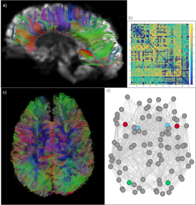
Structural brain network properties and cognitive impairment in adolescents after radiation therapy

Justin Yuan, Melanie Morrison, Angela Jakary, Sabine Mueller, Olga Tymofiyeva, Duan Xu, Janine Lupo
Cranial radiation therapy (CRT) is an effective brain cancer treatment but many patients exhibit cognitive deficits over time. We studied these deficits in adolescent and young adult survivors of pediatric brain cancer with prior CRT using white matter graph network analysis. Executive function and working memory performance were correlated with structural connectivity metrics. Global integration and segregation metrics were associated with neurocognitive deficits, as well as connectivity at domain-specific regions. Our results support past findings of CRT’s negative cognitive effects and suggest that they are driven through structural changes in brain white matter
|
|
0892.
 |
2 |
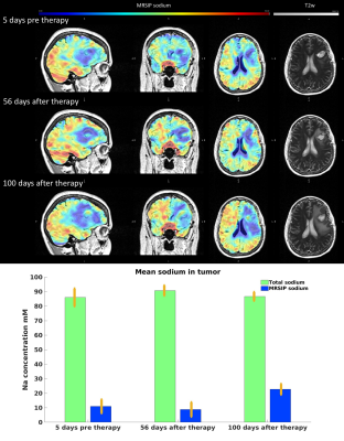
Motion-Restricted Sodium Ion Pools Analysis on Glioblastoma patients with Combinatorial Therapy
Xiuyuan Wang, Yongxian Qian, Rajan Jain, Andrew Chi, Sylvia Kurz, Fernando Boada
Differentiating treatment response from Glioblastoma progression with conventional proton magnetic resonance imaging (MRI) is challenging as it cannot unambiguously differentiate between early therapeutic response and treatment-related pseudo-progression. Based on the stability of sodium’s relaxation rate across human brains, a recent approach for separating motile and motion-restricted sodium ion pools (MRSIP) in the brain was introduced. In this study we evaluate the relationship between MRSIP concentration and the treatment evolution on a pool of glioma patients.
|
 |
0893.
 |
3 |
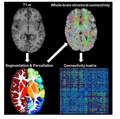
Structural Connectivity abnormality in children treated for Medulloblastoma
Adeoye Oyefiade, Iska Moxon-Emre, Kiran Beera, Jovanka Skocic, Donald Mabbott
Curative treatments for medulloblastoma impart significant toxicity on the developing brain. Though changes to white matter have been described, there remains a limited understanding of the effects of treatment on the structural connectome, which is thought to subserve complex and dynamic behaviors. Identifying compromise within the connectome may elucidate mechanisms of toxicity among survivors. We analyzed connectomic differences between survivors and age-matched controls. We identified two networks situated posteriorly with reduced white matter microstructure in survivors. Our findings complement studies showing long-term effects of treatment on brain structure, and localize these effects to areas around the site of the tumor.
|
|
0894.
 |
4 |
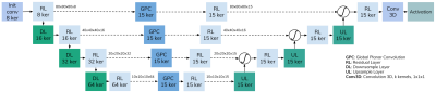
Global Planar Convolutions for improved context aggregation in Brain Tumor Segmentation with MR images
Santi Puch, Irina Sánchez, Aura Hernández, Gemma Piella, Paulo Rodrigues, Vesna Prc?kovska
Brain tumors pose a significant social and economic burden worldwide. A key to improve the quality and expectancy of life of patients with brain tumors is to automate the process of delineation of tumoral structures. In this work we propose the Global Planar Convolution module, a building-block for Convolutional Neural Networks that enhances the context perception capabilities of segmentation networks for brain tumor segmentation. We show that such modules achieve similar performance to equivalent networks with increased depth, and provide an initial inspection of their behavior via interpretation of intermediate feature maps.
|
|
0895.
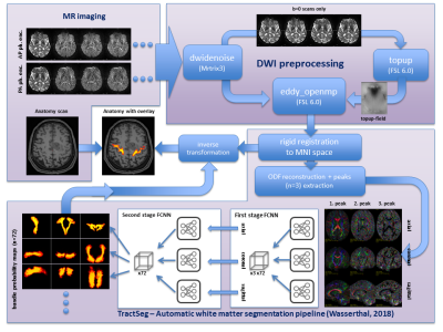 |
5 |
Feasibility study on automated white matter tract segmentation in neurosurgical pre-operative planning
Daniel Güllmar, Rotraud Neumann, Jakob Wasserthal, Jan Walter, Ulf Teichgräber, Thomas Mayer, Jürgen Reichenbach
In neuro-surgical preoperative planning of extirpation of large tumors it is important to locate the paths of critical cerebral nerve fiber bundles (e.g. corticospinal-tract). Manual fiber bundle selection is elaborate, requires expert knowledge and is prone to user errors. Therefore, in this study a fully automatic pipeline for white matter bundle segmentation was setup, incorporating recently published white matter bundle segmentation based using DNN, and tested with 12 patients suffering from large brain lesions. In all cases the position of the corticospinal tracts was evaluated as plausible, although in at least one hemisphere this tract was affected by the lesion.
|
|
0896.
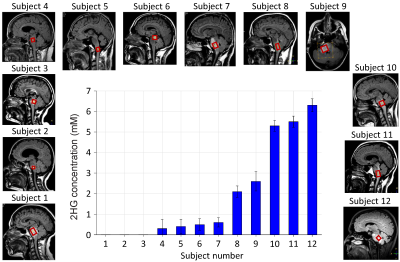 |
6 |
MR spectroscopy of 2-hydroxyglutarate in patients with brainstem tumors in vivo
Changho Choi, Vivek Tiwari, Zhongxu An, Sandeep Ganji, Michael Levy, Edward Pan, Elizabeth Maher, Toral Patel, Bruce Mickey
MRS of 2-hydroxyglutarate (2HG) has the great potential for determining the isocitrate dehydrogenase (IDH) mutational status in brain tumors noninvasively. This clinical role of 2HG MRS may be demonstrated most clearly in patients with brainstem gliomas or deep brain lesions, where surgical biopsy presents significant risk of permanent neurological deficit. We report 2HG MRS data in patients with brainstem tumors. 2HG was evaluated, using a 2HG-optimized TE 97ms PRESS at 3T, in 12 subjects with brainstem lesions in vivo. We also presents data of monitoring the tumor with serial 2HG MRS scans.
|
|
0897.
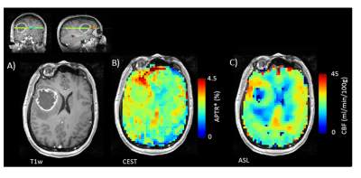 |
7 |
Quantification of regional pathophysiology in Glioblastoma Multiforme
Paula Croal, Kevin Ray, Ruichong Ma, Alex Smith, Moss Zhao, Benjamin Harris, Puneet Plaha, Simon Lord, Nicola Sibson, Michael Chappell
Patients with glioblastoma multiforme (GBM) have extremely poor prognosis due to therapy resistance, aggressiveness, and poor understanding of pathophysiology. Here, we use APT-CEST and ASL MRI to noninvasively probe both pH and perfusion in ten patients with primary GBM, prior to surgical/therapeutic intervention. We observe an overall increase in APT and CBF contrast, consistent with both intracellular alkalosis and angiogenesis. Clustering analysis revealed a strong regional association between pH and CBF in 9/10 patients suggestive of similar spatial disruptions. The ability to image concomitant changes in pH and perfusion may provide a novel way to stratify patients for targeted therapeutics.
|
|
0898.
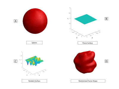 |
8 |
Brain Tumor Simulator: Creating Ground Truth for Evaluation of Complex MR Acquisition and Reconstruction Methodologies
Junzhou Chen, Leah Henze Bancroft, Jorge Jimenez, Anja van der Kolk, Aaron Field, Azam Ahmed, Roberta Strigel, Walter Block
New dynamic MRI methods promise to better characterize and monitor the vexing problem of brain cancer. Assessing these methods and the assumptions they rely upon is difficult as 1) no known gold standard is available for brain tumors and 2) the time window of availability for brain cancer volunteers is narrow. We present a simulator that generates realistic, heterogeneous, tumor models overlaid over normal brain tissue with user-specified permeability parameters. The simulator generates raw data for arbitrary k-space acquisition strategies. Using this simulator, new methodologies for various tumor types and sizes can be assessed before the first volunteer is ever recruited.
|
|
0899.
 |
9 |
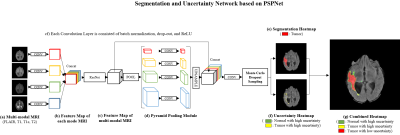
Brain Tumor Segmentation and Uncertainty Quantification Using Monte Carlo dropout sampling
Joohyun Lee, Woojin Jung, Jongho Lee
Deep learning has made tremendous progress in many areas but it is often regarded as a black box with uncertainty in outcome. Therefore, a more reliable method is necessary to be applied in a medical field. In this work, we designed a brain tumor segmentation network that provides uncertainty quantification using Monte Carlo dropout sampling. The proposed method resulted in considerable outcomes and also provided an option for selectively maximizing precision or recall using the uncertainty quantification.
|
|
0900.
 |
10 |
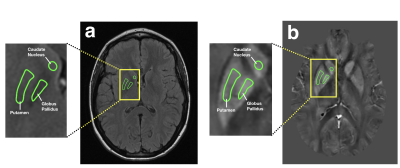
Basal ganglia iron deposition as a biomarker of brain tumor severity
Thomas Reith, Robert Wujek, Robin Karr, Kevin Koch, Mona Al-Gizawiy, Kathleen Schmainda
This study utilized quantitative susceptibility mapping (QSM) to investigate basal ganglia iron deposition in 27 patients diagnosed with gliomas. Basal ganglia QSM values of patients with glioblastomas were found to be higher than those of patients with tumors of lower grades, suggesting that iron deposition in the basal ganglia may serve as a biomarker of brain tumor severity.
|
|
0901.
 |
11 |
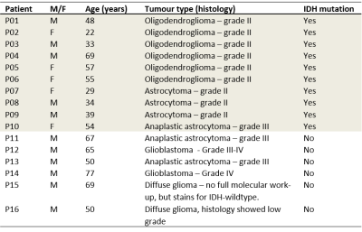
Voxelwise correlation between vascular parameters obtained with ASL and DSC as predictor of IDH-mutation status in non-enhancing glioma
Esther Warnert, Fatih Incekara, Arnaud Vincent, Joost Schouten, Martin van den Bent, Pim French, Hendrikus Dubbink, Johan Kros, Juan-Antonio Hernandez-Tamames, Marion Smits
Previous studies have shown good correlation between ASL and DSC vascular parameters as predictors of glioma grade, indication an option to omit DSC imaging in light of the recent finding of gadolinium deposition in the brain. However, in general these comparative studies were conducted before the recent update of the World Health Organisation classification of brain tumours. This study shows the potential of voxelwise correlations of vascular parameters obtained with ASL and DSC as predictors of IDH-mutation status in non-enhancing glioma and highlights that IDH-mutation status should be included in comparative studies of ASL and DSC vascular parameters in glioma.
|
|
0902.
 |
12 |
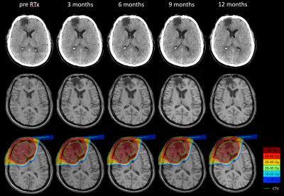
Diffusion and quantitative MR changes in normal appearing brain following radiotherapy.
Felix Raschke, Tim Wesemann, Hannes Wahl, Steffen Appold, Mechthild Krause, Jennifer Linn, Esther Troost
Irradiation of gliomas inevitably involves irradiation of surrounding normal appearing brain. We analysed longitudinal, quantitative MR data of 24 glioma patients before and at 3, 6, 9 and 12 months after radiotherapy and found significant reductions in mean-, axial- and radial diffusivity as well as in T2* in normal appearing white matter. These changes are greater the higher the received dose and progress over time. The diffusion reductions point towards axonal swelling. T2* reductions indicate either increased tissue heterogeneity, e.g. due to microglial activation or changes in tissue oxygenation, e.g. due to vascular alterations.
|
|
0903.
 |
13 |
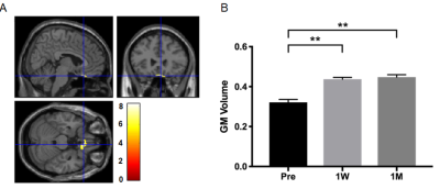
A Brain Morphometric MRI Study on Patients with Suprasellar Tumors: Preoperative and Postoperative Assessment
Qingping Chen, Jianyou Ying, Zhentao Zuo, Rui Wang, Taoyang Yuan, Lu Jin, Chuzhong Li, Songbai Gui, Peng Zhao, Chunhui Liu, Yazhuo Zhang
It has been reported that suprasellar tumors affect patients’ visual field and visual functional network. In this study, longitudinal brain morphometric assessment was performed pre- and post-operation based on 13 suprasellar tumors patients. The gray matter volume of rectus increases, but insular, caudate, and putamen decrease after operation. In addition, the gray matter volume can be predicted by tumor chiasmal lift length. What’s more, at the high-level visual cortex, the surface thickness becomes thicker after operation. We believe this study can improve the preoperative and postoperative assessment of suprasellar tumors in the future.
|
|
0904
 |
14 |
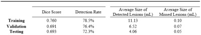
Segmentation of Brain Metastatic Lesions in Magnetic Resonance Imaging using Deep Learning
Video Permission Withheld
Jay Patel, Andrew Beers, Ken Chang, James Brown, Katharina Hoebel, Bruce Rosen, Raymond Huang, Priscilla Brastianos, Elizabeth Gerstner, Jayashree Kalpathy-Cramer
Magnetic resonance imaging plays a key role in assessing the efficacy of treatment for patients with brain metastases by enabling neuroradiologists to track lesions sizes across time points. However, manual segmentation of multiple time-points is prohibitively time-consuming, thus precluding its use in current clinical workflow. In this study, we develop a deep learning approach to automatically segment metastatic lesions, and demonstrate that our predicted segmentation has high agreement with the gold-standard manual segmentation.
|
|
0905.
 |
15 |
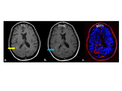
Quantitative deltaT1 (qDT1) as a Replacement for Adjudicated Central ReaderAnalysis: A Sub-Analysis of the RTOG 0625/ACRIN 6677 Multi-Center Brain Tumor Trial
Kathleen Schmainda, Melissa Prah, Zheng Zhang, Bradley Snyder, Scott Rand, Todd Jensen, Daniel Barboriak, Jerrold Boxerman
A semi-automatic method for delineating contrast-agent enhancing brain tumor, called quantitative delta T1 (qDT1), was compared to central reader analysis of clinical trial data. The qDT1 method demonstrated equivalence with expert reads for determination of early tumor progression and proved superior for further distinguishing responders from non-responders/non-progressors at the week 8 time point. Using qDT1 provides a solution to the high percentage of intra- and inter-observer disagreements, while being easier to use and more reliable for the daily clinical assessment of tumor response to therapy, as well as for large scale clinical trials.
|
|
