|
2nd Hour
Poster: The Aging Brain
Power Pitch Poster
Neuro
Tuesday, 14 May 2019
Power Pitch Theater A - Exhibition Hall
09:15 - 10:15
| |
|
Plasma # |
|
0327.
 |
1 |
Targeting brain and cognitive aging with multi-modal imaging and connectome topography profiling
Presentation Not Submitted
Alexander Lowe, Casey Paquola, Reinder Vos de Wael, Sara Lariviere, Shahin Tavakol, Benoit Caldairou, Neda Bernasconi, Andrea Bernasconi, Nathan Spreng, Boris Bernhardt
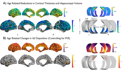
We present an approach to represent and analyze age-related differences in cortical morphology and Aβ uptake based on connectome topography. Studying healthy individuals, we observed age-related reductions in neocortical thickness and atrophy across posterior hippocampal subfields. Additionally, we observed an interplay between aging effects and functional topography in both neocortical and hippocampal regions, with age-related thinning stronger towards unimodal regions and Aβ deposition increasing towards transmodal regions. Similarly, an inverted pattern of volume loss and Aβ deposition was observed along the hippocampal long-axis. Finally, imaging markers were found to predict cognitive performance in a topography-specific manner.
|
|
0328.
 |
2 |
Age-related whole-brain structural changes in relation to cardiovascular risks

Tao Gu, Hui Guo, Min Chen, Xiaowei Song
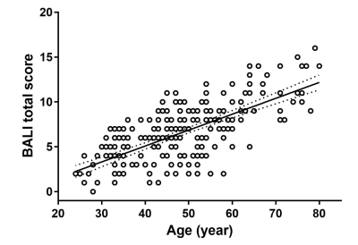
We investigated the relationship between structural brain health with age and cardiovascular risks across the adult life course. A score of the Brain Atrophy and Lesion Index (BALI), which assesses and integrates multiple changes commonly seen on MRI in the aging brain, was generated for each subject from evaluation of T2-weighted MRI. Our data showed that the accumulation of MRI detectable deficits in the brain became evident even in younger adults. Cardiovascular risks strongly affected the whole-brain structural health, in addition to the effect of age.
|
|
0329.
 |
3 |
Differential developmental trajectory of magnetic susceptibility in healthy rhesus macaque brain
Presentation Not Submitted
Jing Wu, Hui Zhang, Yongquan Ye, Shuheng Zhang, Qiang He, Xintian Hu, Nan-jie Gong
Myelination and iron deposition in the deep brain nuclei evolve both spatially and temporally. In this study, we quantitatively evaluated the change of iron content along with age in primate brains using quantitative susceptibility mapping (QSM). All the brain images were acquired from 23 healthy rhesus macaque monkeys (23+/-7.85 y, ranged 2 ~ 29 y) with a 3D five-echo GRE sequence. After analyzing susceptibility maps and R2* maps of the ROIs (including putamen, globus pallidus, caudate nucleus, thalamus, dentate nucleus, red nucleus, substantia nigra), susceptibility in most of the ROIs correlated with the growth of age significantly.
|
 |
0330.
 |
4 |
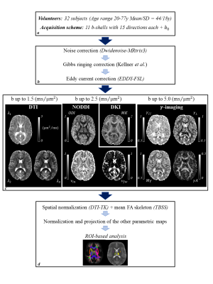
Age-related microstructural and physiological changes in normal brain assessed via anomalous diffusion derived ?, DTI, DKI and NODDI metrics
Michele Guerreri, Marco Palombo, Alessandra Caporale, Emiliano Macaluso, Marco Bozzali, Silvia Capuani
In this study we used γ-metrics, derived from anomalous diffusion signal representation, as well as DTI, NODDI, DKI derived parameters to assess physiological (i.e. the iron content) and microstructural (myelin damage, axonal disintegration, neuron cell loss) modifications in cerebral WM and scGM of middle- and older-aged subjects. We found that γ-metrics are remarkably sensitive and provide complementary information compared to DTI-metrics, MK and NODDI to detect modifications in frontal WM, where substantial changes are expected with aging. Also, the combined use of these techniques may unravel different patterns of modifications of the ageing brain.
|
|
0331.
 |
5 |
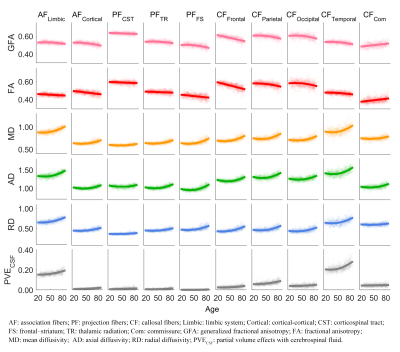
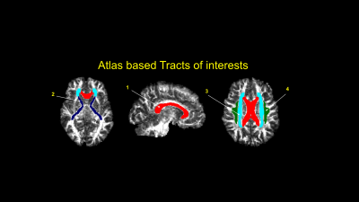
Characteristic Normal Ageing Patterns of White Matter Tracts in 610 Cambridge Centre for Ageing and Neuroscience (Cam-CAN) Healthy Participants
Te-Wei Kao, Yung-Chin Hsu, Wen-Yih Tseng
Previous studies did not clearly characterize normal ageing patterns of white matter tracts across lifespan. Here, we performed tract-specific automatic analysis over the whole brain to measure the diffusion indices on 610 healthy participants recruited from Cam-CAN. We used quadratic or linear models to plot the curves of diffusion indices against age, and calculated the average values and slopes of the curves in 10 subsystems classified from 76 major tracts. Our study characterized different ageing patterns corresponding to different subsystems.
|
|
0332.
 |
6 |
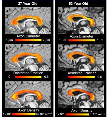
Age-Related Microstructural Alterations in Human Corpus Callosum Measured by High-Gradient Diffusion MRI
Qiuyun Fan, Qiyuan Tian, Ned Ohringer, Aapo Nummenmaa, Thomas Witzel, Sean Tobyne, Eric Klawiter, Bruce Rosen, Lawrence Wald, David Salat, Susie Huang
Cerebral white matter exhibits degenerative changes during normal aging. Noninvasive approaches to measure these microstructural alterations would be invaluable for understanding the substrate and regional variability of age-related white matter degenerations. Recent advances in diffusion MRI have leveraged high gradient strengths to increase sensitivity toward axonal size and density in living human brains. Here, we examined the relationship between age and microstructural properties measured using high-gradient diffusion MRI. We observed an increase in apparent axon diameter and decrease in density with advancing age in the corpus callosum, with changes most pronounced in the genu and relatively absent in the splenium.
|
|
0333.
 |
7 |
The link between vascular stiffness and resting-state fMRI measures in healthy aging
Ahmad Hussein, Jacob Matthews, Zdenka Pausova, Catriona Syme, Christopher MacGowan, Bradley MacIntosh, Tomas Paus, J. Jean Chen
Arterial pulse-wave velocity (PWV) is an established measure of vascular stiffness which is an important risk factor in cardiovascular disease and brain dysfunction. It remains unclear, however, whether PWV variations across age is associated with changes in resting-state fMRI (rs-fMRI) measures, as the fMRI signal is a heavily vascular signal. In this study, we show that PWV has a significant impact on rs-fMRI signal fluctuation amplitude and functional connectivity. Moreover, PWV effects are distinct from those of age, and may not have neuronal underpinnings.
|
|
0334.
 |
8 |
Evaluation of standardized as well as study-specific and age-specific structural T1-weighted brain templates for use in studies on older adults
Abdur Raquib Ridwan, Shengwei Zhang, Mohammad Rakeen Niaz, Xiaoxiao Qi, David A. Bennett, Yongyi Yang, Konstantinos Arfanakis
Atlas-based MRI investigations on older adults often utilize young adult standardized templates, such as those of the ICBM. Additionally, a thorough, quantitative assessment of how available standardized, study-specific and age-specific structural templates perform in studies on older adults has not yet been conducted. Here, a new standardized T1-weighted template was developed specifically for studies on older adults, and was compared to 25 other standardized, study-specific, and age-specific templates, in terms of image quality and inter-subject spatial normalization accuracy.
|
|
0335.
 |
9 |
Arterial stiffness and white matter integrity in the elderly: a diffusion tensor and magnetization transfer imaging study
Atef Badji, Adrián Noriega de la Colina, Agah Karakuzu, Tanguy Duval, Laurence Desjardins-Crépeau, Sven Joubert, Louis Bherer, Maxime Lamarre-cliche, Nikola Stikov, Hélène Girouard, Julien Cohen-Adad
Arterial stiffness is a common condition arising with aging and is associated with an elevated risk for white matter structural abnormalities in the brain. The goal of this study is to combine white matter sensitive techniques (DTI, MTsat) to better understand the impact of arterial stiffness on the white matter microstructure and cognitive health in healthy elderly. Results suggest that arterial stiffness is associated with axon degeneration rather than demyelination. Findings from this study also show that improved executive function performance correlates with white matter metrics. Controlling arterial stiffness might play a role in maintaining the health of white matter axons in the aging brain and thus prevent or slow cognitive decline.
|
|
0336.
 |
10 |
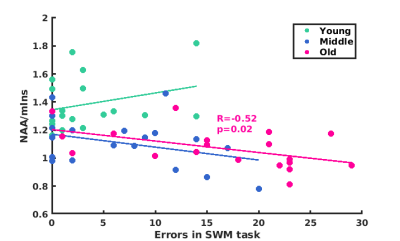
Hippocampal NAA/mIns ratio and spatial working memory across the lifespan
Anna Lind, Carl-Johan Boraxbekk, Hartwig Siebner, Esben Petersen, Anouk Marsman
The NAA/mIns ratio has been proposed as a marker of unhealthy ageing. We found that the NAA/mIns ratio is decreased in the hippocampus for middle-aged and old individuals compared to young individuals. For old individuals, declining NAA/mIns ratios are correlated with poorer performance on a spatial working memory task but this relationship is not found in middle-aged and young individuals. Our results suggest that the NAA/mIns ratio decrease in the hippocampus is observable before the negative functional effects.
|
|
0337.
 |
11 |
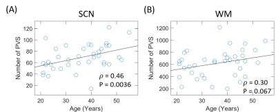
Characterization of perivascular space morphology in healthy volunteers between 21 and 55 years old
Xiaopeng Zong, Chunfeng Lian, Jordan Jimenez, Koji Yamashita, Dinggang Shen, Weili Lin
Perivascular spaces (PVS) are an integral part of the brain’s glymphatic system. Although enlarged PVSs are often observed in older population and patient with neurological diseases, normal PVS morphological features in healthy subjects and their age dependences remain poorly understood. We studied the age-dependence of PVS morphology in healthy volunteers aged 21 – 55. The number and diameters of PVSs were positively correlated with age but exhibited large inter-subject variations. We also found clear spatial heterogeneity in the density of visible PVSs. Further studies are needed to realize the utility of PVS as a potential biomarker for aging and neurological diseases.
|
|
0338.
 |
12 |
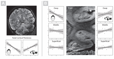
Behavioral Correlates to Laminar Thickness Within the Cortex
Allen Newton, Rankin McGugin, Isabel Gauthier
Cortical thickness changes have been shown to be correlated with a wide variety of behavioral measures. Until recently, methods to probe the laminar changes underlying these large scale cortical changes in vivo have not been available. Here, we present methods to measure laminar thickness within the cortex, and show that sufficient precision exists to observe behavioral correlates within individual layers. Furthermore, our data are consistent with the hypothesis that behavior learned early in life has different laminar thickness correlates than does behavior learned later in life.
|
|
0339.
 |
13 |
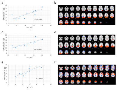
Reduced functional connectivity of resting state networks in the healthy brain is associated with R2*changes consistent with myelin breakdown
James O'Callaghan, Lysia Demetriou, Ayla Mansur, Adam Connolly, Mari Lambrechts, Simon Robinson, Korbinian Eckstein, Lefkos Middleton, Matthew Wall, Courtney Bishop, Roger Gunn, Eugenii Rabiner
Functional connectivity of select resting state networks has been shown to diminish with age. Reported observations of reductions in visual and salience network strength appear to be supported by findings of vulnerability of the associated parietal, occipital, and frontal lobes to structural changes in the healthy brain.
We present data suggesting that measures of R2* in parietal and frontal lobar regions are correlated to visual and salience network connectivity in healthy individuals. These observations may indicate that early breakdown in myelin, associated with R2* shortening in white matter, may be responsible for age related decline in these resting state networks.
|
|
0340.
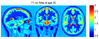 |
14 |
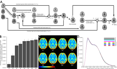
Age-specific Quantitative Brain Atlas Modelling Brain Shape and T1 Changes
Tom Hilbert, Gian-Franco Piredda, Karl Egger, Shan Yang, Tobias Kober
In order to facilitate the use of quantitative mapping approaches in clinical routine, atlases with normative quantitative values are required for comparison on a single-subject basis. To improve such comparison, we propose here to not only model the age-related change in T1, but also the change in brain shape. The method is demonstrated on a dataset of 196 quantitative T1 maps, yielding a brain model with shape and T1 information over the potential lifespan of a patient to assess.
|
|
0341.
 |
15 |
The Relationship between Glutamate and BOLD signal changes During Face-Name Paired-Associates Encoding and Retrieval Memory Task in Healthy Adults concerning age, performance level and genetic risk-A combined 1H-MRS and fMRI study
Hui Zhang, PW Chiu, SWH Wong, GHY Wong, T Liu, Q Chan, HKF Mak
Glutamate is hypothesized to be the neurotransmitter in mediating BOLD fMRI. In this study, fMRI technique was combined with Magnetic Resonance Spectroscopy(MRS) to investigate the relationship between glutamate and the BOLD signal changes during face-name memory task. Three different task-based fMRI face name memory experiments were performed respectively focusing on age difference, performance level and genetic risk. [Glx]abs in left hippocampus of elderly(Experiment1), high-performance(Experiment2) and low genetic risk(Experiment3) showed high correlation with BOLD signal changes in activated regions. On the whole, glutamate appears to be excitatory and lead to compensatory excitations.
|
|
