|
2nd Hour
Poster: Myelin Imaging in Sickness & Health
Power Pitch Poster
Neuro
Monday, 13 May 2019
Power Pitch Theater A - Exhibition Hall
14:45 - 15:45
| |
|
Plasma # |
 |
0112.
 |
1 |
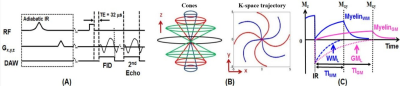
3D Inversion Recovery Ultrashort Echo Time (3D IR-UTE) Magnetic Resonance Imaging of Myelin in Traumatic Brain Injury – a Feasibility Study

Ya-Jun Ma, Adam Searleman, Shanshan Wang, Hyungseok Jang, Jonathan Wong, Eric Chang, Brian Head, Jiang Du
Reduced myelination has been observed in rodents and humans after mild traumatic brain injury (mTBI). However, conventional MR imaging sequences cannot directly detect any signal from myelin. In this study, we aimed to evaluate a 3D IR-UTE sequence for selective imaging of myelin in mice using a standard controlled cortical impact (CCI) model of mTBI with histological confirmation. To demonstrate the clinical feasibility, a translational 3D IR-UTE technique was developed and applied to healthy volunteers and mTBI patients at 3T. The preliminary results demonstrate the feasibility of volumetric myelin mapping using the 3D IR-UTE sequences at 7T and 3T.
|
 |
0113.
 |
2 |
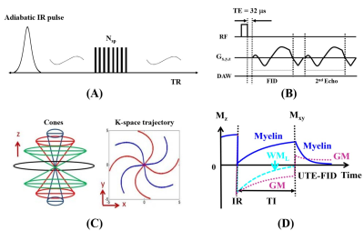
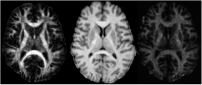
Volumetric Imaging of Myelin in Vivo using 3D Inversion-Recovery Ultrashort Echo Time Cones (3D IR-UTE-Cones) Magnetic Resonance Imaging
Ya-Jun Ma, Adam Searleman, Hyungseok Jang, Shu-Juan Fan, Jonathan Wong, Jody Corey-Bloom, Eric Chang, Jiang Du
To image myelin directly for whole brain on clinical scanners which would provide better characterization of multiple sclerosis (MS) lesions at diagnosis and in response to therapy, we propose a 3D adiabatic inversion recovery prepared ultrashort echo time cones (3D IR-UTE-Cones) sequence for volumetric myelin imaging in vivo with a clinical feasible scan time. The myelin imaging show clearly signal loss in MS lesions for both ex vivo and in vivo brain studies.
|
|
0114.
 |
3 |
Comparison of Myelin- and Axon-Specific Imaging Modalities in Multiple Sclerosis
Sehong Oh, Kunio Nakamura, Kedar Mahajan, Jacqueline Chen, Mark Lowe, Daniel Ontaneda, Bruce Trapp, Ken Sakaie
Multiple sclerosis (MS) is a chronic disease characterized by demyelination and neuronal/axonal pathology. Based on postmortem MRI-pathology correlations lesions found on conventional MRI do not exhibit expected demyelination. There is a need for imaging modalities that have better specificity for myelin, axonal density and axonal health. We compare the properties of fast variants of myelin-specific modalities (myelin water imaging, quantitative magnetization transfer and visualization of short transverse relaxation component) and of axon-specific measures from neurite orientation dispersion and density imaging. These comparisons constitute steps toward developing better imaging biomarkers for MS pathology.
|
|
0115.
 |
4 |
Reproducibility of Practical Imaging-Based Myelin Biomarkers
Sehong Oh, Ken Sakaie
Remyelination therapies are an emerging approach for treating multiple sclerosis, but development of these therapies is hampered by a lack of imaging biomarkers. Imaging with improved specificity to myelin, as compared to conventional MRI, have the potential to act as biomarkers, but current implementations can be time consuming. We examine the performance of fast version of myelin imaging from the perspective of reproducibility, a necessary prerequisite for use in proof-of-concept clinical trials of remyelinating agents.
|
|
0116.
 |
5 |
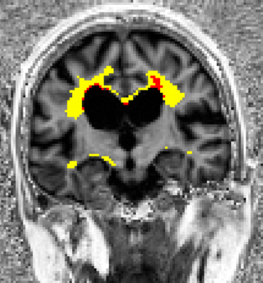
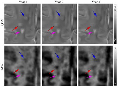
Chronic MS lesions with hyperintense appearance on QSM demonstrate more myelin damage and are long-lasting
Shun Zhang, Thanh Nguyen, Yi Wang, Susan Gauthier
We assessed longitudinal QSM and MWF changes in 307 chronic MS lesions from 41 patients over four years. Lesions were stratified into three groups: hyperintense rim on QSM (rim+), no rim (rim-) and isointense on QSM (QSM-). Rim+ lesions were found to have the lowest MWF. QSM lesions showed a decreasing trend while MWF remains relatively stable over the same period.
|
|
0117.
 |
6 |
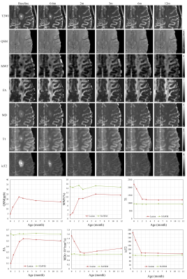
Concurrent remyelination and susceptibility increase in new active MS lesions indicate early iron accrual
Shun Zhang, Yi Wang, Susan Gauthier, Thanh Nguyen
We reported initial results of an ongoing longitudinal study of new enhancing lesions using quantitative MRI including QSM, MWF and DTI. MS patients were followed five times during the first three months of lesion formation. We found a subset of lesions in which QSM rises rapidly and simultaneously with MWF and FA measurements, suggesting increasing iron accumulation within this period.
|
|
0118.
 |
7 |
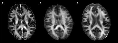
A pediatric brain template for myelin water fraction and diffusion tensor imaging.
Sarah Morris, Richard Holmes, Adam Dvorak, Hanwen Liu, Irene Vavasour, Silvia Mazabel, Burkhard Mädler, Shannon Kolind, David Li, Linda Siegel, Christian Beaulieu, Alex MacKay, Cornelia Laule
Measuring pediatric brain myelination can provide insights into normal brain development as well as many pediatric brain disorders. We have created a myelin water and diffusion tensor template for healthy children aged 9-10 years to be used as a reference in imaging studies. Our template was produced using ANTs software to provide high-quality anatomical alignment of brain structures. ROI analysis revealed significant differences in myelin water fraction between children and adults. We found no significant correlation between myelin water fraction and diffusion tensor metrics across the ROIs investigated, highlighting the complementary information these two techniques provide.
|
|
0119.
 |
8 |
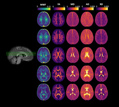
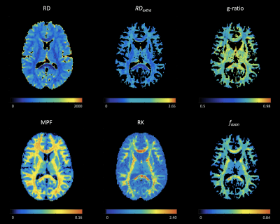
A comparison between diffusion metrics, macromolecular proton fraction, and g-ratio for quantitative in vivo myelin mapping
Yu Sui, Pippa Storey, Alexey Samsonov, Mariana Lazar
Myelination as one of the most reliable indicators of postnatal brain maturation and cognitive ability, supports white matter function by facilitating efficient neural signaling and pathway remodeling. The current study aimed to compare in healthy young adults several recently developed myelin mapping metrics including g-ratio, macromolecular proton fraction, and metrics from diffusion tensor/kurtosis imaging. Relationships between these individual metrics and their specific sensitivity to different aspects of white matter microstructure are discussed.
|
|
0120.
 |
9 |
Test-retest Reliability of Myelin-Sensitive MRI Techniques
Kiara Kunimoto, Bryce Geeraert, R. Lebel, Catherine Lebel
This project evaluated the reliability of quantitative inhomogeneous magnetization transfer (qihMT), a novel myelin-sensitive measure, compared to more common measures, magnetization transfer ratio (MTR) and fractional anisotropy (FA), in 10 healthy adults. A repeated measures ANOVA revealed no significant differences in measure means between scans in 18 white matter regions. Coefficients of variation (CV) demonstrated that FA had the highest reliability, followed by MTR and qihMT. The reduced reliability of qihMT observed here may be mitigated by further optimization of this novel sequence.
|
|
0121.
 |
10 |
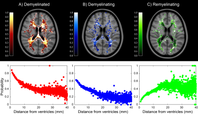
Remyelination is less efficient in periventricular white matter lesions in Multiple Sclerosis
Matteo Tonietto, Emilie Poirion, Caroline Papeix, Michel Bottlaender, Benedetta Bodini, Bruno Stankoff
The objective of this study was to investigate the relationship between the extent of spontaneous remyelination and the distance from ventricular cerebrospinal fluid (CSF) in a group of multiple sclerosis patients. Dynamic remyelination was measured using longitudinal [11C]PiB positron emission tomography and found to be significantly reduced in periventricular white matter lesions, while becoming progressively more extensive with increasing distance from ventricles. Moreover, we found a positive correlation between periventricular remyelination and cortical thickness. These results suggest that CSF-linked factors might interfere with the spontaneous remyelination process in multiple sclerosis patients.
|
|
0122.
 |
11 |
Early life Myelination Mediates the Effects of the APOE Genotype on Cognitive Development
Justin Remer, Douglas Dean, Michaela Voyer, Sean Deoni
A growing focus in Alzheimer’s Disease (AD) research is understanding the earliest preclinical structural changes associated with the disorder. In prior studies our group has shown differences in early myelin content based on APOE genotype and we recently have shown preliminary results that APOE effects early longitudinal myelin and cognitive development. Nevertheless, these results fail to explain how differences in early brain anatomy lead to differences in cognitive development. This is the first study to explore and analyze if differences in early cognitive development based on APOE genotype is explained by differences in early myelin development in a large cohort of healthy neurotypical infants and young children stratified by presence or absence of at least one APOE ε4 allele.
|
|
0123.
 |
12 |
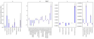
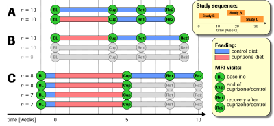
Pitching the Cuprizone Mouse Model for Testing (re-) myelination therapies: robustness and reproducibility
Andreas Bruns, Anna Mechling, Eva Mracsko, Thomas Mueggler, Basil Künnecke
We assessed robustness and reproducibility of the cuprizone mouse model of de- and remyelination for its use in testing novel pharmacological treatments of demyelinating disorders such as multiple sclerosis. In several multimodal MRI studies using independent batches of animals, increases in T2, decreases in MTR and FA and biphasic responses in MK upon cuprizone feeding, as well as partial recoveries after cuprizone withdrawal, showed huge effect sizes and high cross-study consistency, especially in the corpus callosum. Our results substantiate the suitability of the cuprizone mouse model for longitudinal monitoring of the pathology using multimodal MRI.
|
|
0124.
 |
13 |
Multi-parameter quantitative MRI reveals common distribution of myelin in ex-vivo chimpanzee and in-vivo human brains
Daniel Papp, Nicole Eichert, Colin Reveley, Stuart Clare, Rogier Mars
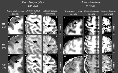
Post-mortem investigations of tissue properties can dramatically extend the biological information that can be obtained from bodies or tissue that cannot be easily investigated in-vivo, as is the case for the majority of species of interest to comparative neuroscience. Here, we show the potential of a quantitive MRI method, multi-parameter mapping, to obtain high-resolution information about tissue properties of large non-human primates that generally cannot be studied anatomically. We compare myelination indices, derived from ex-vivochimpanzee data at 7T to those derived from in-vivo human data at 3T.
|
|
0125.
 |
14 |
Effects of myelin on the water resonance line-shape in postmortem mouse brain
Sean Foxley, Gregory Karczmar, Brian Popko, Pedro Brugarolas, Gregg Wildenberg, Vandana Sampathkumar, Narayanan Kasthuri
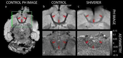
Dysmyelinating diseases are characterized by abnormal myelin formation and function. Such microstructural abnormalities in myelin have been demonstrated to produce measurable effects on the MR signal. This work examines these effects from post-mortem fixed control and shiverer mouse brains on voxel-wise, high-resolution water spectra acquired using a multi-gradient echo pulse sequence. Results demonstrate that components of the spectra are differentially affected by myelin concentration. This suggests that water proton spectra may be sensitive to the tissue microenvironment, specifically myelin, and could serve as potential MRI based biomarkers of dysmyelinating diseases.
|
|
0126.
 |
15 |
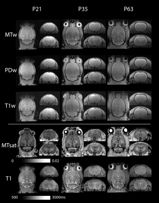
Quantitative multiparametric mapping assessment of the rat brain through adolescence into adulthood
Stephen Sawiak, Bianca Jupp, Jolyon Jones, Peter Zhukovsky, Jeffrey Dalley
We demonstrate high-resolution quantitative multiparametric mapping in the rat: scanning animals before and after adolescence and comparing them to the adult. We produce maps sensitive to myelin signal throughout the brain that complement volumetric information normally used in developmental studies. The aim is to better exploit models of neuropsychiatric disorders, particularly those with developmental components.
|
|
