|
2nd Hour
Poster: Flow & Vessel Imaging
Power Pitch Poster
Cardiovascular
Tuesday, 14 May 2019
Power Pitch Theater A - Exhibition Hall
14:30 - 15:30
| |
|
Plasma # |
 |
0450.
 |
1 |
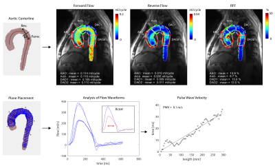
Longitudinal Study of Aortic Stiffness and Flow Reversal in Patients with Cryptogenic Stroke

Kelly Jarvis, Alireza Vali, Amer Ahmed Syed, Kathryn Muldoon, Shyam Prabhakaran, Jeremy Collins, Michael Markl
Atherosclerotic plaque in the descending aorta has emerged as a potential etiology of embolic stroke due to diastolic flow reversal in this region. This study seeks to investigate 1) whether aortic stiffness assessed by pulse wave velocity (PWV) may be related to flow reversal and 2) the effects of medical therapy on PWV and flow reversal. Twenty cryptogenic stroke patients were included in this 4D flow MRI study. We found relationships between age, PWV and flow reversal. No systematic change in PWV or flow reversal was detected for patients taking medications with potential de-stiffening effects.
|
|
0451.
 |
2 |
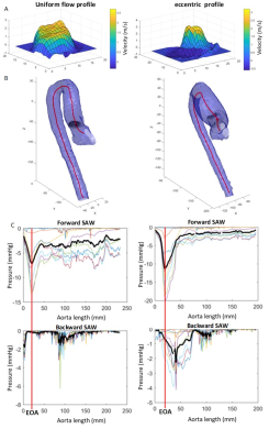
Enhanced non-invasive pressure drop and flow inefficiencies quantification via 4D-flow MRI
Joao Filipe Fernandes, Alessandro Faraci, Saul Myerson, David Alexander Nordsletten, Pablo Lamata
The pressure drop caused by convective effects is the standard clinic method for diagnosis of aortic stenosis using simplified Bernoulli. However simplified Bernoulli results in a reported overestimation that can be resolved with 4D-flow MRI and Simplified Advective Work-energy relative pressure (SAW). This work further refines SAW formulation and proposes an enhanced analysis of the positive and negative components of flow profile. Whereas forward SAW pressure drop correlates better with mean SB (R2=0.951) than full velocity profile by SAW (R2=0.901), backward SAW is totally independent of SB (R2=0.490), which makes the flow efficiency a potential predictive value for disease progression.
|
|
0452.
 |
3 |
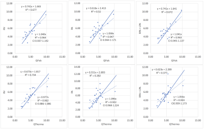
MRI flow and volume quantification with internal validation and correlation to Fick and thermodilution catheterization derived values in pulmonary hypertension
Lindsey Crowe, Anne-Lise Hachulla, Stéphane Noble, Paola Soccal, Maurice Beghetti, Frédéric Lador, Jean-Paul Vallée
Cardiac output (CO) determination is mandatory for the diagnosis work-up of pulmonary hypertension (PH). It is classically obtained by invasive methods such as indirect Fick (Qfick) and thermodilution (Qthermo) performed during right heart catheterisation (RHC). However, non-contrast, non-invasive and reliable methods may be preferred for clinical routine and post-treatment monitoring of patients. This study compared QFlow derived parameters (stroke volumes and cardiac index/ pulmonary and aortic flow) measured non-invasively in PH patients for internal validation of MRI and for comparison to RHC.
|
|
0453.
 |
4 |
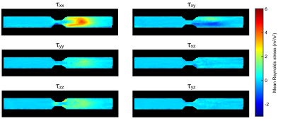
Reynolds Stress Tensor Quantification using a Flow-MRF Approach
Sebastian Flassbeck, Simon Schmidt, Mark Ladd, Sebastian Schmitter
This work demonstrates the feasibility to quantify simultaneously the Reynolds stress tensor and velocities using a Flow-MRF based approach in conditions of turbulent flow. This is achieved by measuring spatially undersampled time-series with pseudo-randomly varying velocity encoding moments, based on the MR Fingerprinting framework.
|
|
0454.
 |
5 |
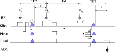
3D MR Velocimetry of Very Slow Flows
Magdoom Kulam Najmudeen, Ahmad Zeinomar, Russell Lonser, Malisa Sarntinoranont, Thomas Mareci
Methods are needed to non-invasively measure in vivo very slow flows, which govern many important physiological processes like brain glymphatic flow. In this study, a new stimulated echo-based phase contrast MRI sequence, robust to phase errors induced by gradient hardware, is used to measure 3D flows as slow as 1 μm/s. The method was validated using a controlled pipe flow experiment. In the absence of induced flow, the method revealed unusual natural convection flows in a water filled tube placed in a wide-bore magnet. The method may be applied to measure important slow flows in vivo.
|
|
0455.
 |
6 |
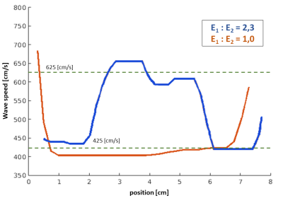
4D-Flow MRI and robust local Pulse Wave Velocity allow the detection of alterations in human aortas
Joaquin Mura, Julio Sotelo, Hernan Mella, Andrew Tran, Tarique Hussain, Bram Ruijsink, Sergio Uribe
An improved version of continuous pulse-wave velocity estimation uses 4D-Flow data with time-delay recovered from a faster formulation using FFT. The novelty also relies upon neglecting regressive time-delay zones, yielding stable and reliable results. Numerical simulations are shown to assess the method. We also present its application in patients with Familial Hypercholesterolemia and Fontan. Consistently with previous findings, adult controls have stiffer aortic walls compared to young controls. Also, Fontan patients appear with stiffer aortic arch than other subjects. More interestingly, all subjects show a softening in the aortic arch respect to the rest of the vessel.
|
|
0456.
 |
7 |
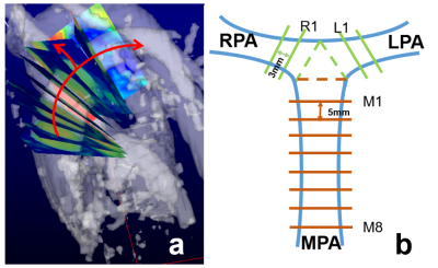
Dilated Pulmonary Arteries and Wall Shear Stress in Patients with Repaired Tetralogy of Fallot.
Han-Jung Liao, Jung-Hsiu Liu, Ming-Ting Wu, Ken-Pen Wang, Mao-Yuan Su, Hsu-Hsia Peng
We aimed to investigate the impact of pulmonary area on wall shear stress (WSS) and oscillatory shear index (OSI) to explore the altered vascular characteristics in patients repaired tetralogy of Fallot (rTOF). rTOF patients with pulmonary dilatation presented decreased axial WSS and increased axial OSI. The correlations between pulmonary area and axial WSS and OSI were missing in rTOF patients, depicting the abnormal endothelial regulation function in response to axial WSS and OSI. In conclusion, the correlation analyses between pulmonary area, axial WSS and OSI might provide helpful information in investigating the altered vascular characteristics in patients with rTOF.
|
|
0457.
 |
8 |
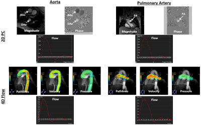
Accelerated whole-chest 4D flow imaging without navigator echo
El-Sayed Ibrahim, Jadranka Stojanovska, Dhiraj Baruah
4D flow MRI allows for thorough evaluation of hemodynamic patterns. Nevertheless, 4D flow imaging is very time consuming from both image acquisition and reconstruction perspectives. In this study, we provide preliminary data about accelerated sub-10-minutes whole-chest 4D flow imaging without navigator echo, based on recent acquisition and reconstruction technical developments, and compare the results to conventional 2D PC imaging across large arteries and atrioventricular valves. Measurements from accelerated 4D flow were in good agreement with conventional 2D PC imaging. The accelerated 4D flow technique would improve MRI cost effectiveness, lead to increased clinical adoption, and provide more patient comfort.
|
|
0458.
 |
9 |
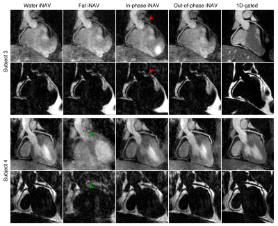
Dual-echo 2D Image Navigators for Respiratory Motion-Corrected Whole-Heart Water/Fat CMRA
Camila Munoz, Gastao Cruz, Radhouene Neji, René Botnar, Claudia Prieto
Whole-heart water/fat coronary MR angiography (CMRA) is a promising technique for improved visualisation of the cardiac anatomy and epicardial and pericardial fat. However, respiratory motion remains a challenge for its integration into clinical routine. Here we propose a respiratory motion-corrected whole-heart water/fat CMRA approach based on dual-echo 2D image navigators (iNAVs) and a combined 2D translational and 3D non-rigid motion corrected reconstruction scheme. Results from healthy subjects indicate that out-of-phase iNAVs produce accurate respiratory translational motion estimation, and that motion-corrected water/fat CMRA images are comparable to reference diaphragmatic-gated images, but are acquired in a significantly shorter scan time.
|
|
0459.
 |
10 |
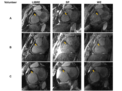
Fat-free free-running coronary MRA at 1.5T using LIBRE water excitation pulses
Nemanja Masala, Jessica Bastiaansen, Lorenzo Di Sopra, Davide Piccini, Jérôme Yerly, Roberto Colotti, Matthias Stuber
Previously published work in fully self-gated free-breathing 3D radial coronary MRA at 1.5T with cardiac-and-respiratory-motion-resolved reconstruction (Free-running framework) suffered from the disadvantage of requiring interrupted bSSFP with ramp-up and fat saturation pre-pulses. Using numerical simulations, in vitro and in vivo scans, we successfully tested the hypothesis that LIBRE, a new water excitation technique, obviates the need for such pre-pulses, improves time efficiency when compared to earlier approaches, and provides both superior fat saturation and vessel delineation relative to more conventional water excitation.
|
|
0460.
 |
11 |
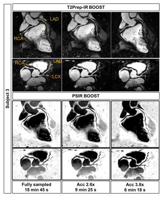
XD-ORCCA for BOOST: accelerated motion-compensated simultaneous bright- and black-blood 3D whole-heart coronary MRI
Teresa Correia, Giulia Ginami, Aurelien Bustin, Radhouene Neji, Rene Botnar, Claudia Prieto
Recently, a novel free-breathing 3D whole-heart sequence, called T2-prepared BOOST, was proposed for non-contrast enhanced bright-blood and black-blood coronary MR imaging, for simultaneous coronary lumen and coronary thrombus/ intraplaque hemorrhage visualization. However, high-resolution fully-sampled BOOST acquisitions require long scan times of ~20min. Here, we propose to use a modified version of XD-ORCCA, a highly efficient respiratory-resolved motion-corrected framework, to accelerate BOOST acquisitions. XD-ORCCA exploits the sparsity in a motion-corrected domain to acquire high-quality respiratory-resolved bright- and black-blood BOOST images in ~6min. Hence, high-resolution free-breathing BOOST imaging is achieved within clinically feasible acquisition times.
|
|
0461.
 |
12 |
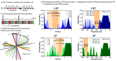
Natively Fat-Suppressed Fast Interrupted Steady State (FISS) for 5D Whole Heart Imaging at 1.5 and 3T
Jessica Bastiaansen, Davide Piccini, Lorenzo Disopra, Christopher Roy, Jérôme Yerly, Robert Edelman, Ioannis Koktzoglou, Matthias Stuber
Fast interrupted steady state (FISS) sequences provide bSSFP signal contrast and concomitant fat signal suppression. In this work, 3D radial FISS was implemented as part of a respiratory self-gated free-breathing cardiac and respiratory motion-resolved 5D imaging framework. Its capabilities for fat suppression were tested and characterized at both 1.5 and 3T. Combined with a XD-GRASP reconstruction, FISS offers a versatile alternative for motion-resolved fat suppressed high-resolution whole-heart anatomical and functional cine imaging with a scan time as low as 8 minutes.
|
|
0462.
 |
13 |
Efficient T2 Mapping of the Carotid Artery using a 3D Stack-of-Stars Variable Flip Angle TSE Pulse Sequence
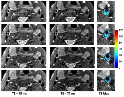
Mahesh Bharath Keerthivasan, Kevin Johnson, Ali Bilgin, Craig Weinkauf, Maria Altbach
We present a radial stack-of-stars TSE pulse sequence with an efficient radial view ordering and optimized refocusing flip angles for 3D T2 mapping of the carotid artery. The technique provides excellent anatomical coverage within clinically acceptable times. The short acquisition time makes the technique less susceptible to motion. Performance of the technique is evaluated using phantoms and in vivo experiments.
|
|
0463.
 |
14 |
Quantitative Measurements of Carotid Atherosclerotic Plaque Compositions using in vivo T1 Mapping: Validation by Histology
Huiyu Qiao, Dongye Li, Hualu Han, Yongjun Han, Jingli Cao, Haikun Qi, Huijun Chen, Tao Wang, Huimin Xu, Xihai Zhao
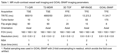
Plaque compositional features are effective indicators for vulnerability and associated with ischemic stroke risk. Multicontrast vessel wall imaging has been utilized to characterize plaque compositions but this technique is time consuming and dependent on reviewer’s experience. Therefore, it is important to characterize plaque compositions with time-efficient imaging approach. This study sought to determine T1 values of carotid plaque components determined by quantitative imaging validated by histology. We found that the T1 values of intraplaque hemorrhage, necrotic core, and loose matrix were distinguishable. Our findings suggest that carotid plaque components might be distinguishable and automatically segmented on quantitative MR imaging.
|
|
0464.
 |
15 |
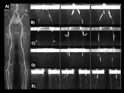
Dynamic Evaluation of Flow in the Lower Extremity Peripheral Arteries using Cine Fast Interrupted Steady-State in Combination with Arterial Spin Labeling.
Emily Aherne, Ioannis Koktzoglou, Benjamin Lind, Robert Edelman
For non-invasive evaluation of peripheral artery disease in the lower extremities prior to revascularization, physicians rely on contrast-enhanced CT angiography and magnetic resonance angiography which generate static images in the arterial phase and do not reveal blood flow. We adapted a prototype non-contrast MRA technique, cine fast interrupted steady-state in combination with arterial spin labeling (cine FISS ASL), to facilitate dynamic visual and quantitative flow evaluation of the lower extremity peripheral arteries. In-plane flow patterns were well visualized and there was very strong positive correlation between peak flow velocities measured by cine FISS ASL and 2D phase contrast MRA.
|
|
