|
2nd Hour
Poster: Myocardial Tissue Characterization
Power Pitch Poster
Cardiovascular
Wednesday, 15 May 2019
Power Pitch Theater A - Exhibition Hall
14:30 - 15:30
| |
|
Plasma # |
 |
0776.
 |
1 |
Quantitative Magnetization Transfer Imaging of Human Myocardial Tissue and Scar

Karina Lopez, Radhouene Neji, Imran Rashid, Shaihan Malik, Rui Pedro Teixeira, Reza Razavi, Claudia Prieto, Sebastien Roujol, Rene Botnar
We have developed a contrast-free single breath-hold 2D MT mapping technique using a two-pool exchange model and dictionary matching to parametrize macromolecular changes associated with myocardial fibrosis. The model was validated in vials with different albumin concentrations and bovine leg. Pool sizes ratio and bound pool T2 maps were obtained from 7 healthy subjects and two patients with ischemic or non-ischemic scar/fibrosis. . Reduced PSR and T2B values were found to correlate with LGE areas in both patients. In vivo quantitative MT mapping of the heart was performed for the first time, showing promising results for the detection of myocardial fibrosis.
|
|
0777.
 |
2 |
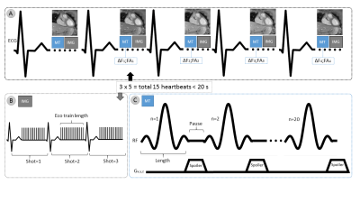
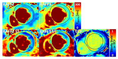
Myocardial Infarct Characterization using Relaxation along Fictitious Field in the nth Rotating Frame
Amir Mirmojarabian, Esa Liukkonen, Victor Casula, Mikko Nissi, Lauri Ahvenjärvi, Juhani Junttila, Timo Liimatainen
Based on the previous promising relaxation along fictitious field in nth frame (RAFFn) findings in myocardial infarct mouse model at 9.4 T, we adjusted RAFFn measurement to standard clinical 1.5 T scanner with γB1 = 500 Hz. We demonstrate with five myocardial infarct patients that fitted steady state from RAFF2 and RAFF3 weighted images can separate the infarct from remote myocardium using clinical main and RF fields. We also demonstrate association between RAFF3 steady state and extra cellular volume that is calculated based on pre and post contrast T1 maps and hematocrit.
|
|
0778.
 |
3 |
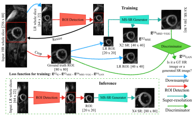
A ROI Focused Multi-Scale Super-Resolution Method for the Diffusion Tensor Cardiac Magnetic Resonance
Jin Zhu, Guang Yang, Pedro Ferreira, Andrew Scott, Sonia Nielles-Vallespin , Jennifer Keegan , Dudley Pennell, Pietro Lio, David Firmin
Diffusion Tensor Cardiovascular Magnetic Resonance (DT-CMR) is a promising contrast-free and non-invasive technique to characterize the tissue integrity and microstructure of the myocardium. However, the complex acquisition protocol results in prolonged scan times and often results in images with low spatial resolution and relatively poor signal-to-noise ratio. Here a novel ROI focused multi-scale super-resolution approach is proposed to improve the apparent spatial resolution of in vivo DT-CMR. Based on simulation studies, our proposed method can achieve increases in apparent spatial resolution by a factor of 4 with preserved image quality and no obvious degradation in the derived DT-CMR parameters.
|
|
0779.
 |
4 |
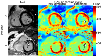
Efficient high-resolution cardiac motion-corrected T1 mapping
Kirsten Becker, Edyta Blaszczyk, Stephanie Funk, Jeanette Schulz-Menger, Tobias Schaeffter, Christoph Kolbitsch
Cardiac T1 mapping provides valuable information on fibrosis in various cardiomyopathies. Commonly, data acquisition is restricted to a small percentage (mid-diastole) of the cardiac cycle to prevent motion artefacts. This leads to low scan efficiency and limits the achievable resolution. We present an 8s T1 mapping approach, which employs a large acquisition window (80% of the cardiac cycle) and corrects for cardiac motion using the same data. The approach was evaluated in native T1 mapping in healthy volunteers and post-contrast T1 mapping in patients. It significantly improved the precision of the obtained T1 maps while successfully minimizing cardiac motion artefacts.
|
|
0780.
 |
5 |
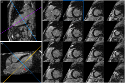
Diffusion tensor cardiovascular magnetic resonance in myocardial infarction: A comparison between high resolution spiral and standard resolution EPI STEAM
Margarita Gorodezky, Pedro Ferreira, Zohya Khalique, Sonia Nielles-Vallespin, Ranil de Silva, Dudley Pennell, Andrew Scott, David Firmin
Diffusion tensor cardiovascular magnetic resonance can provide insight into the function and microstructure of the scar and adjacent region in myocardial infarction (MI). Imaging the thinned infarcted myocardial wall requires a high-resolution acquisition while frequent arrhythmia, shortness of breath and fatigue make the cohort especially challenging for imaging. A high-resolution STEAM acquisition with an interleaved variable density spiral readout and an off-resonance and T2* correction was previously demonstrated in a healthy cohort. Here, this sequence was successfully applied in 7 MI patients at a spatial resolution of 1.8x1.8x8mm3 and compared to a standard resolution EPI sequence.
|
|
0781.
 |
6 |
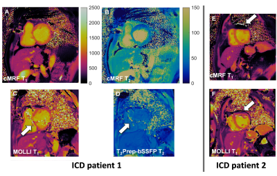
Cardiac Magnetic Resonance Fingerprinting for the Investigation of Suspected Inflammatory Cardiomyopathy
Gabriella Vincenti, Andrew Coristine, Jesse Hamilton, Céline Provins, Juerg Schwitter, Nicole Seiberlich, Ruud van Heeswijk
Inflammatory cardiomyopathy (ICMP) needs to be diagnosed early, and cardiac T1 and T2 mapping both have been shown to increase the accuracy of ICMP diagnosis. Cardiac magnetic resonance fingerprinting (cMRF) can be used to robustly acquire both maps in a single breath-hold, so the goal of this preliminary study was to compare the performance of cMRF and routine parameter mapping in patients with suspected ICMP, including those with implantable cardioverter-defibrillators (ICDs), which often cause significant artifacts. The relaxation times in 24 patients were similar in cMRF and routine mapping, while cMRF may have superior performance in patients with an ICD.
|
|
0782.
 |
7 |
Motion-Compensated 3D Whole-heart Water-Fat Late Gadolinium Enhancement Imaging for Assessment of Myocardial Viability
Camila Munoz, Radhouene Neji, Karl Kunze, Teresa Correia, Christoph Forman, Michaela Schmidt, Imran Rashid, René Botnar, Claudia Prieto
Water/fat 2D LGE imaging has shown promising results for assessment of fibro-fatty infiltration in the myocardium and characterisation of cardiac masses. However, spatial resolution and volumetric coverage within a clinically feasible scan time remain a challenge. Here we propose a highly efficient respiratory motion-compensated 3D whole-heart water/fat inversion recovery (IR)-prepared LGE imaging sequence. Preliminary results from patients with cardiovascular disease demonstrate the feasibility of the approach, with scan duration of ~8 min. Motion-compensated 3D LGE water images offer a good depiction of myocardial scar, while motion-compensated fat images offer complementary diagnostic information, with clear delineation of epicardial and pericardial fat.
|
|
0783.
 |
8 |
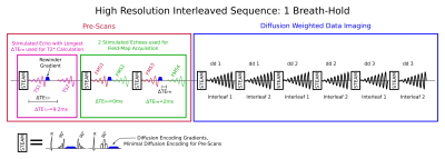
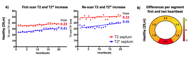
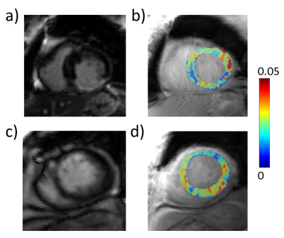
Heartbeat-to-Heartbeat Quantitative Myocardial Oxygenation Imaging within a Single Breath-Hold using a Combined Gradient Echo-Spin Echo EPI (GESE-EPI) Sequence in Patients with Hypertension
Maaike van den Boomen, Gert Jan Snel, Christopher Nguyen, Mary Kate Manhard, David Sosnovik, Rudi Dierckx, Ciprian Catana, David Izquerdo-Garcia, Bruce Rosen, Niek Prakken, Ronald Borra, Kawin Setsompop
Dynamic Cardiac BOLD imaging techniques can suffer from signal variabilities due to changes in heartrate during a breath-hold. We demonstrate that a 2-echo-GESE-EPI sequence can provide T2- and T2*-weighted images simultaneously per heartbeat, but the dynamic changes in these images represent physiological effects mixed with heartrate changes. We then proposed a 5-echo-GESE-EPI for dynamic T2- and T2*-mapping per heartbeat as a readout for myocardial oxygenation. These dynamically acquired T2- and T2*-values were demonstrated to increase over the time of a breath-hold for healthy volunteers, while shown to remain constant and even reverse in hypertension patients.
|
|
0784.
 |
9 |
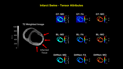
An Analysis of DiffNet Reconstruction Performance in Healthy and Infarcted Cardiac Diffusion Tensor Images
Tyler Cork, Eric Aliotta, Michael Loecher, Luigi Perotti, Daniel Ennis
Cardiac diffusion tensor imaging (cDTI) suffers from low signal-to-noise ratios, which results in tensor variability. In order to decrease tensor variability, the number of diffusion directions or number of averages must increase, consequently increasing the scan time. Recent implementations of artificial neural network (ANN) have proven that a non-linear mapping between diffusion signals and tensors is possible and can decrease tensor variability without increasing scan time. We implement an ANN tensor reconstruction for ex vivo porcine hearts to evaluate if a robust ANN diffusion tensor reconstruction is a beneficial technique to decrease tensor variability at no cost in scan time.
|
|
0785.
 |
10 |
Fully Self-Gated Cardiac and Respiratory Motion-Resolved Isotropic 5D T1 Mapping of the Heart: Preliminary Results
Lorenzo Di Sopra, Christopher Roy, Jessica Bastiaansen, Jérôme Yerly, Davide Piccini, Lionel Arn, Matthias Stuber, Ruud van Heeswijk
Current solutions for T1 mapping rely on 2D images of the heart, and require time-inefficient cardiac gating as well as long breath-holds. To address these drawbacks, a Free-running framework for fully self-gated cardiac and respiratory motion-resolved 5D imaging of the heart was extended to include T1 recovery contrast as a 6th dimension. The framework was tested at 3T in an ISMRM-NIST phantom and demonstrated good agreement between the estimated T1 values and reference standard. Preliminary 3D T1 maps in 3 healthy volunteers showed good resolution of the physiological motion and accurate T1 values of the myocardium and blood.
|
|
0786.
 |
11 |
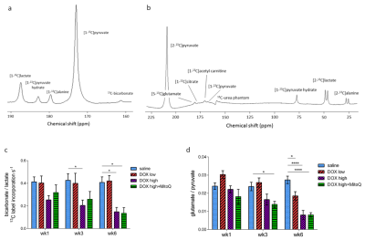
Mitochondrial dysfunction in a rat model of doxorubicin-induced heart failure assessed by hyperpolarized 13C MRS
Kerstin Timm, Charith Perera, Vicky Ball, John Henry, Matthew Kerr, Michael Dodd, Jack Miller, James West, Angela Logan, Julian Griffin, Michael Murphy, Lisa Heather, Damian Tyler
Doxorubicin-chemotherapy can lead to serious cardiac side effects in cancer-patients, culminating in heart failure. Cardiac oxidative stress and impaired energetics are hypothesized to be at the core of this toxicity. We established a clinically relevant rat model of doxorubicin-induced heart failure characterized with CINE MRI by decreased cardiac function. We show here that these functional changes are preceded by a shift from oxidative to anaerobic glucose metabolism measured with hyperpolarized MRS. These changes are likely due to a loss and impairment of mitochondria, which cannot be alleviated with the mitochondrially targeted antioxidant, MitoQ.
|
|
0787.
 |
12 |
Assessment of postinfarct myocardium metabolic degradation with amide proton transfer magnetic resonance imaging at 3 Tesla
Yin Wu, Kaiyue Diao, Jie Liu, Chunchao Xia, Min Ma, Yong He, Hairong Zheng, Yingkun Guo
Myocardium ischemia irritates a series of metabolic degradations, including intracellular acidosis and cell damage. Amide proton transfer (APT) effect is associated with microenvironment acidosis and amide protons inside of cytoplasm. In this study, APT imaging was performed on MI patients at 3 Tesla. Results show significant APT effect reduction from the remote (4.29±2.00%), adjacent (2.74±1.18%) to the infarct (1.43±0.80%) regions (P<0.05), implying progressively metabolic alterations from surrounding tissue to the infarct core. The method provides a novel way for noninvasive assessment of myocardium metabolic degradation postinfarction, promising to facilitate therapeutic decision and treatment evaluation.
|
|
0788.
 |
13 |
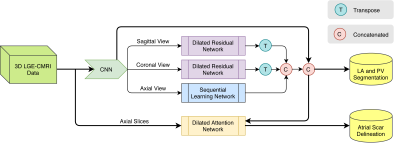
A Deep Learning Based Left Atrium Anatomy Segmentation and Scar Delineation in 3D Late Gadolinium Enhanced CMR Images
Guang Yang, Jun Chen, Zhifan Gao, Shuo Li, Hao Ni, Elsa Angelini, Tom Wong, Raad Mohiaddin, Eva Nyktari, Ricardo Wage, Lei Xu, Yanping Zhang, Xiuquan Du, Heye Zhang, David Firmin, Jennifer Keegan
3D late gadolinium enhanced (LGE) CMR images of left atrial (LA) scar tissue can be used to stratify patients with atrial fibrillation and to guide subsequent ablation therapy. This requires a segmentation of the LA anatomy (usually from an anatomical acquisition) and a further segmentation of the scar tissue within the LA (from a 3D LGE acquisition). We propose a deep learning based framework incorporating multiview information and attention mechanism to solve both LA anatomy and scar segmentations simultaneously from a single 3D LGE acquisition. Compared to existing methods, we show improved segmentation accuracy (mean Dice=93%/87% for LA/scar).
|
|
0789.
 |
14 |
Simultaneous myocardial T1 and T2 mapping in 11 heartbeats using a radial sequence with inversion recovery and T2 preparation
Jiaxin Shao, Ziwu Zhou, Kim-Lien Nguyen, Peng Hu
Myocardial T1 and T2 mapping are promising cardiovascular magnetic resonance (CMR) techniques for quantitative tissue characterization and provide complementary information. Simultaneous T1 and T2 mapping strategies are attractive because of shorter scan time and the potential for inherently co-registered T1/T2 maps. By estimating T1 and T2 at the same time, the T2/T1 bias present in conventional T1 and T2 mapping sequences can be minimized and more accurate T1 and T2 estimation accomplished. In this work, we sought to develop a simultaneous T1 and T2 mapping technique that can achieve high accuracy and precision, without heart-rate dependence, and with high reproducibility comparable to the standard MOLLI and conventional T2 mapping techniques.
|
|
0790.
 |
15 |
Diffusion Tensor CMR a potential early marker of remodelling after myocardial infarction
Sonia Nielles-Vallespin, Pedro Ferreira, Andrew Scott, Dudley Pennell, David Firmin, Andrew Arai, Ranil de Silva
We performed a longitudinal large animal pre-clinical study to assess post-MI temporal alterations of myocardial microstructure, using Diffusion Tensor Cardiac Magnetic Resonance (DT-CMR). In vivo CMR was performed before, at 3 days, and 4 months after the reperfused MI procedure. Remodeled hearts demonstrated reduced right-handed helix angles in the endocardium of the infarct region, as well as a decreased gradient in the healix angle line profiles with associated thinning of the myocardium. Reduced sheetlet angle (E2A) mobility in the acute stage was associated with the development of adverse LV remodeling.
|
|
