|
2nd Hour
Poster: Emerging Body Imaging Technologies & Their Translation
Power Pitch Poster
Body: Breast, Chest, Abdomen, Pelvis
Wednesday, 15 May 2019
Power Pitch Theater C - Exhibition Hall
09:15 - 10:15
| |
|
Plasma # |
|
0696.
 |
31 |
First human imaging studies at 10.5 Tesla: body studies at 450 MHz

Gregory Metzger, Xiaoxuan He, Andrea Grant, Arcan Erturk, Russell Lagore, Gregor Adrinay, Lance DelaBarre, Yigitcan Eryaman, Xiaoping Wu, Edward Auerbach, Pierre-Francois Van de Moortele, Kamil Ugurbil
This work presents the first in vivo human images from a whole body 10.5T MRI system. An initial coil was validated for safe operation at 10.5T allowing anatomic targets in the pelvis and abdomen to be explored. The translation of RF management strategies developed at 7T were employed to tackle the challenges at 10.5T demonstrating that high quality anatomic and quantitative data can be achieved at 450 MHz in the human torso.
|
|
0697.
 |
32 |
Precision medicine advanced by full body imaging biomarkers, whole genomic sequencing and blood-based measures
Natalie Schenker-Ahmed, Y Hou, Christine Swisher, Hung-Chun Yu, Robyn Heister, Lori Napier, Saints Dominguez, Nathaniel Hernandez, Michael Doney, David Karow
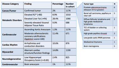
We present the findings from 1190 presumed healthy patients evaluated at a precision medicine clinic. We performed deep quantitative multimodal phenotyping and genotyping comprising quantitative whole-body imaging, whole genome sequencing, and advanced blood-based biomarkers. Within this cohort, medically significant findings included aneurysms, newly identified tumors, coronary artery disease, metabolic disease, cardiac arrhythmia, myocardial disease, and neurodegenerative risk. Forty percent of “healthy” patients had a new clinically significant finding that was not previously known. For this 40%, we are able to provide quantitative continuous metrics that enable individuals to make informed decisions to mitigate their health risks.
|
|
0698.
 |
33 |
Free-breathing Volumetric Body Imaging for Combined Qualitative and Quantitative Tissue Assessment using MR Multitasking
Zixin Deng, Anthony Christodoulou, Nan Wang, Wensha Yang, Lixia Wang, Yi Lao, Fei Han, Xiaoming Bi, Bin Sun, Stephen Pandol, Richard Tuli, Debiao Li, Zhaoyang Fan
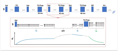
MR has broad clinical applications in body imaging with excellent soft-tissue contrast and qualitative and emerging quantitative assessments, however, respiratory motion has been a critical constraint on the quality of body MR. The aim of this study is to demonstrate the feasibility of a free-breathing volumetric quantitative body imaging technique based on a recently developed MR Multitasking technique. Respiratory phase-resolved T1-, T2-weighted, and T1-, T2-maps can be acquired simultaneously within a single 10-minute scan. Good image quality and comparable T1, T2 measurements to reference scans or published literature were observed in phantom and human studies.
|
|
0699.
 |
34 |
Early Assessment of the Response of Esophageal Squamous Cell Carcinoma to Chemoradiotherapy by Intravoxel Incoherent Motion MRI
Zhengyang Zhou, Jian He, Weibo Chen
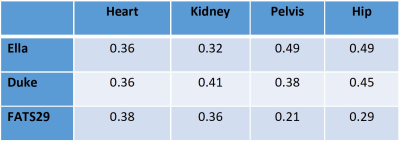
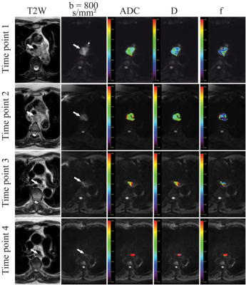
Twenty-three patients with esophageal squamous cell carcinoma (ESCC) underwent intravoxel incoherent motion (IVIM) MRI at four timepoints: pre, mid, end, and post-CRT to assess the value of IVIM parameters in the early assessment of treatment response to CRT. The parameters and their change percentages were compared between complete response (CR) and partial response (PR). ADC, f, %ADC, and %D at mid-CRT in CR group were significantly higher than those in the PR group. D combined with f and ADC had highest area under curve in identifying CR from PR. IVIM parameters proved useful in assessing response to definitive concurrent CRT.
|
 |
0700.
 |
35 |
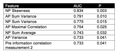
MRI radiomics features for characterization of RCC subtypes
Daniela Said, Stefanie Hectors, Eric Wilck, Ally Rosen, Alp Tuna Beksaç, Ketan Badani, Bachir Taouli
The goal of this study was to evaluate the use of quantitative radiomics features from contrast-enhanced MRI in differentiating common subtypes of solid renal cell carcinomas (RCCs). We found that several radiomics features were associated with common subtypes of RCC. Thus, radiomics features may help in the diagnosis of histologic subtypes of RCC.
|
|
0701.
 |
36 |
Functional Renal Imaging using Urea CEST-MRI
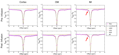
Soo Hyun Shin, Michael Wendland, Moriel Vandsburger
Current diagnosis of renal disease and injury relies on blood urea nitrogen and serum creatinine measurements, which provide generic information about kidney function but do not provide spatial specificity during kidney failure. Urea has previously been used as a vehicle for hyperpolarized MR imaging of renal function, and as a contrast agent of CEST MRI. Here, we examine whether injection of urea can provide CEST contrast for quantitation of renal integrity via probing the spatially varying urea concentrating capacity of a kidney.
|
|
0702.
 |
37 |
Free-breathing renal perfusion measurement with volumetric ASL using variable-density FSE and 4D Compressed-Sensing
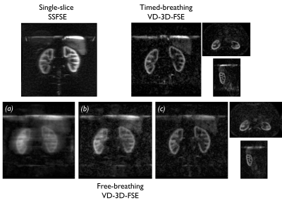
Manuel Taso, Arnaud Guidon, Daniel Litwiller, David Alsop
While single or 2D multi-slice body ASL implementations have been shown to be compatible with free-breathing for abdominal perfusion measurement even in uncooperative clinical populations, free-breathing volumetric encoding has not been reported yet. We propose a free-breathing volumetric ASL acquisition relying on a motion-robust variable-density 3D-FSE sequence with redundant k-space center and pseudo-random variable outer k-space sampling and 4D-Parallel-Imaging-Compressed-Sensing reconstruction. High-quality whole kidneys perfusion images were obtained in less than 5 minutes in free-breathing, potentially extending the clinical applications of non-contrast ASL perfusion in the abdomen.
|
|
0703.
 |
38 |
Quantitative Transport Mapping (QTM) of the Kidney using a Microvascular Network Approximation
Liangdong Zhou, Qihao Zhang, Pascal Spincemaille, Thanh D. Nguyen, Yi Wang
The blood flow quantification is of great importance in the diagnosis of many diseases. The current blood flow quantification methods are mainly based on the Kety’s method, which highly replies on the practically unmeasurable arterial input function (AIF). To void using the AIF as an input, we propose quantitative transport mapping (QTM) that quantitatively models blood flow in tissue according to the underlying biophysics of the transport equation. The inverse problem of QTM is to reconstruct the velocity field of the blood transportation using the time-resolved 4D tracer concentration data. As an example, with the approximation of kidney microvascular network, the renal blood flow (RBF) can be quantified by QTM.
|
|
0704.
 |
39 |
Fatty kidney assessed by renal spectroscopy: a reproducibility study and clinical randomised controlled trial in type 2 diabetes
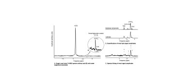
Ilona Dekkers, Maurice Bizino, Paul de Heer, Aiko de Vries, Hildo Lamb
Renal steatosis is a potential biomarker for obesity-related renal disease and this has been suggested as underlying biological pathway of renoprotective effects of Liraglutide in type 2 diabetes (T2DM). Proton magnetic resonance spectroscopy (¹H-MRS) has the ability to non-invasively quantify triglycerides. We examined the reproducibility of ¹H-MRS in healthy volunteers, and explored the application in a clinical trial evaluating the differences in renal triglyceride content after 9-month treatment with liraglutide. We demonstrated that ¹H-MRS is a reproducible technique and comparison with healthy volunteers suggests increased renal triglycerides in T2DM. Renoprotective effects of liraglutide might be based on reduced renal triglycerides.
|
|
0705.
 |
40 |
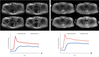
Time-efficient fully 3D non-Cartesian dynamic contrast-enhanced free-breathing MRI of the pelvis with an ultrashort TE cones trajectory
Signy Holmes, Frank Ong, Michael Lustig, Ryan Brunsing, Vipul Sheth, Marcus Alley, Michael Carl, John Pauly, Joseph Cheng, Shreyas Vasanawala
A 3D cones k-space trajectory with golden angle ordering was developed along with a multiscale low rank reconstruction for dynamic contrast enhancement with ultrashort echo time (DCE-UTE) and with fast image reconstruction time. With IRB approval and informed consent the sequence was tested in 21 consecutive subjects referred for 3T contrast-enhanced pelvic MRI. For comparison, a Cartesian DCE SPGR sequence with golden angle radial k-space ordering, soft-gated self navigation, and low rank reconstruction (DCE-SPGR) served as case controls. DCE-UTE performed significantly better than DCE-SPGR in delineation of anatomic structures (p=0.004) and overall perceived image quality (p=0.003). These results indicate a 3D fully noncartesian DCE-UTE sequence and reconstruction improves image quality in pelvic DCE.
|
|
0706.
 |
41 |
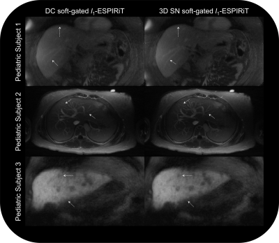
Motion-robust pediatric abdominal angiography using dynamic 3D image self-navigator with 3D cones
Srivathsan Koundinyan, Frank Ong, Dwight Nishimura, Shreyas Vasanawala, Joseph Cheng
Respiratory motion detection using the k-space center (DC signal) can be problematic in pediatric subjects exhibiting highly irregular breathing patterns. To address this problem, we extract respiratory motion information from high frame rate (3.3 Hz), low-resolution (7.7 mm) 3D self-navigators derived from the imaging data (i.e., central portion of 3D cones k-space data) and reconstructed with a multi-scale low-rank framework. Localized motion estimates are obtained from the 3D self-navigators using optical flow registration. We demonstrate that region-specific motion information from 3D self-navigators better mitigates motion artifacts compared to the low-pass filtered DC signal in pediatric abdominal angiography exams.
|
|
0707.
 |
42 |
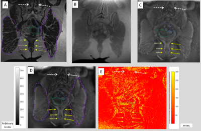
Imaging wound-healing related fibrosis using short inversion time ultra-short TE (STIR-UTE)
Ehud Schmidt, Iga Muradyan, Thomas Benkert, Himanshu Bhat, Aravindan Kolandaivelu, Henry Halperin, Akila Vaswanathan
Collagen deposition occurs during wound-healing processes in several diseases, and following therapy (acute myocardium infarction, radiation induced fibrosis). There is interest in intervening during wound-healing, since chronic scar leads to complications (ventricular tachycardia, gastrointestinal bleeding), with novel medications possibly reducing fibrosis. Intervention requires early detection of diffuse fibrosis (<50% content/voxel) during acute wound-healing. Fibrosis detection with gadolinium-perfusion (LGE, T1 mapping) is problematic during acute disease, due to irregular vascularity. Ultrashort-Time-to-Echo’s (UTE’s) collagen-sensitivity is reduced by fat’s masking signal. With STIR-UTE, we utilize collagen’s short TE and short T1 to suppress fat and map fibrosis in the pelvis, in normal and in post-infarction hearts.
|
|
0708.
 |
43 |
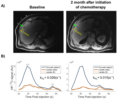
Hyperpolarized 13C MRI of Patients with Metastatic Prostate Cancer to Bone and Liver
Hsin-Yu Chen, Philip Lee, Zi Zhu, Robert Bok, Michael Ohliger, Jeremy Gordon, Mark van Criekinge, Lucas Carvajal, James Slater, Peder Larson, Pamela Munster, Rahul Aggarwal, John Kurhanewicz, Daniel Vigneron
In this feasibility study, hyperpolarized 13C-pyruvate MR exams were conducted on 5 patients who had metastatic prostate cancer to bone or liver. In one man with liver metastasis, serial scans showed a decrease of pyruvate-to-lactate conversion kPL (0.026 to 0.015 s-1) at 2 months after initiation of chemotherapy that was consistent with response based on PSA and RECIST criteria. High kPL was found in patients with bone lesions comparable to that in high-grade primary prostate cancer. Overall, HP-13C MR imaging showed great promise as a biomarker to evaluating treatment responses in metastatic prostate cancer.
|
|
0709
 |
44 |
Utility of simultaneous T1 and T2 mapping with MR Fingerprinting (MRF) for bowel wall imaging in Crohn’s Disease
Video Permission Withheld
Verena Obmann, Nicole Seyfried, Wei-Ching Lo, Ananya Panda, Yun Jiang, Katherine Wright, Preetika Sinh, Jeffrey Katz, Maneesh Dave, Pingfu Fu, Kathleen Ropella-Panagis, Vikas Gulani
The feasibility of using MRF to assess T1 and T2 relaxation times in the intestine for the evaluation of inflammatory bowel disease was explored. 52 patients with Crohn’s Disease underwent MR enterography with MRF acquisitions through the bowel. T1 relaxation times allowed quantitative differentiation between unaffected segments and inflamed segments, p = 0.004. T2 relaxation times further allowed distinction between segments with active and chronic fibrotic changes, p = 0.003.
|
 |
0710.
 |
45 |
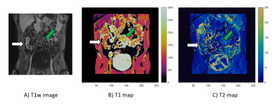
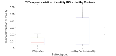
MRI assessed dysmotility and texture analysis in the terminal ileum and small bowel: A pilot study comparison between Irritable Bowel Syndrome (IBS) patients with bloating and healthy controls
Ruaridh Gollifer, Alex Menys, Natalia Zarate-Lopez, Dave Chatoor, Anton Emmanuel, Stuart Taylor, David Atkinson
Gastrointestinal symptoms in irritable bowel syndrome (IBS) occur without any obvious structural gut abnormality. This pilot study is based on abnormal ileo-caecal motility function suggested by wireless capsule data, with potential reflux of caecal contents back into the terminal ileum (TI). We compared constipation-predominant IBS (IBS-C) patients with bloating to healthy controls, through texture analysis and motility measures. We found TI temporal variation of motility was significantly higher in healthy controls. There was also a difference in the TI to small bowel texture analysis contrast ratio, but this was only significant in BTFE sequences.
|
|
