|
2nd Hour
Poster: Liver: Brief but Impactful
Power Pitch Poster
Body: Breast, Chest, Abdomen, Pelvis
Thursday, 16 May 2019
Power Pitch Theater C - Exhibition Hall
09:15 - 10:15
| |
|
Plasma # |
|
1020.
 |
16 |
Repeatability and Reproducibility of Confounder-Corrected R2* as a Biomarker of Liver Iron Concentration: Interim Results from a Multi-Center, Multi-Vendor Study at 1.5T and 3T

Diego Hernando, Ruiyang Zhao, Valentina Taviani, Mounes Aliyari Ghasabeh, Li Pan, Qing Yuan, Stefan Ruschke, Dimitrios Karampinos, Xiaodong Zhong, Ryan Mattison, Ihab Kamel, Ivan Pedrosa, Shreyas Vasanawala, Takeshi Yokoo, Scott Reeder
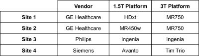
R2* mapping has the potential to provide rapid and reliable quantification of liver iron concentration. Importantly, previous studies have demonstrated that by correcting for relevant confounding factors (eg: noise floor effects and fat) R2* mapping is highly insensitive to the presence of these confounders. However, the repeatability and reproducibility of confounder-corrected R2* across multiple sites and vendors remains unknown. This abstract reports interim results from a multi-center, prospective, NIH-sponsored liver iron quantification study. Our results suggest excellent repeatability and reproducibility of confounder-corrected R2* for liver iron quantification in patients, across four centers, three vendors and at both 1.5T and 3T.
|
 |
1021.
 |
17 |
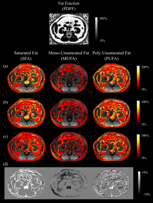
Triglyceride Saturation Estimation using Phase- and Amplitude-Modulated Bipolar MRI
Manuel Schneider, Felix Lugauer, Dominik Nickel, Berthold Kiefer, Sungheon Gene Kim, Linda Moy, Andreas Maier, Mustafa R Bashir
Our purpose was to develop a robust method to estimate triglyceride saturation from bipolar multi-gradient-echo MRI data. Discrepancies in amplitude and phase between even and odd echoes were addressed analytically. The method was demonstrated in abdominal (n=5 patients) and breast (n=1 volunteer) MRI data. Compared to calculating fatty acid composition parameters without modeling amplitude modulations, the proposed method reduced the intensity shift in the readout direction in the estimated parameter maps, but also led to artifacts at water/fat borders.
|
|
1022.
 |
18 |
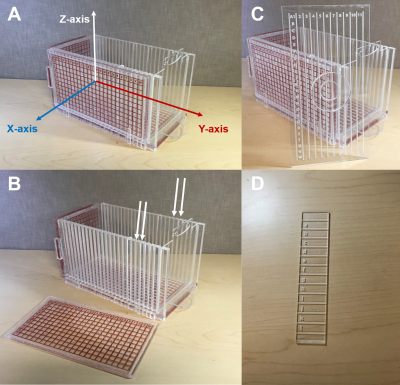
Direct Radiologic-Pathologic Correlation of Liver Lesions with an MR-Compatible Sectioning and Localization Device
Victoria Rendell, Timothy Colgan, Gesine Knobloch, Matthias Muhler, Agnes Loeffler, Rashmi Agni, Krisztian Kovacs, Emily Winslow, Scott Reeder
The purpose of this study was to demonstrate the feasibility of a novel method for precise radiologic-pathologic correlation of liver metastases, in patients undergoing curative resection. Intraoperative gadoxetic acid was administered to patients at the time of partial hepatectomy prior to vascular inflow ligation. Ex vivo MRI was then performed using an MR-compatible sectioning device to identify lesion coordinates to inform pathologic processing. Results from resection specimens in 6 patients with a total of 25 liver lesions are presented. The study’s novel radiologic-pathologic correlation method combines intraoperative gadoxetic acid administration and ex vivo MR imaging to perform accurate lesion localization.
|
|
1023.
 |
19 |
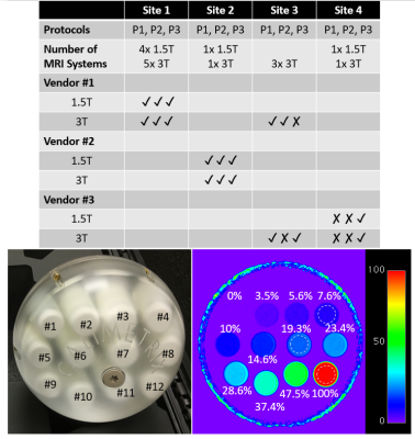
Multi-Site, Multi-Vendor, and Multi-Platform Reproducibility and Accuracy of Quantitative Proton-Density Fat Fraction (PDFF) at 1.5 and 3 Tesla with a Standardized Spherical Phantom: Preliminary Results from a Study by the RSNA QIBA PDFF Committee
Houchun Hu, Takeshi Yokoo, Diego Hernando, Mustafa Bashir, Michael Middleton, Suraj Serai, Daria Malyarenko, Thomas Chenevert, Mark Smith, Walter Henderson, Gavin Hamilton, Yunhong Shu, Claude Sirlin, Jean Tkach, Andrew Trout, Jean Brittain, Scott Reeder, the RSNA QIBA PDFF Committee
Proton Density Fat Fraction (PDFF) is a widely-accepted quantitative MRI biomarker of hepatic steatosis. Formed in 2015, the PDFF Committee of the RSNA QIBA initiative promotes standardized usage of PDFF for clinical care and research. In this work, the committee describes preliminary results from a multi-center, multi-vendor, multi-platform, and multi-protocol study to characterize the accuracy and reproducibility of PDFF as measured by commercially-available 2D and 3D spoiled-gradient-recalled-echo sequences in a standardized phantom with known PDFF reference targets ranging from 0-100%. Results show that confounder-corrected PDFF reconstructed from multi-fat-spectral peak modeling is an accurate and precise quantitative measurement of PDFF, with minimal bias and strong linearity.
|
 |
1024.
 |
20 |
Feasibility of Rapid Automated Liver Quantitative Susceptibility Mapping in a Patient Population
Ramin Jafari, Sujit Sheth, Pascal Spincemaille, Thanh Nguyen, Martin Prince, Yan Wen, Yihao Guo, Kofi Deh, Zhe Liu, Daniel Margolis, Gary Brittenham, Andrea Kierans, Yi Wang
Accurate measurement of the liver iron concentration (LIC) is needed to guide iron-chelating therapy for patients with transfusional iron overload. In this work, we investigate the feasibility of automated quantitative susceptibility mapping (QSM) to measure the LIC in clinical practice. We propose to incorporate In-phase echo acquisition for rapid, robust initialization of the field in water-fat separation problem (T2*-IDEAL).
|
|
1025.
 |
21 |
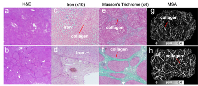
Imaging Diamagnetic Susceptibility of Collagen in Hepatic Fibrosis using Susceptibility Tensor Imaging
Hongjiang Wei, Chunlei Liu
We propose to quantify susceptibility anisotropy of collagen in the liver using susceptibility tensor imaging (STI). Magnetic susceptibility anisotropy measured by STI differentiates the anisotropic collagen fibers which is associated with the severity of liver fibrosis from iron deposition. The results are compared with histology to inform the interpretation of collagen content in the liver fibrosis tissues. Our results suggest that magnetic susceptibility anisotropy is a quantitative marker for collagen content in cirrhotic liver disease.
|
|
1026.
 |
22 |
Liver glycogen concentration and body hydration status are predictors of liver shMOLLI T1 measurements
Ferenc Mozes, Ladislav Valkovic, Michael Pavlides, Matthew Robson, Elizabeth Tunnicliffe
Liver T1 measurements can be used to characterise liver disease but are also sensitive to physiologically normal liver changes. In this work, a set of metabolic interventions were performed to assess the effect of liver glycogen concentration and body hydration status on liver shMOLLI T1 measurements in healthy volunteers. Glycogen showed an in vivo relaxivity in keeping with literature phantom data and hydration status showed a smaller effect. Our results will impact the way T1 measurements of participants with impaired glycogen storage are interpreted, as well as instructions given to participants before their scans.
|
|
1027.
 |
23 |
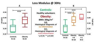
Detection of Early Hepatic Inflammation in Obese Patients with MR Elastography (MRE)
Jiahui Li, Alina Allen, Sudhakar K. Venkatesh, Taofic Mounajjed, Jun Chen, Kevin J. Glaser, Armando Manduca, Vijay Shah, Richard L. Ehman, Meng Yin
We performed multifrequency MRE in 10 controls and 88 patients underwent bariatric surgeries. A total of 38 patients were reassessed one year later with MRE and biopsy. At the initial exam, MRE-assessed loss modulus (imaginary component of the complex shear modulus) at 30Hz was significantly higher in obese patients compared with controls, even those without elevated liver fat. The elevation in loss modulus became normalized after treatment. In summary, MRE-assessed loss modulus shows promise as a sensitive indicator of early hepatic inflammation or cell injury in obese patients, pointing to a potential role in selection of patients for bariatric surgery.
|
|
1028.
 |
24 |
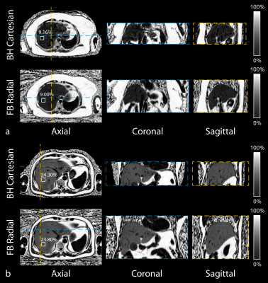
Free-Breathing Liver Fat Quantification in Adults with NAFLD using a 3D Stack-Of-Radial MRI Technique
Tess Armstrong, Xiaodong Zhong, Holden Wu
Non-alcoholic fatty liver disease (NAFLD) is the most common chronic liver disease worldwide and can lead to liver failure. Conventional Cartesian MRI techniques can quantify liver fat. However, Cartesian MRI requires breath-holding to avoid respiratory motion artifacts in the liver, which may be challenging for many patients. Therefore, we evaluated the accuracy and repeatability of a recently developed free-breathing 3D stack-of-radial liver fat quantification technique in adults with NAFLD at 3 T. The new free-breathing technique demonstrated good repeatability and accuracy compared to conventional breath-holding Cartesian MRI and breath-holding MR spectroscopy.
|
|
1029.
 |
25 |
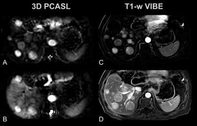
3D Arterial Spin Labeling Imaging of Arterial and Portal-Venous Perfusion in Human Liver at 3 Tesla under Free Breathing: Preliminary Results
Petros Martirosian, Bernd Kühn, Jakob Weiss, Ulrich Grosse, Rüdiger Hoffmann, Martin Schwartz, Berthold Kiefer, Konstantin Nikolaou, Fritz Schick
Measurement of hepatic arterial perfusion is important for assessment of chronic liver diseases and characterization of liver lesions. The aim of this study was to investigate the capability of a 3D-PCASL sequence for measurement of arterial, portal-venous and global perfusion of the liver under free respiration. It is demonstrated that the presented method provides high quality ASL perfusion images of the liver under free breathing conditions. Results in a patient with hepatocellular carcinoma clearly indicate high and quantitatively measurable arterial perfusion of lesions, although direct measurement of the clearly lower ASL signal of arterial perfusion in normal liver tissue is usually limited by background noise.
|
|
1030.
 |
26 |
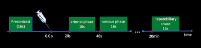
Low Flow Continuous Oxygen Inhalation May Avoid the Incidence of “Transient Severe Motion” (TSM) after Administration of Gadoxetate Disodium
Xiaoqian Jia, Jianxin Guo, Xiaocheng Wei, Jingtao Sun, Xianjun Li, Yangyang Han, Ming Guo, Jian Yang
In this study, we try to provide patients with continuous low flow oxygen inhalation during dynamic contrast-enhanced MR liver imaging to reduce “Transient Severe Motion” (TSM) after administration of gadoxetate disodium. The image quality was quantitatively assessed regarding to motion artifacts for each dynamic phases. The incidences of TSM were also compared between experimental and control groups. The results show that continuous oxygen inhalation at low flow rate can significantly reduce motion artifact and may avoid the occurrence of TSM.
|
|
1031.
 |
27 |
Gadoxetic acid-enhanced MRI: assessment of arterial phase artifacts and hepatobiliary uptake in a large series.
Naik Vietti Violi, Mathew Cherny, Pamela Argiriadi, Ally Rosen, Amanda Weiss, Gabriela Hernandez-Meza, Maxwell Segall, Jonathon Rosenblatt, Shingo Kihira, Sara Lewis, Bachir Taouli
In this study, we retrospectively evaluated the prevalence of artifacts during the arterial phase presumed to be related to transient severe motion during liver MRI with gadoxetic acid as well as the quality of contrast uptake during hepatobiliary phase in a single large center study. While we found a relatively high rate of motion artifacts on a single arterial phase (9.8%), it was rarely present in both arterial phases (2.3%). We also observed poor contrast uptake during hepatobiliary phase in 1.6% of cases. TSM and poor contrast uptake during HBP was found in only 0.7% of patients.
|
|
1032.
 |
28 |
Characterization of Abdominal Neoplasms using a Fast Inversion Recovery Radial SSFP T1 Mapping Technique
Mahesh Bharath Keerthivasan, Zhitao Li, Jean-Philippe Galons, Kevin Johnson, Bobby Kalb, Diego Martin, Ali Bilgin, Maria Altbach
We present a rapid technique for high-resolution multi-slice T1 mapping of the abdomen using the inversion recovery radial steady-state free-precision (IR-radSSFP) pulse sequence. We propose a joint two-component fit to estimate accurate T1 maps in the presence of partial volume. The utility of the sequence has been demonstrated for the characterization of abdominal neoplasms using data acquired on 22 clinical patients.
|
|
1033.
 |
29 |
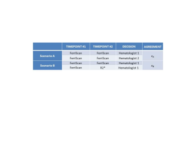
Comparison of FerriScan- versus R2*-Derived Liver Iron Concentration (LIC) for Clinical Decision Making in Patients with Liver Iron Overload Disease
Marshall Sussman, Kevin Kuo, Richard Ward, George Tomlinson, Kartik Jhaveri
This study compared clinical decision making between FerriScan and an R2*-based method in patients with iron overload disease. FerriScan is the current gold standard. However, it is expensive, and data must be transferred offsite. FerriScan and R2* techniques were both used to manage (simulated) patient chelator dosing. The agreement between different reviewers assessing the same FerriScan data was the same as the agreement when the same reviewer used R2*- compared to FerriScan-derived LIC values. Thus, switching to R2*-based decision making from FerriScan is no more likely to cause a difference in patient management than switching from one hematologist to another.
|
|
1034.
 |
30 |
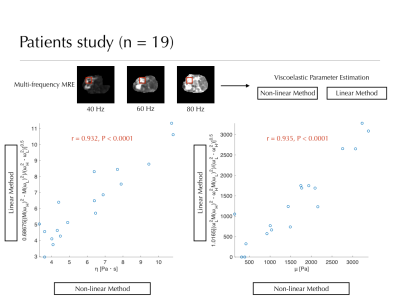
Alternative linear post-processing method for calculating viscoelastic parameters of the liver from multi-frequency MR elastograms.
Akira Yamada, Hayato Hayashihara, Yoshihiro Kitoh, Yasuo Adachi, Aya Shiobara, Yasunari Fujinaga
An alternative linear post-processing method that enables fast and precise calculation of viscoelastic parameters (viscosity, η and elasticity, μ) of the liver from multi-frequency MR elastograms has been shown in this study. Correlation of viscoelastic parameters between linear and non-linear calculation methods was significantly high in phantom (r = 0.980 for μ, r = 0.983 for η) and patients study (r = 0.932 for μ, r = 0.935 for η). Calculation speed by linear method (0.42 s) was significantly faster than that by non-linear method (47.14 s). The proposed method will promote clinical application of viscoelastic analysis of liver diseases using MR elastography.
|
|
