|
2nd Hour
Poster: MSK Power Poster
Power Pitch Poster
Musculoskeletal
Monday, 13 May 2019
Power Pitch Theater B - Exhibition Hall
14:45 - 15:45
| |
|
Plasma # |
 |
0127.
 |
16 |
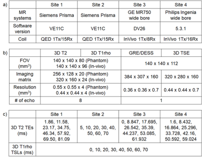
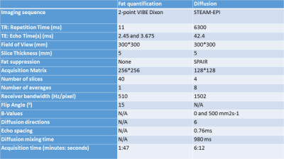
Multi-vendor multi-site T1? and T2 quantification of knee cartilage

Jeehun Kim, Kenji Mamoto, Richard Lartey, Kaipin Xu, Matthew Tanaka, Emma Bahroos, Carl Winalski, Thomas Link, Peter Hardy, Qi Peng, Angie Botto-van Bemden, Kecheng Liu, Robert Peters, Can Wu, Xiaojuan Li
T1ρ and T2 relaxation times are promising biomarkers for early detection of osteoarthritis due to its sensitivity to cartilage degeneration. Good reliability is essential for these quantitative measures to be widely applicable in clinical trials. We implemented MAPSS T1ρ and T2 imaging on multiple platforms (Siemens, GE, Philips) and evaluated the intra-site repeatability and inter-site reproducibility of T1ρ and T2 data in a multi-site multi-vendor setting.
|
 |
0128.
 |
17 |
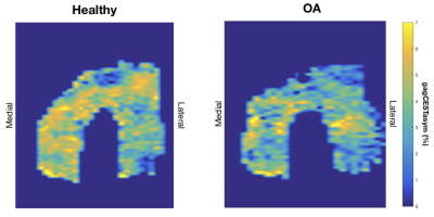
GagCEST at 3T Can Detect Cartilage Differences Between Healthy and OA Subjects
Elka Rubin, Lauren Watkins, Valentina Mazzoli, Arjun Desai, Gabe Ho, Feliks Kogan, Scott Ulrich, Julie Kolesar, Scott Delp, Gary Beaupre, Garry Gold
Chemical exchange saturation transfer of GAG (gagCEST) is a quantitative MR technique that is a useful biomarker for assessing GAG content at 7T. However, its utility at 3T remains unclear. In this study, we compare gagCEST asymmetry values of healthy and osteoarthritic subjects scanned at 3T. Comparisons between healthy and OA subjects indicate a significant difference in the average gagCEST signal across the medial and lateral anterior and medial weight-bearing regions of the femoral cartilage. The results of this study suggest that there is potential for use of gagCEST in the study of OA at 3T.
|
|
0129.
 |
18 |
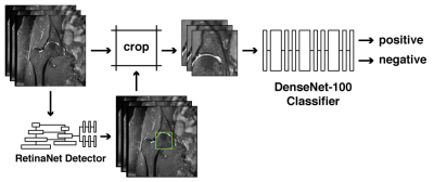
Deep Learning Pipeline for Automated Identification of Osteoarthritic Degenerative Changes in the Hip
Eugene Ozhinsky, Radhika Tibrewala, Rutwik Shah, Sarah Foreman, Valentina Pedoia, Sharmila Majumdar
Manual identification of bone and cartilage abnormalities in MR images can be laborious and time consuming. The goal of this study was to develop a fully automated deep learning pipeline to identify morphological and degenerative changes in patients with hip osteoarthritis (OA). It included an object detection deep convolutional neural network (DCNN) that generated cropped images of the hip joint and a classification DCNN that identified the presence of morphological bone and cartilage changes.
|
|
0130.
 |
19 |
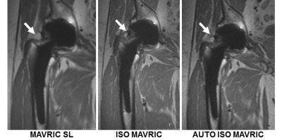
Added Clinical Value of Isotropic MAVRIC SL Acquisitions of Total Hip Arthroplasties
Kelly Zochowski, Jacky Cheung, Mauro Miranda, Erin Argentieri, Bin Lin, S. Kaushik, Alissa Burge, Matthew Koff, Hollis Potter
MAVRIC SL, a multi-spectral MRI imaging sequence, reduces metallic susceptibility artifact to improve visualization near joint arthroplasty by acquiring 24 spectral bins of off-resonance data. Many implants require fewer bins, and this study uses a calibration scan to determine the number of bins necessary to permit an isotropic MAVRIC acquisition and a reduced TR isotropic MAVRIC acquisition. The isotropic MAVRIC images decreased blurring and improved visualization of the periprosthetic bone and synovium while retaining image quality. Lowering the TR decreased scan time but affected image interpretation. Isotropic MAVRIC acquisitions may improve the diagnostic capability of MAVRIC SL images.
|
|
0131.
 |
20 |
Five-minute single sequence comprehensive 4D pediatric ankle MRI with T2 Shuffling
Jonathan Tamir, Fida Wishah, Jesse Sandberg, Marcus Alley, Michael Lustig, Shreyas Vasanawala
The use of volumetric acquisitions for musculoskeletal MR in clinical settings has been limited due to blurring artifacts from T2 decay. T2 Shuffling (T2Sh), a redesigned 3D fast spin-echo technique that mitigates blurring, has been successfully applied to pediatric knee MRI in a clinical setting and used to streamline the pediatric knee exam. This work assesses a shortened T2Sh scan for pediatric ankle MRI with total scan time under 5 minutes. Our results show that T2Sh has the potential to provide a comprehensive diagnostic protocol in place of the conventional long 2D exam.
|
|
0132.
 |
21 |
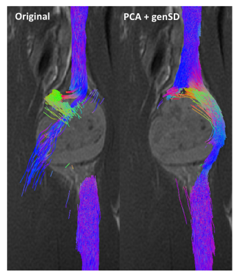
Denoising of Diffusion MRI Improves Peripheral Nerve Conspicuity and Reproducibility
Jaemin Shin, Jahnavi Curlin, Ek Tsoon Tan, Maggie Fung, Darryl Sneag
This study evaluated q-space-based (genSD) and principal component analysis (PCA) denoising techniques to enhance SNR for peripheral nerve diffusion MRI of 10 healthy knees. The combination of both methods (PCA+genSD) was compared with a PCA-only method as well as the average of 10 repetitions. The combined denoising method showed improved performance with respect to SNR, peripheral nerve conspicuity and reproducibility.
|
|
0133.
 |
22 |
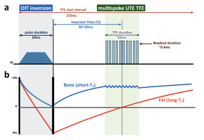
Robust Motion-Compensated Lumbar Spine Bone imaging using 3D UTE with Broadband Inversion Recovery Pulse and k-space Weighted Navigator Gating
Masami Yoneyama, Iain Ball, Ben Kennedy, Takayuki Sakai, Atsuya Watanabe, Marc Van Cauteren
We proposed a new technique for the lumbar spine MR bone imaging based on broadband inversion recovery prepared segmented multispoke UTE sequence with k-space weighted navigator gating (3D BoneVIEW) for assessment of low back pain. 3D BoneVIEW provided robust bone imaging with sufficient background suppression and without respiratory artifacts. This sequence has a great potential to help more accurate assessment of the low back pain as an alternative to CT imaging.
|
|
0134.
 |
23 |
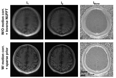
Self-navigated, rapid 3D UTE for motion-robust skull imaging
Hyunyeol Lee, Xia Zhao, Hee Kwon Song, Rosaline Zhang, Scott Barttlett, Felix Wehrli
Solid-state MRI via 3D UTE or ZTE methods has potential for bone-selective imaging as a radiation-free alternative to computed tomography, particularly for children with craniofacial abnormalities. However, relatively long scan times make the technique vulnerable to artifacts from involuntary subject movements, thereby impairing image quality. Here, we developed a self-navigated, rapid 3D UTE technique by combining a retrospective motion detection and correction approach with sparsity-constrained image reconstruction. Results from in vivo studies demonstrate the proposed method's feasibility in achieving motion-corrected whole-skull images at 1 mm isotropic resolution in 2.1 minutes scan time.
|
|
0135.
 |
24 |
Fat Suppression in UTE Imaging of Short T2 Tissues Using a Novel Soft-hard Composite RF Pulse
Ya-Jun Ma, Saeed Jerban, Hyungseok jang, Eric Chang, Jiang Du
Fat suppression is very important for both high contrast morphological imaging and accurate quantitative MR imaging. However, the conventional fat suppression methods such as chemical shift-based fat saturation are not well-suited for short T2 imaging due to the large direct saturation of short T2 tissues with broad spectra. The purpose of this study was to design a novel fat suppression pulse for ultrashort echo time (UTE) imaging of short T2 tissues with well-preserved short T2 signals using a soft-hard composite pulse.
|
|
0136.
 |
25 |
Cortical bone quantifications using ultrashort echo time MR imaging (UTE-MRI) correlate well with histomorphometric assessment of bone microstructure
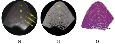
Saeed Jerban, Yajun Ma, Jonathan Wong, Amin Nazaran, Adam Searleman, Lidi Wan, Judith Williams, Eric Chang, Jiang Du
Ultrashort echo time magnetic resonance imaging (UTE-MRI) has been used to assess cortical bone porosity, as validated routinely with high resolution micro computed tomography (μCT). This study investigated the correlations between UTE-MRI-based quantifications and histomorphometric measures, as well as between UTE-MRI-based quantifications and μCT results. MRI properties showed strong correlations with both histomorphometric and μCT-based porosities. Only UTE-MRI could assess small pore (<40 μm) variations with moderate correlations. Major porosity changes were from large pores in studied specimens; therefore, μCT employment is likely adequate to validate UTE-MRI biomarkers. However, UTE-MRI techniques can assess pores below the detectable range by μCT, porosities which might contribute differently to bone mechanics.
|
|
0137.
 |
26 |
A Quantitative, Multiparametric Method for Bone Edema and Adiposity Characterisation in Inflamed Trabecular Bone
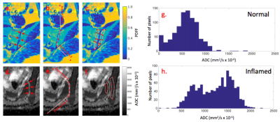
Timothy Bray, Naomi Sakai, Alexandra Dudek, Kannan Rajesparan, Corinne Fisher, Coziana Ciurtin, Debajit Sen, Alan Bainbridge, Margaret Hall-Craggs
MRI is increasingly used to identify and monitor inflammation in patients with inflammatory diseases involving the skeleton, such as spondyloarthritis. However, conventional image interpretation by radiologists provides only indirect information about the inflammatory process and lacks reproducibility. Here, we describe a partially-automated multiparametric MRI tool for quantifying and characterising both active and chronic inflammation in spondyloarthritis, relying on histographic analysis of apparent diffusion coefficient (ADC) and proton density fat fraction (PDFF) maps. We show that histographic analysis improves performance compared to simple averaging, and, further, that ADC and PDFF provide distinct, complementary information regarding active inflammation and structural damage respectively.
|
|
0138.
 |
27 |
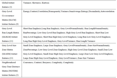
Texture analysis based on diffusional kurtosis imaging for the differentiation of benign and malignant bone tumors
Ying Li, Cuiping Ren, Jingliang Cheng, Zhizheng Zhuo
This work investigated and evaluated the role of textures extracted from magnetic resonance (MR) diffusion kurtosis imaging (DKI) in characterizing the bone tumors, and furtherly evaluate the ability of these textures to differentiate benign and malignant tumors by using support vector machine classifiers (SVM), which might be helpful for clinical diagnosis and studies. The texture parameters have the ability to character the bone tumor and SVM classifier showed good performance in the differentiation of benign and malignant bone tumors.
|
|
0139.
 |
28 |
Pilot study on facioscapulohumeral muscular dystrophy patients with dynamic phase contrast imaging of electrically stimulated quadriceps muscles
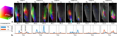
Xeni Deligianni, Francesco Santini, Giorgio Tasca, Mauro Monforte, Francesca Solazzo, Raimondo Vitale, Paolo Felisaz, Giancarlo Germani, Niels Bergsland, Enzo Ricci, Anna Pichiecchio
Facioscapulohumeral muscular dystrophy is characterized by a peculiar non-linear muscle-by-muscle involvement and is very hard to predict. The purpose of this study was to use dynamic phase contrast MR imaging of electrically stimulated quadriceps muscle to characterize the elastic potential of the muscle in FSHD patients and contribute to the understanding of this challenging disease. Velocity, strain, and strain rate were analyzed and compared to the results of physical examination.
|
|
0140.
 |
29 |
Quantitative MRI Measurements can Distinguish Myositis From Healthy Control Muscle.
Matthew Farrow, Ai Lyn Tan, Paul Emery, Maya Buch, Andrew Grainger, Steven Tanner, John Biglands
Myositis is an autoimmune inflammatory muscle disease which can decrease quality of life and increase mortality. Clinical presentation includes muscle weakness, changes in muscle microstructure, myosteatosis and myalgia. Current diagnosis is reliant on subjective clinical examinations, blood tests and invasive biopsies. Quantitative MRI techniques such as diffusion and fat fraction measurements are sensitive to changes within the muscle. 10 myositis patients and 16 healthy controls underwent scans of the thigh. Significant differences were found in fat fraction and diffusion measurements between myositis patients and healthy controls, implying these measures have potential as biomarkers in the diagnosis and management of myositis.
|
|
0141.
 |
30 |
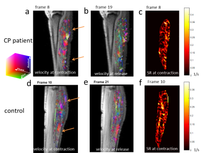
Imaging of calf muscle contraction in pediatric patients with cerebral palsy and healthy children by dynamic phase contrast MRI
Claudia Weidensteiner, Xeni Deligianni, Francesco Santini, Tanja Haas, Philipp Madoerin, Oliver Bieri, Katrin Bracht-Schweizer, Erich Rutz, Meritxell Garcia, Reinald Brunner
Aim of this study is to investigate the feasibility of phase contrast imaging for assessment of muscle function in children with cerebral palsy (CP). Time-resolved cine phase contrast MRI was synchronized with electrical muscle stimulation of the calf muscle at a clinical 3T MRI scanner. 11 healthy and 4 children with hemiparetic CP were scanned. Dynamic velocity, strain, and strain rate maps were reconstructed. Synchronous dynamic PC MRI of electrically stimulated muscle is feasible in children, even in CP patients and might provide further insight into the health status of their muscles.
|
|
