|
2nd Hour
Poster: Other Nuclei MR: Looking at Other Resonances
Power Pitch Poster
Spectroscopy & Non-Proton MR
Tuesday, 14 May 2019
Power Pitch Theater C - Exhibition Hall
14:30 - 15:30
| |
|
Plasma # |
|
0480.
 |
31 |
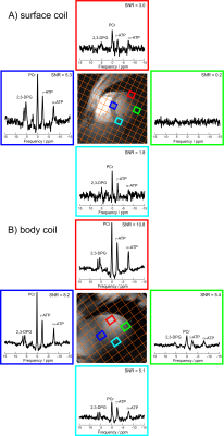
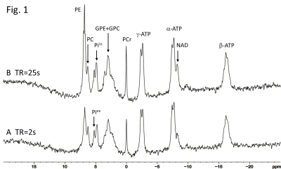
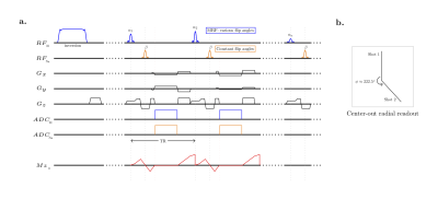
First assessment of cardiac energy metabolism in the lateral and inferior segments of the left ventricle in vivo at 7T

Ladislav Valkovic, Jane Ellis, Lucian Purvis, Albrecht Schmid, Stefan Neubauer, Christopher Rodgers
Using a novel whole-body transmit coil with an integrated 30-element receive array, powered by a 35kW RF power amplifier, we show the feasibility of whole-heart cardiac 31P-MRS at 7T. PSF simulations were performed to directly compare the new coil to a published protocol using a 16-element surface coil. The whole-body coil delivers analysable spectra from all left ventricular segments in the hearts of 3 volunteers, whereas the 16-element surface coil allowed probing energetics only in the proximal half of the left ventricle. This coil will enable new studies of focal disease and improve the SNR for regionally homogenous disease investigations.
|
 |
0481.
 |
32 |
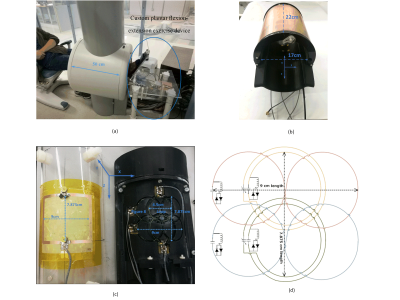
Dynamic 31P MRS of Skeletal Muscle with a 1 Telsa Extremity Scanner
Minyu Gu, Travis Carrell, John Bosshard, Clayton Cruthirds, Nicolaas Deutz, Marielle Engelen, Mary McDougall, Steven Wright
The potential of using low-field MRI scanners as a platform to enable low-cost 31P studies outside of the conventional hospital environment was examined. All experiments were conducted on an ONI 1T scanner with a custom broadband multichannel receiver. A transmit-only birdcage and receive-only four-element array were designed and built to enhance the SNR and improve linewidth. Preliminary phantom and in vivo results show the proposed design improved the linewidth from 0.71 ppm to 0.33 ppm, SNR was increased by approximately 2 times, and inorganic phosphate and phosphocreatine exchange were observed during the volunteer’s exercise.
|
|
0482.
 |
33 |
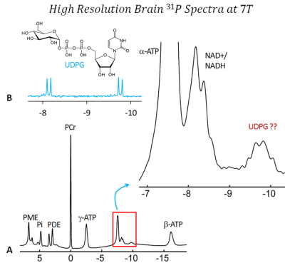
Human Brain 31P NMR Spectra at 7T: UDP-Glucose Assignment Revisited
Jimin Ren, A Dean Sherry, Craig Malloy
In a typical human brain 31P NMR spectrum, a small peak is often present at ~-9.7 ppm, near the α-ATP and NAD+/NADH signals. This 31P resonance, accounting for ~1/30th of the brain α-ATP signal, has been considered to be a doublet and assigned to UDPG. Here we present strong evidence to show that the -9.7 ppm signal is in fact a finely structured quartet. This finding has direct impact on current 31P NMR method for evaluation of brain redox, which requires UDPG correction. It may also have implication on our interpretation of brain energy metabolism based on 31P NMR data.
|
|
0483.
 |
34 |
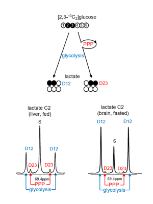
Activity of the Pentose Phosphate Pathway Is Increased in Hepatoma Using a Novel Tracer, [2,3-13C2]glucose
Min Hee Lee, Craig Malloy, Ian Corbin, JunJie Li, Eunsook Jin
The pentose phosphate pathway (PPP) was investigated in a rat model of hepatoma and results were compared to normal liver and other tissues. A novel and specific tracer of the PPP, [2,3-13C2]glucose, is introduced. The resulting isotopomers are informative because [1,2-13C2]lactate arises only from glycolysis and [2,3-13C2]lactate arises only from the PPP. The PPP was more active in the fed vs. fasted state in most tissues. These results correlated with mRNA expression of key enzymes in the PPP, and flux through both the PPP and glycolysis was substantially increased in hepatoma compared to healthy liver.
|
|
0484.
 |
35 |
Phosphorous and Proton MR Spectroscopy Study of Creatine Transporter Deficiency
Shizhe Li, Simona Bianconi, Jan Willem van der Veen, JoEllyn Stolinski, An Dang Du, Kim Cecil, Porter Forbes, Jun Shen
The X- linked Creatine Transporter Deficiency (CTD) is one of three types of the cerebral creatine deficiency disorders. It is caused by mutations in the X-linked gene SLC6A8. We report the first combined 31P and 1H MRS study of CTD patients. Relative concentrations of key metabolites were quantitatively analyzed. The PCr/total-phosphate and total-Cr/NAA ratios were found to be markedly reduced in CTD patients. By combining the proton and 31P data we found that the relative reduction in PCr in CTD patients is much less than that of total Cr.
|
|
0485.
 |
36 |
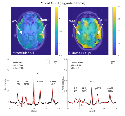
Volumetric mapping of intra- and extracellular pH in glioma patients using 31P MRSI at 7 Tesla
Andreas Korzowski, Nina Weinfurtner, Sebastian Mueller, Johannes Breitling, Steffen Goerke, Heinz-Peter Schlemmer, Mark Ladd, Daniel Paech, Peter Bachert
In vivo phosphorus magnetic resonance spectroscopic imaging (31P MRSI) enables the non-invasive mapping of absolute intra- and extracellular pH values of the human brain. However, achieving reasonable spatial resolution for studies of brain tumor patients is challenging due to low phosphate concentrations. In this study we demonstrate that 31P MRSI at 7 Tesla enables volumetric mapping of intra- and extracellular pH for studies of brain tumor patients. Volumetric pH maps with actual voxel size of 8 ml can be obtained within 30 minutes acquisition duration, and may provide novel insight into the pathophysiology of brain tumors.
|
|
0486.
 |
37 |
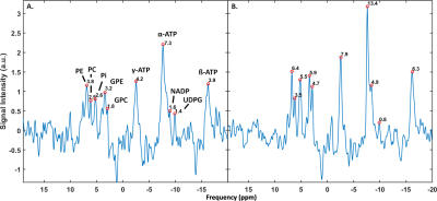
Full coverage 31P MRSI of the liver with a body coil at 7T
Quincy van Houtum, Catalina Arteaga de Castro, Dennis Klomp, Wybe van der Kemp
We demonstrate increased 31P metabolite sensitivity by acquiring 31P signals over a large volume in the liver of a volunteer using a 31P whole body birdcage coil at 7T and minimize spatial reach of muscle signal leakage plus investigate frequency alignment due to B0 homogeneities. Sufficient SNR could be obtained in weighted average spectrum of all liver voxels and of four local liver voxels. Correcting B0 inhomogeneities by aligning resulted in a two-fold and 30% increase compared to non-aligned for the liver voxel average and the local voxel average respectively. The full setup allowed for full liver coverage and minimized muscle signal leakage.
|
|
0487.
 |
38 |
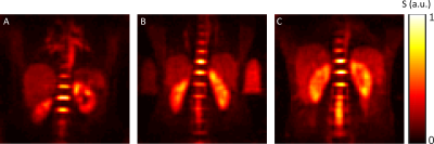
Quantitative 23Na MRI of the human liver at 7 Tesla
Johanna Lott, Nicolas Behl, Armin Nagel, Reiner Umathum, Peter Bachert, Mark Ladd, Tanja Platt
Sodium (23Na) MRI has been proposed as a potential imaging modality for the characterization of hepatic tumors and for monitoring therapy response. Up to now, only one study has been performed on implanted hepatocellular carcinomas in rats. In the present work, in-vivo 23Na MRI of the healthy human liver is performed and to the best of our knowledge, the hepatic tissue sodium concentration is estimated for the first time. For quantitative 23Na MRI correction methods were applied such as self-gating and B1+ correction. The mean sodium concentration for three volunteers was estimated to be (27±5) mM in the liver.
|
 |
0488.
 |
39 |
Simultaneous proton MR fingerprinting and sodium imaging
Zidan Yu, Guillaume Madelin, Daniel Sodickson, Martijn Cloos
Sodium MRI can provide unique metabolic information to study the human body and its afflictions. However, the low intrinsic signal to noise ratio of sodium MRI limits the resolution of the sodium images to 3-5 mm isotropic and necessitates long acquisition times (~10-20 min). The necessity to perform 1H and 23Na acquisitions sequentially also prolongs the total scan time and limits applications of combined proton-sodium imaging. In this work, we demonstrate a technique to simultaneously acquire sodium images and multi-parametric proton maps in one single scan.
|
|
0489.
 |
40 |
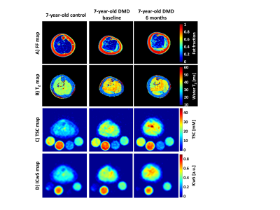
Longitudinal structural change in skeletal muscle tissue of Duchenne muscular dystrophy patients based on 1H- and 23Na-MRI
Teresa Gerhalter, Lena Gast, Benjamin Marty, Regina Trollmann, Frank Roemer, Frederik Laun, Michael Uder, Pierre Carlier, Armin Nagel
Duchenne muscular dystrophy (DMD) is a hereditary neuromuscular disease leading to progressive muscle wasting. Here, young DMD boys were examined twice within six months with a MRI protocol that included commonly used biomarkers such as fat fraction derived from the Dixon method and water T2 as well as 23Na MRI indices. Sodium anomalies were commonly observed and developed even in absence of fatty degenerative changes and water T2increases over the observational period. Although limited in the small number of subjects, the data supports that 23Na could be used to characterize early dystrophic muscle alteration in a longitudinal fashion.
|
|
0490.
 |
41 |
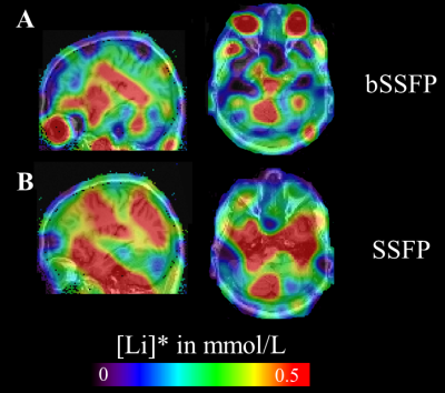
bSSFP vs SSFP acquisitions of 7Li MRI at 7T: Comparison of sensitivity and quantification accuracy
Jacques Stout, Franck Mauconduit, Franz Hozer, Arthur Coste, Sandro Romanzetti, Cécile Rabrait-Lerman, Franck Bellivier, Edouard Duchesnay, Fawzi Boumezbeur
3D 7Li SSFP and balanced SSFP approaches have been compared by acquiring both datasets from seven euthymic bipolar disorder (BD) patients at 7T. Quantification was performed using the phantom replacement approach accounting for global T1 and T2 relaxation effects. With both methods, heterogeneous brain Li distributions were observed with marked differences in the eyes notably. However, strong correlations between averaged apparent lithium concentrations could be established across all BD patients. While the bSSFP approach is a viable and practical option for 7Li MRI, a more realistic quantification pipeline should be considered in the future.
|
|
0491.
 |
42 |
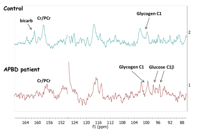
Glycogen detection in human brain via natural abundance 13C MRS at 7T
Sergey Cheshkov, Ivan Dimitrov, Brandy Verhalen, A. Dean Sherry, Berge Minassian, Craig Malloy
Large quantities of abnormally-branched brain glycogen are hypothesized to be accumulating in disorders such as Lafora disease and Adult Polyglucosan Body Disease (APBD). However, non-invasive tools for brain glycogen detection in vivo are lacking. In this work we have used natural abundance 13C MRS at 7T with NOE, to detect glycogen in both normal and APBD brain. Qualitative comparison of the respective glycogen C1 signals in these two cases indicates no dramatic increase of the detectable glycogen concentration in APBD. To our knowledge this is the first human cerebral glycogen detection via natural abundance 13C MRS.
|
|
0492.
 |
43 |
On the magnetic field dependence of deuterium metabolic imaging (DMI)
Robin de Graaf, Arjan Hendriks, Dennis Klomp, Chathura Kumaragamage, Dimitri Welting, Catalina Arteaga de Castro, Peter Brown, Scott McIntyre, Terence Nixon, Jeanine Prompers, Henk De Feyter
Deuterium metabolic imaging (DMI) is a novel, MR-based method to spatially map metabolism. DMI has been shown to provide robust and sensitive maps of cerebral glucose metabolism in healthy volunteers and patients with brain tumors. Here the magnetic field dependence of DMI in terms of sensitivity and resolution is investigated. Using RF coils sized for animal and human studies on magnetic fields ranging from 4T to 11.7T, a supralinear magnetic field dependence of the DMI sensitivity is established. The increased sensitivity of DMI at 7T compared to 4T allows the acquisition of human brain DMI at a 1 mL spatial resolution.
|
|
0493
 |
44 |
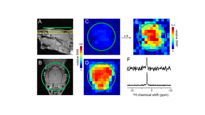
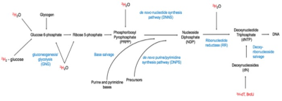
Deuterated water labeling followed by deuterium MRI for visualization of tumors in vivo
Video Permission Withheld
Nataliya Buxbaum, Keita Saito, Hellmut Merkle, Kathrynne Warrick, Natella Maglakelidze, Donald Farthing, Kazu Yamamoto, Nobu Oshima, Murali Cherukuri, Ronald Gress
In vivo DNA labeling with deuterated water (2H2O) has been used for cell kinetics research and more recently to image rapidly proliferating immune cells in the context of graft-versus-host disease. Using a custom dual-resonance coil (1H-2H) we demonstrate that this approach can be applied to the in vivo detection of tumors via MRI in a xenograft tumor mouse model. Therefore, this novel imaging technique could serve as a sensitive, safe, and non-radioactive method of tumor detection with significant impact on the field of oncology.
|
|
0494.
 |
45 |
Evaluation of trifluoroacetic acid as a theranostic fluorine-19 MRI agent for chemical ablation of solid tissue.
Samuel Einstein, Emily Thompson, Chunxiao Guo, Elizabeth Whitley, Erik Cressman, James Bankson
Chemical ablation therapies are an established treatment for hepatocellular carcinoma, but accurate mapping and monitoring of the ablative agent’s distribution is critical to improving outcomes. We evaluated the theranostic application of trifluoroacetic acid (TFA) as an ablative agent. Fluorine-19 MRI was optimized to image the agent with excellent sensitivity and cine 19F-MRI was developed to demonstrate the feasibility of real-time injection monitoring. Ablation of ex vivo liver tissue demonstrated TFA to be both effective and imageable, even at low concentrations. We conclude that TFA is a promising theranostic agent for ablation of solid tissue.
|
|
