|
2nd Hour
Poster: Molecular & Metabolic Imaging
Power Pitch Poster
Molecular Imaging
Monday, 13 May 2019
Power Pitch Theater C - Exhibition Hall
17:00 - 18:00
| |
|
Plasma # |
|
0252.
 |
31 |
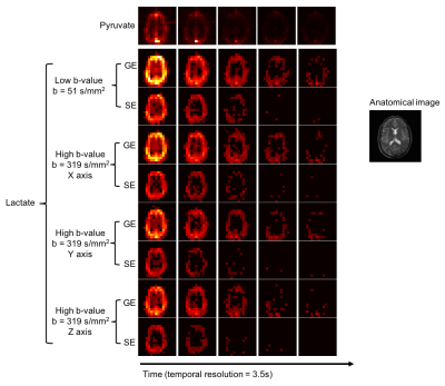
Probing Cerebral Lactate Compartmentalization with Hyperpolarized Diffusion Weighted 13C MRI

Jeremy Gordon, Shuyu Tang, Xucheng Zhu, Daniel Vigneron, Peder Larson
Hyperpolarized 13C MRI has been used to non-invasively measure metabolism in real-time. However, perfusion and transporter expression can impact the compartmentalization of metabolites. In this work, we investigated the feasibility of diffusion weighted imaging of lactate generated from HP [1-13C]pyruvate in the human brain to assess lactate efflux and compartmentalization in a healthy volunteer. Whole brain lactate ADC values were 0.37?10-3 mm2/s, 0.29?10-3 mm2/s, and 0.41?10-3 mm2/s when diffusion gradients were applied in the X, Y, and Z direction, respectively, demonstrating the feasibility of diffusion weighted HP 13C MRI in a clinical setting.
|
|
0253
 |
32 |
Imaging a hallmark of cancer: hyperpolarized [U-2H, U-13C]-glucose and hyperpolarized [1-13C]-dehydroascorbic acid can monitor TERT expression in gliomas
Video Permission Withheld
Pavithra Viswanath, Georgios Batsios, Russell Pieper, Sabrina Ronen
Expression of telomerase reverse transcriptase (TERT) is a fundamental hallmark of cancer. Identification of imaging biomarkers of TERT expression will facilitate non-invasive assessment of tumor burden and response to therapy. Our studies in glioma indicate that TERT expression leads to increased redox capacity characterized by elevated 1H-MRS-detectable glutathione and NADPH. Concomitantly, TERT increases 13C-MRS-detectable flux of glucose through the pentose phosphate pathway, which provides NADPH. Importantly, hyperpolarized [U-2H, U-13C]-glucose and hyperpolarized [1-13C]-dehydroascorbic acid can image these alterations in glucose and redox metabolism. Our study identifies potential non-invasive translational metabolic imaging probes of TERT expression in glioma and possibly other cancers.
|
 |
0254.
 |
33 |
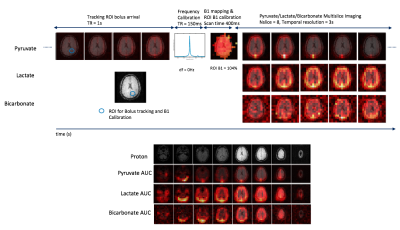
Initial experience of applying bolus tracking and real-time B0/B1 calibration for human hyperpolarized 13C imaging
Shuyu Tang, Jeremy Gordon, Robert Bok, James Slater, Jane Wang, Daniel Vigneron, Peder Larson
A new hyperpolarized 13C MRI approach with bolus tracking and real-time B0/B1 calibration was developed and tested in 4 human HP 13C MRI studies of brain, prostate, kidney and pancreas metabolism. The use of this framework demonstrated improved accuracy and robustness for human hyperpolarized 13C imaging.
|
 |
0255.
 |
34 |
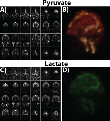
Hyperpolarized C-13 bSSFP Imaging of the Human Brain
Eugene Milshteyn, Cornelius von Morze, Jeremy Gordon, Galen Reed, Adam Autry, Hsin-Yu Chen, Daniele Mammoli, Robert Bok, James Slater, Mark Van Criekinge, Lucas Carvajal, Duan Xu, Peder Larson, Sarah Nelson, John Kurhanewicz, Daniel Vigneron
Current early phase clinical trials of hyperpolarized C-13 imaging of the brain, prostate, and liver have demonstrated the exceptional ability to rapidly visualize metabolism of pyruvate at high spatiotemporal resolutions. Prior human studies have used MRSI, EPSI, EPI, and spiral MRI and MRSI. In this study, we investigated for the first time the bSSFP sequence with its high SNR efficiency for human hyperpolarized C-13 imaging of pyruvate and lactate. This research showed the ability to acquire dynamic 1-1.5cm isotropic bSSFP images of the human brain in a clinical setting.
|
|
0256.
 |
35 |
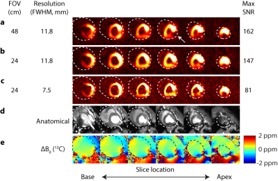
Imaging the circumferential hyperpolarized 13C-bicarbonate distribution in the normal heart
Angus Lau, Albert Chen, Charles Cunningham
A typical observation in hyperpolarized 13C cardiac imaging is that the bicarbonate images are not circumferential, near the apex, even in healthy subjects, which precludes interrogation of metabolism in the posterior myocardium. A common explanation for the signal drop-off is anterior receive coil sensitivity. In this abstract, we demonstrate that this is not necessarily the case. A significant source of reduced signal results from B0 inhomogeneity in the posterior wall. Shortened readout durations which achieve higher kmax are shown to enable high resolution imaging of the circumferential bicarbonate distribution in pigs.
|
|
0257.
 |
36 |
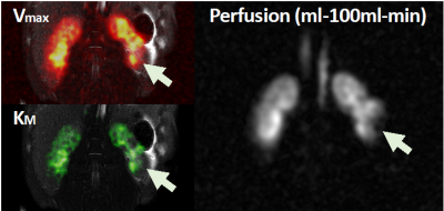
Hyperpolarized [1-13C]pyruvate MRS in a large animal model of partial renal obstruction supports clinical use in patients
Esben Hansen, Uffe Kjærgaard, Rasmus Tougaard, Rolf Schulte, Hans Stødkilde-Jørgensen, Christoffer Laustsen
Hyperpolarized [1-13C]pyruvate was used in this study to explorer the capability of detecting ischemic injury in the kidneys and to present the heterogeneity from the different kidney compartments. This was done using Michaelis–Menten kinetics by obtaining data in the an saturation recovery setup. Ischemic injuries was observed and in close correlation to standard perfusion MRI. The results from this study shows promise for the human introduction of this method.
|
|
0258.
 |
37 |
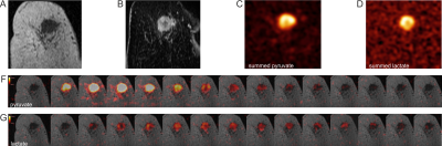
Imaging metabolic heterogeneity in breast cancer using hyperpolarized 13C-MRSI
Ramona Woitek, Mary McLean, James Grist, Raquel Manzano Garcia, Turid Torheim, Elena Provenzano, Oscar Rueda, Andrew Gill, Andrew Patterson, Frank Riemer, Joshua Kaggie, Stephan Ursprung, Fulvio Zaccagna, Surrin Deen, Marie-Christine Laurent, Matthew Locke, Amy Frary, Sarah Hilborne, Chris Boursnell, Titus Lanz, Amy Schiller, Ilse Patterson, Bruno Carmo, Rhys Slough, Richard Baird, Evis Sala, Bristi Basu, Jean Abraham, Suet-Feung Chin, Martin Graves, Fiona Gilbert, Carlos Caldas, Kevin Brindle, Ferdia Gallagher
13C magnetic resonance spectroscopic imaging (13C-MRSI) is a promising technique for elucidating metabolic heterogeneity in breast cancer. We used 13C-MRSI to evaluate the extent of glycolysis in different histologic and molecular breast cancer subtypes and correlated these findings with the expression of a key transmembrane transporter (MCT1) and glycolytic enzyme (LDHA). In addition to a strong correlation between glycolysis and tumor volume, there was higher expression of MCT1 and LDHA as well as CAIX, a hypoxia marker, in the more glycolytic tumors. This is the first study in humans to demonstrate the relationship between intertumoral heterogeneity on gene expression analysis and 13C-MRSI.
|
|
0259.
 |
38 |
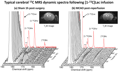
Kinetic Modeling of Hyperpolarized [1-13C]Lactate Metabolism in a Mouse Model of Ischemic Stroke
Thanh Phong Lê, Lara Buscemi, Elise Vinckenbosch, Mario Lepore, Rolf Gruetter , Lorenz Hirt , Jean-Noël Hyacinthe , Mor Mishkovsky
Stroke is the second cause of death and third leading cause of disability worldwide. Lactate injection was found to provide neuroprotection in preclinical models of ischemic stroke. Alteration of the metabolism induced by ischemia can be measured in real time using magnetic resonance with hyperpolarized 13C labeled probes.
This study aims at investigating the feasibility of quantifying changes in the kinetics of hyperpolarized [1-13C]lactate metabolism following ischemia in a mouse model of stroke in order to assess the potential of hyperpolarized lactate as a theranostic agent for stroke.
|
|
0260.
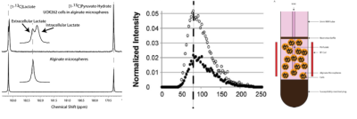 |
39 |
Assessment of intracellular lactate relaxation, production and efflux rates in cells using hyperpolarized 13C MR
Fayyaz Ahamed, Mark Van Criekinge, Zhen Wang, John Kurhanewicz, Peder Larson, Renuka Sriram
Enzymatic conversions can now be measured with hyperpolarized 13C MR on a sub-minute time scale. Using this technology we have shown that renal cell carcinoma cells of varying aggressiveness in 3D culture in bioreactors (5mm NMR tube) can monitor both lactate production and its efflux in real time. Using this platform, we have robustly characterized certain parameters that are difficult to measure in vivo, such as intracellular longitudinal relaxation time and kinetic transport rate. Further validation of these measures were obtained by fitting the same model to data from cells treated with transporter inhibitor.
|
|
0261.
 |
40 |
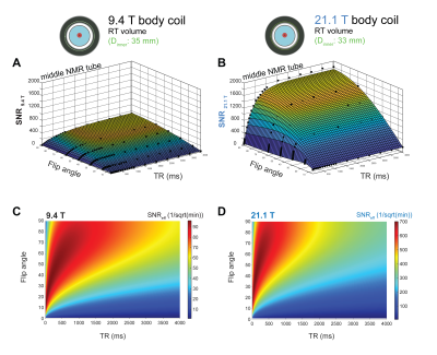
Fluorine-19 MR at 21.1 Tesla for Enhanced Detection of Brain Inflammation
Sonia Waiczies, Jens Rosenberg, Paula Ramos Delgado, Ludger Starke, Joao dos Santos Periquito , Christian Prinz, Jason Millward, Andre Kuehne, Helmar Waiczies, Andreas Pohlmann, Thoralf Niendorf
Fluorine-19 (19F) MR methods are invaluable for several applications including detection of brain inflammation but suffer from inherently low signal-to noise ratio (SNR). A magnetic field increase from 9.4 to 21.1 Tesla was studied as strategy for increasing signal sensitivity. As a result of an SNR increase, inflammation regions undetected at 9.4T were revealed at 21.1T. Although the SNR gain at 21.1T does not reach that achieved with a cryogenic quadrature RF surface probe (19F-CRP) at 9.4T, increased sensitivity was observed throughout the whole field of view at 21.1T, from ventral to dorsal head regions.
|
|
0262.
 |
41 |
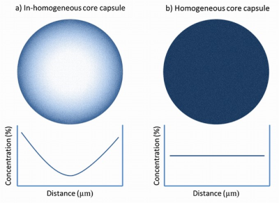
Fluorine MRI-Visible Mixed-Alginate Gradient Fluorocapsules for Image-Guided Diabetes Treatment
Dian Arifin, Genaro Paredes-Juarez, Paul de Vos, Jeff Bulte
A promising treatment of auto-immune juvenile diabetes is transplantation of beta islet cells. Islets can be encapsulated inside semi-permeable microcapsules to protect them against the patients’ immune system. Low islet survival and the lack of means to monitor the implants are major issues. We employed mixed-alginate gradient (MAG) microcapsules that better support human islet viability compared to currently used microcapsules. By labeling the capsules with clinically used agent CS-1000, we created MAG fluorocapsules which appeared as hot spots in mice on 19F MRI. MAG fluorocapsules offer a drug-free means to treat diabetic patients long-term while enabling imaging of transplanted islets.
|
|
0263.
 |
42 |
Efficient Dictionary-based Attenuation Correction for combined PET-MR systems
Matteo Cencini, Guido Buonincontri, Michela Tosetti
In many diagnostic applications, a correct photon attenuation correction is crucial for quantifying the uptake of a PET tracer. While in PET/CT scanners attenuation map is readily estimated from CT Hounsfield units, in combined PET/MRI scanners it must be obtained by processing of high resolution images. This map must then be co-registered to low resolution PET map, wasting acquisition time. Here, we propose an efficient approach based on a fast transient-state acquisition and a three-component signal model: this allow to obtain tissue fraction maps, which can be used to estimate attenuation map, directly at PET resolution.
|
|
0264.
 |
43 |
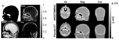
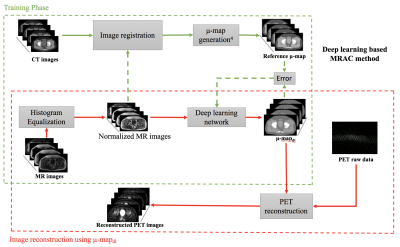
An MR based PET attenuation correction using a deep learning approach and evaluation in prostate cancer patients
Andrii Pozaruk, Kamlesh Pawar, Shenpeng Li, Alexandra Carey, Yen-Cheng Henry Pan, Viswanath P Sudarshan, Marian Cholewa, Jeremy Grummet, Zhaolin Chen, Gary Egan
Accurate Magnetic Resonance (MR) imaging based attenuation correction is crucial for quantitative Positron Emission Tomography (PET) in simultaneous MR/PET imaging. However, due to a lack of robust MR bone imaging methods, MR based attenuation correction remains a critical issue in MR/PET image reconstruction. In this work, we developed and evaluated a deep learning (DL) based MR based attenuation correction method for improved MR/PET quantification accuracy in prostatic cancer imaging.
|
|
0265.
 |
44 |
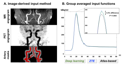
Impact of attenuation correction on image-derived input function and cerebral blood flow quantification with simultaneous [15O]-water PET/MRI
Trine Hjoernevik, Mohammad Khalighi, Sandeep Kaushik, Yosuke Ishii, Greg Zaharchuk, Audrey Fan
This study evaluated the impact of attenuation correction (AC) on image-derived input functions (IDIF) and kinetic modeling of cerebral blood flow (CBF) parameters for simultaneous [15O]-water PET/MRI in the brain. Atlas-based AC led to 4.3% underestimation of the IDIF peak and 8-18% overestimation of absolute CBF in different brain perfusion states. On the other hand, zero echo time (ZTE)-based AC provided reproducible quantification of absolute CBF, comparable to the deep learning AC reference that was trained on real CT images. Attenuation correction is an important consideration for IDIF calculation and parametric mapping with PET/MRI; and ZTE-based and deep learning-based AC provide suitable quantitative accuracy for [15O]-water studies.
|
|
0266.
 |
45 |
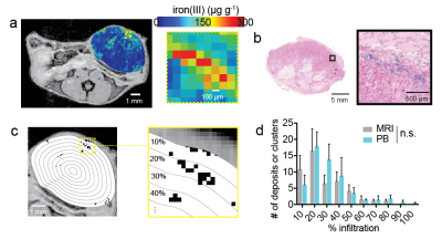
Spatial profiling of endogenous cellular iron MRI contrast by machine vision classifies macrophage infiltration in breast cancer models of metabolic- and immune-therapy
Avigdor Leftin, Jason Koutcher
Tumor macrophage response to therapy is conventionally detected with iron nanoparticle-enhanced MRI. However, endogenous cellular iron detection methods exist that can bypass caveats intrinsic to contrast agent use. We demonstrate that contrast-agent free multi-gradient echo R2* relaxometry iron MRI and machine vision analysis approaches can map tumor macrophage iron deposits and detect cellular response to metabolic and immunotherapy in preclinical models of breast cancer. Feasibility of endogenous macrophage imaging is shown, and the value of the cellular MRI biomarker is demonstrated as a function of treatment and tumor model.
|
|
