|
2nd Hour
Poster: Mix All Physical Properties With Water & Shake Well
Power Pitch Poster
Contrast Mechanisms
Wednesday, 15 May 2019
Power Pitch Theater B - Exhibition Hall
09:15 - 10:15
| |
|
Plasma # |
 |
0681.
 |
16 |
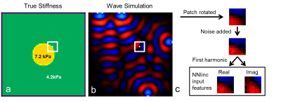
Inhomogeneous Neural Network Inversion (NNI_inh) for stiffness estimation in Magnetic Resonance Elastography

Jonathan Scott, Arvin Arani, Armando Manduca, John Huston III, Richard Ehman, Murphy Matthew
Stiffness estimates in small focal lesions by magnetic resonance elastography are often inaccurate. One factor contributing to these errors is the assumption of local material homogeneity made by most inversion algorithms. Here we describe an artificial neural network based inversion technique that accounts for material inhomogeneity (NNI_inh) and evaluate it in simulation, phantom, and in-vivo experiments. NNI_inh provides higher contrast-to-noise ratio for inclusions and may provide clearer delineation of inclusion boundaries when compared to two inversion algorithms that assume local material homogeneity. Preliminary clinical results in a case of hepatocellular carcinoma are also shown.
|
|
0682.
 |
17 |
How tumour pressure components are related to tumour blood perfusion and mechanical properties
Gwenaël Pagé, Marion Tardieu, Laurent Besret, Bernard Van Beers, Philippe Garteiser
The purpose of this study was to assess the influence of tumour pressure components (solid stress and IFP) on blood perfusion and mechanical properties. MR elastography and perfusion measurements (FAIR method) were performed in mice with tumoursxenografted subcutaneously. Tumours pressure components were measured with a catheter-transducer system. The results suggest that solid stress is the major pressure component. Both solid and fluid pressures influence perfusion and by collapsing vessels could be obstacles to drug delivery. However, the MR elastography results suggest that increased solid stress and increased mechanical properties are two distinct tumour characteristics.
|
|
0683.
 |
18 |
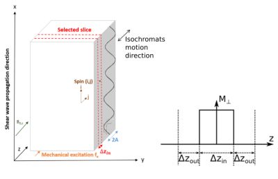
Constant gradient magnetic resonance elastography experiments on phantom and bovine liver.
Pilar Sango Solanas, Eric Van Reeth, Pauline M. Lefebvre, Hélène Ratiney, Elisabeth Brusseau, Denis Grenier, Steffen J. Glaser, Dominique Sugny, Olivier Beuf, Kevin Tse-Ve-Koon
Magnetic Resonance Elastography (MRE) is performed by the application of motion-sensitive gradients. In this study, RF pulses are designed with an optimal control algorithm to obtain a desired magnetization phase distribution. Such pulse, in presence of a constant gradient, allows to simultaneously perform spatially selective excitation and motion encoding. This offers some advantages when compared to standard MRE encoding strategy. Simulations, phantom and ex vivo experiments show that phase-to-noise ratios are improved. These results demonstrate that optimal control-based pulses can be used to encode motion in the MRE excitation phase with relevant advantages for further in vivo liver rat studies.
|
|
0684.
 |
19 |
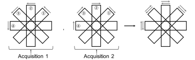
Motion robust distortion-free Arterial Spin Labeling
Jörn Huber, Marco Vicari, Matthias Günther
An improved reconstruction pipeline for arterial spin labeling (ASL) by means of a 3D GRASE PROPELLER (3DGP) acquisition is presented. Quality of ASL perfusion images is affected by motion artefacts, which can be mitigated by self-referred motion correction with 3DGP. However, since 3DGP is based on an EPI readout, geometric distortion is introduced in images, especially for long echo train lengths (ETL). The proposed method allows self-referred correction of motion as well as geometric distortion by playing each PROPELLER brick twice with toggled phase-encoded direction. The Topup technique then enables distortion correction followed by PROPELLER motion correction.
|
|
0685.
 |
20 |
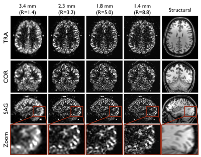
High Resolution Perfusion Imaging using Golden Angle Radial Arterial Spin Labelling
Thomas Okell, Mark Chiew
Arterial spin labeling perfusion imaging at clinical field strengths is generally confined to relatively coarse voxels (~4 mm), preventing the investigation of perfusion variations on small spatial scales and leading to problems with partial volume effects. In this work we demonstrate the ability of a golden angle radial approach combined with a regularized reconstruction technique to produce time-resolved perfusion images with isotropic voxel sizes lower than 2 mm. This was shown to improve grey matter definition and reduce partial volume effects.
|
|
0686.
 |
21 |
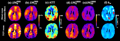
Mapping water exchange across the blood-brain barrier using three-dimensional diffusion-prepared arterial spin labeled perfusion MRI
Xingfeng Shao, Samantha J Ma, Marlene Casey, Lina D’Orazio, John M Ringman, Danny J.J. Wang
We present a novel pulse sequence and modeling algorithm to quantify the water exchange rate (kw) across the BBB without contrast, and to evaluate its clinical utility in a cohort of elderly subjects at risk of cerebral small vessel disease (SVD). A diffusion preparation module was integrated with pseudo-continuous ASL (pCASL) with background suppressed 3D GRASE readout. The results showed good reproducibility of kw measurements (ICC=0.75) in elderly subjects with repeated scans ~2 weeks apart. Average kw was increased in subjects with diabetes and hypercholesterolemia, and was correlated with vascular risk factors, cognitive function and white matter hyperintensities.
|
|
0687.
 |
22 |
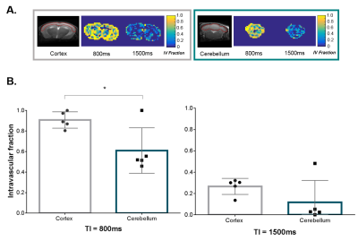
Regional Variation of Water Permeability at the Blood-Brain Interface in the Mouse Brain using Multi-TE ASL: The Role of Aquaporin-4
Yolanda Ohene, Ian Harrison, Payam Nahavandi , Ozama Ismail, Phoebe Evans , Eleanor Bird, Ole Ottersen , Erlend Nagelhus, David Thomas , Mark Lythgoe, Jack Wells
We apply a multi-TE ASL technique to the mouse brain to assess the regional variation of water permeability at the blood brain interface, and measure the expression of brain AQP4 water channels as a marker of water transport. We report a significant decrease in the intravascular fraction of the ASL signal in the cerebellum compared to the cortex, 0.61 (± 0.22) and 0.90 (± 0.08) respectively, which is consistent with a marked increase (~400%) in Aqp4 expression in the cerebellum. This technique is a promising tool to better understand the dynamic role of AQP4 in pathological conditions.
|
|
0688.
 |
23 |
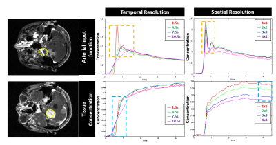
Ultra-High Resolution Whole-Brain DCE MRI makes Significant Difference in Estimating Permeability Maps for Cancer Patients
Joon Sik Park, Eun Ji Lim, Seung Hong Choi, Chul-Ho Sohn, Jaeseok Park
An ultra-high resolution, whole-brain DCE MRI with a temporal resolution of 1.5sec (3~4 times higher than conventional routine) and a spatial resolution of isotropic 1.0mm3 (5~6 times higher than conventional routine) is introduced in a real clinical setting to address problems related to arterial input function and partial volumes for precise estimation of vascular permeability information in brain cancer patients. Compared with conventional routine vendor-provided method, the proposed ultra-high resolution DCE MRI produces significant differences in permeability maps particularly for both Ktrans and ve.
|
 |
0689.
 |
24 |
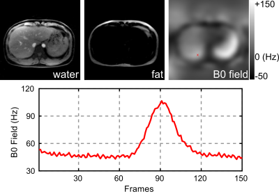
Dynamic Water/Fat Separation and Field Inhomogeneity Mapping at a Temporal Resolution of 40 ms
Zhengguo Tan, Dirk Voit, Jost Kollmeier, Martin Uecker, Jens Frahm
To achieve dynamic water/fat separation even in the presence of rapid physiological motions and large magnetic field inhomogeneities, this work presents a multi-echo multi-spoke radial FLASH sequence and a model-based non-linear inverse reconstruction. Asymmetric echoes are integrated into the sequence to shorten echo times. A spatial-smoothness constraint on field inhomogeneity maps is developed to counteract local minima in the non-convex inverse problem.
|
|
0690.
 |
25 |
Water/fat separation for distortion-free EPI with point spread function encoding
Zhangxuan Hu, Yishi Wang, Zijing Dong, Hua Guo
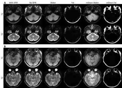
Effective removal of chemical-shift artifacts in EPI is a challenging problem especially with severe field inhomogeneity. In this study, water/fat separation thus fat suppression is achieved using the intrinsic signals of point spread function (PSF) encoded EPI (PSF-EPI), in which chemical-shift encoding is realized in the intermediate images with different time shifts. The results from three imaging regions show that this PSF-EPI based method can separate water/fat robustly. Thus fat signals can be effectively removed from EPI even with severe field inhomogeneity.
|
|
0691.
 |
26 |
Brain-component mapping with Inversion-Recovery bSSFP
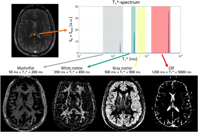
Julian Pfister, Martin Blaimer, Walter Kullmann, Andreas Bartsch, Peter Jakob, Felix Breuer
An inversion recovery (IR) bSSFP measurement allows to calculate quantitative parameter maps assuming mono-exponential signals. However, the measured signals often show multi-exponential behavior due to partial volume effects or tissue microstructure. With IR bSSFP it is possible to extract a spectrum of the apparent relaxation times T1* and hence to identify multiple components in each voxel. By integration over specific T1* ranges, different brain-components like white matter, gray matter or CSF can be mapped. In this work, we demonstrate that even short-living components such as myelin water are detected providing helpful information for diagnostic purposes, e.g. in neurodegenerative diseases.
|
|
0692.
 |
27 |
Ultra-high temporal resolution on the inversion recovery curve: new insight into T1 relaxometry of the human brain
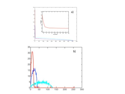
Ana-Maria Oros, Anna Weglage, N. Jon Shah
Tissue T1 relaxation is considered to be monoexponential. This is, however, an assumption, since it is seldom measured with sufficiently dense sampling of the relaxation curve. We hypothesized that a spatially-resolved investigation of tissue T1 relaxation curve with ultra-high temporal resolution might reveal previously unidentified characteristics of this fundamental NMR parameter. We imaged 10 healthy volunteers using a Look-Locker sequence with 460 time points 17ms apart on the inversion recovery curve. Denoising using PCA and NNLS decomposition of the recovery curves revealed, among others, a moderately short T1 component (4-500ms) in both WM and GM, tentatively assigned to myelin water.
|
|
0693.
 |
28 |
Improved Body Quantitative Susceptibility Mapping by Using a Variable-Layer Single-Min-Cut Graph-Cut Algorithm for Field-Mapping
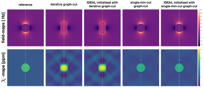
Christof Böhm, Maximilian Diefenbach, Jakob Meineke, Axel Haase, Dimitrios Karampinos
Body QSM relies on accurate magnetic field-mapping that accounts for the presence of fat and is robust to large background fields. This work proposes the application of a single-min-cut graph-cut-based pipeline aiming at accurate, non-smoothed and unwrapped field-mapping without the need for further computationally expensive or problematic voxel-wise fitting or unwrapping techniques. The used body QSM pipeline achieves higher accuracy in field-map and susceptibility values, which is demonstrated in a numerical phantom and in in-vivo spine datasets.
|
|
0694.
 |
29 |
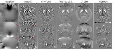
Single-step Quantitative Susceptibility Mapping Reconstruction without Brain Extraction using Deep Learning
Hongjiang Wei, Steven Cao, Fuhua Yan, Kristen Yeom, Chunlei Liu
We propose a deep learning-based single-step QSM reconstruction method that directly estimates magnetic susceptibility from total phase images without brain extraction. 209 healthy subjects with ages ranging from 11 to 82 years were employed for network training. The trained network provides superior image quality and high reconstruction speed compared to other single-step QSM methods. In addition, the elimination of the brain extraction step makes it possible to estimate susceptibility near the boundaries and explore the magnetic susceptibility of whole brain vascular images.
|
|
0695.
 |
30 |
Estimation of low frequency conductivity from Larmor frequency conductivity
Ulrich Katscher, Christoph Leussler, Nick Flaeschner, Fabian Wenzel
EEG source localization requires the conductivity map of the patient's head. Patient-individual conductivity maps can be measured in the framework of MRI using "Electric Properties Tomography" (EPT). However, EEG source localization requires the conductivity belonging to low frequency (LF) range instead radiofrequency (RF) range, as it is the case for EPT. To solve this problem, this study suggests two EPT measurements at different B0, and to transform the resulting conductivities from RF to LF via a simplified Cole-Cole-model. The approach has been validated with organic and non-organic phantoms as well as with volunteer brain experiments.
|
|
