|
2nd Hour
Poster: Cutting Edge CEST
Power Pitch Poster
Contrast Mechanisms
Monday, 13 May 2019
Power Pitch Theater C - Exhibition Hall
14:45 - 15:45
| |
|
Plasma # |
|
0142.
 |
31 |
Accelerate Parallel CEST Imaging with Dynamic Convolutional Recurrent Neural Network

Huajun She, Quan Chen, Shuo Li, Kang Yan, Xudong Chen, Xi Chen, Yuan Feng, Jochen Keupp, Robert Lenkinski, Elena Vinogradov, Yiping Du
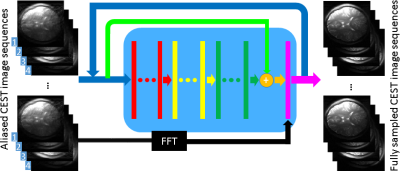
CEST is a new contrast mechanism in MRI. However, a successful application of CEST is hampered by its slow acquisition. This work investigates accelerating parallel CEST imaging using dynamic convolutional recurrent neural networks. This work is the first try to apply recurrent neural networks to accelerate CEST imaging, which jointly learns the spatial and Z-spectral features. The in vivo brain results show that the proposed method demonstrates a much better reconstruction quality of the human brain MTRasym maps than the traditional dynamic compressed sensing method, while the reconstruction time is one hundred times shorter.
|
|
0143.
 |
32 |
Denoising of Z-spectra for stable CEST MRI using principal component analysis
Johannes Breitling, Anagha Deshmane, Steffen Goerke, Kai Herz, Mark Ladd, Klaus Scheffler, Peter Bachert, Moritz Zaiss
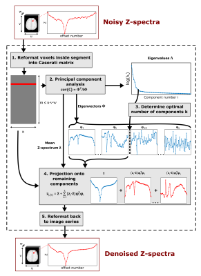
Chemical exchange saturation transfer (CEST) MRI allows for the indirect detection of low-concentration biomolecules by their saturation transfer to the abundant water pool. However, reliable quantification of CEST effects remains challenging and requires a high image signal-to-noise ratio. In this study, we show that principle component analysis can provide a denoising capability which is comparable or better than 6-fold averaging. Principle component analysis allows identifying similarities across all noisy Z-spectra, and thus, extracting the relevant information. The resulting denoised Z-spectra provide a more stable basis for quantification of selective CEST effects, without requiring additional measurements.
|
|
0144.
 |
33 |
Homogenous Excitation in Whole Brain CEST: Combination of Snapshot CEST and Multiple Interleaved Mode Saturation
Andrzej Liebert, Moritz Zaiss, Rene Gumbrecht, Patrick Liebig, Benjamin Schmitt, Frederik Laun, Arnd Doerfler, Michael Uder, Armin Nagel
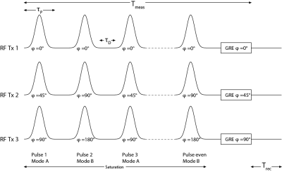
To perform Chemical Exchange Saturation Transfer MRI of the whole brain a homogeneous saturation and fast readout are required. To achieve a fast and robust 3D acquisition a spiral-centric-reordered GRE readout was used. In addition, a Multiple Interleaved Mode Saturation scheme was applied to mitigate B1+-inhomogeneity effects of the CEST saturation. Combination of these two methods allows acquiring a homogenous CEST contrast in a volume of approximately 220x220x45mm3 within an acquisition time of 7 min 26s.
|
 |
0145.
 |
34 |
A Novel Approach for Improved CEST Imaging with Real-Time Frequency Drift Correction
Ruibin Liu, Hongxi Zhang, Weiming Niu, Can Lai, Qiuping Ding, Weibo Chen, Sayuan Liang, Jinyuan Zhou, Dan Wu, Yi Zhang
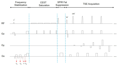
Chemical Exchange Saturation Transfer (CEST) imaging is highly sensitive to temporal B0 drift. Here, we proposed a novel frequency-stabilized CEST (FS-CEST) imaging sequence by adding a frequency stabilization module to the conventional non-frequency-stabilized CEST (NFS-CEST) sequence for correcting artifacts due to B0 drift in real time. The FS-CEST sequence was implemented in phantoms and 26 human volunteers, and generated substantially more stable magnetization transfer ratio asymmetry (MTRasym) spectra and amide proton transfer weighted (APTw) images than the conventional NFS-CEST sequence. The FS-CEST sequence provides an effective approach for B0 drift correction without additional scan time.
|
|
0146.
 |
35 |
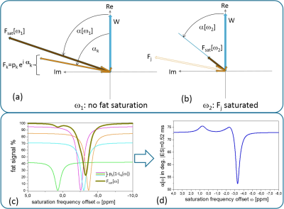
Z-Spectral Water-Fat Separation for APTw MRI in the Body using efficient Single-Echo Acquisitions
Jochen Keupp, Elena Vinogradov, Holger Eggers
An APTw/CEST-MRI technique to obtain water-only Z-spectra in the presence of fat is described. Water-fat separation in CEST is complicated by partial saturation of spectral fat components. Here, the saturation frequency-dependent water-fat phases are calculated using a multi-peak saturation model. A Z-spectrum acquisition with a single echo-shift is combined with a reference acquisition (S0) using 3 echo-shifts for water-fraction and B0-mapping. A B0-corrected, water-only Z-spectrum is obtained by complex rotations and weighted subtraction according to the water-fat phase. Volunteer examinations at 3T are shown (breast and abdominal). The single echo-shift technique offers a time-efficient means for water-fat separation in APTw/CEST-MRI.
|
|
0147.
 |
36 |
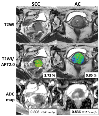
Amide proton transfer (APT) imaging of uterine cervical cancer; prediction of histological findings
Keisuke Ishimatsu, Akihiro Nishie, Yukihisa Takayama, Yoshiki Asayama, Yasuhiro Ushijima, Daisuke Kakihara, Tomohiro Nakayama, Koichiro Morita, Seiichiro Takao, Osamu Togao, Yoshihiro Ohishi, Kenzo Sonoda, Jochen Keupp, Hiroshi Honda
It is important to diagnose histological type and existence of parametrial invasion in uterine cervical cancer as correctly as possible because these factors are important in choosing treatment strategies or predicting prognosis. The objective of our study is to investigate whether amide proton transfer (APT) imaging is useful for evaluation of uterine cervical cancer. We compared the APT signal of uterine cervical cancer with different histological findings (histological type and existence of parametrial invasion) using three different durations of presaturation pulse.
|
|
0148.
 |
37 |
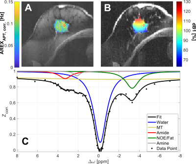
Fat corrected APT-CEST in the human breast at 7 Tesla: application to mamma carcinoma and dependency on menstrual cycle
Ferdinand Zimmermann, Andreas Korzowski, Lisa Loi, Johannes Breitling, Jan-Eric Meissner, Moritz Zaiss, Sebastian Bickelhaupt, Heinz-Peter Schlemmer, Mark Ladd, Peter Bachert, Daniel Paech, Sarah Schott, Steffen Goerke
The fat correction method enables robust APT-CEST quantification in the human breast and proved its suitability for examinations in vivo. We present to the extent of our knowledge the first APT-CEST contrast corrected for fat signal contribution, spillover, B1 field inhomogeneities and T1 relaxation in a breast cancer patient. The CEST contrast increased threefold compared to the measurement of a healthy volunteer. Repeated CEST imaging over the course of one menstrual cycle in one healthy woman did not reveal a hormonal correlation of APT contrast. A clinical study in healthy premenopausal volunteers will now investigate the dependency on menstrual cycle.
|
|
0149.
 |
38 |
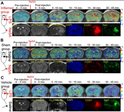
NaSA-CEST MRI: comparison with Gd-enhanced contrast for imaging brain inflammation
Xiaolei Song, Yanrong Chen, Chenwang Jin, Tao Liu, Chengyan Chu, Yuguo Li, Yue Yuan, Xiaowei He, Piotr Walczak, Jeff Bulte
Sodium salicylate (NaSA), a nonsteroidal anti-inflammatory drug and the main metabolite of aspirin, accumulates specifically in inflamed tissue. Since NaSA can be detected with CEST-MRI at millimolar concentrations, we investigated the use of NaSA-enhanced CEST MRI for in vivo mapping of brain inflammation, induced by intracerebral injection of lipopolysaccharide in mice. NaSA-CEST shows signal enhancement in the inflamed LPS-injected hemisphere, which was not observed in two control groups. NaSA-CEST exhibits distinct signal kinetics and enhanced regions from that of Gd-enhanced MRI, and shows correlations with histological staining of inflammatory markers, indicating its potential as a new platform for imaging neuroinflammation.
|
|
0150.
 |
39 |
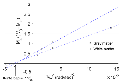
Proton exchange rate increases in MS lesions
Mehran Shaghaghi, Weiwei Chen, Alessandro Scotti, Haiqi Ye, Yan Zhang, Wenzhen Zhu, Kejia Cai
We have evaluated the performance of magnetic resonance proton exchange (Kex) imaging in vivo in characterization of gray matter, white matter as well as MS lesions. With informed consent, 10 control and 8 MS diagnosed subjects underwent a brain MRI on a 3T clinical-scanner. Kex maps were generated by pixel-wise ?tting of the omega plot constructed from four different saturation power (B1=1, 2, 3 & 4 µT). Kex values from gray matter (GM), white matter (WM) and lesions were calculated, and two-tailed paired Student’s T-test was used to classify each group. Kex was able to demarcates each region. The combined use of Kex mapping has potential to improve the early and speci?c diagnosis of MS.
|
|
0151.
 |
40 |
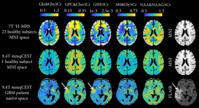
Multicolor metabolic quantitative CEST (mmqCEST): high resolution imaging of brain metabolites
Vitaliy Khlebnikov, Alex Bhogal, Mark Schuppert, Moritz Zaiss, Tobias Lindig, Benjamin Bender, Ulrike Ernemann, Klaus Scheffler, Peter Luijten, Hans Hoogduin, Dennis Klomp, Jeanine Prompers
Multicolor metabolic quantitative CEST (mmqCEST): high resolution imaging of brain metabolites
|
|
0152.
 |
41 |
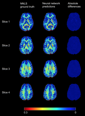
150× acceleration of myelin water imaging data analysis by a neural network
Hanwen Liu, Qing-san Xiang, Roger Tam, Alex MacKay, John Kramer, Cornelia Laule
In-vivo information of myelin content is desirable for studying many brain diseases and injuries which damage myelin. Myelin water imaging (MWI) is a validated and quantitative MR method to myelin. However, the data post-processing of MWI is mathematically complex and computationally demanding. The analysis typically takes several hours for a whole brain analysis, which limits its clinical applications. Our objective was to train a neural network as an alternative method for the MWI data analysis. We found this novel approach can accelerate MWI data analysis by over 150 times.
|
|
0153.
 |
42 |
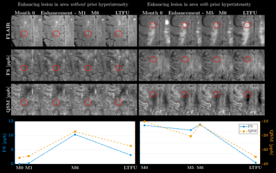
Contrasting frequency shifts and QSM in a longitudinal analysis of MS lesions to determine the nature of MR frequency and QSM signal changes
Vanessa Wiggermann, Enedino Hernandez-Torres, Irene Vavasour, Cornelia Laule, David Li, Anthony Traboulsee, Alexander MacKay, Alexander Rauscher
Magnetic susceptibility and MR frequency shifts in MS lesions are sensitive measures of tissue damage. However, the sensitivity of FS and QSM to magnetic susceptibility effects as well as changes in tissue microarchitecture complicate data interpretation in biological systems. By contrasting QSM and resonance frequency shift maps these two mechanisms may be differentiated. We observed that the signal shifts at enhancement are reflective of microstructural changes indicating formation of myelin debris, as similar FS and QSM changes were observed. Signal reductions in MS lesions 5yrs-post-enhancement however are only present on QSM, suggesting removal of myelin debris and axonal loss as the underlying mechanisms.
|
|
0154.
 |
43 |
Impact of unilateral carotid artery stenosis on white matter fiber orientation effects of mq-BOLD derived oxygen extraction fraction
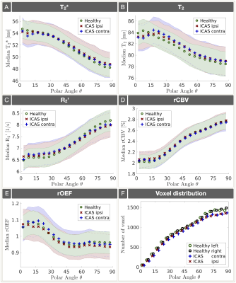
Stephan Kaczmarz, Jens Goettler, Andreas Hock, Claus Zimmer, Fahmeed Hyder, Christine Preibisch
Assessment of relative oxygen extraction fraction (rOEF) in white matter (WM) by multiparametric quantitative-BOLD (mq-BOLD) has highest clinical relevance, but was so far limited due to known WM anisotropy effects. Here, we present data from a clinical study in 29 internal carotid artery stenosis (ICAS) patients and 30 age-matched healthy controls (HC). The major aim was to characterise the ICAS impact on T2*, T2, R2’, rCBV and rOEF orientation dependencies in WM. Our results show very similar rOEF orientation dependencies for ICAS-patients compared to HC and low average rOEF orientation errors of 4.5% indicating potentially meaningful rOEF evaluations in WM.
|
|
0155.
 |
44 |
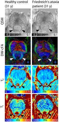
Quantitative assessment of the degeneration of the superior cerebellar peduncle in Friedreich’s ataxia at 7 T: susceptibility, diffusion anisotropy, and T2 and T1 relaxometry
Sina Straub, Julian Emmerich, Stephanie Mangesius, Elisabetta Indelicato, Mark Ladd, Sylvia Boesch, Elke Gizewski
Friedreich’s ataxia is a rare disease involving degenerative processes within white matter fiber tracts, spinal nerves and the cerebellum. A correlation of patients’ clinical status and superior cerebellar peduncle atrophy has been shown in MR volumetry studies. The ongoing ultra-high field study presented here assesses the degeneration of the superior cerebellar peduncle in Friedreich’s ataxia with quantitative MR parameters – susceptibility, diffusion anisotropy, and T2 and T1 relaxometry. Statistically significant differences between fractional anisotropy as well as T2 values in patients and healthy controls could be observed, indicating that these quantitative MRI methods potentially provide valuable biomarkers to assess the course of Friedreich’s ataxia.
|
|
0156.
 |
45 |
Glucose Uptake in Mouse Brain Detected by MRI Frequency Shifts with a Jump-Return Sequence
Zhiliang Wei, Haifeng Zeng, Lin Chen, Kannie Chan, Xiang Xu, Issel Lim, Xu Li, Hanzhang Lu, Peter van Zijl, Jiadi Xu
Neuronal activity relies on glucose metabolism for energy maintenance and abnormalities in glucose uptake and metabolism constitute a potential biomarker for many disorders, including neurodegenerative diseases. Existing MRI techniques for monitoring glucose uptake and transportation often suffer from insufficient detection sensitivity. Here, we demonstrate a jump-return MRI (JR-MRI) method with high sensitivity for monitoring glucose uptake via tracing the water-frequency shift induced by chemical exchange. Conventional MRS was performed to validate the delivery of glucose to the brain.
|
|
