|
2nd Hour
Poster: Microstructure Modelling & Validation
Power Pitch Poster
Diffusion
Thursday, 16 May 2019
Power Pitch Theater A - Exhibition Hall
09:15 - 10:15
| |
|
Plasma # |
|
1005.
 |
1 |
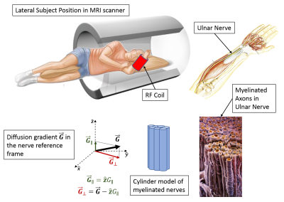
The relationship between diffusion MRI-derived axon diameters and conduction velocities in human peripheral nerves

Alexandru Avram, Zhen Ni, Felipe Vial, Amber Simmons, Adam Bernstein, Giorgio Leodori, Sinisa Pajevic, Richard Coppola, Mark Hallett, Peter Basser
From diffusion-weighted MRIs with large q-values we estimated average axon diameters (AADs) in the median and ulnar nerves of healthy volunteers. We described signals using a tissue model of restricted and hindered diffusion and employed the multiple correlation function framework to account for all gradient pulses. Nerve conduction velocity distributions in the ulnar nerves were measured using electrophysiological techniques. Along these nerves, AADs correlated with average nerve conduction velocities supporting the linear dependence with propagation speed previously reported in the classical literature. This work presents pilot data suggesting the possibility of inferring inter-cortical latencies from whole-brain diffusion MRI.
|
|
1006
 |
2 |
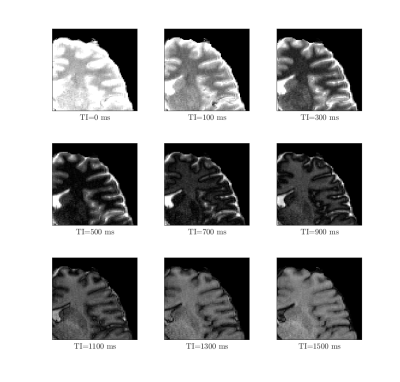
Exploring diffusion properties in neocortical grey matter using inversion recovery diffusion weighted imaging
Video Permission Withheld
Fakhereh Movahedian Attar, Bibek Dhital, Luke Edwards, Nikolaus Weiskopf
We measured inversion recovery prepared diffusion signal for grey matter. We found that the mean diffusivity (MD) in grey matter drops significantly for TIs higher than the nulling TI. MD in GM dropped from around 0.8×10⁻³ mm2/s before the null point to around 0.6×10⁻³ mm2/s immediately after the null point. Our findings suggest that the neocortical gray matter could be characterized using a two dimensional T1-diffusion correlation measurement.
|
|
1007.
 |
3 |
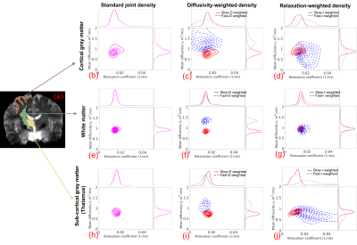
Joint RElaxation-Diffusion Imaging Moments (REDIM) to probe tissue microstructure
Lipeng Ning, Borjan Gagoski, Daniel Park, Filip Szczepankiewicz, Carl-Fredrik Westin, Yogesh Rathi
The joint probability distribution of diffusivity and T2-relaxation coefficient provides useful information to characterize tissue microstructure. A standard approach for estimating this joint distribution relies on estimating the inverse Laplace transform from a large number of measurements which is not only infeasible in clinical settings but also numerically unstable. In this work, we introduce a novel approach, termed REDIM, to probe tissue microstructure using the statistical properties of a family of scaled distribution functions. In particular, we use specific functions to zoom into the joint probability function to robustly estimate different features of the underlying diffusion-relaxation processes. We show that this approach can be reliably implemented for use with in-vivo diffusion MRI (dMRI) data.
|
|
1008.
 |
4 |
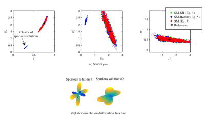
Biophysical modeling of the white matter: from theory towards clinical practice
Rafael Henriques, Chantal Tax, Noam Shemesh, Jelle Veraart
We address the degeneracy of the diffusion standard model and improve the precision and accuracy in parameter estimation, thus promoting clinical applicability. Acquisition of additional data is not required; instead, we introduce a more robust and accurate estimator by fitting the standard model directly to diffusion-weighted data rather than its rotational invariants. We are able to overcome the implicit assumption of data being shelled in terms of b-values. This enables the correction of gradient nonlinearities to avoid biases in model parameter estimation, whereas revising the optimal experimental design demonstrates that non-shelled encoding schemes are favorable in terms of achievable precision.
|
|
1009.
 |
5 |
Effective medium theory of multiple diffusion encoding
Sune Jespersen, Els Fieremans, Dmitry Novikov
We consider the effect of tissue heterogeneity on diffusion acquisitions with multiple encodings. Previously, these signals were analyzed in Gaussian compartments, or disconnected pores. Here we assume a more realistic situation where a compartment cannot be considered Gaussian (uniform) at finite diffusion times, and derive the 4th-order contributions in the diffusion weightings, that distinguish the double-diffusion-encoding (DDE) signal from its single-encoding counterpart. We specifically identify terms odd in the DDE diffusion wave-vectors, which emerge due to local tissue heterogeneity but absent when compartments are Gaussian. Our results are expressed via the dynamical exponent related to the disorder universality class, and agree with Monte Carlo simulations.
|
|
1010.
 |
6 |
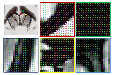
Deep learning 3D white matter fiber orientation from 2D histology: pulling 3D rabbits out of 2D hats
Kurt Schilling, Vishwesh Nath, Samuel Remedios, Roza Bayrak, Yurui Gao, Justin Blaber, Adam Anderson, Bennett Landman
Most histological analysis of tissue microstructure is inherently 2D. In this work, we implement a deep learning approach to extract 3D microstructural measures from 2D microscopy images. Specifically, we train a neural network to estimate 3D fiber orientation distributions from myelin-stained micrographs. We apply this technique to an entirely unseen brain, which suggests the potential to use this methodology on consecutive 2D slices to investigate 3D structural connectivity using “myelin-stained-tractography” at resolutions much higher than possible with current diffusion MRI practices. There is potential to use similar techniques to estimate a number of 3D metrics from common 2D histological contrasts.
|
 |
1011.
 |
7 |
Multi-diffusion time DWI to detect altered microvessel structure in a rat model of hypertension
Lauren Scott, Ben Dickie, Shelley Rawson, Graham Coutts, Tim Burnett, Geoff Parker, Stuart Allan, Laura Parkes
The role of microvascular pathology in the development and progression of dementia is currently unclear. Non-invasive methods for imaging microvessel structure are needed to study cerebrovascular alterations in-vivo. The time for water molecules to change direction relative to the diffusion time alters the measured pseudo-diffusion coefficient of intra-voxel incoherent motion (IVIM). Using multi-diffusion time multi-b-value data and a velocity autocorrelation model, the capillary segment length (l) can estimated. In this study we validate hippocampal l against vascular corrosion casts, then apply the method to study vascular structure and function in a rat model of hypertension in comparison with age-matched controls.
|
|
1012.
 |
8 |
Revisiting double diffusion encoding MRS in the mouse brain at 11.7 T: what microstructural features is metabolite diffusion sensitive to?
Mélissa Vincent, Julien Valette
Double diffusion encoding MRS (DDE-MRS) was shown to be highly sensitive to cell microstructure, in particular cell fiber diameter. Here we revisit the ability of DDE-MRS to probe microstructure by quantifying DDE-MRS signal for six metabolites with high accuracy. We show that signal modulation differs for neuronal and glial metabolites, yielding larger fiber diameters for glia when using a model of isotropically oriented cylinders, as previously reported. However, fiber diameters appear overestimated. Further data acquisition and modeling suggests DDE-MRS is not only sensitive to fiber diameter but also to other microstructural features, such as cell body diameter or fiber length.
|
|
1013.
 |
9 |
Validating the single fODF and single microstructure kernel assumption in vivo using local SHARD features
Daan Christiaens, Jelle Veraart, Lucilio Cordero-Grande, Anthony Price, Jana Hutter, Joseph Hajnal, J-Donald Tournier
Most biophysical models of diffusion in the white matter assume that the diffusion MRI signal can be written as the convolution of a single fibre orientation distribution function and a single microstructural kernel, thereby ignoring microstructural differences between fascicles being captured in the same voxel. Here, we validate this assumption by various model selection approaches, i.e., Relative variance explained, F-test of 1- and 2-component models, and Component noise likelihood, and conclude that biologically more realistic, but mathematically more complex versions of the Diffusion Standard Model do not significantly improve the goodness-of-fit, even in case of rich diffusion MR data.
|
 |
1014.
 |
10 |
Effect of cell complexity and size on diffusion MRI signal: a simulation study
Andrada Ianus, Ross Callaghan, Daniel Alexander, Hui Zhang, Marco Palombo
Mapping tissue microstructure in gray matter is becoming an important field of research in diffusion MRI. Previous studies have focused on characterizing different features separately, such as neurite density, branching order or soma size. However, the combined effect of different cell morphologies and sizes on the diffusion MRI signal has not yet been investigated. This work employs numerical simulations in more realistic cell configurations to investigate the effect of soma diameter, branch diameter and branching order, on the dMRI signal and its time dependence.
|
|
1015.
 |
11 |
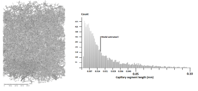
Quantitative characterization of a capillary-network MRI phantom using restricted diffusion analysis
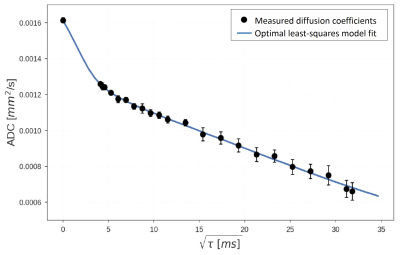
Astrid Mayr, Moritz Schneider, Thomas Gaass, Katia Parodi, Jens Ricke, Julien Dinkel, Olaf Dietrich
An artificial 3D capillary-network phantom (made out of melt-spun sugar fibers) was characterized with diffusion-time-dependent diffusion-weighted MRI. Restricted diffusion was analyzed based on a two-compartment (cylinders and spheres) model; diffusion coefficients were simulated with a simple random-walk model of spins in 2D and 3D spherical geometries. The best model fit agreed very well with the measured diffusion-time-dependent diffusion coefficients and resulted in a mean diameter of 142±2 µm of the spheres and a mean diameter of 9.7±0.7 µm of the capillary tubes (cylinders) with a capillary fraction of fcyl=35±1%.
|
|
1016.
 |
12 |
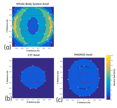
High Gradient Amplitude and High Slew Rate Oscillating Gradient Diffusion-Encoding for Human Brain Imaging
Ek Tan, Tim Sprenger, Eric Fiveland, Yihe Hua, Matt Bernstein, Vincent Ho, Thomas Foo
Two high amplitude and slew rate head-gradient MRI systems (C3T: 80mT/m, 700T/m/s, and MAGNUS: 200 mT/m, 500 T/m/s) with significantly better performance than clinical whole-body MRI have been developed. These allow for microstructure-sensitive, oscillating-gradient-spin-echo (OGSE) diffusion-encoding to be feasibly applied for human brain imaging. An analysis of waveforms at varying gradient performances reveals that peripheral nerve stimulation and gradient slew rates limit OGSE frequencies, while gradient strength limits b-values and echo times. In vivo human imaging was performed at 3T, demonstrating significantly increased measured diffusivity in OGSE diffusion tensor imaging (DTI) as compared to standard DTI.
|
|
1017.
 |
13 |
Mapping tumour response to radiotherapy using diffusion model comparison
Damien McHugh, Grazyna Lipowska-Bhalla, Muhammad Babur, Yvonne Watson, Penny Hubbard Cristinacce, Josephine Naish, Jamie Honeychurch, Kaye Williams, James O'Connor, Geoff Parker
This work evaluates how the suitability of two diffusion MRI models varies spatially within tumours at the voxel level and in response to radiotherapy, potentially allowing inference of qualitatively different tumour microenvironments. Models of restricted and free diffusion were compared, with regions well-described by the former hypothesised to reflect cellular tissue, and those well-described by the latter expected to reflect necrosis or oedema. Results suggest spatial and radiotherapy-related variation in the models’ suitability for describing diffusion in tumours, with a post-therapy decrease in the proportion of tissue characterised by restricted diffusion. Within restricted diffusion regions, microstructural parameters were sensitive to radiotherapy-induced changes.
|
|
1018.
 |
14 |
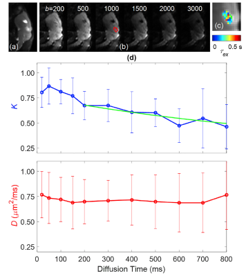
Comparison of Tumor Water Exchange Rate and Intracellular Water Lifetime Measured by Diffusion MRI
Jin Zhang, Sungheon Kim
In vivo measurement of cellular-interstitial water exchange rate remains non-trivial. Its development is also hampered by the lack of a gold standard method for validation. In this study, we have used two complementary diffusion MRI methods to measure the water exchange rates. Time-dependent diffusional kurtosis imaging was used to measure the water exchange rate (τexτex)
between the intra- and extra-cellular spaces, while a constant gradient diffusion MRI experiment was used to measure the intracellular water lifetime (τiτi).
These two measurements were conducted using GL261, a mouse glioma tumor model.
|
|
1019.
 |
15 |
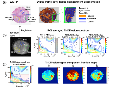
Characterization of Prostate Microstructure Using Diffusion-Relaxation Correlation Spectrum Imaging and Comparison to Digital Histopathology
Zhaohuan Zhang, Clara Magyar, Alan Priester, Sepideh Shakeri, Sohrab Afshari Mirak, Amirhossein Mohammadian Bajgiran, Anthony Sisk, Kyunghyun Sung, Robert Reiter, Dieter Enzmann, Steven Raman, Holden Wu
Prostate microstructural MRI has the potential to improve prostate cancer (PCa) detection and characterization by resolving the signal signatures of sub-voxel microscopic tissue compartments. Recently, a new model, Diffusion-Relaxation Correlation Spectrum Imaging (DR-CSI), was developed but has not been applied to prostate imaging. In this work, we investigated Prostate DR-CSI and compared DR-CSI features (signal component fractions) to histopathology features (microscopic tissue compartments) derived from digital pathology, to evaluate this new multi-component signal model for prostate microstructure characterization.
|
|
