|
2nd Hour
Poster: Diffusion: Applications
Power Pitch Poster
Diffusion
Tuesday, 14 May 2019
Power Pitch Theater B - Exhibition Hall
09:15 - 10:15
| |
|
Plasma # |
|
0342.
 |
16 |
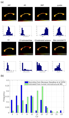
Mapping axonal conduction velocities from in vivo MRI data

Mark Drakesmith, Umesh Rudrapatna, Derek Jones
The ability to infer axonal conduction velocities (CV) non-invasively from in vivo neuroimaging is of huge biological importance. Having previously shown that accurate CV estimates are feasible with MRI-measurable parameters, we show here the sensitivity of MRI-derived CV estimates to modelling errors and acquisition noise. We show that for the parameters typically seen in white matter axons, there is less than 5% error in CV estimates. Application to a human diffusion/relaxometry dataset generates CV estimates in corpus callosum that are close to those observed in electrophysiology literature. This illustrates further the feasibility of estimating CV from in vivo microstructural MRI.
|
|
0343.
 |
17 |
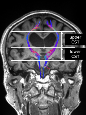
Evaluating microstructure of the corticospinal tract in normal pressure hydrocephalus with diffusion MRI using oscillating gradient spin-echo
Ryusuke Irie, Kouhei Kamiya, Masaaki Hori, Michimasa Suzuki, Koji Kamagata, Tomoko Maekawa, Shohei Fujita, Yuki Takenaka, Issei Fukunaga, Madoka Nakajima, Masakazu Miyajima, Katsutoshi Murata, Shigeki Aoki
We evaluated the corticospinal tract (CST) of normal pressure hydrocephalus (NPH) patients using the oscillating gradient spin-echo (OGSE) method. Diffusion time can be reduced greatly with OGSE and properties of the microstructure can be evaluated in more detail by collecting data using multiple diffusion times. The diffusivity of the CST increased and the diffusion time dependence was small in NPH patients compared with healthy controls. It is possible to estimate the structural change of CST in NPH by using multiple diffusion times with OGSE.
|
 |
0344.
 |
18 |
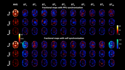
Investigation of the dependence of free water and pseudo-diffusion MRI estimates on the cardiac cycle
Alberto De Luca, Suzanne Franklin, Carlo Lucci, Jeroen Hendrikse, Martijn Froeling, Alexander Leemans
With diffusion MRI (dMRI) data at multiple diffusion weightings it is possible to quantify the relative fractions of multiple water pools. In this work, we investigated changes in free water diffusion and micro- and macro-vascular pseudo-diffusion during the cardiac cycle. Further, we propose a data driven method to bin the dMRI signals according to the cardiac phase. dMRI at 4 diffusion weightings was acquired 80 times with short repetition time. A multi-exponential fit of the binned data showed increases of free water in white matter and periventricular areas, and opposite increases/decreases for micro- and macro-vascular pseudo-diffusion in grey matter, respectively.
|
|
0345.
 |
19 |
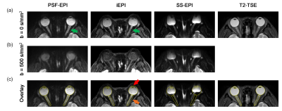
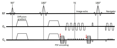
Distortion-free diffusion-weighted imaging of the eyeballs and the optic nerves using PSF-EPI
Jieying Zhang, Yishi Wang, Yuhui Xiong, Hua Guo
Distortion-free diffusion-weighted imaging can provide high fidelity information for the diagnosis of ophthalmological diseases. Traditional echo-planar imaging technique suffers from geometric distortion in the skull base. This study applies a point spread function encoded technique in eyeball imaging. This technique can generate distortion-free DWI of the eyeballs and the optic nerves with high efficiency. Its resistance to geometric distortion and ability to generate accurate ADCs were validated on phantom and healthy subjects. This technique has the potential in providing accurate information for diagnosis and characterizing diseases that are difficult to distinguish by traditional imaging methods.
|
|
0346.
 |
20 |
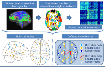
Defining subnetworks by rich-club architecture in 16p11.2 deletion syndrome reveals differential structural white-matter alterations
Ai Wern Chung, Banu Ahtam, P. Ellen Grant, Kiho Im
16p22.1 deletion syndrome has been implicated in disorders such as autism and is associated with developmental deficits, including delay in language acquisition. Widespread DTI white-matter alterations have been identified in patients but the structural organisation using network theory has yet to be investigated. Using rich-club nodes to stratify the connectome into rich-club, feeder and seeder subnetworks, we compute graph topological measures in children with 16p11.2 deletion. While rich-club regions were similar to those in Controls, differential alterations in connectivity and topology suggest a reorganisation of subnetworks in patients with the feeder subnetwork possibly compensating for deficits in the rich-club.
|
|
0347.
 |
21 |
Topological reorganizations of the white matter structural networks in early blind adolescents
Zhifeng Zhou, Leilei Shi, Xia Liu, Long Qian, Hengguo Li
Previous neuroimaging studies have revealed functional neuroplasticity after visual deprivation. However, the structural reorganization of the brain induced by visual deprivation is rarely reported, and the mechanism of structural neuroplasticity remains unclear. This study is one of the first attempts to investigate structural neuroplasticity in the white matter networks at both the global and the local node levels using the Diffusion Tensor Imaging data and the graph theory analysis method. The results demostrate disrupted brain structure and reorganized brain structural networks in early blind adolecents, which provide evidence of structural neuroplasticity induced by visual deprevation.
|
|
0348.
 |
22 |
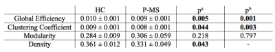
Using microstructure informed tractography to reduce discrepancy in the density of structural connectomes. An application to Multiple Sclerosis.
Simona Schiavi, Maria Petracca, Matteo Battocchio, Mohamed Mounir El Mendili, Matilde Inglese, Alessandro Daducci
Graph theory is a valuable framework to study brain connectivity and has been widely applied to investigate pathological conditions such as Multiple Sclerosis (MS). However, differences in topology between groups of subjects could be affected by discrepancy in density. Here we propose to use microstructure informed tractography to directly account for such density differences and provide a fair comparison between MS patients and healthy subjects. Our results show the capability of the proposed method to control for density and support the appropriateness of global efficiency and clustering coefficient for the characterization of comprehensive functions such as cognition in MS.
|
|
0349.
 |
23 |
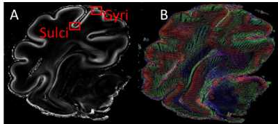
Gyri and sulci microstructural differences in the sheep brain during cortical folding assessed by diffusion MRI.
Yohan van de Looij, Sebastian Quezada, David Walker, Nadia Hale, Mary Tolcos, Petra Hüppi, Stéphane Sizonenko
Despite a large number of studies assessing cerebral development, some of the events underlying folding of the cerebral cortex remain unclear, especially those concerning micro-architectural differences between outward (gyri) and inward (sulci) folds. The aim of this work was twofold: 1) depict cortical microstructure differences between gyri and sulci and 2) assess the potential of NODDI in the understanding of cortical folding. Gyri and sulci present different maturation timelines leading to microstructural differences. Diffusion imaging is a powerful tool to probe accurately these differences. These results are of high interest for the understanding of cortical folding process.
|
|
0350.
 |
24 |
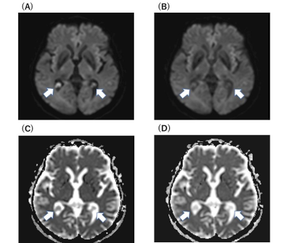
Cholesteatomas Detection using PSF-encoded EPI DWI
Guangqi Li, Yishi Wang, Yingying Shang, Yufei Qiao, Hua Guo
DWI has proved to be valuable in detection of cholesteatoma, but there still remain challenges. Susceptibility artifacts and geometric distortions in single-shot EPI DWI affect the identification of subtle nidus. TSE-based DWI can reduce susceptibility-induced artifacts, but the disadvantages are SNR degradation and T2-blurring. In this work, point-spread-function (PSF) encoded EPI DWI was employed to overcome the aforementioned limitations for cholesteatomas detection. This technique has the capability to acquire high-quality, distortion- and blurring-free diffusion images with more accurate lesion boundary.
|
|
0351.
 |
25 |
Diffusion Time Dependence in the Evaluation of Choroid Plexus Cysts
Yutaka Ikenouchi, Tomoko Maekawa, Masaaki Hori, Katsutoshi Murata, Thorsten Feiweiwer, Christina Andica , Shohei Fujita, Ryusuke Irie, Akifumi Hagiwara, Kouhei Kamiya, Akihiko Wada, Shigeki Aoki
To obtain information regarding the internal structure of choroid plexus cysts (CPCs) that appear hyperintense on DWI, we investigated the ADC values acquired with a shorter diffusion time using an oscillating gradient spin-echo (OGSE). The ADC values for twenty-seven patients with CPCs were measured with effective diffusion time (Δeff) of 6.5 and 35.2 ms. The ADC values of CPCs were significantly higher at the Δeff of 6.5 ms than with Δeff of 35.2 ms. The dependence of ADC values on the diffusion time in CPCs suggested spatially restricted diffusion.
|
 |
0352.
 |
26 |
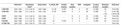
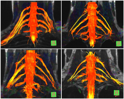
Fast High-Resolution Diffusion Tensor Imaging of the Cervical Spinal Cord Using Point-Spread-Function Encoded EPI (PSF-EPI)
Sisi Li, Yishi Wang, Yong Hai, Hanwen Zhang, Hua Guo
Diffusion tensor imaging (DTI) of spinal cord holds great promise to aid diagnosis of spine-related diseases. However, it is greatly limited in clinics by the physiological motion induced artifacts and susceptibility inhomogeneity induced distortions. To reduce distortions in DTI, various multi-shot EPI (MS-EPI) methods have been proposed. However, the acquisition time is prolonged due to multiple shots. In this study, we achieved cervical spine DTI using only 6 shots based on a distortion- and blurring- free MS-EPI technique, Point-Spread-Function Encoded EPI (PSF-EPI). The acquisition time is within 5 minutes. The efficacy of PSF-EPI is demonstrated on healthy volunteers and patients.
|
|
0353.
 |
27 |
Diffusion Tensor Imaging Tractography for Diagnosing Traumatic Brachial Plexus Root Avulsions: A Proof-of-Concept Study
Ryckie Wade, Steven Tanner, John Ridgway, James Rankine, Irvin Teh, David Shelley, Brian Chaka, Grainne Bourke
Due to limitations in the accuracy of clinical MRI, adults with traumatic brachial plexus injuries (BPI) undergo major exploratory surgery to define their injury. Early exploration and reconstruction restores limb function and improves quality of life. Diffusion tensor imaging (DTI) tractography offers the potential to replace exploratory surgery by assessing the continuity of the roots of the brachial plexus. Twenty healthy adults were used for sequence development on a 3 Tesla system and the protocol validated on 12 patients with known patterns of BPI. DTI reliably reconstructs the normal and injured brachial plexus with high fidelity and superior diagnostic accuracy.
|
|
0354.
 |
28 |
Diffusion kurtosis imaging in quantitative diagnosis of nonalcoholic fatty liver disease: a rabbit model study
Xianfu Luo, Chang li, Weiqiang Dou, Yun Peng, Jing Ye
We aimed to investigate if diffusion kurtosis imaging (DKI) can be applied to assess nonalcoholic fatty liver disease (NAFLD) by providing fractional anisotropy (FA), mean diffusion (MD) and mean kurtosis (MK). We used DKI derived parameters to analyze NAFLD in a rabbit model and compared them with the apparent-diffusion-coefficient (ADC) from a mono-exponential diffusion-weighted imaging model. While FA showed comparable results with the severity of NAFLD group, MD and MK indicated more robust performance in the diagnosis of nonalcoholic steatohepatitis (NASH) compared with ADC. We therefore demonstrated that DKI had potential in stratifying NAFLD and early diagnosis of NASH.
|
|
0355.
 |
29 |
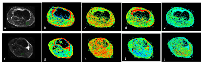
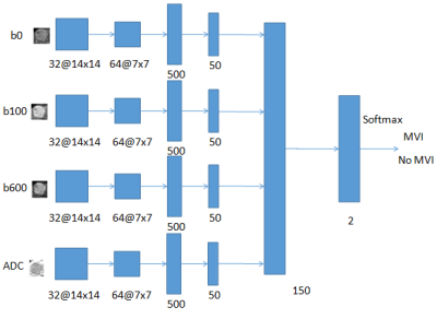
Can preoperative Diffusion-weighted MR predict the Microvascular invasion of hepatocellular carcinoma? A deep learning evaluation
Wu Zhou, Hanqiu Ju, Wanwei Jian, Hui Huang, Shaoyang Men, Honglai Zhang
Microvascular invasion (MVI) of hepatocellualr carcinoma(HCC) is regarded as the most important factor associated with the success of curative resection and the outcome after liver transplation. As the MVI prediction can only be ultimately determined by the histopathological features of the tumor cells, numerous works have attempted to predict the MVI of HCC based on noninvasively preoperative images. Diffusion-weighted MR has also shown to be effective for MVI prediction based on signal intensities of lesions and the apparent diffusion coefficient (ADC). In this work, the emerging deep learning technique is used for MVI prediction of HCC based on DWI.
|
|
0356.
 |
30 |
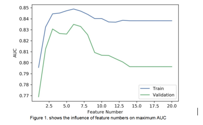
Exploring the Application of an Multi-Advanced-Diffusion-Model-Based Radiomics Method in Grading of Intestinal Fibrosis of Crohn's Disease
Li Huang, Xuehua Li, Siyun Huang, Mengzhu Wang, Canhui Sun, Shiting Feng, Xu Yan, Yang Song, Ziping Li
In the present study, we investigated a multiple diffusion-model-based radiomics model in grading of bowel fibrosis of Crohn's disease (CD) patients. We used histogram features derived from parameters of DWI, IVIM and DKI models and chose SVM as the classifier to construct a prediction model. The results showed that the most accurate prediction was achieved by incorporating the following 6 features into a nomogram, including the DKI-related histogram parameters (mean D, mean K, 10th percentiles of K) and IVIM-related parameters (mean D0-2000, mean D*0-2000, 90th percentiles of f), with AUC and accuracy reached 0.835 and 0.833, respectively.
|
|
