|
2nd Hour
Poster: Diffusion MRI: Acquisition, Reconstruction & Artefact Correction
Power Pitch Poster
Diffusion
Monday, 13 May 2019
Power Pitch Theater A - Exhibition Hall
17:00 - 18:00
| |
|
Plasma # |
|
0222.
 |
1 |
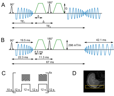
A GRANDIOSE sequence to time-lock BOLD and diffusion-weighted fMRI contrasts in humans using ultra-strong gradients and spirals

Suryanarayana Umesh Rudrapatna, Lars Mueller, Melissa Wright, Derek Jones, Richard Wise
Diffusion-weighted fMRI (dfMRI) has been suggested to provide more direct and specific correlates to neuronal activation than BOLD fMRI. However, its underpinnings are debated. A sequence that captures BOLD and dfMRI contrasts simultaneously can play a vital role in elucidating the dfMRI contrast mechanisms. Hence, we developed a sequence that leverages the ultra-strong gradients for diffusion-weighting and spiral-in and spiral-out trajectories to acquire BOLD and dfMRI contrasts near-simultaneously. We demonstrate its functionality using visual stimulation in humans. This novel sequence enables a direct comparison between BOLD and dfMRI contrasts and offers new opportunities to improve our understanding of these contrasts.
|
 |
0223
 |
2 |
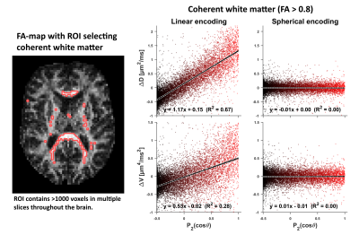
Is spherical diffusion encoding rotation invariant? An investigation of diffusion time-dependence in the healthy brain
Video Permission Withheld
Filip Szczepankiewicz, Samo Lasic, Markus Nilsson, Henrik Lundell, Carl-Fredrik Westin, Daniel Topgaard
Recent advances in diffusion weighted MRI have reignited interest in spherical (or isotropic) diffusion encoding. For such encoding to reach high efficiency (minimal echo time), the gradient waveforms have irregular shapes, by design. As such, they lack a well-defined diffusion time and can even be spectrally anisotropic. Most analysis methods based on such encoding assume that diffusion is multi-Gaussian, i.e., that the diffusion is not time-dependent. Since this is a central assumption, we investigate if spherical diffusion encoding is indeed rotation invariant, or if the diffusion time anisotropy has a discernible effect on the diffusion weighted signal in healthy brain.
|
|
0224.
 |
3 |
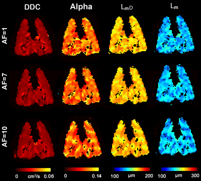
Application of an Extended Stretched-Exponential Model for Morphometric Analysis of Accelerated Diffusion-weighted 129Xe MRI of the Rat Lung
Alexei Ouriadov, Matthew Fox, Andras Lindenmaier, Elaine Stirrat, Grace Parraga, Giles Santyr
Hyperpolarized 129Xe pulmonary MRI is poised for clinical translation due in part to the clinical-relevance of 129Xe MRI biomarkers of lung disease. A rapid multi-b diffusion-weighted 129Xe MRI requires for clinical morphometry due to the challenges in acquiring a fully-sampled dataset during the relatively short 10-16sec breath-holds. Therefore, in this proof-of-concept evaluation, our objective was to measure morphometry estimates in a small group of control-rats as well as rats with early stage radiation-induced-lung-injury, and compare the stretched-exponential-model based morphometry estimates for three different cases: 1) fully-sampled k-space 2) 85% retrospectively under-sampled k-space, (acceleration factor (AF)=7), and 3) 90% retrospectively under-sampled (AF=10) k-space.
|
|
0225.
 |
4 |
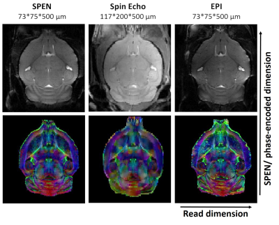
SPatiotemporal ENcoding (SPEN) at ultra-high fields: Applications to high resolution (<100 µm isotropic) in vivo mouse brain DTI
Maxime Yon, Qingjia Bao, Rafael Henriques, Noam Shemesh, Lucio Frydman
SPEN can be a valuable alternative to Spin Echo EPI, especially at very high fields where sensitivity is maximized but magnetic field susceptibility artifacts become important. This work presents a new Paravision® 6 software package capable of acquiring and processing SPEN data, incorporating single/multishot acquisitions, motion correction, image zooming and multiple contrast possibilities such as CEST, diffusion or multi-echo acquisition. The power of this method is exemplified with in vivo 2D and 3D diffusion tensor imaging (DTI) studies on whole and zoomed mouse brain regions, acquired at 15.2T utilizing a cryoprobe and reaching isotropic resolution of 75 µm.
|
|
0226.
 |
5 |
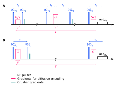
Storing phase information in the longitudinal direction: Experimental verification of double diffusion encoding with stimulated echoes applied to closed pores
Kerstin Demberg, Frederik Laun, Peter Bachert, Tristan Kuder
When applying double diffusion encoding (DDE) to arbitrarily shaped closed pores, non-vanishing imaginary parts in the diffusion signal arise, which allow determining the average shape of the pores in the considered volume element. Key limitations in such experiments are the available gradient strength and reaching the diffusion long-time limit restricted by T2 decay. When incorporating stimulated echoes into DDE-sequences, the slower T1 relaxation can be exploited to reach the long-time limit in larger pores demanding lower gradient strengths. We present experimental verification that phase information can be stored in longitudinal magnetization direction thus preserving complex signals under application of stimulated echoes.
|
|
0227.
 |
6 |
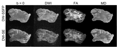
Diffusion acquisition methods for unfixed ex vivo neonatal brain scan at 7T: sequence optimisation and preliminary experiments
Wenchuan Wu, Jerome Sallet, Rogier Mars, Benjamin Tendler, Matteo Bastiani, Sebastian Rieger, Daniel Papp, Jacques-Donald Tournier, Joseph Hajnal, Karla Miller
In this work we present preliminary investigations into the use of diffusion-weighted steady state free procession (DW-SSFP) sequences and conventional diffusion-weighted spin-echo (DW-SE) sequences for unfixed neonate brains. We conduct an exploratory experiment to demonstrate the feasibility of existing dMRI methods on an unfixed porcine brain. We then propose a framework to match DW-SE and DW-SSFP contrast in a way that predicts optimal performance of each, in order to compare between methods.
|
|
0228.
 |
7 |
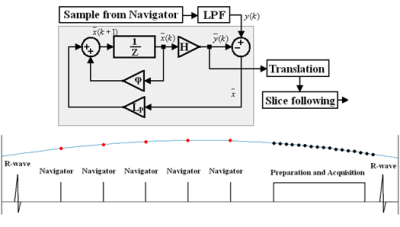
Towards robust free-breathing cardiac DTI
Stephen Jermy, Aaron Hess, Ntobeko Ntusi, Ernesta Meintjes, Elizabeth Tunnicliffe
A prospective respiratory motion correction control system, capable of performing of slice tracking, was implemented in a spin echo diffusion weighted sequence to perform free-breathing acquisitions. The performance of the motion correction control system was compared against common respiratory motion compensation techniques, namely breath-holds, respiratory gating, and standard slice tracking. The values of all the free-breathing techniques varied from the breath-hold data, however, the motion correction control system produced very consistent results. The slice tracking methods were able to significantly reduce the acquisition time (by 50%), compared to the respiratory gating technique.
|
|
0229.
 |
8 |
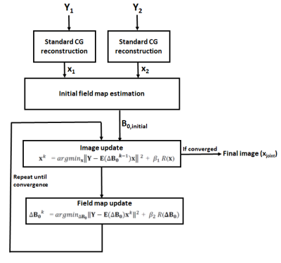
Model based joint B0 and image estimation framework for dynamic field mapping and signal pile-up correction in prostate diffusion MRI
Muhammad Usman, Antonis Matakos, Lebina Kakkar, Simon Arridge, David Atkinson
Prostate diffusion MRI is recognized as a potential biomarker for tumour detection but currently it is unusable in some patients due to significant distortions. We proposed a novel model based joint image and B0 reconstruction framework that can correct these distortions by using data acquired from opposite phase encoding gradient directions. Using sampling time shift between the two acquisitions, the proposed method is robust against any dynamic changes in the off resonance effects in the prostate-rectal air region.
|
|
0230.
 |
9 |
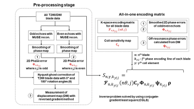
Self-calibrated and Collaborative Propeller-EPI Reconstruction (SCOPER) for High-Quality Diffusion-Tensor Imaging
Xiaoxi Liu, Di Cui, Erpeng Dai, Edward S. Hui, Queenie Chan, Hing-Chiu Chang
Propeller-EPI is a self-navigated multi-shot technique for high-resolution diffusion-tensor imaging. However, corrections for 2D Nyquist ghost and distortion for each blade data are necessary to obtain high-quality image. In this study, we aim to develop a self-calibrated and collaborative Propeller-EPI reconstruction (SCOPER) framework that can 1) allows the estimation of phase errors and off-resonance map from the blade data, and 2) collaboratively reconstruct fully-corrected Propeller-EPI image from all blade data and the thereof. Our results demonstrated that SCOPER shows improved SNR performance, compared with conventional Propeller-EPI reconstruction pipelines.
|
|
0231.
 |
10 |
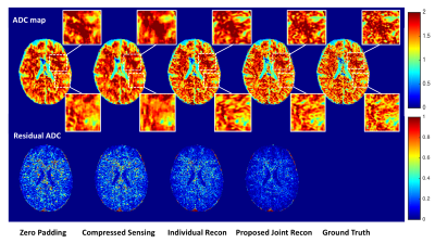
Simultaneous Reconstruction of Multiple b-Values DWI using a Joint Convolutional Neural Network
Chengyan Wang, Yucheng Liang, Yuan Wu, Danni Yang, Yiping P. Du
This study presented a joint convolutional neural network (CNN) architecture for the reconstruction of multiple b-values diffusion-weighted (DW) images simultaneously. The proposed joint-net is able to extract high-level anatomical correlations among multi-contrast images and correct misalignment between images by adding a spatial transformation layer. Experimental results show that the proposed algorithm outperforms single image reconstruction network and compressed sensing algorithm with improved image quality. The training process of the joint-net is much more efficient compared to individual training for each b-value image. Besides, combination of data consistency and the joint-net enables precise characterization of brain tumor in a patient study.
|
|
0232.
 |
11 |
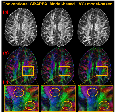
Improved gSlider Reconstruction for Isotropic High-Resolution DTI Using A Model-Based Method and Virtual Coil Concept
Simin Liu, Erpeng Dai, Zijing Dong, Hua Guo
Recently, generalized SLIce Dithered Enhanced Resolution (gSlider) has been proposed as a new acquisition strategy for isotropic high-resolution DTI. In this study, to further boost the SNR performance and reconstruction accuracy, the model-based DTI reconstruction method was merged into the gSlider reconstruction procedure. Moreover, virtual coil (VC) concept was also integrated into the proposed method to further improve the SNR of gSlider reconstruction. Compared with conventional GRAPPA, the superiority of the model-based method has been demonstrated by in vivo results, especially when in combination with the virtual coil concept.
|
|
0233.
 |
12 |
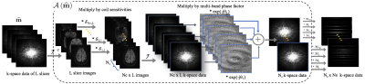
SMS-MUSSELS: A Navigator-free Reconstruction for Slice-accelerated Multi-shot Diffusion Imaging
Merry Mani, Mathews Jacob, Graeme McKinnon, Baolian Yang, Brian Rutt, Adam Kerr, Vincent Magnotta
Multi-shot diffusion-weighted (msDW) imaging schemes can improve the spatial resolution of DWIs. However, the multi-shot readout spans multiple TRs which reduce the time efficiency of msDWI. Hence the practical utility of multi-shot methods for high angular resolution imaging for fiber tracking purposes currently remains limited. To improve the time efficiency of msDWI, SMS acceleration can be employed. However, the DWIs from SMS-accelerated msDW acquisition will exhibit severe artifacts due to slice aliasing and inter-shot phase inconsistencies arising from the msDW encoding. We present a novel navigator-free reconstruction method to simultaneously slice unfold and jointly recover multi-shot diffusion data from an SMS-accelerated msDW acquisition. The method is shown to effectively reconstruct 4-shot DW data accelerated at a multi-band factor of 3.
|
|
0234.
 |
13 |
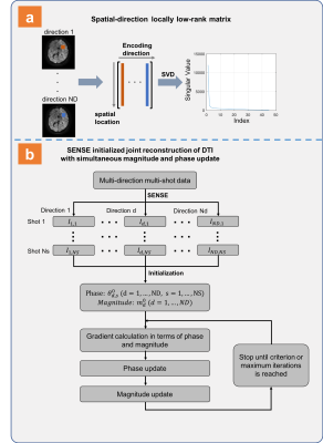
A nonlinear model for DTI reconstruction with locally low-rank regularization
Yuxin Hu, Qiyuan Tian, Grant Yang, Jennifer McNab, Bruce Daniel, Brian Hargreaves
We developed a nonlinear model with simultaneous phase and magnitude updates for iterative multi-shot DWI reconstruction. In addition, locally low-rank regularization along the diffusion encoding direction was included in the proposed model to utilize angular correlation for DTI reconstruction. In-vivo high-resolution and high b-value images have been acquired to validate the proposed method and the proposed method significantly reduces image noise.
|
|
0235.
 |
14 |
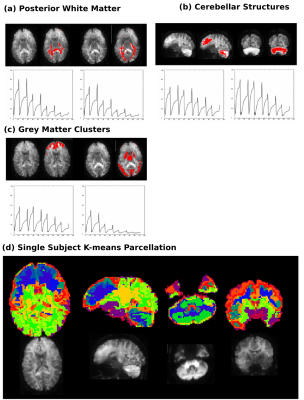
ZEBRA – from rich-multi-dimensional data to anatomical profiles
Jana Hutter, Jonathan O'Muircheartaigh, Paddy Slator, Daan Christiaens, Sophie Arulkumaran, Lucilio Cordero Grande, Rui Pedro Teixeira, Anthony Price, J-Donald Tournier, Joseph Hajnal
An efficient joint multi-parametric diffusion-relaxometry MRI acquisition, ZEBRA, is presented. Improvements to optimize the joint sampling in several dimensions include logarithmic TI sampling, superblock strategies and globally and locally optimized gradient schemes. These are introduced together with a proposed whole-brain protocol (resolution 2.5mm isotropic). The data is analysed by an assumption free clustering step – designed to extract tissue information and anatomical profiles directly from the signal. Depiction of several clusters – including the deep grey matter and cerebellar substructures - illustrate the richness of the obtained data.
|
|
0236.
 |
15 |
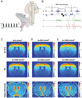
In-vivo diffusion-fMRI using Incomplete Initial Nutation Diffusion Imaging (INDI)
Daniel Nunes, Noam Shemesh
Diffusion functional-MRI (dfMRI) is thought to capture microstructural changes associated with neural activity. The water apparent diffusion coefficient decrease observed upon neuronal activity is hypothesized to be cell-swelling dependent. Yet, one of the confounding factors for dfMRI is that zero and nonzero b-values need to be acquired to deliver accurate changes in diffusivity and those images are typically separated by at least one repetition time. Incomplete initial Nutation Diffusion Imaging (INDI) was proposed as a method to acquire two images with different diffusion-weighting with separation of <50ms. Here, we performed INDI-fMRI experiments to report mean diffusivity changes using forepaw-stimulated rats.
|
|
