|
2nd Hour
Poster: Task-Based fMRI & fMRI Acquisition Methods
Power Pitch Poster
fMRI
Tuesday, 14 May 2019
Power Pitch Theater C - Exhibition Hall
09:15 - 10:15
| |
|
Plasma # |
 |
0357.
 |
31 |
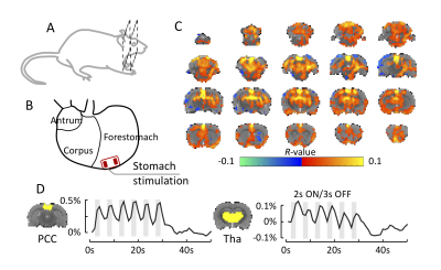
The Predominant Role of the Vagus Nerve in Gastric Electrical Stimulation Evoked Gut-Brain Axis

Jiayue Cao, Kun-Han Lu, Christina Hendren, Robert Phillips, Deborah Jaffey, Terry Powley, Zhongming Liu
The gut-brain axis serves the bidirectional communication between the enteric nervous system and the central nervous system and links the gastrointestinal function with cognitive and emotional centers of the brain. Three different pathways have been identified to be responsible for the gut-brain axis, including the chemical modulation, the spinal nerve, and the vagus nerve. However, the functional role of each pathway is unclear and still under investigation. Using functional magnetic resonance imaging (fMRI), we report that gastric electrical stimulation (GES) can modulate the gut-brain axis and evokes fMRI activity in multiple brain regions including both cognitive and sensory systems. Most of the GES evoked brain regions are attributable to the vagal function, suggesting a central role of the vagus nerve in the gut-brain axis, especially in modulating the cognitive and emotional centers.
|
|
0358.
 |
32 |
In-Vivo Current Mapping in Transcranial Direct Current Stimulation (tDCS) – A Concurrent tDCS-MRCDI Study
Anita Jwa, Jonathan Goodman, Gary Glover
Transcranial direct current stimulation (tDCS) is gaining momentum both in the research community and in the general public as an attractive tool for neuromodulation. However, the mechanism of tDCS in the brain is not fully understood. In this study, we conducted a concurrent tDCS–magnetic resonance current density imaging (MRCDI) experiment to measure the primary direction and magnitude of current in a human brain undergoing direct current stimulation. Our results show that current flow deviates significantly from its desired distribution based on human head models and that the current mostly flows through the white matter and cerebrospinal fluid.
|
|
0359.
 |
33 |
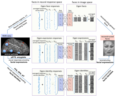
Reconstructing multi-dimensional face attributes from fMRI signals
Hui Zhang, Zixiang Wei, Xueping Wang, Yunhong Wang
We develop a new framework that can reconstruct the perceived faces from functional MRI signals. Inspired by the psychological evidences that face processing is transmitted along two distinct neuroanatomical visual pathways in human brain, our framework can efficiently extract the multidimensional information of facial expression and facial identity information from functional MRI signals for precise face image reconstruction.
|
|
0360.
 |
34 |
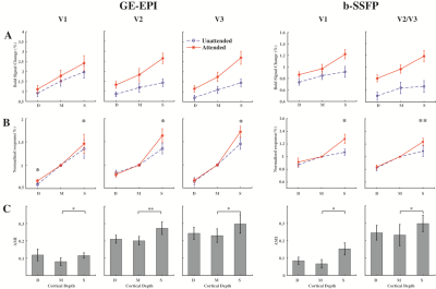
Attention modulation of layer-specific signals in human visual cortex
Chengwen Liu, Chencan Qian, Zihao Zhang, Kaibao Sun, Jing An, Danny J.J. Wang, Peng Zhang
Attention mechanisms at different cortical layers of the human visual cortex remain poorly understood. Here we investigated the attention modulation of layer-specific activities in the human visual cortex, using submillimeter-resolution BOLD fMRI at 7 Tesla. Results showed that compared to the middle-layer activities, attention increased signals in the superficial and deep layers of V1, and in the superficial layers of V2 and V3. Contrast modulation was strongest in the middle layer of V1, consistent with the feedforward input from the LGN. These findings suggest that top-down spatial attention mainly modulates output signals in the superficial layers of human visual cortex.
|
|
0361.
 |
35 |
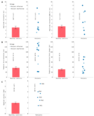
Assessing somatotopic and mototopic organisation in Focal Hand Dystonia using high-resolution 7T fMRI
Daisie Pakenham, Michael Asghar, Paul Glover, George O'Neill, Ayan Sengupta, Denis Schluppeck, Rosa Sanchez-Panchuelo, Miles Humberstone, Susan Francis
7T fMRI provides a non-invasive method to study somatotopy and mototopy. Here, healthy controls (HCs) and Focal Hand Dystonia (FHD) patients 4-weeks post-Botox injection undergo behavioural and fMRI assessment. Behavioural measures include temporal discrimination, amplitude thresholding and spatial acuity assessment. fMRI includes a somatosensory travelling wave and event-related paradigm, and mototopy travelling wave paradigm. In FHD patients, amplitude threshold, temporal discrimination threshold and spatial acuity was increased compared to HCs. Maps of somatotopy and mototopy are shown for FHD patients and HCs along with digit separation. FHD patients will be rescanned 3 months post-Botox to determine whether changes are evident.
|
|
0362.
 |
36 |
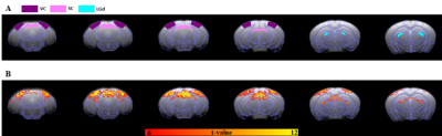
Characteristics of mouse BOLD fMRI responding to visual stimulation
Ngoc Anh Dinh, WonBeom Jung, Seong-Gi Kim
While the most common BOLD fMRI studies in mice have focused on evoking BOLD response in the somatosensory system, the functional response characteristic of mouse visual system is not well explored. Here, we investigated BOLD fMRI in mice with visual stimuli varying flash frequency in the range of 1 to 10Hz and pulse width from 1 to 50ms under ketamine/xylazine anesthesia at 9.4T.
|
|
0363.
 |
37 |
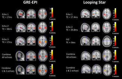
Looping Star silent fMRI: a platform for improving studies of auditory processing
Nikou Damestani, David Lythgoe, Florian Wiesinger, Ana Beatriz Solana, Steven Williams, Fernando Zelaya
Conventional functional magnetic resonance imaging (fMRI) produces acoustic noise levels comparable to a running chainsaw. This presents numerous challenges for functional data interpretation, providing a substantial confound for auditory processing. Recently, a novel imaging technique known as “Looping Star” has been developed, which reduces this acoustic noise to the amplitude of normal conversation. We applied this acquisition technique with an auditory paradigm for the first time, comparing it with conventional fMRI. We established that it displays good functional sensitivity in spite of reduced signal-to-fluctuation-noise, alongside functional localisation free from inflow effects. This technique could revolutionise future investigations of acoustic processing.
|
|
0364.
 |
38 |
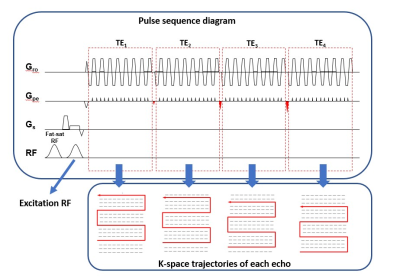
Reconstruction of highly-accelerated multi-echo fMRI using parametric POCS based multiplexed sensitivity-encoding (POCSMUSE)
Shihui Chen, Mei-Lan Chu, Queenie Chan, Hing-Chiu Chang
Multi-echo fMRI is an emerging technique that can improve the fidelity and interpretability of fMRI, such as differentiating BOLD and non-BOLD signals. In multi-echo fMRI acquisition, high acceleration factor for parallel imaging (i.e., R = 3) was used to achieve reasonable TE interval and maintain the same resolution as single-echo acquisition. The accelerated multi-echo data are reconstructed using SENSE, which may suffer from undesired noise amplification. In this study, we proposed a multi-echo multi-segment EPI (MEMS-EPI) technique to acquire multi-echo fMRI with high acceleration factor, and then used parametric POCSMUSE algorithm to reconstruct the data.
|
|
0365.
 |
39 |
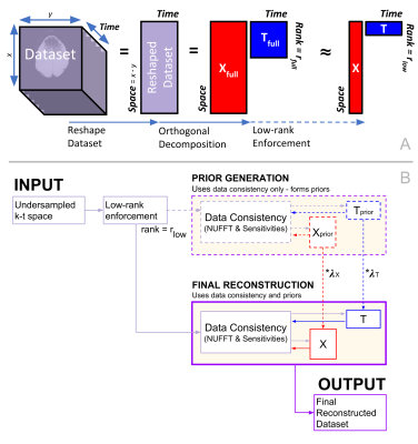
Improving k-t PERRI: a low-rank data-driven fMRI k-t acceleration method
Harry Mason, Karla Miller, Nadine Graedel, Mark Chiew
FMRI data quality requires both good image fidelity (conferring spatial specificity), and high temporal resolution (conferring statistical robustness). We present an image reconstruction algorithm that aims to achieve both aims through a spatio-temporal (k-t) image reconstruction. Our approach utilises low-rank reconstruction algorithms and 3D golden angle k-space sampling. Using golden-angle sampling, we demonstrate that data-driven spatial and temporal priors can be incorporated into reconstruction. We demonstrate improvement over previously-proposed methods (k-t FASTER and k-t PSF) that correspond to special cases of our prior-based reconstruction. These results have great potential to improve on time-independent reconstructions currently in use.
|
|
0366.
 |
40 |
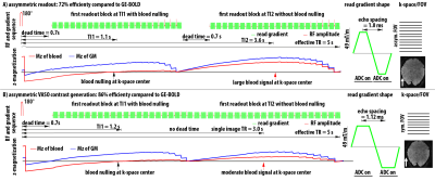
Measuring layer-dependent CBV fMRI in the visual system
Laurentius Huber, Elisha Merriam, Insub Kim, Yuhui Chai, Sriranga Kashyap, Jonathan Polimeni, Zvi Roth, Won Mok Shim, Seong-Gi Kim, Dimo Ivanov, Benedikt Poser, Peter Bandettini
With recent developments of ultra-high field MRI and high-resolution CBV-sensitive VASO sequences, it became possible to measure activity changes across cortical depths in brain areas of large cortical thicknesses (4mm in M1). Applications of layer-dependent CBV-fMRI in the visual cortex, however, have been complicated by long arterial-arrival-times, coverage requirements, and lower cortical thickness (1.9mm). Here, we developed new sequence setups for layer-dependent acquisition of CBV changes during tasks and resting-state. We find that the proposed large-coverage VASO protocols with 0.8mm (iso) resolutions can pinpoint feedforward and feedback input into V1 from LGN and V5 during tasks and resting-state.
|
|
0367.
 |
41 |
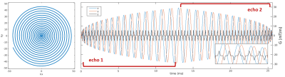
Dual-echo simultaneous multi-slice spiral acquisition for simultaneous CBF and BOLD fMRI at 7T
Denizhan Kurban, Gilad Liberman, Sriranga Kashyap, Dimo Ivanov, Benedikt Poser
Dual-echo acquisition with arterial spin labelling (ASL) allows for simultaneous measurements of CBF and BOLD signal changes which are helpful to understand the cerebral haemodynamics in health and disease. At ultra-high field (≥7T), however, this becomes impractical with established EPI based techniques since the readout duration precludes the use of appropriate echo times at commonly desired spatial resolutions. Single-shot spiral imaging presents a promising alternative: it allows the CBF signal to be acquired at very short TE while providing a flexible choice of TE for the second BOLD echo. In this work we show a dual-echo simultaneous multi-slice spiral out-in sequence for the acquisition of CBF and BOLD signals.
|
|
0368.
 |
42 |
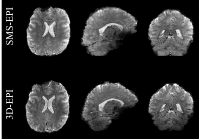
Comparison of SMS-EPI versus 3D-EPI in an fMRI localizer study at HCP-style resolution and TR, using parallel transmission Universal Pulses at 7T
Caroline Le Ster, Franck Mauconduit, Antonio Moreno, Vincent Gras, Rüdiger Stirnberg, Benedikt Poser, Alexandre Vignaud, Evelyn Eger, Florent Meyniel, Stanislas Dehaene, Nicolas Boulant
Simultaneous-Multi-Slice (SMS) and 3D-EPI sequences with parallel-transmit B1 homogenization by means of calibration-free Universal Pulses were evaluated at 7T and at HCP-style resolution and repetition time (1.6mm iso, TR=1120ms). Comparison was based on sensitivity and specificity of activation detection in a multi-modal functional localizer task paradigm. When including nuisance regressors in the analysis, the SMS-EPI shows superior sensitivity but lower specificity than the 3D-EPI, with overall comparable performance when weighting the two metrics equally, but with significantly smaller SAR demands and sound pressure levels for the 3D-EPI.
|
|
0369.
 |
43 |
BOLD temporal SNR bias and variance across the HCP population as a function of cortical B0-orientation and orientation variability
Olivia Viessmann, Jingyuan Chen, Kawin Setsompop, Lawrence Wald, Jonathan Polimeni
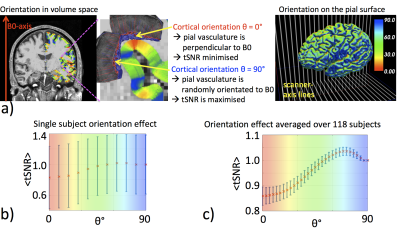
BOLD fMRI signals vary with the orientation of the cortex to the B0-field as extravascular susceptibility effects vary with the orientation of the cortical pial vasculature. This creates regional tSNR biases with cortical folding. Certain cortical folds are more homogenous across the population than others and orientation variability across subjects should introduce tSNR variability at the group-level. Here, we use HCP 3T rs-fMRI data to show that B0-orientation contributes to within-subject tSNR bias and orientation variability contributes to tSNR variance across subjects. We found that functional connectivity networks with more perpendicular orientation exhibit higher tSNR and networks with high orientation consistency have lower tSNR variability.
|
|
0370.
 |
44 |
Evaluation of Sparse Sampling Approaches for 3D Functional MRI
Michelle Karker, Claire Lin, Jeffrey Fessler, Douglas Noll
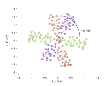
This work evaluates the performance of a novel sampling pattern compared to common undersampling techniques with the ultimate goal of obtaining high spatiotemporal resolution 3D fMRI via sparse sampling and model-based reconstruction. We examine a novel 3D Cartesian sampling pattern with random phase encodings in ky-kz rotated by the golden angle, resulting in a variable density sampling of k-space, with dense sampling of the high-signal energy k-space center. The functional activation and noise amplification (g-factor) results of this sampling technique are compared to those of standard sampling regimes.
|
|
0371.
 |
45 |
Microstrip Array Insert for Head Coils: Towards Layer fMRI at High Fields
Burak Akin, Ali Özen
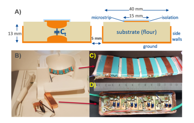
Rx coils for high field strengths are mostly based on loop coil design and cover relatively large FOVs, limiting the achievable voxel size due to the SNR and acquisition time constraints. Microstrip coils have limited sensitivity profile along the B1 direction, thus are useful for resolving cortical activity. In this study, we introduce flexible microstrip array insert coils (MSAi) that are compatible with existing volume coils. We demonstrate measurement of BOLD and highly confined activity wide spread in calcarine sulcus and motor cortex using MSAi at 3 T
|
|
