|
2nd Hour
Poster: Interventional & RF: Safety & Solutions
Power Pitch Poster
Interventional MRI
Wednesday, 15 May 2019
Power Pitch Theater B - Exhibition Hall
14:30 - 15:30
| |
|
Plasma # |
|
0791.
 |
16 |
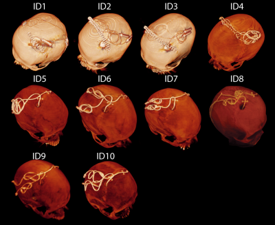
Reconfigurable coil technology can substantially reduce RF heating of bilateral deep brain simulation leads during MRI at 1.5 T: First in-vitro studies with realistic implant trajectories

Laleh Golestanirad, Boris Keil, John Kirsch, Julie Pilitsis , Lawrence Wald
Patients with deep brain stimulation (DBS) implants significantly benefit from MRI, however their access is restricted in these patients because of safety concerns due to RF heating of the leads. Recently we introduced a patient-adjustable reconfigurable MRI coil system that significantly reduced the SAR at the tip of single DBS leads (unilateral) in simulation studies during MRI at 1.5T. Here we present the first in-vitro measurements of RF heating-reduction performance of the coil system showing a significant reduction in heating of realistic bilateral DBS implants.
|
 |
0792.
 |
17 |
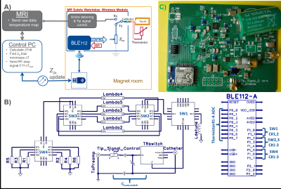
MR Safety Watchdog for Safe Active Catheters: Wireless Impedance Controller with Real-time Feedback
Ali Özen, Berk Silemek, Thomas Lottner, Ergin Atalar, Michael Bock
RF-induced heating of implants or devices can be controlled through manipulation of their termination impedance which is dependent on the dielectric properties of the medium and the incident field configurations. We designed a wireless module that modifies the input impedance of an active catheter to keep the temperature increase below a threshold, ΔTmax. The wireless module monitors local heating at the tip of the catheter or can receive data from an external temperature measurement device to search for the optimal impedance that minimizes temperature rise. RF transmission is blocked via a feedback system when ΔTmax is exceeded.
|
|
0793.
 |
18 |
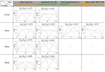
Explaining current patterns on linear metallic implants during MRI exams using the transfer matrix.
Janot Tokaya, Peter Stijnman, Peter Luijten, Cornelis Berg, Alexander Raaijmakers
Currents induced on relatively(<20cm) short linear metallic implants typically occur in certain patterns. Knowledge of these patterns can help current monitoring and identification of hazardous exposure conditions. The transfer matrix (TM) of an implant predicts what patterns will appear. The eigenvalues of the TM indicate which modes are induced naturally. The projection of the eigenvectors onto realistic incident electric fields obtained from electromagnetic simulations explain which modes are likely and effectively excited. Over 80% of electric field distributions excite the first eigenmode of the investigated structures more efficiently. Moreover, all severe currents follow the pattern of this first eigenmode.
|
|
0794.
 |
19 |
Lower risk of hearing loss without sacrificing image quality in fetal MR imaging: a feasibility study using acoustic reduction technique
Le Cao, Ting Liu, Junjun Li, Jingtao Sun, Jianxin Guo, Xiaocheng Wei, Jian Yang
3.0T MR scanner can achieve superior image quality depicting fetal anatomic details over 1.5T, but may poses higher risk of adverse impact on fetal auditory development due to its intrinsically higher acoustic noise level. This comparative study investigated the value of acoustic noise reduction technique in fetal exam. The result shows the technique can acquire equivalent quality images in 3.0T scanner, meanwhile decrease hearing loss risk in fetal head examinations compared with the conventional method.
|
|
0795.
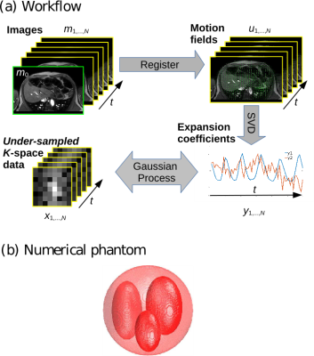 |
20 |
Acquisition, reconstruction and uncertainty quantification of 3D non-rigid motion fields directly from k-space data at 100 Hz frame rate
Alessandro Sbrizzi, Niek Huttinga, Cornelis van den Berg
We introduce a method based on Gaussian Processes to rapidly acquire and reconstruct non-rigid motion fields of the human body directly from few points in k-space. Overall, the acquisition and processing time is less than 10 milliseconds for 3D applications leading to 100 Hz frame rate. Uncertainty quantification is also provided, making the method suitable for clinical real-time applications such as MR-guided radiation therapy.
|
|
0796.
 |
21 |
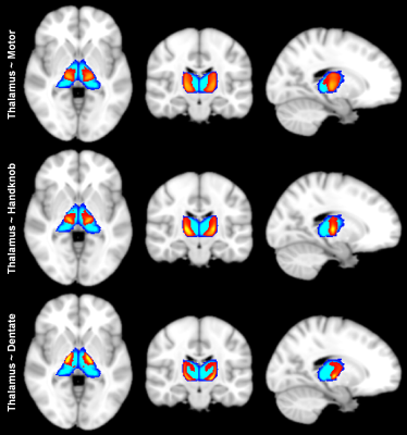
Creating a diffusion tractography-based atlas of human thalamic ventral intermediate nucleus aided by deep learning
Qiyuan Tian, Chanon Ngamsombat, Berkin Bilgic, Qiuyun Fan, Yuxin Hu, Jennifer McNab, Thomas Witzel, Kawin Setsompop, Jonathan Polimeni, Susie Huang
Tremor suppression in the hands of patients with essential tremor can be achieved by lesioning the ventral intermediate nucleus (Vim) of the thalamus using transcranial MR-guided focused ultrasound. Recent work has shown that diffusion MR tractography identifies the Vim more precisely and predicts the degree of tremor suppression. Here, we trained a convolutional neural network (CNN) to automatically segment relevant regions-of-interest including the thalamus, red nucleus, dentate nucleus and handknob region for probabilistic tractography to identify the Vim. We applied the CNN to 200 HCP healthy subjects and created a tractography-based atlas of Vim location, which could aid in neurosurgical guidance.
|
|
0797.
 |
22 |
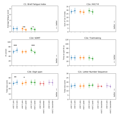
Safety evaluation of human exposure to a 10.5T whole body magnet: protocol design and preliminary results.
Andrea Grant, Jeramy Kulesa, Xiaoxuan He, Pierre-Francios Van de Moortele, Yigitcan Eryaman, Gregor Adriany, Michelle Hartwig, Cheryl Olman, Sarah Bedell, Lin Zhang, Margaret Koeritzer, Meredith Adams, Thomas Henry, Gregory Metzger, Kamil Ugurbil
We present the initial results of our 10.5T safety study measuring the impact on cognitive, vestibular, and physiological metrics. Data from 17 subjects show evidence of short term (same day) but no long term (weeks) fatigue, increased nystagmus upon initial exposure to static field, with no relevant effect on physiological measures when at isocenter.
|
|
0798.
 |
23 |
Residual magnetization of human subjects after exposure to magnetic fields
George Hutchinson, Niall Holmes, Matthew Brookes, Richard Bowtell
It is known that subjects who have recently had an MRI scan can produce greater "magnetic noise" in magnetoencephalography (MEG) studies. We investigated this phenomenon, which may be related to remanent magnetization of magnetite particles that have been identified in post mortem tissue, by analyzing the field variation produced by controlled head movements in a MEG scanner, before and after subjects had been exposed to the local magnetic field of a small permanent magnet or inserted in a 3T magnet. The results show significant variability across subjects, but with a general elevation of the measured field variation after field exposure.
|
|
0799.
 |
24 |
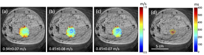
Magnetic resonance shear wave elastography in cadaver breast
Allison Payne, Lorne Hofstetter, Riley Haag-Roeger, Henrik Odeen, Dennis Parker
Tissue stiffness is a known marker of malignancy and has been shown to change due to thermal therapies. A shear wave elastography technique compatible with magnetic resonance guided focused ultrasound technology is demonstrated in a cadaver breast specimen. Volumetric shear wave speed maps acquired using a multiple-point sonication pattern before and after an MRgFUS ablation demonstrate that a change in shear wave speed can be detected. This technique provides a functional stiffness metric that is complementary to currently used monitoring and assessment techniques in MRgFUS treatments.
|
|
0800.
 |
25 |
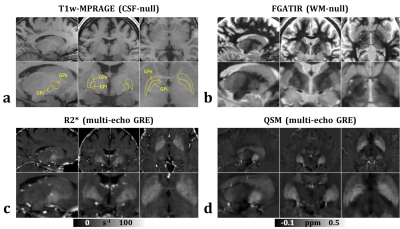
Localization of GPi for MRgFUS pallidotomy: a comparison between high-resolution FGATIR, R2* and QSM at 3 T
Hongfu Sun, M. Ethan MacDonald, Erin Mazerolle, Kristin Sabourin, G. Bruce Pike
Precise localization of the internal globus pallidus (GPi) is critical for MRgFUS pallidotomy for movement disorders such as Parkinson’s disease. In this study, high-resolution FGATIR, R2* and QSM are compared for localizing GPi in six healthy subjects (age from 21 to 41). All three methods displayed some image contrasts in the GP area. QSM demonstrated the best delineation of GPi from the internal capsule, which is generally considered a risk zone for pallidotomy. GPi also appeared smaller in FGATIR, where GPi was hypointense, than in QSM, where GPi was hyperintense.
|
|
0801.
 |
26 |
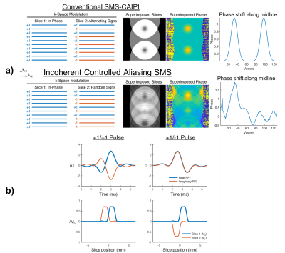
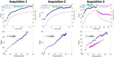
Simultaneous Multislice MRI Temperature Imaging with a Single Receiver Coil
Kristin Quah, Megan Poorman, William Grissom
The incoherent controlled aliasing SMS method is introduced which increases volume coverage in real-time MR thermometry by acquiring multiple slices simultaneously, and requires only one receiver coil. RF pulses flip the slice phases between TRs, creating incoherent hotspot aliasing. Unaliased heating maps are then recovered from each slice using a sparsity-promoting temperature reconstruction.
|
|
0802.
 |
27 |
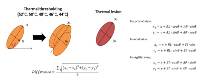
Predicting final lesion size using Thermometry information during MR-guided focused ultrasound treatment of Parkinson’s Disease
Sijia Guo, Jiachen Zhuo, Rao Gullapalli, Dheeraj Gandhi
Recently, the MR-guided focused ultrasound technology (MRgFUS) offers the possibility to perform subthalamic thermal ablation with reduced risks and optimized accuracy. However, the ability to reliably predict lesion size is still evolving. The goal of this study was to improve the predictability of lesion size by thermal thresholding areas on MR thermometry during treatment procedures.
|
|
0803
 |
28 |
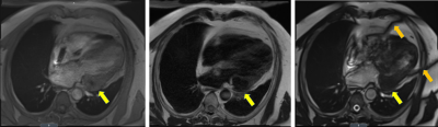
First in Human: MRI-guided radiation therapy of the heart with implantable cardiac defibrillator
Video Permission Withheld
H Michael Gach, Olga Green, Phillip Cuculich, Erin Wittland, Areti Marko, Molly Luchtefeld, Jill Entwistle, Deshan Yang, David Wilber, Sasa Mutic, Clifford Robinson
Low-field (0.35 T) magnetic resonance imaging guided radiation therapy (MR-IGRT) was used for the first time to treat a patient with a cardiovascular implantable electronic device (CIED) and a cardiac fibroma located on the left ventricle. Stereotactic body radiation therapy (SBRT) was delivered in 5 fractions at a dose of 700 cGy/fraction with MRI-based real-time tumor tracking and beam gating. Lessons learned include the need for metal artifact suppression, higher MRI temporal resolution, and MRI safety workflows adapted to radiation oncology.
|
|
0804.
 |
29 |
Feasibility of Thermo-Acoustic Ultrasound for Non-invasive Monitoring of Temperature at Lead Tips During MRI
Neerav Dixit, John Pauly, Greig Scott
The amplification of local SAR at device lead tips that causes RF-induced heating has previously been shown to be detectable using thermo-acoustic ultrasound (TAUS). Due to the temperature dependence of the thermal expansion coefficient in tissue, the thermo-acoustic pressure generated at a lead tip is a function of the temperature at the lead tip. By observing how the TAUS signal amplitude changes over time, real-time tracking of lead tip temperature during an MRI scan should be possible. Here, we experimentally demonstrate the temperature-dependence of the TAUS signal, showing the feasibility of TAUS for temperature monitoring at lead tips during MRI.
|
|
0805.
 |
30 |
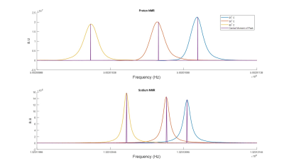
Multinuclear Absolute MR Thermometry
Leeor Alon, Emilia Silletta, Alexej Jerschow, Guillaume Madelin
Absolute MR thermometry has been unachievable clinically since the advent of MR and MR practitioners mostly rely on relative measurement of thermal changes using the proton resonance frequency shift method. Here, we introduce the JAMS method for reconstruction of absolute temperature using multinuclear frequency measurements. The method takes advantage of different frequency shifts with temperature of different nuclei (e.g. proton and sodium) for the reconstruction. Theory of the method is presented and proof-of-principle experiments validating the approach.
|
|
