27th ISMRM Annual Meeting • 11-16 May 2019 • Montréal, QC, Canada
| Sunrise Session Quantitative MRI: Quantitative Susceptibility Mapping |
||||||||||||
|
Quantitative MRI: Quantitative Susceptibility Mapping
Sunrise Session ORGANIZERS: José Marques, Sebastian Kozerke, Ileana Hancu
Wednesday, 15 May 2019
Skill Level: Intermediate to Advanced
Session Number: S-W-08
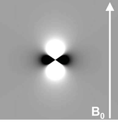
Overview Extend the understanding of the MR signal evolution beyond the simple phenemenological single-pool relaxation models. During this educational course, the role of tissue physical properties such as magnetic susceptibility and electric conductivity and how they affect imaging and relaxation processes will be addressed. Furthermore, the role of diffusion, perfusion, flow in the observed signal and how they can be encoded and measured in MRI will be discussed. Target Audience Scientists who are interested in developing/improving quantitative MR approaches. Educational Objectives As a result of attending this course, participants should be able to: - Develop MR pulse sequence design to implement practical encoding approaches of the various physical properties of tissues; - Apply decoding equation/reconstruction to calculate tissue magnetic susceptibility, electrical conductivity, relaxation, diffusion, perfusion, flow; and - Compare various software solutions for data reconstruction/processing.
|
||||||||||||
| The International Society for Magnetic Resonance in Medicine is accredited by the Accreditation Council for Continuing Medical Education to provide continuing medical education for physicians. |