|
Mapping the Complementary Roles of Myelin & Microstructure in White Matter
Combined Educational & Scientific Session
ORGANIZERS: Noam Shemesh, Alexander Leemans, Jongho Lee
Thursday, 16 May 2019
| Room 516C-E |
13:45 - 15:45 |
Moderators: Nikola Stikov, Adrienne Dula |
Skill Level: Intermediate
Session Number: TH-05
Overview
White matter of the brain has a unique microstructural composition that divides white matter into multiple compartments. This session will introduce a few quantitative microstructural imaging methods that measure the white matter compartments (e.g. myelin and axon).
Target Audience
Physicians and scientists wanting to explore methodologies for quantitative microstructural imaging techniques for white matter; physicians and scientists engaged in advanced white matter imaging for scientific and clinical research.
Educational Objectives
As a result of attending this course, participants should be able to:
- Describe the microstructural composition of white matter;
- Summarize various white matter microstructural imaging methods with their imaging targets; and
- Explain the technical limitations of the methods.
13:45
|
|
Diffusion Goes Multi-Modal: Myelin Mapping

Mark Does
Myelin plays a central role in determining MRI contrast in neuronal tissues, and, consequently, there are many MRI approaches measuring myelin content. This presentation will review several common approaches as well as some recent and developing approaches.
|
14:15
|
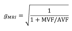 |
g-Ratio Mapping: Challenges & Validation
Jennifer Campbell, Ilana Leppert, G. Bruce Pike
The fiber g-ratio is the ratio of the inner to the outer diameter of the myelin sheath of a myelinated axon. In healthy neural tissue, it is optimized for speed of signal conduction, cellular energetics, and spatial constraints. The g-ratio is a fundamental contributor to functionally relevant neuronal properties such as conduction velocity. g-Ratio imaging has the potential to allow us to investigate in vivo g-ratio changes in health and disease. This lecture details the methodology for imaging the g-ratio with MRI, describes the known challenges in doing so, presents validation work to date, and discusses challenges in validating the technique.
|
14:45
|
1115.
 |
Characterizing diffusion of myelin water in the living human brain using ultra-strong gradients and spiral readout
Chantal Tax, Umesh Rudrapatna, Lars Mueller, Derek Jones
Myelin is a key white matter compartment, but myelin water is beyond the detection of conventional diffusion MRI methods because of its short T2. Here we combine ultra-strong gradients and spiral readout to achieve very short echo times (TE=30ms) at very high diffusion weighting (b=6000s/mm^2), with the aim of achieving significant sensitivity to the diffusion of myelin water in the living human brain. We investigated the challenge of disentangling 3 distinct compartments – including the short T2 component from myelin.
|
14:57
|
1116.
 |
A Novel MRI technique for quantifying myelin in mice brain white matter
Ella Wilczynski, Efrat Sasson, Uzi Eliav, Uri Nevo, Gil Navon
In vivo myelin imaging in MS could provide a tracking tool for demyelination and facilitating the development of new therapeutic agents that may promote remyelination. We propose to use a recently reported MRI sequence, MEX, which measures a signal linearly dependent on the myelin protons fraction in the tissue, F. The sequence was used on cuprizone fed (n=7) and control mice (n=4). The cuprizone mice exhibit significant decrease of F by 25% in the Corpus Callosum (WM), and no change in GM. This study provides a proof of usefulness of this method, for demonstrating quantification of brain white matter demyelination.
|
15:09
|
1117.
 |
Simple scheme for correcting bias in axonal water fraction due to differences in compartmental transverse relaxation times
Emilie McKinnon, Jens Jensen
Neglecting differences in compartmental transverse relaxation times when modeling diffusion MRI data may affect the accuracy of estimates for microstructural parameters. Here we propose a straightforward method for correcting the bias in the axonal water fraction (AWF), as calculated from the fiber ball white matter (FBWM) model, due to T2 differences between the intra-axonal and extra-axonal compartments. This correction scheme simply requires that one additional high b-value shell be acquired at a different echo time than for the standard dataset needed for FBWM. AWF values were found to be 16% lower, on average, after the T2 correction.
|
15:21
|
1118.
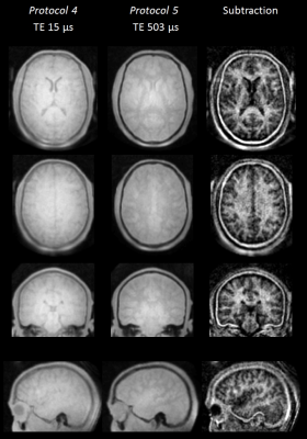 |
Advances in direct myelin imaging
Markus Weiger, Romain Froidevaux, David Brunner, Manuela Rösler, Klaas Pruessmann
Direct imaging of myelin is of great interest for improved diagnosis of neurodegenerative diseases. However, MR signals from myelin exhibit ultra-short T2 values in a range of 8 μs - 1 ms with a large fraction below 100 μs. Due to restrictions in sequence timing, current short-T2 imaging approaches cannot sufficiently capture these very short signals. In the present work, advanced short-T2 methodology and hardware are employed to actually image the majority of the ultra-short-T2 components in the brain. The abilities of the approach are proven in excised brain tissue and applied in vivo.
|
15:33
|
1119.
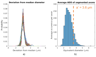 |
Uncovering 3D Axonal Morphologies with Synchrotron Imaging: Impact on Microstructure Imaging with Diffusion MRI
Mariam Andersson, Hans Martin Kjer, Jonathan Rafael-Patino, Vedrana Andersen Dahl, Alexandra Pacureanu, Jean-Philippe Thiran, Martin Bech, Anders Bjorholm Dahl, Tim B. Dyrby
In this study, we segmented large axons from high resolution 3D synchrotron images of the monkey splenium. We simulate the intra-axonal MRI diffusion signal of both the segmented geometry and a simplified, corresponding cylindrical geometry, and calculate the corresponding axon diameter indices. The axon diameter index is well estimated in the simplified geometry, but is overestimated in the more complex segmented geometry, potentially due to variations in axon diameter and non-uniform trajectories. Lastly, we present the observation that segmented axons, which all have average diameters >2 µm, seem to experience similar absolute diameter variations.
|
15:45
|
|
Adjournment |
|
