|
Value of MRI
Combined Educational & Scientific Session
ORGANIZERS: Vikas Gulani, James Pipe
Thursday, 16 May 2019
| Room 512A-H |
16:00 - 18:00 |
Moderators: Sapna Rawal, Susie Huang |
Skill Level: Basic to Intermediate
Session Number: TH-07
Overview
This session continues the work started in the ISMRM Value Initiative, introduced in previous ISMRM meetings. Health care systems around the world are grappling with the growing cost of health care. All participants in the healthcare chain need to be able to understand major models of health care delivery such as fee for service and value based care. In the context of imaging, there is a growing need for researchers and clinicians to understand and be able to measure the downstream value added by imaging methods. This session seeks to fill these needs. The session will begin with a brief introduction, followed by three long format talks. Two talks will focus on cost effectiveness analysis, with an introductory and a more in-depth talk with a specific example of such analysis. The third teaching talk will focus on different health care delivery models, and how these relate to imaging. After these talks, the session will continue into proffered papers on studying the value of MRI from multiple perspectives.
Target Audience
Scientists and physicians interested in determining or improving the value of MRI in the healthcare system, either from a technical or a clinical perspective.
Educational Objectives
As a result of attending this course, participants should be able to:
-Describe models of health care delivery, in particular fee for service, and value based care;
-Identify the implications of various models of healthcare delivery on imaging;
-Describe basic principles of cost effectiveness analysis, including metrics of healthcare outcomes, and models employed in such analysis in determining whether or not a particular intervention is cost effective;
-Apply cost effectiveness analysis in the context of a specific imaging problem; and
-Recall ongoing research in MRI, around the subject of value, both in the technical and in the clinical realm.
16:00
|
|
Reimbursement Models & Their Impact on Imaging

Richard Ehman
Reimbursement Models & Their Impact on Imaging
|
16:20
|
 |
Cost-Effectiveness Analysis: An Introduction
Pari Pandharipande
Overview of talk 1. Effectiveness
2. Utility, Quality-Adjusted Life Year (QALY)
3. Cost-Effectiveness
4. Willingness-to-pay (WTP) threshold
|
16:40
|
|
Cost Effectiveness Principles in MRI: Optimizing MRI from the Cost Effectiveness Perspective
Stella Kang
This educational presentation will describe the importance of studying MRI cost effectiveness as it relates to population-level health outcomes, patient-centered care, and costs to the healthcare system. Examples of work in MRI, spanning innovation and common clinical practice, will serve to illustrate applications of cost effectiveness analysis. Cost effectiveness analysis, a standardized means of comparing medical interventions and tests, quantifies the clinical effectiveness relative to cumulative costs. Furthermore, research goals can be prioritized based on targets for maximized value.
|
17:00
 |
1185.
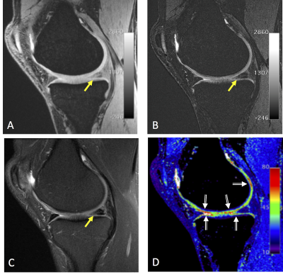 |
5-Minute Quantitative Double-Echo in Steady-State for High-Value Diagnostic Knee MRI: Combining an Efficient Multi-Contrast Acquisition with Quantitative Imaging and Artificial Intelligence
Akshay Chaudhari, Murray Grissom, Zhongnan Fang, Jin Lee, Garry Gold, Brian Hargreaves, Kathryn Stevens
There exists interest in rapid diagnostic knee magnetic resonance imaging (MRI) protocols in a push towards ‘high-value radiology’. Recent efforts for expediting knee MRI involve accelerating 2D fast spin echo (FSE) sequences, which precludes multiplanar reformations, or using 3D FSE sequences, which can cause image blurring. To overcome these limitations, we show how a 5-minute quantitative double-echo steady-state (qDESS) sequence generates high-resolution and multi-contrast images using deep-learning-based super-resolution, along with automatic T2 relaxation time measurements. In a preliminary study with 25 patients, we demonstrate how qDESS can perform rapid and accurate diagnostic knee MRI using rich structural and quantitative information.
|
17:04
 |
1186.
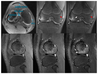 |
Introduction of Targeted Rapid Knee MRI exam using T2 Shuffling into Clinical Practice: Retrospective Analysis on Image Quality, Charges, and Scan Time
Jonathan Tamir, Michael Lustig, Valentina Taviani, Marcus Alley, Kendall O'Brien, Becki Perkins, Lori Hart, Fida Wishah, Jesse Sandberg, Michael Anderson, Javier Turek, Theodore Willke, Shreyas Vasanawala
Volumetric fast spin echo (FSE) of the knee using T2 Shuffling (T2Sh) has previously been described as comparable to traditional 2D imaging. T2Sh has the added advantage of being a rapid single-scan 4D multi-plane reformattable sequence for pediatric knee examinations. This study investigates the feasibility and effectiveness of a targeted rapid pediatric knee MRI exam after introduction into clinical practice, with the goal of reducing cost and enabling same-day MRI access.
|
17:08
|
1187.
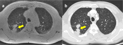 |
Pulmonary High-Resolution ZTE MR Imaging: Comparison with Low-Dose Computed Tomography for the Assessment of Pulmonary Nodules
Did Not Present
Qin Peng, Yao Huang, Wei Tang, Ning Wu, Xiaocheng Wei, Kaiyu Wang
We evaluated the capability of pulmonary MR imaging of zero echo time (zTE) in assessing patients with pulmonary nodules detected by low-dose computed tomography (LDCT). Consequently, pulmonary thin-section MR imaging with zTE was useful in nodule detection and was considered at least as effective as low-dose thin-section CT. This excellent acquisition method of zTE-MRI is turned out to be radiation-free, quiet and relatively insensitive to cardio-respiratory motion, which is important for the detection and follow-up of pulmonary nodules.
|
17:12
|
1188.
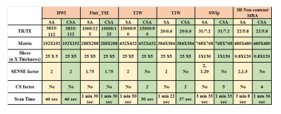 |
Comprehensive stroke protocol of less than six minutes: using Compressed-SENSE with valued addition of SWIp and Non-Contrast-3D-MRA
Rupsa Bhattacharjee, R Karthick Raj, Radha Krishan Verma, Rakesh K Gupta
Acute-stroke-management needs intervention to be done within certain time-window-period. A stroke-MRI-protocol should preferably be within six minutes as reported in previous studies. Objective of this study is to design a comprehensive stroke protocol that is less than six minutes, but also includes SWI and 3D Non-contrast-MR-angio which can help in diagnosing parameters such as tissue viability, site of occlusion, collateral status, stroke-penumbra and silent microbleeds. This is achieved by using compressed-SENSE, technique that combines parallel imaging and compressed sensing by adopting balanced-sampling method, thus providing excellent time-reduction as well as adequate and diagnostic image quality.
|
17:16
|
1189.
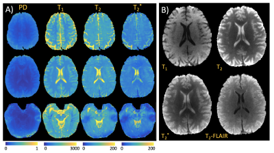 |
Combined T1, T2, and T2* mapping using a multi-inversion multi-echo spin and gradient echo EPI sequence
Mary Kate Manhard, Congyu Liao, Jason Stockmann, Daniel Park, SoHyun Han, Jonathan Polimeni, Berkin Bilgic, Kawin Setsompop
Rapid EPI acquisitions are limited by geometric distortion and blurring due to long readouts. Here we incorporate several recent advances in reconstruction approaches and hardware technology to mitigate some of these errors, and implement a multi-echo multi-inversion EPI-based sequence that can be used to find quantitative proton density, T1, T2, and T2* maps. In addition, high quality clinical contrasts can be generated from these maps including T2-, T2*-, T1-, and FLAIR-weighted images. Our protocol provides high-quality, whole-brain, multi-contrast maps with minimal distortion in scan times of 1-3 minutes.
|
17:20
|
1190.
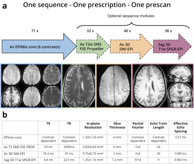 |
EPIMix 3.0
Tim Sprenger, Ola Norbeck, Johan Berglund, Henric Rydén, Enrico Avventi, Stefan Skare
EPIMix is a multi-contrast MRI sequence that can produce T1-FLAIR, T2w, T2*w, T2-FLAIR, DWI, ADC images of the brain in about 70 s. To make EPIMix closer to a single-scan brain MRI exam, we propose optional supplemental sequence modules. The add-on sequences include T2w SMS FSE Propeller (distortion free), 3D SWI-EPI, and 3D T1w SPGR-EPI (isotropic resolution), which all have higher resolution and less distortions compared to EPIMix. With this extension to EPIMix, nine images series can be produced in 3 min total scan time.
|
17:24
|
1191.
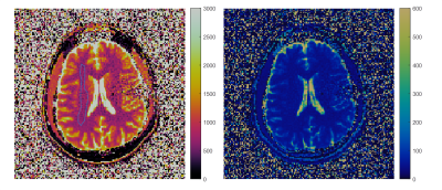 |
Increasing the Value of Legacy MRI Scanners with Magnetic Resonance Fingerprinting
Brendan Eck, Wei-ching Lo, Yun Jiang, Kecheng Liu, Vikas Gulani, Nicole Seiberlich
Few of the 32,000 MRI scanners around the world are equipped with the latest hardware and software required for advanced imaging. Magnetic Resonance Fingerprinting (MRF) presents an opportunity to expand the value of the existing MRI install base as it is rapid, quantitative, and does not require the latest gradient systems or multi-channel receiver coils. In this work, we demonstrate repeatable and reproducible T1 and T2 MRF maps on a 16-year-old MRI scanner as a proof of principle towards implementing MRF on cheap, legacy MRI scanners.
|
17:28
|
1192.
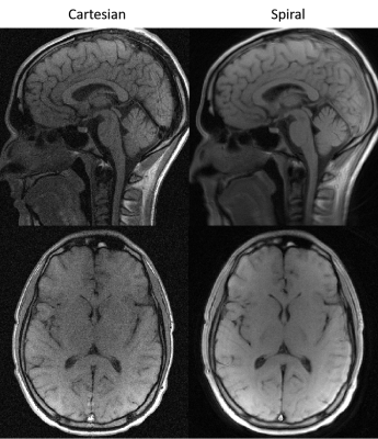 |
Spin-echo imaging at 0.55T using a spiral trajectory
Rajiv Ramasawmy, Daniel Herzka, Matthew Restivo, Ipshitta Bhattacharya, Robert Lederman, Adrienne Campbell-Washburn
Low field MRI combined with modern system engineering is attractive for high quality imaging with reduced production and operation costs. Moreover, there are potential clinical opportunities afforded by the reduced SAR and reduced local susceptibility. However, for a low field system to be clinically useful, neuroimaging must be feasible. Here we explore the potential of spiral neuroimaging at 0.55T to regain SNR that is sacrificed by moving to low field. We show that we can use a 30 ms spiral readout at 0.55T without the need for off-resonance correction and generate a 2.6x gain in SNR compared to Cartesian imaging protocols.
|
17:32
|
1193.
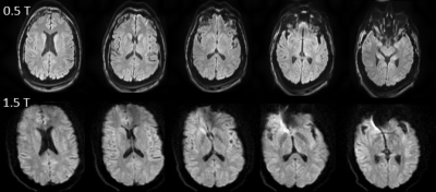 |
High-Performance Diffusion Imaging on a 0.5T System
Jeff Stainsby, Chad Harris, Geron Bindseil, Curtis Wiens, Phil Beatty, Andrew Curtis
Diffusion imaging is a valuable tool in the identification of neurological diseases, especially in an acute setting. Large high-field systems can be challenging to site in an acute setting. Mid-field systems have various siting advantages but historically can suffer from sub-par imaging performance. In this work we compare diffusion weighted imaging performance on a 0.5 T system with a high-performance gradient system to a typical 1.5 T clinical scanner. We demonstrate the ability to achieve comparable imaging performance both analytically as well as through imaging examples.
|
17:36
|
1194.
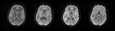 |
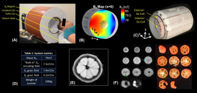
Imaging at 0.5 T with high-performance system components
Jeff Stainsby, Geron Bindseil, Ian Connell, Gilbert Thevathasan, Andrew Curtis, Phil Beatty, Chad Harris, Curtis Wiens, Alex Panther
While the predominant MR market share is taken up with high-field systems (1.0T and above), there are benefits to imaging at mid-field. Mid-field systems can have a compact fringe field, low weight, compact design facilitating siting, low susceptibility-based geometric distortion and reduced RF heating with or without the presence of implants. Mid-field systems have historically been associated with sub-par image quality due to lower signal-to-noise behavior coupled with an absence of state-of-the-art hardware. Here we present a head-only, superconducting mid-field system designed with high-performance system components aimed at achieving comparable image quality to typical 1.5T systems in comparable scan times.
|
17:40
|
1195.
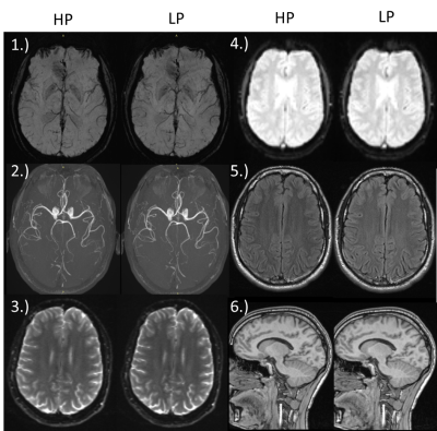 |
On the value of high-performance gradients for clinical MR imaging
Marius Menza, Maximilian Russe, Jürgen Hennig, Maxim Zaitsev
High-performance imaging remains to be one of the hot topics in scientific and clinical development over the recent decades. High-performance gradients are also commonly believed to promise several good image quality and rapid acquisition times. In this project we question the hypothesis that strong gradient systems are advantageous for standard clinical imaging and diagnostics. More specifically we aim to analyse the impact of distinct reduced gradient performance parameters on imaging quality of stroke, cardiac and liver protocols compared to the state-of-the-art high-performance gradient systems at 1.5T, while keeping parameters and measurement time as close as possible.
|
17:44
|
1196.
 |
Feasibility of brain pathology assessment with diffusion imaging on a portable scanner using a fixed encoding field
Jason Stockmann, Patrick McDaniel, Christopher Vuahgn, Clarissa Cooley, Lawrence Wald
Diffusion-weighted MRI is widely used for diagnosing brain pathology, but is typically not available close to the site of acute brain injury due to size, weight, power, and exclusion-zone constraints. Recently-developed lightweight brain scanners have achieved portability by using inhomogeneous B0 fields and by correcting for field imperfections during image reconstruction. Building on work done by the petroleum industry with single-sided borehole magnets, we use the built-in spatial encoding field of a portable brain scanner for both diffusion-encoding and spatial-encoding. We also introduce a method for using dummy refocusing pulses to vary the b-value without changing TEeff and T2-contrast.
|
17:48
|
1197.
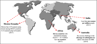 |
Self-administered exam using Autonomous Magnetic Resonance Imaging (AMRI)
Keerthi Sravan Ravi, Sairam Geethanath, John Vaughan Jr.
Scanner density expressed as number of scanners per million people could be used to quantify access to MRI. Globally, scanner density distribution is heterogenous and shortage of skilled manpower to operate systems is one of the barriers in delivering MR. The imr-framework software library transforms a standard MR system into an autonomous scanner (“AMRI”). This work demonstrates self-administering an MR exam leveraging the AMRI implementation. Self-administered exams could alleviate some of the challenges associated with delivering MR imaging to financially constrained geographies. Current and future work involves deployment of AMRI in Kolkata, India.
|
17:52
|
1198.
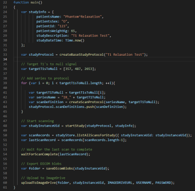 |
How MRI as a Micro-Service Architecture can Accelerate Research and Commercialization
Stewart Bright, Philip Beatty, David Kennedy, Andrea Vargas
We have developed an MRI system having a modern micro-service style distributed software architecture, with the intent of building a flexible, sustainable platform. To describe the architecture, we provide two demonstrations: 1) fully scripted, perform a phantom experiment, extract the images, and upload them to a cloud server for analysis, and 2) update a protocol, append to the queue of scans to be acquired at the simplified operator console, all remotely from another user interface. Through these demonstrations and discussion of its current uses, we aim to illustrate how it could help accelerate collaborative research and commercialization efforts.
|
17:56
|
|
Adjournment |
|
