|
Prostate MRI: New Acquisition & Post-Processing Techniques
Combined Educational & Scientific Session
ORGANIZERS: Daniel Margolis, Claude Sirlin, Utaroh Motosugi
Tuesday, 14 May 2019
| Room 516C-E |
08:15 - 10:15 |
Moderators: Tom Scheenen, Piotr Kozlowski |
Skill Level: Basic to Advanced
Session Number: TU-03
Overview
A combined educational and scientific session will present the state of the art in new acquisition techniques followed by an example and then describe how post-processing improves the performance and value of prostate MRI with two examples of novel post-processing
Target Audience
Body radiologists, MRI physicists and engineers, and image processing scientists.
Educational Objectives
As a result of attending this course, participants should be able to:
- Identify new image acquisition techniques that may be clinically available; and
- Explain how image post-processing improves performance and adds value.
08:15
|
|
Overview of Image Analysis for Prostate MRI

Kirsten Selnæs
Prostate cancer is one of the most frequently diagnosed cancers worldwide and multiparametric MRI is an important part of the diagnostic work-up of prostate cancer patients. Suggestions for standardization of acquisition and interpretation of the images are provided in the PI-RADS v. 2 system, which is increasingly used in clinical practice. New approaches for image analysis, where quantitative features are extracted from the images, allow for the quantitative and multidimensional nature of image data to be exploited. Various machine learning approaches has been used in a wide range of applications within evaluation of prostate mpMRI.
|
08:45
 |
0375.
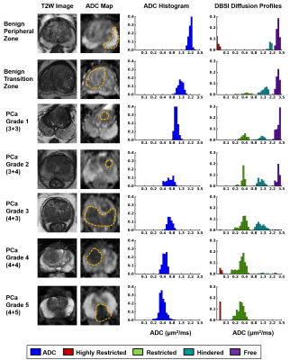 |
Noninvasive Prostate Cancer Grading Using Diffusion MR Structural Fingerprints
Zezhong Ye, Qingsong Yang, Joshua Lin, Peng Sun, Chunyu Song, Ajit George, Sam Gary, Jeffrey Viox, Ruimeng Yang, Jie Zhan, Joseph Ippolito, Jianping Lu, Sheng-Kwei Song
Prostate cancer (PCa) is second most common cause of cancer death among American men. Curently clinicians rely on needle biopsies for PCa grading, although biopsy Gleason scores often differ from those of pathological analyses. We demonstrated modified-DBSI captured and quantified heterogeneous diffusion fingerprints reflecting prostatic histopathology, capable of noninvasively grading PCa with high accuracy. The diagnostic power of modified-DBSI could prevent low-grade cancer patients from undergoing unnecessary and costly invasive procedures, offering patients more reliable assessments on cancer progression during active surveillance, and helping patients and clinicians to determine the most appropriate treatment options.
|
08:57
|
0376.
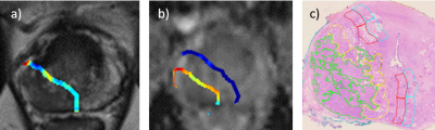 |
Peri-tumoral radiomics on 3T MRI discriminative of D’Amico prostate cancer risk categories show association with epithelium, lumen and stromal densities on whole mount pathology
Rakesh Shiradkar, Ahmad Algohary, Xavier Farre, Patrick Leo, Harri Merisaari, Pekka Taimen, Hannu Aronen, Peter Bostrom, Ivan Jambor, Anant Madabhushi
There is currently increasing interest in looking at role of radiomic features within the peri-tumoral region for disease characterization. In this work, we explore association of peri-tumoral radiomic features of prostate extracted from mpMRI with D’Amico risk. Additionally, we explore morphologic basis of these peri-tumoral features by analyzing the region on whole mount pathology. We observed greater epithelial content in high-risk compared to low, intermediate-risk lesions and vice versa with stroma. This heterogeneity within the peri-tumoral region may be captured by radiomic features that suggest peri-tumoral region of prostate may contain important information associated with risk of prostate cancer progression.
|
09:09
|
|
Advances in Image Acquisition
Video Permission Withheld
Antonio Westphalen
Existing prostate cancer imaging and processing techniques are constantly improving, while new ones are being developed. This talk will focus on the potential benefits of four techniques: hybrid multidimensional MR imaging, luminal water imaging, multi-shot diffusion-weighted imaging, and restriction spectrum imaging.
|
09:39
|
0377.
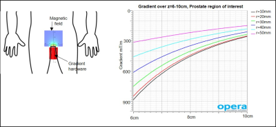 |
Prospects of a Dedicated Nonlinear Gradient for Prostate DWI
Jeffrey Weinreb, Gigi Galiana
DWI is a crucial contrast for prostate cancer, not only for detection but also biopsy guidance and monitoring. Unfortunately, DWI of prostate is plagued by very low SNR, caused by the long TEs needed to encode diffusion with standard imaging gradients. Here we show that by abandoning typical requirements of an imaging gradient, especially nonlinearity, very strong diffusion weighting can be locally achieved using a single standard amplifier, greatly improving DWI image quality. Experiments verify the feasibility of DWI with nonlinear gradients. Though prostate is an ideal first target, this approach could find application in many other organs.
|
09:51
|
0378.
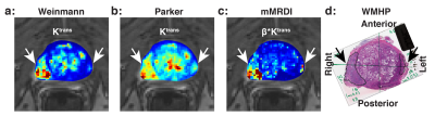 |
Evaluation of Prostate Cancer Detection using Modified MR Dispersion Imaging
Xinran Zhong, KyungHyun Sung
A modified version of MR dispersion imaging, mMRDI, is introduced to achieve an effective estimation of intravascular dispersion parameter while maintaining low computational complexity. A total of 53 patients who underwent 3T mp-MRI exams prior to robotic assisted radical prostatectomy are included for evaluation. The estimation of the additional intravascular dispersion related parameter offers minimum model fitting errors and improved delineation between clinically significant prostate cancer and normal tissue assessed by the receiver operating curve analysis (AUC = 0.92).
|
10:03
|
0379.
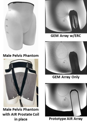 |
Evaluation of an eight element AIR coil array for MRI in the prostate
Phillip Rossman, Eric Borisch, Roger Grimm, Brent Warndahl, Kiaran McGee, Stephen Riederer
Obtaining sufficient SNR in MRI of the prostate can be problematic without the use of an endorectal coil (ERC). We have constructed a lightweight, highly flexible receive-only surface coil array consisting of 8 recently developed AIR coil elements (GE Healthcare, Waukesha, WI, USA). This work compares the AIR coil array to commercial arrays commonly used for prostate imaging with and without the inclusion of an ERC. Initial phantom results show that the AIR coil array has approximately 2.5-3× higher SNR than the GE 32 channel anterior body/GEM posterior array when no ERC is used and approaches ERC SNR performance.
|
10:33
|
|
Adjournment |
|
