Combined Educational & Scientific Session
Improved Motion Correction & Effective Free-Breathing Approaches Back to Program-at-a-Glance Back to Program-at-a-Glance
|
|
Improved Motion Correction & Effective Free-Breathing Approaches
Combined Educational & Scientific Session
ORGANIZERS: Bernd Wintersperger, Jenny Keegan
Tuesday, 14 May 2019
| Room 513A-C |
15:45 - 17:45 |
Moderators: Andrew Scott, Matthias Stuber |
Skill Level: Basic to Intermediate
Session Number: Tu-07
Overview
This session will review conventional and advanced approaches to cardiac and respiratory motion correction in cardiovascular MRI.
Target Audience
Clinicians and scientists wishing to understand how cardiac and respiratory motion can be corrected for, both on commercial clinical scanners and in research applications.
Educational Objectives
As a result of attending this course, participants should be able to:
- Recognize how the heart moves with the respiratory and cardiac cycles;
- Compare how the motion affects image quality for different sequences and different k-space paths;
- Describe conventional methods of motion correction; and
- Explain emerging highly advanced techniques that are highly efficient and may provide cardiac and/or respiratory motion-resolved imaging.
15:45
|
 |
Motion of the Heart with the Respiratory & Cardiac Cycles: Conventional Approaches to Motion Correction

Leon Axel
Respiratory and cardiac motion can result in degradation of MR images. We review the basic aspects of these motions, and some standard means that can be used to monitor them and control their effects on imaging.
|
16:10
|
|
Self Assessment Module (SAM) |
16:15
|
 |
Motion of the Heart with the Respiratory & Cardiac Cycles: Advanced Motion Correction Techniques
Claudia Prieto
MRI acquisition is slow compared to physiological motion, thus the extensive cardiac- and respiratory- induced motion of the heart during the acquisition period can degrade image quality by introducing ghosting and/or blurring like motion artefacts. Several cardiac and respiratory motion compensation techniques have been proposed to overcome this problem. These techniques are based on minimizing, correcting or resolving the motion during the acquisition. This talk will include examples of recently introduced advanced methods to deal with cardiac and respiratory motion, discussing their strengths and limitations.
|
16:45
 |
0605.
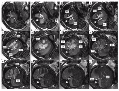 |
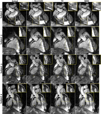
Imaging Fetal Congenital Heart Disease Using Motion Compensated Cardiovascular Magnetic Resonance Imaging
Christopher Roy, Davide Marini, David Lloyd, Wadi Mawad, Shi-Joon Yoo, Eric Schrauben, Edgar Jaeggi, Mike Seed, Christopher Macgowan
In pregnancies where fetal congenital heart disease is suspected during routine obstetric ultrasound, a thorough echocardiographic workup is required to assess the fetal cardiac anatomy and function. MRI has been increasingly proposed as an adjunct diagnostic tool to evaluate the fetus. In this work, we present a newly developed method for reconstructing high-resolution dynamic MR images of the fetal heart, evaluate this modality in the context of visualizing cardiac abnormalities, and compare to echocardiography. We show that MRI of the fetal heart has the potential to compliment echocardiography in the assessment of congenital heart disease.
|
17:05
 |
0606.
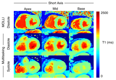 |
3D Multitasking for Non-ECG, Free-Breathing, Simultaneous Cardiac Motion-Resolved T1 Mapping and Function Evaluation
Video Permission Withheld
Jaime Shaw, Xiaoming Bi, Debiao Li, Anthony Christodoulou
Myocardial T1 mapping characterizes focal and diffuse fibrosis. We have recently shown cardiac motion-resolved 2D T1 maps with free breathing and without ECG, greatly simplifying workflow. In this study, we extend this approach to 3D and redesign the acquisition to improve spatial resolution and sampling efficiency compared to 2D. We also show the feasibility of assessing function from the same cardiac-resolved 3D Multitasking scan used for T1 mapping. T1 maps were compared to 2D MOLLI and function was compared to a breath-hold cine sequence.
|
17:25
|
0607.
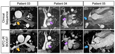 |
Non-contrast enhanced simultaneous bright- and black-blood 3D whole-heart MRI for the assessment of arterial and venous anatomy in patients with congenital heart disease.
Giulia Ginami, Imran Rashid, Harith Alam, Radhouene Neji, Israel Valverde, Alessandra Frigiola, René Botnar, Claudia Prieto
Bright- and black-blood MRI sequences provide complementary diagnostic information in patients with congenital heart disease (CHD). Typically, contrast agent administration is needed to depict structures such as the pulmonary veins or regions of disturbed blood flow. However, contrast-enhanced imaging is not ideal for regular screening, while the acquisition of bright- and black-blood sequences in a sequential fashion remains sub-optimal. Here we propose a motion-compensated 3D whole-heart sequence that provides co-registered bright- and black-blood volumes without the need for contrast agent administration. The sequence was tested in patients with CHD and showed high-quality delineation of both arterial and venous structures.
|
17:45
|
|
Adjournment |
|
Back to Program-at-a-Glance |  Back to Top Back to Top |
| The International Society for Magnetic Resonance in Medicine is accredited by the Accreditation Council for Continuing Medical Education to provide continuing medical education for physicians. |
