|
Stroke: Early Signs & Late Recovery
Combined Educational & Scientific Session
ORGANIZERS: Meiyun Wang, Andre Obenaus
Wednesday, 15 May 2019
| Room 513A-C |
13:30 - 15:30 |
Moderators: Yan Bai, Meiyun Wang |
Skill Level: Basic to Advanced
Session Number: W-05
Overview
Stroke is one of the leading cause of deaths in the world. This combined scientific and educational session focuses on the applications of advanced MRIs in the early diagnosis, guiding therapy, and evaluation of late functional recovery in stroke.
Target Audience
Trainees and scientists, as well as neuroradiologists, neurologists, and other clinical professionals interested in understanding the latest developments in stroke imaging.
Educational Objectives
As a result of attending this course, participants should be able to:
- Discuss early diagnosis and evaluation of stroke using neuroimaging;
- Describe how the brain re-organizes during functional recovery; and
- Discuss current and emerging imaging techniques.
13:30
|
|
Assessment of Functional Recovery After Stroke: Connectivity

Arno Villringer
Symptoms after stroke are not only determined by location and extent of a lesion, but also by alterations of connectivity. Such changes have been assessed with functional connectivity MRI (fcMRI) based on resting-state recordings. Here, different fcMRI approaches are reviewed ranging from interhemispheric connectivity in one network, simultaneous assessment of many networks to methods based on graph theory. A recent approach overcomes the notion of “fixed separate connectivity networks” but assumes a multidimensional connectivity space (Margulies et al. PNAS 2016). As a perspective, fcMRI will allow to tailor brain stimulation therapies to the pathophysiological state of the individual patient.
|
13:50
|
|
Mapping Asymmetrically Prominent Cortical Veins & Microbleeds Using SWI & QSM
E. Mark Haacke
|
14:05
|
|
Self Assessment Module (SAM) |
14:10
|
|
Intracranial Collaterals: The Role of MRI
Xin Lou
Studies have found collateral circulation was correlated with progression of stroke and clinical outcome. Many advanced imaging approaches appeared regarding the intracranial collaterals, including DSA, CT and MR. Specifically, the newly developed MR sequences, including multiphase contrast enhanced MR angiography, ASL and vessel-encoded ASL etc. have been used in the visualization of collaterals. Additionally, some conventional MR sequences have also been found useful. Learning what MR can do in the field of collaterals is of paramount importance. Application of MR techniques in intracranial collaterals may help elucidate pathophysiological characteristics of collaterals and enhance the therapeutic and prognostic performance in stroke patients.
|
14:30
 |
0826.
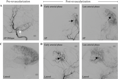 |
Choroid plexus perfusion MRI indicates cerebrospinal fluid production changes after surgically-manipulated vascular tone: implications for glymphatic flow
Skylar Johnson, Sarah Lants, Meher Juttukonda, Colin McKnight, Daniel Claassen, Manus Donahue
The objective of this study was to determine if a feedback circuit between CSF production and arterial health may exist in the human brain. Sequential measurements of CSF volume were obtained, as well as cortical and choroid plexus function measured from pseudo-continuous arterial spin labeling (pCASL) before and after clinically-indicated indirect surgical revascularization. Regression analyses were used to evaluate dependence of study parameters on time. Following surgically-induced angiogenesis, fronto-parietal perfusion increased, which paralleled a reduction in choroid plexus perfusion. This could reflect a homeostatic mechanism where improved perivascular flow and more robust waste clearance prompts decreased choroid plexus CSF production.
|
14:42
|
0827.
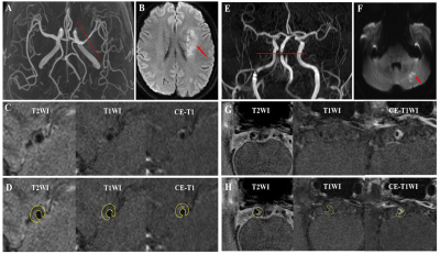 |
Radiomic analysis on symptomatic and asymptomatic intracranial atherosclerotic plaque using high resolution MRI
Zhang Shi, Chengcheng Zhu, Bing Tian, Jianping Lu, QI Liu
This study aims to evaluate a quantitative radiomic approach including texture analysis based on HR-MRI to differentiate acute symptomatic plaque from asymptomatic plaque. 158 patients with middle cerebral artery and basilar artery stenosis underwent HR-MRI. The stenosis value, plaque area/burden, lumen area, intraplaque hemorrhage (IPH), contrast enhancement ratio and 109 quantitative radiomic features were extracted. Multivariate logistic analysis and a random forest model were performed. The result was shown that smoking, IPH and enhancement ratio were independently associated with symptomatic plaques. Radiomic features in T2, T1 and CE-T1 images were associated with symptomatic plaques, whose AUC respectively are 0.801,0.835 and 0.846. The combined all radiomic approach had a significantly higher AUC of 0.953. Combination of all features reached an AUC of 0.976, with accuracy of 87.4%. Radiomic analysis accurately distinguished between acutely symptomatic plaques and asymptomatic plaques.
|
14:54
|
0828.
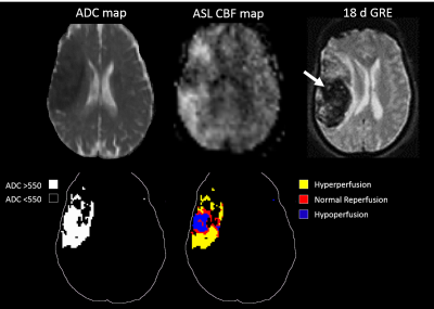 |
Reperfusion into Severely Damaged Brain Tissue is Associated with Impending Parenchymal Hemorrhage in Recanalized Acute Ischemic Stroke Patients
Samantha Ma, Songlin Yu, David Liebeskind, Xin Qiao, Lirong Yan, Xin Lou, Jeffrey Saver, Noriko Salamon, Danny Wang
While timely reperfusion can reduce more extensive brain tissue injury by salvaging reversibly damaged penumbra, late recanalization also carries the risk of causing additional and substantial brain damage, such as ischemia-reperfusion injury, compared with no revascularization. The reperfusion status after thrombolysis within the DWI lesion territory and its relationship with HT is unclear, leaving the details regarding post-treatment care unspecified. In this study, we aim to quantify the volume of reperfusion into severely damaged brain tissue and investigate its predictive value for impending parenchymal hematoma following recanalization.
|
15:06
 |
0829.
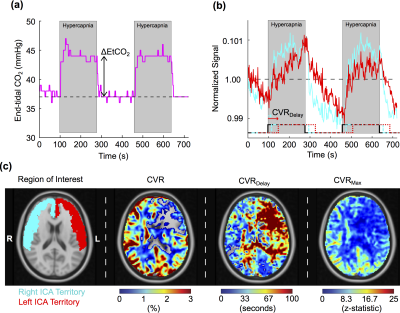 |
Supervised Learning Techniques Applied to Multi-modal Functional Neuroimaging Data Have Promise for Infarct Prediction
Spencer Waddle, Meher Juttukonda, Sarah Lants, Larry Davis, Rohan Chitale, Matthew Fusco, Lori Jordan, Manus Donahue
Common functional imaging approaches such as cerebral blood flow-weighted arterial spin labeling and cerebrovascular reactivity-weighted blood oxygenation level-dependent MRI are susceptible to quantification errors when applied to patients with significant arterial steno-occlusive disease, due to artifacts that result from delayed blood arrival and arteriolar rigidity. Recently it was suggested that the artifacts from standard quantitation approaches can be exploited together with machine learning algorithms to localize regions of hemodynamic impairment as defined by gold standard catheter angiography. Here, we investigate whether similar algorithms can be applied to identify spatial regions that progress to infarction in a longitudinal study.
|
15:18
|
0830.
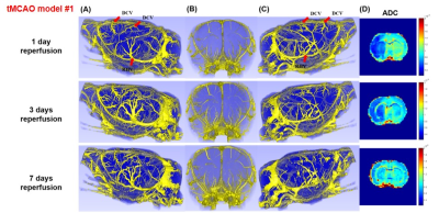 |
Dual Contrast MRI for Visualization of Whole Brain Macro and Microvascular Remodeling in Rat Ischemic Stroke Model
MungSoo Kang, DongKyu Lee, SeokHa Jin, HyungJoon Cho
Because of therapeutic and prognostic significance, the visualizations and quantifications of vascular remodelings are crucial for ischemic stroke brains. In this work, dual contrast MRI was implemented on tMCAO rats as an ischemic stroke model to visualize whole-brain macro- and micro-vascular remodeling. In additional to R1 based UTE-MRA, which showed macrovascular remodeling, simultaneously acquired VSI, Q and MVD metrics from ΔR2-ΔR2*-MRI showed microvascular remodeling in ischemic reperfused rat brains.
|
15:30
|
|
Adjournment |
|
