|
Cell Tracking & Molecular Imaging Using Exogenous Agents
Combined Educational & Scientific Session
ORGANIZERS: Ronald Ouwerkerk, Kannie WY Chan, Yi-Fen Yen
Wednesday, 15 May 2019
| Room 516C-E |
13:30 - 15:30 |
Moderators: Guanshu Liu, Nirbhay Yadav |
Skill Level: Basic to Intermediate
Session Number: W-07
Overview
An overview of methods and applications of MR based cell tracking methods using contrast agents or bioorganic contrast enhancement. Mechanisms based on gadolinium, super-paramagnetic ion, fluorine, and methods based on chemical exchange saturation transfer are discussed. The current and future clinical applications are also discussed.
Target Audience
Educational Objectives
As a result of attending this course, participants should be able to:
- Identify methods available to probe cell distribution, cell survival, cell function, cell differentiation, and cellular enzymatic activity; and
- List which probes are suitable for humans use and which probes have made it into clinical trials.
13:30
|
|
Methods for Cell Tracking

Erik Shapiro
MRI has proven to be a powerful imaging modality for non-invasive, whole body imaging, with meaningful image resolution for studying cellular dynamics. MRI contrast is generated by capitalizing on differences in water molecule microenvironment, including diffusion rate and direction, magnetic field differences, and water content. In order to use MRI to distinguish a unique population of cells from other cells in the body, such as a cell transplant, or the infiltration of immune cells to a disease, one of these properties needs to be altered, or a tracer needs to be introduced. This will generate contrast or signal from these cells.
|
14:00
|
|
Clinical Applications of MR Cell Tracking
Video Permission Withheld
Jeff Bulte
MRI is expected to play a key role in evaluating the outcome of clinical trials based on stem and immune cell therapy. In order to facilitate and implement the translation of these therapies into the clinic, it will be necessary to monitor the immediate cellular engraftment, subsequent biodistribution and migration, and cell survival and differentiation non-invasively over time.
|
14:30
|
0821.
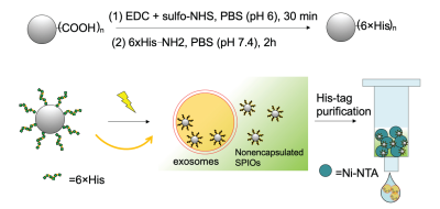 |
In vivo MRI tracking of stem-cell-derived extracellular vesicles
Zheng Han, Senquan Liu, Zheng Ding, Chuheng Chen, Zelong Chen, Linzhao Cheng, Guanshu Liu
Induced pluripotent stem cell (iPSC) derived extracellular vesicles (EVs) constitute a new class of cell-free regenerative medicine. Non-invasive tracking of the delivery and in vivo distribution of EVs is highly desirable. Traditional labeling methods suffer from either low labeling efficiency or difficulty with purification. Here, we report a new labeling strategy using surface-functionalized magnetic particles, in which the nonencapsulated magnetic particles can be easily separated from the labeled EVs, allowing the preparation of magnetically labeled EVs with high purity. We then demonstrate in vivo tracking of magnetically labeled EVs in the kidney and liver using MRI.
|
14:42
|
0822.
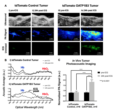 |
Multimodality OATP1B3-enhanced reporter gene imaging of engineered cells via fluorescence, photoacoustic, and magnetic resonance imaging
Nivin Nyström, Lawrence Yip, Jeffrey Carson, Timothy Scholl, John Ronald
Reporter genes can generate information on cell fate in vivo. Multimodality imaging can mitigate limitations of individual imaging modalities; thus, multimodality reporters are highly sought. We present a trimodality reporter gene system based on human Organic Anion-Transporting Polypeptide 1b3 (Oatp1b3) and its ability to take up indocyanine green (ICG) for fluorescence and photoacoustic imaging, as well as gadolinium ethoxy benzyl diethylenetriamine pentaacetic acid (Gd-EOB-DTPA) for MRI. This technology should find utility in tracking cell- and gene-based therapies for preclinical studies, and due to the human-derivation of the reporter gene and already clinically-utilized probes, presents with high translational potential.
|
14:54
|
0823.
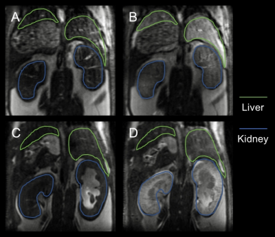 |
Chimeric Mouse Model for MRI Contrast Agent Evaluation
Faryal Mir, Ryan Tomaszewski, Dorela Shuboni-Mulligan, Christiane Mallett, Jeremy Hix, Nicholas Ether, Erik Shapiro
While rodents are the primary models for contrast agent evaluation, there is considerable variability in pharmacokinetics of contrast agents in mice vs humans. OATPs play a role in in vivo pharmacokinetics in mice and humans. This study provides evidence that the OATP1B1/1B3 knock-in mouse is a more useful screening tool for novel MRI contrast agents destined for clinical use as compared to the traditionally used wild-type models.
|
15:06
 |
0824.
 |
MEMRI Can Quantify Myocardial Infarct Size Earlier Than LGE-MRI
Nur Hayati Jasmin, May Zaw Thin, Valerie Taylor, Christopher Pope, Mark Lythgoe, Sean Davidson, Daniel Stuckey
Late gadolinium enhanced MRI (LGE-MRI) is an established method for quantification of infarct size after myocardial infarction but is non-specific and reflects the increased membrane rupture and extracellular volume (ECV) that occurs several hours after myocardial infarction. Manganese (Mn2+) is an efficient intracellular MR contrast agent, which acts as an analogue of Calcium (Ca2+) and can provide information on cell viability. The present study shows that manganese-enhanced MRI (MEMRI) can quantify the final infarct size earlier than LGE-MRI.
|
15:18
|
0825.
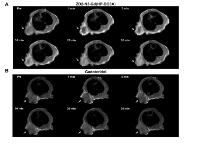 |
Magnetic resonance molecular imaging of EDB fibronectin with a ZD2 peptide Gd(HP-DO3A) conjugate for detecting pancreatic cancer
Peter Qiao, Nadia Ayat, Amita Vaidya, Songqi Gao, Han Zheng, Samuel Chou, Zheng Rong Lu
Pancreatic cancer (PC) carries a poor prognosis, partially due to lack of early diagnosis. To address this unmet need, the potential of magnetic resonance molecular imaging of extradomain B fibronectin with an oligopeptide targeted MRI contrast agent was assessed in mouse models of PC. Four PC cell lines were assayed for EDB-FN expression in vitro and found to overexpress EDB-FN. Analysis of ZD2 binding and contrast enhanced MRI demonstrated over two-fold improvement in CNR compared to non-targeted gadoteridol, with little non-specific binding. Histological analysis revealed the expression of EDB-FN in the tumor microenvironment and confirmed the features observed on MRI
|
15:30
|
|
Adjournment |
|
