Weekend Educational Session
Myelin Back to Program-at-a-Glance Back to Program-at-a-Glance
|
|
Myelin
Weekend Course
ORGANIZERS: Cornelia Laule, Alex MacKay
Saturday, 11 May 2019
| Room 516C-E |
13:30 - 17:00 |
Moderators: Cornelia Laule, Alex MacKay |
Skill Level: Basic to Intermediate
Session Number: WE-12
Overview
The goal of this course is to introduce the field of myelin imaging. The session will begin with an overview of the role of myelin in normal brain function and clinical value of being able to image myelin. Each of the next 7 presentations will cover different approaches to imaging myelin.
Target Audience
Clinicians who would like to be able to monitor myelin in demyelinating diseases and acquired injuries; neuroscientists who are interested in understanding myelin in normal brain function and want to learn what imaging techniques are available to measure myelin; and physicists and engineers who want to learn how to image myelin.
Educational Objectives
As a result of attending this course, participants should be able to:
- List the pros and cons of the available MR techniques for measuring myelin;
- Describe how inhomogeneity in B0 and B1 influence the different myelin imaging techniques;
- Explain how changes in CNS environment due to non-myelin pathology influence the different myelin imaging techniques; and
- Identify which of the available MR techniques for measuring myelin is most appropriate for each participant's application.
13:30
|
|
What Is Myelin & Why Is It Important to Image?

Samuel Ludwin
|
13:50
|
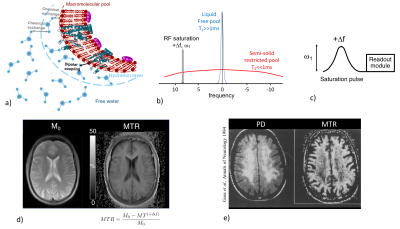 |
Magnetization Transfer Techniques
Guillaume Duhamel
The potential of Magnetization Transfer techniques (MTR, qMT and ihMT) for myelin imaging is presented
|
14:10
|
|
Myelin Water Imaging
Shannon Kolind
Myelin water imaging (MWI) provides quantitative measurements specific to myelin by separating the MRI signal into contributions from the various water pools present within a voxel. In central nervous system tissue these water pools generally correspond to intra- and extra-cellular water, which relaxes slowly, and water trapped between the myelin bilayers, which relaxes quickly. The fraction of water corresponding to the water trapped within the myelin sheath, the myelin water fraction (MWF), provides a quantitative measure related to myelin content. This course will discuss acquisition and analysis techniques as well as common artefacts and pitfalls.
|
14:30
|
|
T1 & T1w/T2w
Christine Tardif
This talk describes two methods used for imaging myelin content in vivo: T1 mapping and T1-weighted/T2-weighted (T1w/T2w) signal ratio imaging. Their advantages and limitations will be discussed in comparison to other techniques presented in the Myelin Imaging session.
|
14:50
|
|
Break & Meet the Teachers |
15:20
|
|
Ultra Short TE
Mark Does
Magnetic resonance imaging (MRI) contrast can be highly sensitive to the presence of myelin; however, quantitative MRI approaches to measure myelin content remain a challenge. With ultra-short echo time (UTE) MRI, it may be possible to directly image the rapidly-relaxing non-aqueous protons in the phospholipid bilayers that comprise myelin. UTE-MRI brings with it technical challenges, and the MRI characteristics of the ultra-short T2 signal from myelin is not yet well understood. Several studies have presented UTE-MRI of brain thought to reflect myelin, but questions remain and further work is needed to validate this approach.
|
15:40
|
 |
Synthetic Myelin Imaging
Marcel Warntjes
Synthetic MR imaging is a method that creates conventional T1W, T2W and FLAIR images by measuring the T1 and T2 relaxation times. The method can only resolve slow relaxation components, but by using a model for myelin including magnetization exchange, it can infer the presence of myelin. The advantage of the method is the short scan time, where typically full head coverage is obtained in 6 minutes. A new sequence is developed where even a 1 mm isotropic resolution can be obtained in that scan time. An overview is provided of the measurements, modeling and clinical application of myelin detection using synthetic MRI.
|
16:00
|
 |
T2*
Dong-Hyun Kim
Recently, there has been numerous research on myelin water imaging via T2* contrast, e.g. based on multi gradient-echo imaging. In this talk, I will present the methodology of T2* based myelin water imaging. Acquisition and reconstruction methods will be presented. Also, the pros and cons of T2* based myelin water imaging and challenges will be discussed.
|
16:20
|
|
Exploring myelin content changes with Positron Emission Tomography: application to multiple sclerosis
Benedetta Bodini
Measuring myelin content changes in living patients is essential to understand the mechanisms underlying demyelinating diseases such as multiple sclerosis (MS). Several advanced MRI techniques have proven to be extremely sensitive for detecting microstructural changes affecting brain tissues of patients with MS. However, since similar changes in the physical properties of tissues detected by MRI can result from multiple pathological processes indistinguishable from each other, the interpretation of imaging data acquired with MRI remains challenged by an intrinsic suboptimal specificity. Being based on the use of radiolabeled compounds selectively binding to specific biological targets, positron emission tomography (PET) offers the highest possible specificity to explore myelin content changes in the human brain.
|
16:40
|
|
Panel Discussion |
17:00
|
|
Adjournment & Meet the Teachers |
|
Back to Program-at-a-Glance |  Back to Top Back to Top |
| The International Society for Magnetic Resonance in Medicine is accredited by the Accreditation Council for Continuing Medical Education to provide continuing medical education for physicians. |
