Weekend Educational Session
Vascular Educational Back to Program-at-a-Glance Back to Program-at-a-Glance
|
|
Vascular Educational
Weekend Course
ORGANIZERS: Winfried Willinek, Neville Gai
Sunday, 12 May 2019
| Room 518A-C |
08:00 - 12:00 |
Moderators: Giles Roditi, Oliver Wieben |
Skill Level: Basic to Intermediate
Session Number: WE-16
Overview
This course reviews the essential components of a vascular MR exam including contrast and non-contrast techniques, flow and vessel wall imaging. Both established and more recent approaches will be discussed, together with clinical applications.
Target Audience
Clinicians and scientists who wish to understand the clinical needs and applications of vascular imaging and to gain an understanding of the technical foundations of both established practice and more recent developments.
Educational Objectives
As a result of attending this course, participants should be able to:
- Recognize the technical basis of the methods used in a vascular MR exam;
- Describe the clinical applications of the techniques used in vascular imaging; and
- Describe recent advances in vascular imaging and their benefits.
08:00
|
|
Contrast Agents

Tim Leiner
|
08:30
|
|
Non-Contrast Enhanced
Video Permission Withheld
Mitsue Miyazaki
Various established non-contrast enhanced MRA techniques such as time-of-flight (TOF), quiescent interval single-shot (QISS), fresh blood imaging (FBI), and a flow-in spin labeling are discussed with their characteristic features. In addition, recent on-going research techniques are introduced such as flow-sensitive dephasing (FSD), velocity-selective inversion preparation with 3D bSSFP, radial QISS, and radial fast interrupted steady-state (FISS).
|
09:00
|
|
Contrast-Enhanced
Jeremy Collins
Contrast-enhanced MR angiography is an adaptable imaging technique that can be tailored to the clinical question posed. CE-MRA relies on subtracted and unsubtracted techniques applied to single station, multi station, and time-resolved CE-MRA. CE-MR Angiography is considered a reference standard for arterial evalation. More recent developments rely on imaging in the steady state with ECG-gating, applying acceleration schema to shorten imaging time without compromising the spatial resolution.
|
09:30
|
|
Flow Hemodynamics
Susanne Schnell
Phase contrast MRI and its utilization to measure blood flow will be explained. Spins that move during an MRI acquisition exhibit different imaging characteristics compared to stationary spins. Flowing spins, for example from flowing blood, appear as an artifact in the image. However, by understanding these characteristics of flowing spins, their appearance can be utilized for angiographic purposes. 2D Phase contrast imaging is sensitized to flow by using a series of bipolar gradients to affect the phase signal of spins that flow with a uniform velocity in the direction parallel to the gradients. By utilizing ECG gating, blood flow velocities can be measured in a time-resolved manner. 2D phase contrast can be extended to a time-resolved 3D volume acquisition with 3-directional velocity encoding, which is called 4D flow MRI. This encoding of velocity enables quantification of flow hemodynamics. Furthermore, some potential sources of error will be discussed, such as misalignment of flow, velocity aliasing and phase offset errors.
|
10:00
|
|
Break & Meet the Teachers |
10:30
|
|
Supraaortic & Intracranial
Bum Soo Kim
This talk will describe the sequences used for MRA of intracranial and supraaortic vessels, and clinical application to cerebrovascular diseases, focusing on the contributions that MRA can make to diagnosis and follow-up.
|
11:00
|
 |
Chest & Abdominal
Video Permission Withheld
Mark Schiebler
This syllabus and the accompanying slides serve as an introduction to the use of pulmonary MRA for the primary diagnosis of pulmonary embolism. It is our hope that you will be able to start up a program at your institution based on this information.
|
11:30
|
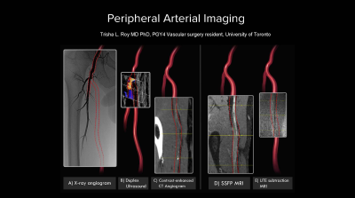 |
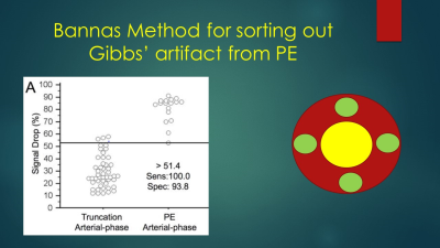
Peripheral
Trisha Roy
This peripheral arterial educational session will review the pathophysiology and management of peripheral arterial disease, current imaging modalities, gaps in knowledge and opportunities for MRI to address the many remaining questions in this field.
|
12:00
|
|
Lunch & Meet the Teachers |
|
Back to Program-at-a-Glance |  Back to Top Back to Top |
| The International Society for Magnetic Resonance in Medicine is accredited by the Accreditation Council for Continuing Medical Education to provide continuing medical education for physicians. |
