
Combined Educational & Scientific Session
Renal MRI: Past, Present & Future
Session Topic: MRI of the Kidneys
Session Sub-Topic: Renal MRI: Past, Present & Future
Combined Educational & Scientific Session
ORGANIZERS: Christoffer Laustsen, Daniel Margolis, Mustafa Shadi Bashir
| Wednesday Parallel 3 Live Q&A | Wednesday, 12 August 2020, 15:15 - 16:00 UTC | Moderators: Steven Sourbron & Octavia Bane |
Skill Level: Basic to Advanced
Session Number: C-M-01
Overview
This session will provide an overview of the state-of-the-art, emerging MR biomarkers and the clinical relevance of MRI biomarkers of renal diseases.
Target Audience
Basic researchers and clinicians interested in renal MRI.
Educational Objectives
As a result of attending this course, participants should be able to:
- Name basic imaging strategies for naive, AKI and CKD applications;
- Describe the emerging imaging strategies for naive, AKI and CKD applications;
- Explain the functional imaging strategies for naive, AKI and CKD applications; and
- Identify anatomic and functional characteristics of suspicious renal lesions.
 |
State-of-the-Art Focal Kidney MR Biomarkers
Brian Allen
Many solid renal neoplasms have characteristic features on imaging. Imaging biomarkers can be used to non-invasively identify histology of the most common subtypes of renal cell carcinoma and can be used to assess response to therapy. No one biomarker can accurately differentiate all renal masses, as some renal masses have features in common, necessitating attention to all images, phases, or sequences.
|
|
| Emerging Focal Kidney Biomarkers
Cornelius von Morze
|
||
 |
0949.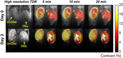 |
Iopamidol CEST MR Urography for Urinary Tract Obstructions
KowsalyaDevi Pavuluri1, Shaowei Bo1, Farazad Sedaghat1, Max Kates2, and Michael T McMahon1,3
Video Permission Withheld
1The Russell H. Morgan Department of Radiology and Radiological Science, The Johns Hopkins University School of Medicine, Baltimore, MD, United States, 2Department of Urology, The Johns Hopkins University School of Medicine, Baltimore, MD, United States, 3F.M. Kirby Research Center for Functional Brain Imaging, Kennedy Krieger Institute, Baltimore, MD, United States
Urinary tract obstructions (UTOs) are impairments in urine flow which can lead to pain, infection and irreversible kidney damage if left undiagnosed or untreated. Chemical exchange saturation transfer (CEST) is a novel MRI contrast mechanism that is particularly sensitive to environmental changes including changes in pH values. In this study we developed a protocol by administering the FDA approved iopamidol to obtain dynamic pH and perfusion MRI contrast maps of the kidneys and compared these with iopamidol administered multi-phase CT in a unilateral urinary obstruction mouse model.
|
0950.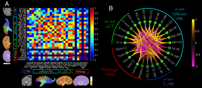 |
T2 and diffusion tensor imaging of kidney disease in an epicutaneous TLR7 enhanced lupus mouse model
Luke Xie1, Vineela D. Gandham1, Kai H. Barck1, Eric Suto1, Wyne P. Lee1, Oded Foreman1, Richard A. D. Carano1, Alex J. De Crespigny1, and Robby M. Weimer1
Video Permission Withheld
1Genentech, South San Francisco, CA, United States
System lupus erythematosus (SLE) is an autoimmune disease that can lead to lupus nephritis and glomerulonephritis. Studies have evaluated kidneys from SLE patients using diffusion-weighted imaging. However, specific MRI metrics most related to the underlying disease has not been identified. In this study, we evaluate a lupus model with MRI and determine the physical properties that contribute to the MRI signal. This is achieved through structural analysis of glomeruli with whole kidney 3D micro-CT and pathological evaluation of glomeruli, tubules, interstitium, and arterioles. Finally, a comprehensive correlation analysis is performed to determine top MRI metrics most sensitive to the disease.
|
|
| Emerging Functional Kidney MR Biomarkers
Steffen Ringgaard
Non-invasive assessment of kidney function and microstructure is important for diagnosis and treatment monitoring of patients with kidney diseases. Besides its ability to make high-resolution diagnostic images, MR also has the potential for evaluating a number of functional parameters. In this lecture we will discuss the most promising of these MR biomarkers for assessing kidney function and microstructure, and we will briefly touch up on some arising methodologies. This includes ASL, phase contrast, BOLD imaging, diffusion imaging, relaxation mapping and some non-proton methods.
|
||
|
0951.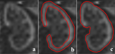 |
Pulsed Arterial Spin Labeling and Pseudo-Continuous Arterial Spin Labeling MRI for Diagnosis of Renal Insufficiency
Zhiyong Lin1, Rui Wang1, Jing Liu1, Jinxia Zhu2, Chengwen Liu2, Bernd Kühn3, and Xiaoying Wang1
1Department of Radiology, Peking University First Hospital, Beijing, China, 2MR Collaboration, Siemens Healthcare, Ltd., Beijing, China, 3Siemens Healthcare GmbH, Erlangen, Germany
Patients with renal artery stenosis (RAS) exhibit changes in renal artery hemodynamics. This study investigated the clinical value of pulsed arterial spin labeling (PASL) and pseudo-continuous arterial spin labeling (pCASL) in diagnosing and grading renal insufficiency in patients with RAS. PASL performed better for measuring renal blood flow (RBF) in the renal cortex to provide differential diagnosis of renal function, while the RBF values obtained with pCASL were more closely correlated with the glomerular filtration rate (GFR). These findings indicate that PASL and pCASL MRI have utility for diagnosing and grading renal insufficiency in patients with RAS.
|
|
0952.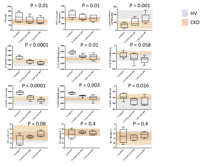 |
Assessment of Acute Kidney Injury and associated longitudinal changes with recovery using multiparametric renal MRI
Charlotte Elizabeth Buchanan1, Huda Mahmoud2, Eleanor F Cox1, Rebecca Noble2, Benjamin L Prestwich1, Isma Kazmi 2, Maarten W Taal2, Nicholas Selby2, and Susan T Francis1
1Sir Peter Mansfield Imaging Centre, University of Nottingham, Nottingham, United Kingdom, 2Centre for Kidney Research and Innovation, University of Nottingham, Derby, United Kingdom
Acute kidney injury (AKI) is defined clinically using serum creatinine. We use multiparametric renal MRI to assess longitudinal changes in AKI. Nine participants were assessed at time of AKI, 7 were re-scanned at 3-months and 1-year. At peak AKI, total kidney volume (TKV) and cortex and medulla T1 were elevated, and cortex perfusion reduced compared to HVs. After 3-months, TKV reduced compared to peak AKI, cortex and medulla T1 remained slightly elevated compared to HVs. Perfusion remained reduced compared to HVs after 1-year. MRI showed incomplete recovery at 3 months, despite normalisation of biochemistry, providing potential to identify maladaptive repair.
|
 Watch the Video