
Combined Educational & Scientific Session
Spinal Cord: Cool MR Tools & How to Use Them
Session Topic: Spinal Cord, Head and Neck
Session Sub-Topic: Spinal Cord: Cool MR Tools & How to Use Them
Combined Educational & Scientific Session
ORGANIZERS: Cornelia Laule, John Port
| Thursday Parallel 2 Live Q&A | Thursday, 13 August 2020, 15:05 - 15:50 UTC | Moderators: Seth Smith & Cornelia Laule |
Skill Level: Basic to Intermediate
Session Number: C-M-02
Overview
This course will present information about spinal cord sequences and the tools used to analyse spinal cord imaging.
Target Audience
Clinicians and scientists interested in studying the spinal cord. This course may appeal to researchers studying MS and other causes of transverse myelitis.
Educational Objectives
As a result of attending this course, participants should be able to:
- Describe the various optimal MR sequences for imaging the spinal cord;
- List technical challenges associated with obtaining good MRI images of the spinal cord; and
- Discuss the pros and cons of the various spinal cord imaging analysis packages.
| A Soup of MR Sequences for the Spinal Cord
Virginie Callot
This presentation is intended to give a non-exhaustive overview of what can be done in the spinal cord using quantitative MRI. « Classical » sequences that can be robustly used will be described. For each of these sequence families, more advanced techniques will be briefly underlined. Sequences providing functional, metabolic and vascular information will also be discussed. We will finish with a brief overview of recent advances in SC MRI at 7T. From this « soup » of sequences, attendees should be able to extract the best ingredients and recipes for their own investigation. |
||
1166.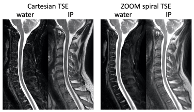 |
A ZOOM spiral TSE technique for spinal imaging
Zhiqiang Li1, Ryan K Robison2, Melvyn B Ooi1,3, and John P Karis1
1Neuroradiology, Barrow Neurological Institute, Phoenix, AZ, United States, 2Radiology, Phoenix Children's Hospital, Phoenix, AZ, United States, 3Philips Healthcare, Gainesville, FL, United States
Spine is a challenging area for MRI due to both anatomical features and motion. Spiral TSE has been proposed for spinal MRI. Aliasing in spiral spinal MRI is mitigated by combining oversampling with saturation band, with the latter not always achieving consistent and good signal suppression. In this work, the ZOOM technique has been incorporated into spiral TSE. Phantom and in vivo results demonstrate better signal suppression with ZOOM spiral TSE compared to spiral TSE acquired with a saturation band.
|
|
1167.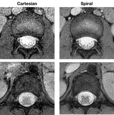 |
Axial T2*-Weighted Spiral MRI of the Spine at 1.5T
Ryan Robison1, Amber Pokorney1, Michael Kuwabara1, and Patricia Cornejo1
1Phoenix Children's Hospital, Phoenix, AZ, United States
This study evaluates spiral as an alternative to Cartesian in axial T2*-weighted gradient echo imaging of the spine. SNR and CNR measurements were performed on spine data from a healthy volunteer. Expert reviewer ratings were also performed on data from 5 patients. Both the SNR/CNR measurements and reviewer ratings indicate that spiral can yield superior image quality without significant artifacts for a given scan time.
|
|
1168.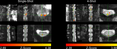 |
Spatial Specificity of BOLD Signal in the Spinal Cord at 7T Using a Noxious Thermal Stimulus
Alan C Seifert1,2,3 and S Johanna Vannesjo4,5
1Biomedical Engineering and Imaging Institute, Icahn School of Medicine at Mount Sinai, New York, NY, United States, 2Department of Radiology, Icahn School of Medicine at Mount Sinai, New York, NY, United States, 3Graduate School of Biomedical Sciences, Icahn School of Medicine at Mount Sinai, New York, NY, United States, 4Spinal Cord Injury Center, University Hospital Balgrist, University of Zurich, Zurich, Switzerland, 5Wellcome Centre for Integrative Neuroimaging, FMRIB, University of Oxford, Oxford, United Kingdom
BOLD signal in gradient-echo images is a combination of macrovascular and microvascular contributions, where the macrovascular component, arising from larger veins draining the activated tissue, is less specific to the site of activation. In this work, we image activation produced in the cervical spinal cord by a noxious thermal stimulus at 7T. We consistently observed activation in the dorsal white matter medial to the dorsal horn, rather than in the gray matter itself. However, due to the relatively straightforward venous architecture of the spinal cord, this observed displaced activation does remain closely related to the true site of neuronal activation.
|
|
| Spicing Up Your Soup: Analysis Techniques for the Spinal Cord
Benjamin De Leener
|
||
1169.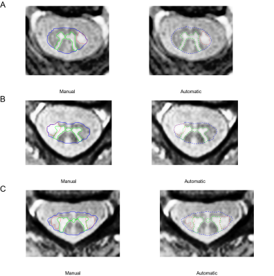 |
Automatic Quantification Pipeline for Spinal Cord Grey and White Matter in Multiple Sclerosis
Charidimos Tsagkas1,2,3, Antal Horvath4, Alexandra Todea5, Jannis Mueller1,2, Anna Altermatt2,3, Marina Leimbacher6, Simon Pezold4, Matthias Weigel2,4,7, Tanja Haas7, Michael Amann1,3,4, Ludwig Kappos1,2, Till Sprenger1,8, Philippe Cattin4,
Cristina Granziera1,2,4, and Katrin Parmar1,2
1Neurologic Clinic and Policlinic, Departments of Medicine, Biomedical Engineering and Clinical Research, University Hospital Basel, University of Basel, Basel, Switzerland, 2Translational Imaging in Neurology (ThINK) Basel, Department of Medicine and Biomedical Engineering, University Hospital Basel, University of Basel, Basel, Switzerland, 3Medical Image Analysis Center (MIAC AG), Basel, Switzerland, 4Department of Biomedical Engineering, University of Basel, Allschwil, Switzerland, 5Division of Diagnostic and Interventional Neuroradiology, Department of Radiology and Nuclear Medicine, University Hospital Basel, University of Basel, Basel, Switzerland, 6Medical Faculty, University of Basel, Basel, Switzerland, 7Division of Radiological Physics, Department of Radiology, University Hospital Basel, University of Basel, Basel, Switzerland, 8Department of Neurology, DKD Helios Klinik Wiesbaden, Wiesbaden, Germany
Currently, there is no gold-standard for spinal cord (SC) grey and white matter (GM/WM) quantification in multiple sclerosis (MS). In this work, the cervical SC of 24 MS patients and 24 healthy controls (HC) was scanned on a 3T MRI-system using averaged magnetization inversion recovery acquisitions. Manual segmentations were provided to train a “Multi-Dimensional Gated Recurrent Unit” neural network for subsequent automatic SC GM/WM/lesion segmentation. Accuracy of automatic segmentations was high and decreased in the order WM→GM→lesions and HC→MS. MS patients had reduced SC GM and WM compared to HC. Finally, SC GM, WM and lesions correlated with physical disability.
|
|
 |
1170.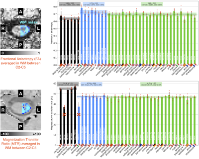 |
Quantitative MRI of the spinal cord: reproducibility and normative values across 40 sites
Eva Alonso-Ortiz1, Charley Gros1, Alexandru Foias1, Mihael Abramovic2, Christoph Arneitz2, Nicole Atcheson3, Laura Barlow4, Robert Barry5,6,7, Markus Barth3, Marco Battiston8, Christian Buchel9, Matthew Budde10, Virginie Callot11,12,
Benjamin De Leener13,14,15, Maxime Descoteaux16,17, Paulo Loureiro de Sousa18, Dostal Marek19, Julien Doyon15, Adam Dvorak20, Falk Eippert21, Karla Epperson22, Jürgen Finsterbusch9, Issei Fukunaga23, Claudia Wheeler-Kingshott8,24,25, Giancarlo Germani26, Guillaume Gilbert27,
Francesco Grussu28,29, Akifumi Hagiwara23, Pierre-Gilles Henry30, Tomas Horak31, Masaaki Hori23, James Joers30, K Kamiya32, Haleh Karbasforoushan33, Ali Khatibi34,35, Joo-Won Kim36, Nawal Kinany37, Hagen Kitzler38, S Kolind39,
Joe Yazhuo Kong40,41,42, Petr Kudlička31, Paul Kuntke43, Nyoman Kurniawan3, Slawomir Kusmia44, Rene Labounek45,46, Maria Marcella Laganà47, Corree Laule48, Christine Law49, Christophe Lenglet30, Tobias Leutritz21, Yaou Liu50,51, Sara Llufriu52,
Sean Mackey53, Eloy Martinez52, Igor Nestrasil30,45, Nico Papinutto54, Daniel Papp55, Deborah Pareto56, Todd Parrish57, Anna Pichiecchio26,58, Alex Rovira Cañellas56, Marc Ruitenberg59, Rebecca Samson28, Giorgio Savini26, Maryam Seif60,
Alan Seifert36, Alex Smith55, Z A Smith57, Elisabeth Solana52, Y Suzuki61, G Tackley44, Alexandra Tinnermann9, Jan Valosek46, Marios Yiannakas28, Kenneth Weber62, Nikolaus Weiskopf21, Richard Wise44, P O Wyss2, Junqian Xu36,
and Julien Cohen-Adad1,63
1NeuroPoly Lab, Institute of Biomedical Engineering, Polytechnique Montreal, Montreal, QC, Canada, 2Department of Radiology, Swiss Paraplegic Centre, Nottwil, Switzerland, 3Centre for Advanced Imaging, The University of Queensland, Brisbane, Australia, 4Department of Radiology, University of British Columbia, Vancouver, BC, Canada, 5Athinoula A. Martinos Center for Biomedical Imaging, Department of Radiology, Massachusetts General Hospital, Charlestown, MD, United States, 6Department of Radiology, Harvard Medical School, Boston, MA, United States, 7Harvard–Massachusetts Institute of Technology Health Sciences & Technology, Cambridge, MA, United States, 8Queen Square MS Centre, Queen Square Institute of Neurology, Faculty of Brain Sciences, University College London, London, United Kingdom, 9Institute of Systems Neuroscience, University Medical Center Hamburg-Eppendorf, Hamburg, Germany, 10Department of Neurosurgery, Medical College of Wisconsin, Milwaukee, WI, United States, 11CNRS, CRMBM, Aix-Marseille University, Marseille, France, 12APHM, Hopital Universitaire Timone, CEMEREM, Marseille, France, 13Department of Computer and Software Engineering, Polytechnique Montreal, Montreal, QC, Canada, 14CHU Sainte-Justine Research Centre, Montreal, QC, Canada, 15Montreal Neurological Institute, McGill University, Montreal, QC, Canada, 16CIMS, Centre de Recherche CHUS, Sherbrooke, QC, Canada, 17Sherbrooke Connectivity Imaging Lab (SCIL), Computer Science Department,, Université de Sherbrooke, Sherbrooke, QC, Canada, 18CNRS, ICube, FMTS, Université de Strasbourg, Strasbourg, France, 19University Hospital Brno, Brno, Czech Republic, 20Department of Physics and Astronomy, University of British Columbia, Vancouver, BC, Canada, 21Max Planck Institute for Human Cognitive and Brain Sciences, Leipzig, Germany, 22Richard M. Lucas Center, Stanford University School of Medicine, Stanford, CA, United States, 23Department of Radiology, Juntendo University School of Medicine, Tokyo, Japan, 24Department of Brain and Behavioural Sciences, University of Pavia, Pavia, Italy, 25Brain MRI 3T Research Centre, IRCCS Mondino Foundation, Pavia, Italy, 26Neuroradiology Unit, IRCCS Mondino Foundation, Pavia, Italy, 27MR Clinical Science, Philips Healthcare, Markham, ON, Canada, 28Queen Square MS Centre, Queen Square Institute of Neurology, Faculty of Brain Sciences, Faculty of Brain Sciences, University College London, London, United Kingdom, 29Centre for Medical Image Computing, Department of Computer Science, University College London, London, United Kingdom, 30Center for Magnetic Resonance Research, Department of Radiology, University of Minnesota, Minneapolis, MN, United States, 31CEITEC - Central European Institute of Technology, Brno, Czech Republic, 32University of Tokyo, Tokyo, Japan, 33Interdepartmental Neuroscience Program, Northwestern University School of Medicine, Chicago, IL, United States, 34Department of Neurology and Neurosurgery, McGill University, Montreal, QC, Canada, 35Centre of Precision Rehabilitation for Spinal Pain (CPR Spine), School of Sport, Exercise and Rehabilitation Sciences, College of Life and Environmental Sciences, University of Birmingham, Birmingham, United Kingdom, 36Translational and Molecular Imaging Institute, Department of Radiology, Icahn School of Medicine at Mount Sinai, New York, NY, United States, 37Center for Neuroprosthetics, Institute of Bioengineering, Ecole Polytechnique Fédérale de Lausanne, Lausanne, Switzerland, 38Department of Neuroradiology, Technische Universität Dresden, Dresden, Germany, 39Departments of Medicine (Neurology), Physics & Astronomy, Radiology, University of British Columbia, Vancouver, BC, Canada, 40CAS Key Laboratory of Behavioral Science, Institute of Psychology, Chinese Academy of Sciences, Beijing, China, 41Department of Psychology, University of Chinese Academy of Sciences, Beijing, China, 42Wellcome Centre for Integrative Neuroimaging, University of Oxford, Oxford, United Kingdom, 43University Hospital Carl Gustav Carus, Dresden, Germany, 44CUBRIC, Cardiff University, Walles, United Kingdom, 45Division of Clinical Behavioral Neuroscience, Department of Pediatrics, University of Minnesota, Minneapolis, MN, United States, 46Departments of Neurology and Biomedical Engineering, University Hospital Olomouc, Olomouc, Czech Republic, 47IRCCS Fondazione Don Carlo Gnocchi, Milan, Italy, 48Departments of Pathology & Laboratory Medicine, Physics & Astronomy, Radiology; International Collaboration on Repair Discoveries (ICORD), University of British Columbia, Vancouver, BC, Canada, 49Department of Anesthesiology, Perioperative and Pain Medicine, Stanford University School of Medicine, Palo Alto, CA, United States, 50Department of Radiology, Beijing Tiantan Hospital, Capital Medical University, Beijing, China, 51Tiantan Image Research Center, China National Clinical Research Center for Neurological Diseases, Beijing, China, 52Center of Neuroimmunology, Laboratory of Advanced Imaging in Neuroimmunological Diseases, Hospital Clinic Barcelona, Institut d'Investigacions Biomediques August Pi i Sunyer (IDIBAPS) and Universitat de Barcelona, Barcelona, Spain, 53Stanford University School of Medicine, Stanford, CA, United States, 54Department of Neurology, University of California San Francisco, San Francisco, CA, United States, 55Wellcome Centre For Integrative Neuroimaging, FMRIB, NDCN, University of Oxford, Oxford, United Kingdom, 56Neuroradiology Section, Vall Hebron University Hospital, Barcelona, Spain, 57Feinberg School of Medicine, Northwestern University School of Medicine, Chicago, IL, United States, 58Department of Brain and Behavioural Neuroscience, University of Pavia, Pavia, Italy, 59School of Biomedical Sciences, Faculty of Medicine, The University of Queensland, Brisbane, Australia, 60Spinal Cord Injury Center Balgrist, University of Zurich, Zurich, Switzerland, 61Department of Radiology, University of Tokyo, Tokyo, Japan, 62Systems Neuroscience and Pain Laboratory, Stanford University, Stanford, CA, United States, 63Functional Neuroimaging Unit, CRIUGM, University of Montreal, Montreal, QC, Canada
Normative quantitative MRI values are useful for establishing diagnosis in individuals with a suspected disease. Building on the recent creation of an open-access multi-center database (n=248 subjects) of spinal cord MRI, we processed those data to extract quantitative metrics that are commonly used (cross-sectional area, diffusion and magnetization transfer metrics). Inter-vendor (Siemens, Philips, GE), inter- and intra-site coefficients of variation (COV) were calculated. Overall results suggest that the spinal cord generic acquisition protocol is reproducible across sites and vendors (COVs within 2-8%). The data and processing pipeline are publicly available at https://spine-generic.readthedocs.io/.
|
 Watch the Video