
Combined Educational & Scientific Session
Hyperpolarized 13C Metabolic Imaging for Clinical Research
Session Topic: Hyperpolarized MR
Session Sub-Topic: Hyperpolarized 13C Metabolic Imaging for Clinical Research
Combined Educational & Scientific Session
ORGANIZERS: Yi-Fen Yen, Christoffer Laustsen, Malgorzata Marjanska
| Tuesday Parallel 5 Live Q&A | Tuesday, 11 August 2020, 15:15 - 16:00 UTC | Moderators: Jeremy Gordon & Jack Miller |
Skill Level: Basic to Intermediate
Session Number: C-Th-01
Overview
Hyperpolarized 13C metabolic imaging is a novel technique to investigate metabolic alterations in disease. 2020 will mark 10 years since the first in-human hyperpolarized 13C study. This emerging technique has since been demonstrated in a wide variety of clinical trials. The goal of this session is to educate clinical and basic scientists on the technology and clinical applications of this novel imaging modality, as well as its future outlook.
Target Audience
Clinicians, physicists, chemists, and engineers interested in learning about how this technique works, including results of clinical trials and future outlook.
Educational Objectives
As a result of attending this course, participants should be able to:
- Explain the function of a polarizer;
- Name at least one cancer application that has been demonstrated by this technique; and
- Name at least one non-cancer application that has been demonstrated by this technique.
| From Mouse to Man: The Value & Future Outlook
Kayvan Keshari
|
||
| Hyperpolarized 13C Imaging in 2030: A Clinician’s View
Ferdia Gallagher
|
||
 |
0687.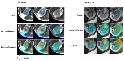 |
Initial Experience of Hyperpolarized 13C Pyruvate MRI in Patients with Renal Tumors
Shuyu Tang1, Peder E.Z. Larson1, Maxwell Meng1, James Slater1, Jeremy Gordon1, Daniel B. Vigneron1, and Zhen J. Wang1
1University of California, San Francisco, San Francisco, CA, United States
We present our initial experience of applying HP 13C pyruvate MRI in patients with renal tumors. Distinct tumor metabolic pattern and heterogeneity can be observed on HP 13C pyruvate MRI. Our data from subjects with two injections also suggests that the metabolite measurements are reproducible. This initial experience paves the way for this metabolic imaging technique to be applied for differentiating between benign renal tumors, low grade RCCs and high grade RCCs.
|
0688.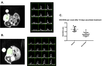 |
Hyperpolarized [1-13C] dehydroascorbic acid imaging of ascorbate-mediated oxidative stress in pancreatic cancer
Nathaniel Kim1, Arsen Mamakhanyan1, Kristin Granlund1, Elisa de Stanchina2, Manish Shah3, Lewis Cantley3,4, and Kayvan A. Keshari1,3
1Department of Radiology, Memorial Sloan Kettering Cancer Center, New York, NY, United States, 2Antitumor Assessment Core Facility, Memorial Sloan Kettering Cancer Center, New York, NY, United States, 3Weill Cornell Medical College, New York, NY, United States, 4Meyer Cancer Center, Weill Cornell Medical College, New York, NY, United States
We investigated hyperpolarized [1-13C] dehydroascorbic acid (HP DHA) as an imaging agent for probing oxidative stress in patient derived xenograft models (PDXs) of pancreatic cancer. By increasing the T1 via D2O solvation and increasing the dose administered via awake mouse injection, conversion of DHA to ascorbate was readily observed in BRAF and KRAS mutant cancers. HP DHA was then used to characterize oxidative stress in these PDX models and their biochemical mechanism of response to ascorbate therapy. Changes in DHA/ascorbate metabolism were measured in these tumor models, demonstrating a proof of concept method for assessing ascorbate therapy in pancreatic cancer.
|
|
 |
0689.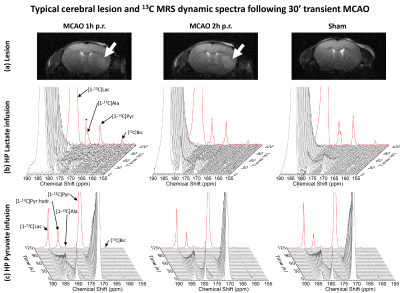 |
Metabolism of the hyperpolarized neuroprotective agents [1-13C] lactate and [1-13C] pyruvate in a mouse model of transient ischemic stroke
Thanh Phong Lê1,2, Lara Buscemi3, Elise Vinckenbosch1, Mario Lepore4, Lorenz Hirt3, Jean-Noël Hyacinthe1,5, and Mor Mishkovsky2
1Geneva School of Health Sciences, HES-SO University of Applied Sciences and Arts Western Switzerland, Geneva, Switzerland, 2Laboratory of Functional and Metabolic Imaging, École polytechnique fédérale de Lausanne (EPFL), Lausanne, Switzerland, 3Department of Clinical Neurosciences, Lausanne University Hospital (CHUV), Lausanne, Switzerland, 4Center for Biomedical Imaging - Animal Imaging and Technology (CIBM-AIT), École polytechnique fédérale de Lausanne (EPFL), Lausanne, Switzerland, 5Image Guided Intervention Laboratory, University of Geneva (UNIGE), Geneva, Switzerland
Stroke is a major cause of death and disability. Neuroprotective strategies could ameliorate patient recovery. Pyruvate and lactate were found neuroprotectant in preclinical studies of stroke models. Hyperpolarized 13C MRI provides a new way for real-time molecular imaging. In this work, we hyperpolarize those neuroprotective agents to study changes of their metabolism when administered at their therapeutic dose after ischemic stroke. We found that the metabolism of hyperpolarized lactate is significantly altered after transient cerebral ischemia, whereas moderate changes were depicted with hyperpolarized pyruvate. Those imply that hyperpolarized lactate would potentially be a better theranostic biosensor for stroke.
|
|
0690.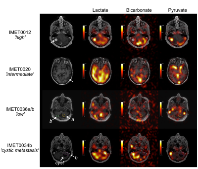 |
Assessment of Intracranial Metastases in Patients using Hyperpolarized 13C MRI
Casey Y Lee1,2, Hany Soliman3, Benjamin J Geraghty1,2, Nadia D Bragagnolo1,2, Albert P Chen4, William J Perks5, Arjun Sahgal3, Michael W Chan6, Sean Symons7, and Charles H Cunningham1,2
1Medical Biophysics, University of Toronto, Toronto, ON, Canada, 2Physical Sciences, Sunnybrook Research Institute, Toronto, ON, Canada, 3Radiation Oncology, Sunnybrook Health Sciences Centre, Toronto, ON, Canada, 4GE Healthcare Technologies, Toronto, ON, Canada, 5Pharmacy, Sunnybrook Health Sciences Centre, Toronto, ON, Canada, 6Diagostic Imaging, Trillium Health Partners, Mississauga, ON, Canada, 7Radiology, Sunnybrook Health Sciences Centre, Toronto, ON, Canada
Hyperpolarized 13C MRI was used to acquire images of [1-13C]lactate and 13C-bicarbonate from the injected [1-13C]pyruvate in 8 patients with brain metastases. Lesions were manually contoured and the mean tumor 13C-lactate signal was converted to a z-score by extending the approach previously described in Lee et al. (2019). As expected, the z-score ranks of the anatomical regions were less concordant in patients compared to controls. A range of lactate z-scores were observed in metastatic lesions, showing metabolic heterogeneity consistent with the known heterogeneity in metastatic features and clinical status. The lesions with the highest and 5th highest lactate z-scores progressed.
|
|
0691.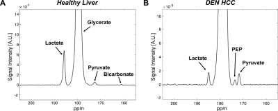 |
Hyperpolarized [1-13C] Glycerate as Probe to Assess Glycolytic Activity in a Rat Model of Hepatocellular Carcinoma
Jun Chen1, Evan LaGue2, Junjie Li1, Edward Hackett1, Ian Corbin1, Kelvin Billingsley2, and Jae Mo Park3,4
1AIRC, UT Southwestern Medical Center at Dallas, Dallas, TX, United States, 2Chemistry and Biochemistry, California State University Fullerton, Fullerton, CA, United States, 3UT Southwestern Medical Center at Dallas, Dallas, TX, United States, 4Electrical Engineering, University of Texas at Dallas, Richardson, TX, United States
Hyperpolarized [1-13C] glycerate was used to study the in vivo glycolytic activity in a rat model of hepatocellular carcinoma (HCC). Carbon-13 labeled glycolytic intermediate phosphorenolpyruvate (PEP) was detected in the tumor in addition to pyruvate and lactate peaks. The in vivo results were confirmed by high resolution 13C NMR spectra of tissue extracts, after steady-state infusion of [2,3-13C2] glycerate. The results illustrate the potential of [1-13C] glycerate as a metabolic probe for assessing glycolytic flux.
|
0692.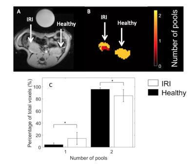 |
Hyperpolarized 13C Urea laplacian relaxation processing reveals differences between healthy and ischemic renal T2 relaxation.
James Timothy Grist1, Christian Mariager2, and Christoffer Laustsen2
1University of Birmingham, Birmingham, United Kingdom, 2Aarhus University, Aarhus, Denmark Hyperpoalrized 13C urea T2 relaxometry has been previously used to assess the diabetic and ischemic kidney. In this study we utilise a novel fitting method (Laplacian) to visualise the extent of damage, through a reduction in bi-exponential relaxation behaviour, in a rodent model of renal ischemia.
This opens up a number of potential pre-clinical and clinical uses of hyperpoalrized 13C urea imaging providing a novel, and useful, readout of renal ischemia. |
 Watch the Video