
Combined Educational & Scientific Session
Value of MRI
Session Topic: Educational Q&A: Value
Session Sub-Topic: Value of MRI
Combined Educational & Scientific Session
ORGANIZERS: Vikas Gulani, Krishna Nayak
| Monday Parallel 1 Live Q&A | Monday, 10 August 2020, 15:15 - 16:00 UTC | Moderators: Nicole Seiberlich & Kathryn Keenan |
Skill Level: Basic to Advanced
Session Number: C-Th-03
Overview
Measuring value of MRI is becoming a major goal of the MR community. Comparative effectiveness approaches allow the realization of this goal, but these studies have not traditionally been explored in the ISMRM community. In this session, we will start with an introduction to comparative effectiveness and then follow up with a case study of this work applied to a specific MRI problem.
This will be followed by a one-hour scientific session exploring the value of MRI in general.
Target Audience
All of ISMRM.
Educational Objectives
As a result of attending this course, participants should be able to:
- Explain the need to measure value in MRI;
- Explain how a comparable effectiveness approach can be used to compare the cost effectiveness of competing approaches to a clinical problem;
- Set up a model for comparing multiple approaches to a clinical problem; and
- Define and use the Incremental Cost Effectiveness Ratio to compare clinical problems.
| Introduction to Comparative Effectiveness Research
Nicholas Schiltz
Comparative and cost-effectiveness approaches can be used to assess the value of MRI, but these studies have not traditionally been explored in the ISMRM community. This session will provide an introduction to comparative and cost-effectiveness research (CER), with particular focus on decision model approaches. Topics covered include: best practices for setting up a decision tree model, key measures and interpretation including incremental-cost effectiveness ratio, and the value of information. At the end of the session, audience members should know how to create an outline of a model for their own research questions to assess the value of MRI.
|
||
| Case Study of Applying Comparative Effectiveness Research in MRI: Prostate MRI Before Biopsy
Shivani Pahwa
|
||
 |
0294.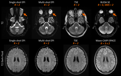 |
A comprehensive distortion-free 2-minute brain MR examination using BUDA and Wave-CAIPI
Wei-Ching Lo1, Kawin Setsompop2,3,4,5, Congyu Liao2, Susie Yi Huang2,3,4,5, John Conklin2,3,4, Stephen F. Cauley2,3,4, Wei Liu6, Bryan Clifford1, Steffen Bollmann1, Xiaozhi Cao2,7, Zijing Zhang2,8, Daniel Polak2,3,9,10, Daniel Nicolas Splitthoff9,
Thorsten Feiweier9, Qiyuan Tian2,3,4, Jaejin Cho2, John E. Kirsch2,3,4, Shivraman Giri1, Otto Rapalino3,4, Pamela W. Schaefer3,4, Larry L. Wald2, and Berkin Bilgic2
1Siemens Medical Solutions, Boston, MA, United States, 2Athinoula A. Martinos Center for Biomedical Imaging, Department of Radiology, Massachusetts General Hospital, Boston, MA, United States, 3Department of Radiology, Massachusetts General Hospital, Boston, MA, United States, 4Harvard Medical School, Boston, MA, United States, 5Harvard-MIT Division of Health Sciences and Technology, Massachusetts Institute of Technology, Boston, MA, United States, 6Siemens Shenzhen Magnetic Resonance Ltd., Shenzhen, China, 7Center for Brain Imaging Science and Technology, Biomedical Engineering, Zhejiang University, Hangzhou, Zhejiang, China, 8State Key Laboratory of Modern Optical Instrumentation, College of Optical Science and Engineering, Zhejiang University, Hangzhou, Zhejiang, China, 9Siemens Healtcare GmbH, Erlangen, Germany, 10Department of Physics and Astronomy, Heidelberg University, Heidelberg, Germany
We utilize Wave-CAIPI and BUDA techniques to develop a rapid 2-minute protocol that produces high in-plane resolution and distortion-free axial images for comprehensive evaluation of the brain. The protocol includes 3D T1-weighted Wave-CAIPI MPRAGE, 3D dark-fluid T2-weighted Wave-CAIPI SPACE-FLAIR, 2D T2*-weighted gradient echo BUDA, 2D T2-weighted and diffusion-weighted spin-echo BUDA. The results of the optimized protocol demonstrate comparable image quality, tissue contrast, and spatial resolution to standard clinical scans while keeping the total scan time to less than 2 minutes. The advanced acquisition and reconstruction framework presented here offers a path toward increasing clinical acceptance of ultrafast brain examinations.
|
0295.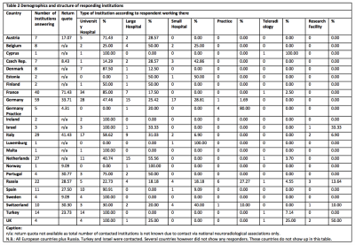 |
THE LONG ROAD FROM INVENTION TO IMPLEMENTATION: A PAN-EUROPEAN NEURORADIOLOGICAL SURVEY ON QUANTITATIVE MRI TECHNIQUES IN CLINICAL PRACTICE
Vera Catharina Keil1, Marion Smits2,3, Steffi Thust3,4, Jan Petr5, Laszlo Solymosi1, and Elia Manfrini1,6
1Neuroradiology, University Hospital Bonn, Bonn, Germany, 2Department of Radiology and Nuclear Medicine (Ne-515), Erasmus MC, Rotterdam, Netherlands, 3Lysholm Department of Neuroradiology, National Hospital for Neurology and Neurosurgery Queen Square, London, United Kingdom, 4Department of Brain Rehabilitation and Repair, UCL Institute of Neurology Queen Square, London, United Kingdom, 5Institute of Radiopharmaceutical Cancer Research, Helmholtz-Zentrum Dresden-Rossendorf, Dresden, Germany, 6Universita Politecnica delle Marche. Facolta di Medicina e Chirurgia, Ancona, Italy
This pan-European online survey study revealed that clinically working Neuroradiologists appreciate the additional diagnostic accuracy rendered by quantitative MRI techniques. However, the clinical implementation of many techniques is hampered by a lack of knowledge on how to acquire, post-process and interpret results of multiple quantitative MRI techniques including ASL, CEST/APT, IVIM and others. With exception of DSC and DWI in tumor imaging and stroke, the number of indications is also still limited especially regarding head/neck Radiology and neurodegenerative diseases.
|
|
0296.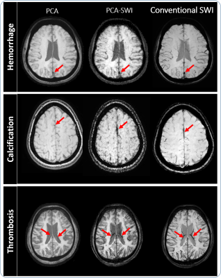 |
Free lunch may not exist, but free contrast does: Calculation of Susceptibility weighted Images from data acquired for Phase Contrast Angiography
Yogesh kannan Mariappan1, Jaladhar Neelavalli1, Nehul Makani1,2, Narayana Krishna Rolla1, Karthik Gopalakrishnan1, Nalini Pagadala1,2, and Jitendar Saini3
1Philips Healthcare, Bengaluru, India, 2Indian Institute of Technology, Madras, Chennai, India, 3National Institute for Mental Health and Neuroscience, Bengaluru, India
In Phase contrast Angiography (PCA), the flow is encoded as additional phase onto the background phase typically using a Fast field echo (FFE) based pulse sequence. This flow dependent phase is then extracted and is used in further downstream processing. The background phase is typically discarded. In this work, this background phase is processed to obtain Susceptibility weighted Imaging (SWI) contrast. Our preliminary results indicate that the additional SWI contrast (PCA-SWI) images can potentially provide clinically significant information like hemorrhage, calcification and thrombus etc. and are similar to the results obtained from conventional SWI images.
|
|
|
0297.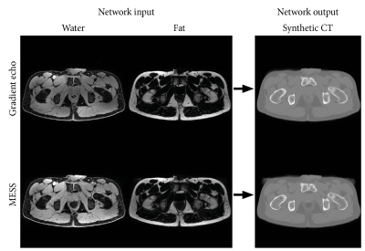 |
Multiple-echo steady-state for MR-only prostate radiotherapy: Combined T1/T2-weighted imaging, water-fat separation, and synthetic CT
Frank Zijlstra1, Mateusz C Florkow1, and Peter R Seevinck1,2
1Image Sciences Institute, UMC Utrecht, Utrecht, Netherlands, 2MRIguidance BV, Utrecht, Netherlands
We propose an efficient sequence for the acquisition of multiple contrasts for MR-only radiotherapy. By including T2-weighted echoes to a gradient echo sequence, this sequence provides T1- and T2-weighted imaging and water-fat separation in a single 4 minute acquisition. A previously trained deep neural network for synthetic CT generation was successfully applied to this new sequence, demonstrating that synthetic CT-based dose calculations can be performed. Furthermore, increased contrast between the anatomies of interest shows promise for (automatic) segmentation.
|
|
0298.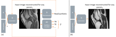 |
Acquisition Parameter Conditioned Generative Adversarial Network for Enhanced MR Image Synthesis
Jonas Denck1,2,3, George William Ferguson3, Jens Guehring3, Andreas Maier1, and Eva Rothgang2
1Pattern Recognition Lab, Department of Computer Science, Friedrich-Alexander-Universität Erlangen-Nürnberg, Erlangen, Germany, 2Technical University of Applied Sciences Amberg-Weiden, Amberg, Germany, 3Siemens Healthineers, Erlangen, Germany
Current approaches for the synthesis of MR images are only trained on MR images with a specific set of acquisition parameter values, limiting the clinical value of these methods. We therefore trained a generative adversarial network (GAN) to generate synthetic MR knee images conditioned on various acquisition parameters (TR, TE, imaging orientation). This enables us to synthesize MR images with adjustable image contrast. This work can support radiologists and technologists during the parameterization of MR sequences, can serve as a valuable tool for radiology training, and can be used for customized data generation to support AI training.
|
 |
0299.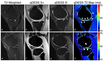 |
Rapid High-Value Diagnostic and Quantitative Knee MRI: A Prospective Artificial Intelligence Study
Akshay S Chaudhari1, Murray Grissom2, Zhongnan Fang3, Bragi Sveinsson4, Jin Hyung Lee1, Garry E Gold1, Brian A Hargreaves1, and Kathryn J Stevens1
1Stanford University, Stanford, CA, United States, 2Santa Clara Valley Medical Center, San Jose, CA, United States, 3LVIS Corporation, Palo Alto, CA, United States, 4Harvard Medical School, Boston, MA, United States
Knee MRI protocols usually require 20+ minutes of scan time, leading to great interest in expedited and high-value imaging examinations. Moreover, despite the popularity of quantitative imaging for osteoarthritis, it is not routinely implemented clinically. In this study, we use a 5-minute quantitative double-echo steady-state (qDESS) sequence that produces simultaneous morphological images and T2 relaxation time measurements. We prospectively enhance the slice-resolution of qDESS using deep learning. We show that qDESS provided high diagnostic accuracy compared to both diagnostic knee MRI and surgical findings. Additionally, automatic T2 maps increased reader diagnostic confidence and sensitivity to cartilage lesions.
|
0300.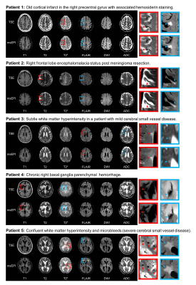 |
A comprehensive multi-shot EPI protocol for high-quality clinical brain imaging in 3 minutes
John Conklin1, Bryan Clifford2, Steffen Bollmann2, Wei-Ching Lo2, Berkin Bilgic3, Stephen Cauley3, Kawin Setsompop3, Thorsten Feiweier4, John Kirsch3, R. Gilberto Gonzalez3, Pamela Schaefer3, Otto Rapalino3, and Susie Huang3
1Radiology, Massachusetts General Hospital, Boston, MA, United States, 2Siemens Medical Solutions, Boston, MA, United States, 3Massachusetts General Hospital, Boston, MA, United States, 4Siemens Healthcare GmbH, Erlangen, Germany
A comprehensive 3 minute whole-brain MRI exam based on multi-shot echoplanar imaging (ms-EPI) was optimized and evaluated in 5 patients with different clinical pathologies. This approach minimizes artifacts associated with single-shot echoplanar imaging, and provides image quality similar to that of a 10-minute clinical reference protocol based on turbo spin-echo imaging.
|
|
0301.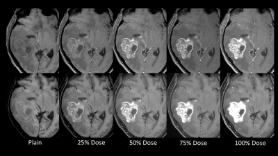 |
Possibility of the Reduction of Gd Dose using Spiral Spin-Echo Method for Contrast Enhanced Scans at 3.0T
Ravi Varma Dandu1, Rithika Varma Dandu2, Karthick Raj Rajendran3, Narayana Rolla4, and Indrajit Saha5
1Citi Neuro Centre, Hyderabad, India, 2RV College of Engineering, Bengaluru, India, 3Philips Healthcare, Eindhoven, Netherlands, 4Philips Healthcare, Bangalore, India, 5Philips Healthcare, Gurgaon, India
This study compares the performance of spin echo T1 with spiral k-space filling and three other techniques, for post contrast T1-weighted imaging of the brain. The lesion enhancement in each technique was evaluated after incremental fractional doses of gadolinium injection. The enhancement achieved on T1-FFE and T1-TSE techniques with full dose contrast could be achieved with 50% to 75% dose contrast in spiral imaging. Spiral imaging can thus be used to reduce the dose of injected contrast medium (by at least 25% and up to 50%) without compromising on the diagnostic quality of the post contrast study.
|
|
0302.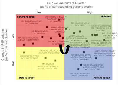 |
Determining the Value of Fit-for-Purpose MRI Exams of Multiple Sclerosis
Arijitt Borthakur1, Megan Frame1, Kristen Martin1, Melissa Mueller Gildea1, Charles E. Kahn, Jr1, and Mitchell D. Schnall1
1Radiology, Perelman School of Medicine at the University of Pennsylvania, Philadelphia, PA, United States
We compare the time savings and reduction in radiologists’ workload after implementing ten new fit-for-purpose MRI protocols for imaging of multiple sclerosis patients at our academic medical center. A 2x2 framework was created to provide a method to monitor implementation of new imaging protocols in order to gauge success of adoption as well as determine areas for operational improvement.
|
|
0303.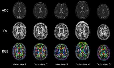 |
Feasibility of Diffusion Tensor Imaging at 0.5T
Curtis N Wiens1, Chad T Harris1, Andrew T Curtis1, Philip J Beatty1, and Jeff A Stainsby1
1Research and Development, Synaptive Medical, Toronto, ON, Canada
This work examined the feasibility of diffusion tensor imaging (DTI) at 0.5T, a technique performed almost exclusively at field strengths of at least 1.5T. 2D diffusion-weighted axial spin-echo echo-planar imaging and 3D T1 weighted acquisitions were performed in the NIST isotropic diffusion phantom, a DTI phantom, and 5 healthy volunteers on a head-specific 0.5T MRI system. ADC measurements of the NIST phantom were in excellent agreement with previously recorded 3T measurements while DTI processing and tractography performed using Modus Plan was successful in all of the volunteers.
|
|
0304.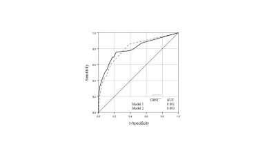 |
LI-RADS category 5 hepatocellular carcinoma: preoperative gadoxetic acid–enhanced MRI to predict early recurrence after curative resection
Hong Wei1, Hanyu Jiang1, and Bin Song1
1Department of Radiology, Sichuan University West China Hospital, Chengdu, China
This study aimed to investigate whether LI-RADS v2018 could indicate some prognostic information for high-risk patients with LR-5 hepatocellular carcinoma (HCC). We retrospectively evaluated 125 patients who underwent gadoxetic acid–enhanced MR examination within 1 month before surgical resection for HCC. The Cox proportional hazards model revealed that corona enhancement, peritumoral hypointensity on hepatobiliary phase, multifocality and serum alpha-fetoprotein were independent risk factors for early recurrence. The combined model derived from predictive biomarkers showed good performance, which could be used to effectively predict early recurrence after curative hepatectomy for LR-5 HCC.
|
 Watch the Video