
Combined Educational & Scientific Session
Microstructure: What Scales Are You Probing?
Session Topic: Diffusion Modeling, Tractography and Applications
Session Sub-Topic: Microstructure: What Scales Are You Probing?
Combined Educational & Scientific Session
ORGANIZERS: Carl-Fredrik Westin
| Wednesday Parallel 4 Live Q&A | Wednesday, 12 August 2020, 14:30 - 15:15 UTC | Moderators: Carl-Fredrik Westin & Dmitry Novikov |
Skill Level: Basic to Advanced
Session Number: C-W-02
Overview
Tissue microstructure imaging with MRI has become one of the major areas of interest within the quantitative MRI community. However, how do you really know you are imaging microstructure? You need to be able to reliably identify and quantify the relevant tissue features at the micrometer scale. This requires a considerable effort in developing adequate models and validation tools. The two educational lectures of this CES will be devoted to the overview of the frontiers of modeling and validation, and the subsequent five scientific presentations will highlight the cutting-edge research in this exciting area.
Target Audience
Scientists and clinicians interested in understanding of the cellular-level origins of the MRI signal.
Educational Objectives
As a result of attending this course, participants should be able to:
- Describe the recent progress in microstructure modeling;
- Describe modeling assumptions, pitfalls, and unresolved problems;
- Outline an overview of the model validation strategies; and
- Discuss the recent progress in tissue microstructure imaging.
 |
Probing Microstructure Lengths Scales with Diffusion: Theory
Valerij Kiselev
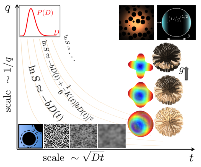
Regimes of diffusion weighting are discussed starting with the simplest measurement with narrow gradient pulses. Such measurements can be classified on a plane of diffusion time and the wave vector induced by the diffusion-sensitizing gradients. Beyond this simple picture are gradients with a finite duration, which radically change the signal behavior for the closed compartment. Versatile diffusion weighting scheme, the successors of the double diffusion encoding, are discussed under the overarching idea of geometry matching between the gradient encoding and the targeted cell population.
|
|
| Probing Microstructure Lengths Scales with Diffusion: Application
Joseph Ackerman
This didactic lecture will highlight illustrative measurements with physically-realizable model systems that provide simplifying parsimonious dMRI test platforms and deliver parameters useful for the modeling of microstructurally-complex real tissues. These dMRI test platforms will include: (i) perfused, cultured (in vitro), microbead-adherent HeLa cells; (ii) perfused, cultured (in vitro), microbead-adherent neurons and glia (aka, “brains-on-beads”); (iii) Xenopus laevis oocyte (aka, frog egg); and (iv) intracellular N-acetylaspartate (NAA) in rat brain in vivo.
|
||
 |
0843.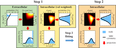 |
Non-invasive mapping of non-parametric cell size distributions using MRI-cytometry
Junzhong Xu1, Xiaoyu Jiang1, Sean P Devan2, Lori R Arlinghaus1, Eliot T McKinley1, Jingping Xie1, Zhongliang Zu1, Qing Wang3, A Bapsi Chakravarthy1, Yong Wang3, and John C Gore1
1Vanderbilt University Medical Center, Nashville, TN, United States, 2Vanderbilt University, Nashville, TN, United States, 3Washingon University, St. Louis, MO, United States
Non-invasive mapping of cell size distribution provides a unique means to probe biological tissues. We introduce a diffusion MRI based framework that does not require prior assumptions on distribution functions to provide tissue microstructural properties including non-cell-volume-weighted cell size distributions. We validated this approach, which we call MRI-cytometry, comprehensively using computer simulations in silico, cultured cells in vitro, and animal xenografts in vivo. We then demonstrate the implementation of MRI-cytometry in imaging breast cancer patients using clinical 3T MRI, indicating its potential clinical application such as more specific assessments of tumor status and therapeutic responses.
|
0844.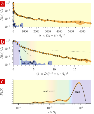 |
Unprecedented diffusion weighting and exchange resolution of cellular and sub-cellular structures in live and fixed neural tissue
Nathan Hu Williamson1, Rea Ravin1, Dan Benjamini1, Hellmut Merkle2, Melanie Falgairolle2, Michael J O'Donovan2, Dvir Blivis2, Dave Ide2, Teddy Cai1, Nima Ghorashi3, Ruiliang Bai1,4, and Peter Basser1
1Eunice Kennedy Shriver National Institutes of Child Health and Human Development, National Institutes of Health, Bethesda, MD, United States, 2National Institute of Neurological Disorders and Stroke, National Institutes of Health, Bethesda, MD, United States, 3National Heart, Lung, and Blood Institute, National Institutes of Health, Bethesda, MD, United States, 4College of Biomedical Engineering and Instrument Science, Zhejiang University, Hangzhou, China
Diffusion and exchange methods are developed using the large static gradient produced by a single-sided permanent magnet and provide resolution to water within sub-micron membrane structures. Using tissue delipidation methods, we show that water diffusion is restricted solely by lipid membranes. Most of the diffusion signal can be assigned to water in tissue which is far from membranes. The remaining 25% can be assigned to water restricted within membrane structures at the cellular, organelle, and vesicle levels. Diffusion exchange spectroscopy measures water exchanging between membrane structures and free environments at 100 s−1.
|
|
|
0845.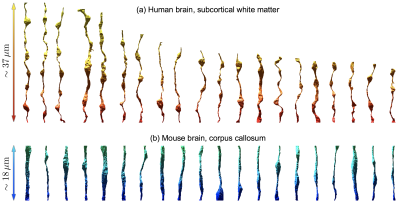 |
Random walk simulations of diffusion in human brain white matter from 3d EM validate diffusion time-dependence transverse and parallel to axons
Hong-Hsi Lee1, Qiyuan Tian2, Chanon Ngamsombat2, Daniel R Berger3, Jeff W Lichtman3, Susie Y Huang2, Dmitry S Novikov1, and Els Fieremans1
1Center for Biomedical Imaging, Department of Radiology, NYU School of Medicine, New York, NY, United States, 2Athinoula A. Martinos Center for Biomedical Imaging, Massachusetts General Hospital, Charlestown, MA, United States, 3Department of Molecular and Cellular Biology, Harvard University, Cambridge, MA, United States
We perform for the first time Monte Carlo simulations inside realistic human intra-axonal space segmented from electron microscopy. We observe non-Gaussian time-dependent diffusion in both radial and axial directions, and validate analytical models of these phenomena. We also compare the results in human axons with similar simulations in mouse axons, and discuss how the differences in axonal parameters (mean radius, radius variation, undulations) explain the differences in the simulation results between the human and mouse.
|
0846.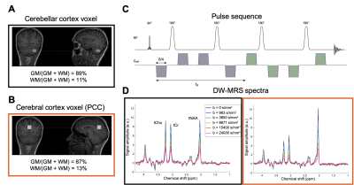 |
Characterizing the fine microstructure of cerebellar and cerebral cortex non-invasively with metabolite diffusion-weighted MRS
Marco Palombo1, Cecile Gallea2,3, Guglielmo Genovese3,4, Stephane Lehericy2,3, and Francesca Branzoli3,4
1Centre for Medical Image Computing, Department of Computer Science, University College London, London, United Kingdom, 2Team "Movement Investigations and Therapeutics", Brain and Spine Institute - ICM, Paris, France, 3INSERM U 1127, CNRS UMR 7225, Sorbonne University, Paris, France, 4Brain and Spine Institute - ICM, Centre for NeuroImaging Research - CENIR, Paris, France
Diffusion-weighted magnetic resonance spectroscopy (DW-MRS) performed at ultra-high b-values enables the quantification of fine cell microstructural features such as dendritic spine density. Here, we measured in-vivo the diffusion of total N-acetyl-aspartate (tNAA) and choline compounds (tCho) in the human cerebellar and cerebral cortex at 3 T, up to a b-value of 24 ms/μm2. We used biophysical modelling and numerical simulations to interpret the metabolite signal attenuation with the b-value. The diffusion of tNAA, a mostly neuronal metabolite, is compatible with a larger presence of spines and highly restricting granular cell soma in cerebellar compared to cerebral cortex.
|
|
|
0847.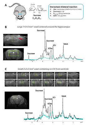 |
Characterizing extracellular diffusion properties using diffusion-weighted MRS of sucrose injected in the mouse brain
Mélissa Vincent1,2, Mylène Gaudin1,2, Covadonga Lucas-Torres3, Océane Guillemaud1,2, Carole Escartin1,2, Alan Wong3, and Julien Valette1,2
1Molecular Imaging Research Centre (MIRCen), Commissariat à l'Energie Atomique et aux Energies Alternatives (CEA), Fontenay-aux-Roses, France, 2UMR 9199, Neurodegenerative Diseases Laboratory, Centre National de la Recherche Scientifique (CNRS), Université Paris-Sud, Université Paris-Saclay, Fontenay-aux-Roses, France, 3NIMBE/ Laboratoire de Structure et Dynamique par Résonance Magnétique (LSDRM), Commissariat à l'Energie Atomique et aux Energies Alternatives (CEA), Gif-sur-Yvette, France
Diffusion in the extracellular space is assessed by state-of-the-art diffusion-weighted MRS techniques following intracerebral injection of sucrose, which predominantly remains in the extracellular space. Sucrose diffusion appears to be not strictly Gaussian and different from intracellular metabolites diffusion. Signal attenuation is stronger and deviates from mono-exponential attenuation at very high b-values, suggesting the presence of some highly restricted pool. The ADC is higher and decreases when augmenting td, indicating that the tortuosity regime is not reached yet. Lastly, unlike intracellular metabolites, sucrose diffusion does not exhibit microstructural anisotropy in double-diffusion-encoding experiments.
|
 Watch the Video