
Combined Educational & Scientific Session
Multi-Contrast & High Dimensionality Cardiovascular MRI
Session Topic: Novel imaging techniques for CMR
Session Sub-Topic: Multi-Contrast & High Dimensionality Cardiovascular MRI
Combined Educational & Scientific Session
ORGANIZERS: Jennifer Steeden, Bernd Wintersperger
| Thursday Parallel 3 Live Q&A | Thursday, 13 August 2020, 14:20 - 15:05 UTC | Moderators: Anthony Christodoulou & Eddy Solomon |
Skill Level: Intermediate to Advanced
Session Number: C-W-03
Overview
MR fingerprinting and high-dimensional MRI provide insight into the cardiovascular system beyond standard 2D/3D imaging. At this CES session, we will address the technology and refinements, clinical implications, and future directions for multi-contrast and multidimensional cardiovascular imaging MRI.
Target Audience
The audience for this CES session spans all of MR science including, but not limited to: MR physics, biomedical engineering, electrical engineering/computer science, clinical radiology, basic and clinical scientists, clinical radiologists, basic and clinical postdoctoral researchers, graduate students, and MR technologists.
Educational Objectives
As a result of attending this course, participants should be able to:
- Recall the currently utilized acquisition strategies in MRF and high-dimensional imaging and the principles behind sequence design;
- Explain the reconstruction methods approaches being utilized in MRF and high-dimensional imaging; and
- Summarize the developing clinical applications of MRF and high-dimensional imaging.
| Fingerprinting: Concept & State-of-the Art Techniques
Yong Chen
Magnetic Resonance Fingerprinting is a novel imaging method for rapid quantitative imaging. This presentation will first cover the basic concepts of Magnetic Resonance Fingerprinting and then introduce its extension for cardiac imaging. We will further discuss recent advances in cardiac Magnetic Resonance Fingerprinting and the future directions in clinical applications.
|
||
| High-Dimensionality Imaging: What More Does It Give Us?
Ricardo Otazo
A recent paradigm shift in MRI has seen the capture of multiple dynamic dimensions in a single acquisition. At first glance, adding new dimensions would appear to make MRI more challenging, but recent developments in compressed sensing and low-rank tensor imaging have shown that this multidimensional image structure can be exploited to improve over conventional imaging performance and enable access to new physiological information of clinical interest. This talk will present the most significant developments in this paradigm shift along with relevant clinical applications.
|
||
1081.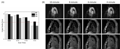 |
MR Multitasking based Multi-dimensional Assessment of Cardiovascular System (MT-MACS) Technique: Feasibility on the Thoracic Aorta
Zhehao Hu1,2, Anthony G. Christodoulou1, Nan Wang1,2, Shlee S. Song3, Marcel M. Maya4, Mariko L. Ishimori5, Lindsy J. Forbess5, Jiayu Xiao1, Xiaoming Bi6, Fei Han6, Debiao Li1,2,7, and Zhaoyang Fan1,2,7
1Biomedical Imaging Research Institute, Cedars-Sinai Medical Center, Los Angeles, CA, United States, 2Bioengineering Department, University of California, Los Angeles, Los Angeles, CA, United States, 3Department of Neurology, Cedars-Sinai Medical Center, Los Angeles, CA, United States, 4Department of Imaging, Cedars-Sinai Medical Center, Los Angeles, CA, United States, 5Department of Rheumatology, Cedars-Sinai Medical Center, Los Angeles, CA, United States, 6Siemens Healthineers, Los Angeles, CA, United States, 7Department of Medicine, University of California, Los Angeles, Los Angeles, CA, United States
Thoracic aortic diseases are one of the most common causes of cardiovascular morbidity and mortality, where imaging plays a central role in diagnosis. As a noninvasive technique, MR imaging has the potential to provide a comprehensive evaluation of the thoracic aorta from various aspects. However, clinical adoption of this modality is hindered by several limitations, i.e. long scan time and cumbersome setup for accommodating motion during data acquisition. In this work, we present an MR MultiTasking based 3D Multi-dimensional Assessment of Cardiovascular System (MT-MACS) technique that allows for ECG- and navigator-free thoracic aortic imaging within 6 minutes.
|
|
 |
1082.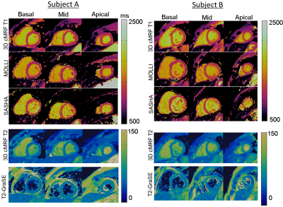 |
3D Free-breathing Cardiac Magnetic Resonance Fingerprinting
Gastao Cruz1, Olivier Jaubert1, Haikun Qi1, Aurelien Bustin1, Giorgia Milotta1, Torben Schneider2, Peter Koken3, Mariya Doneva3, René M. Botnar1, and Claudia Prieto1
1Biomedical Engineering Department, School of Biomedical Engineering and Imaging Sciences, King's College London, London, United Kingdom, 2Philips Healthcare, Guildford, United Kingdom, 3Philips Research Hamburg, Hamburg, Germany
2D cardiac Magnetic Resonance Fingerprinting (cMRF) has been proposed for simultaneous and co-registered T1/T2 mapping using ECG-triggering and breath-holding. However, 2D cMRF provides limited coverage of the heart and is sensitive to residual through-plane respiratory motion. Here we propose respiratory motion-compensated 3D cMRF to enable whole-heart myocardial T1/T2 mapping in a single free-breathing scan. Respiratory bellows driven localized autofocus is proposed for beat-to-beat translational motion correction and patch-based low rank MRF reconstruction is employed to minimise residual aliasing. 3D cMRF enabled whole-heart T1/T2 mapping in ~7min scan time with comparable map quality to conventional 2D MOLLI, SASHA and T2-GraSE.
|
1083.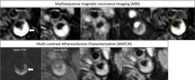 |
Quantification of carotid plaque compositon with Multi-contrast Atherosclerosis Characterization (MATCH) versus multisequence carotid MRI
Mohamed Kassem1,2, Ellen Boswijk2, Jochem Van der Pol2, Rik PM Moonen2, Jan Bucerius2, Zhaoyang Fan3, and M Eline Kooi1,2
1Radiology and Nuclear medicine, CARIM School for Cardiovascular Diseases, Maastricht, Netherlands, 2Radiology and Nuclear medicine, Maastricht university Medical Centre, Maastricht, Netherlands, 3Cedars-Sinai Medical Center, Biomedical Imaging Research Institute, Los Angeles, CA, United States
Multisequence MRI protocol usually includes an MP-RAGE sequence for the identification of plaque compositions. Multisequence MRI has some limitations including long scan time and image mis-registration errors. Multi-contrast Atherosclerosis Characterization (MATCH) was developed to overcome the above limitations. Eighteen patients with ≥2 mm carotid plaques underwent 3.0T carotid MRI including conventional multisequence and MATCH. For the artery-based component detection, excellent agreement was obtained for LRNC, substantial for IPH and slight agreement for calcifications. No significant difference between MATCH and conventional MRI was shown in measurement of volume of LRNC/IPH, IPH, calcifications, percentage wall volume and normalized wall index.
|
|
1084.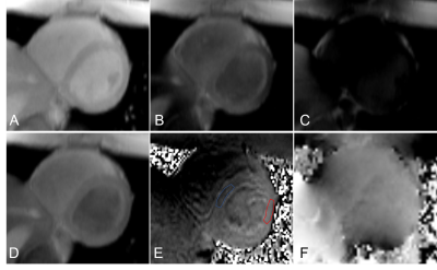 |
B1 Inhomogeneity at 3T causes spatially non-reproducible and inaccurate cardiac creatine CEST-contrast in healthy controls
Wissam AlGhuraibawi1, Kevin Godines1, Mark Velasquez1, Sinyeob Ahn2, Wolfgang Rehwald3, and Moriel Vandsburger1
1Bioengineering, University of California Berkeley, Berkeley, CA, United States, 2Siemens Healthineers, Concord, CA, United States, 3Siemens Healthineers, Durham, NC, United States
CEST-MRI is an emerging molecular imaging method for non-invasive assessment of cardiomyocyte metabolites. In cardiac CEST-MRI, spatial B1 inhomogeneity across the myocardium significantly reduces the accuracy of measured CEST contrasts. Deviation from the prescribed B1 leads to altered creatine CEST contrast due to both reduced labeling efficiency and heightened magnetization transfer and direct water direct saturation across the heart. The final impact is measurement of falsely and substantially reduced creatine CEST contrast in the healthy heart.
|
|
|
1085.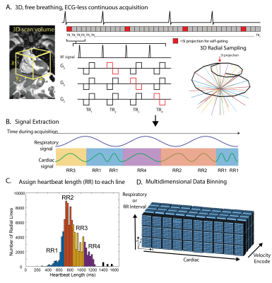 |
Decoding the Effects of Rhythm on Hemodynamics in Patients with Atrial Fibrillation Using a 5D Flow Framework
Liliana Ma1,2, Jerome Yerly3, Lorenzo Di Sopra3, Davide Piccini3,4, Rod Passman5, Philip Greenland5, Daniel Kim1,2, Matthias Stuber3, and Michael Markl1,2
1Department of Radiology, Northwestern University, Feinberg School of Medicine, Chicago, IL, United States, 2Department of Biomedical Engineering, Northwestern University, Evanston, IL, United States, 3Department of Radiology, Lausanne University Hospital (CHUV) and University of Lausanne (UNIL), Lausanne, Switzerland, 4Siemens Healthcare, Lausanne, Switzerland, 5Department of Medicine and Preventive Medicine, Northwestern University, Feinberg School of Medicine, Chicago, IL, United States
Atrial fibrillation (AF) is the most common cardiac arrhythmia and is associated with increased risk of ischemic stroke. Arrhythmic heartbeats in AF may alter atrial flow characteristics and the influence of differences in heart rates on LA 3D hemodynamics has not yet been systematically investigated. Recently, a fully self-gated free-running 5D flow (4D flow+respiration) framework was introduced and validated. The purpose of this study was to expand the 5D flow framework to explore the influence of heart rates on thrombogenic hemodynamic parameters in AF patients.
|
1086.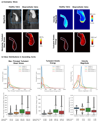 |
5D Flow Tensor MRI with Multipoint Encoding for Efficient Mapping of Reynolds Stresses in the In-vivo Aorta
Jonas Walheim1, Hannes Dillinger1, Alexander Gotschy1,2,3, and Sebastian Kozerke1
1Institute for Biomedical Engineering, University and ETH Zurich, Zurich, Switzerland, Zurich, Switzerland, 2Great Ormond Street Hospital, London, United Kingdom, 3Department of Cardiology, University Hospital Zurich, Zurich, Switzerland
In-vivo 5D Flow Tensor MRI with multipoint encoding for accurate assessment of Reynolds stresses in the aorta is presented. Based on distributions of turbulence intensity in healthy and pathological flows, a 6-directional multipoint encoding with 3 different encoding strengths is proposed. Using a 5D Flow compressed sensing acquisition in-vivo data are collected in 10 minutes irrespective of breathing motion. Data obtained in aortic valve patients and healthy controls demonstrate the feasibility of the method to quantify turbulence in healthy and pathological flow.
|
 Watch the Video