
Digital Poster Session
Neuro: Multiple Sclerosis and Myelin
Neuro
1368 -1380 Multiple Sclerosis and Myelin - Multiple Sclerosis/Neuromyelitis Optica & Cognitive Impairment in MS
1381 -1396 Multiple Sclerosis and Myelin - Multiple Sclerosis: QSM, Relaxation & Diffusion Imaging
1397 -1411 Multiple Sclerosis and Myelin - Multiple Sclerosis: Outside the Cerebral White Matter
1412 -1425 Multiple Sclerosis and Myelin - Multiple Sclerosis: Inside the Cerebral White Matter
1426 -1439 Multiple Sclerosis and Myelin - Myelin Imaging: Acquisition to Application
1368.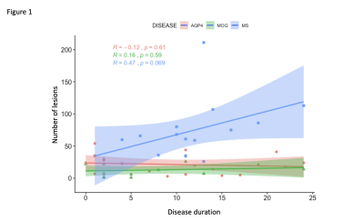 |
Contrasting brain MRI features in Ab-mediated demyelinating diseases and MS: investigating the pathogenesis of brain damage
Silvia Messina1, Romina Mariano1, Adriana Roca-Fernandez1, Ana Cavey1, Rosie Everett2, Sandra Reeve2, Maria Isabel Leite1, Mark Jenkinson1,3, and Jacqueline Palace1
1Nuffield Department of Clinical Neurosciences, University of Oxford, Oxford, United Kingdom, 2Oxford University Hospital NHS Foundation Trust, Oxford, United Kingdom, 3University of Adelaide, Adelaide, Australia
The previous non-conventional MRI studies included both AQP4 positive and negative patients, thus yielding differing results, partly explained by cohort heterogeneity (i.e. antibodies status). No studies are available in MOG-Ab disease. We compared, using non-conventional imaging, brain MRI findings in 73 subjects with AQP4-ab, MOG-Ab disease, Multiple Sclerosis and in healthy controls. We found that MOG-Ab disease have a lower lesions load when compared to MS and AQP4-Ab disease and we demonstrated, for the first time, that significant changes occur in the deep grey matter while no significant changes were detected in the non-lesional white matter in MOG-Ab disease.
|
|
1369.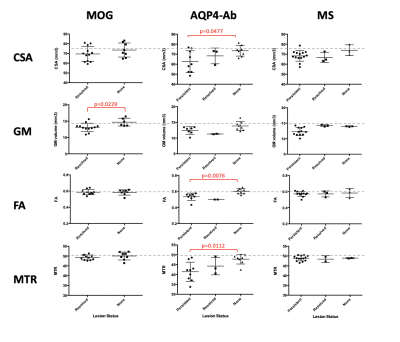 |
Multi-modal Cervical Cord MRI in MOGAD, AQP4-Ab NMOSD & MS
Romina Mariano1, Silvia Messina1, Adriana Roca-Fernandez1, Ana Cavey1, Rosie Everett2, Maria Isabel Leite1, Yazhuo Kong1,3, and Jacqueline Palace1
1Nuffield Department of Clinical Neurosciences, University of Oxford, Oxford, United Kingdom, 2Oxford University Hospitals NHS Foundation Trust, Oxford, United Kingdom, 3The Key Laboratory of Behaviour Science, Institute of Psychology, Chinese Academy of Sciences, Beijing, China
Spinal cord involvement is an important feature of antibody-mediated demyelination, as in AQP4-Ab-positive NMOSD and MOGAD, as well as in MS. In this study of 80 participants (20 MOGAD, 20 AQP4-Ab, 20 MS, 20 health volunteers) we use multimodal cervical cord MRI to show that MOGAD may predominantly affect the grey matter; AQP4-Ab disease shows the most severely affected cord, localised to lesional areas; and that MRI metrics in the cervical cord do not differentiate these three conditions but have clinical relevance in their association with disability and pain scores, independent of disease type.
|
|
1370.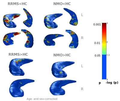 |
Hippocampal regional vulnerability to damage differs between MS and neuromyelitis optica
Elisabetta Pagani1, Laura Cacciaguerra1,2, Gianna C. Riccitelli1, Marta Radaelli2, Massimo Filippi1,2,3, and Maria A. Rocca1,2
Video Permission Withheld
1Neuroimaging Research Unit, Institute of Experimental Neurology, Division of Neuroscience, IRCCS San Raffaele Scientific Institute, Milan, Italy, 2Neurology Unit, IRCCS San Raffaele Scientific Institute, Milan, Italy, 3Vita-Salute San Raffaele University, Milan, Italy
Aim of the study was to characterize regional hippocampal volumetric alterations in multiple sclerosis (MS) and neuromyelitis optica spectrum disorders (NMOSD) and to estimate correlations with MRI measures of inflammation and hippocampal disconnection. Brain T2 and T1 lesions and hippocampi were manually segmented; major hippocampal connections were reconstructed with tractography. Compared to healthy controls, NMOSD patients showed mild atrophy, whereas MS patients had diffuse hippocampal atrophy. Dentate gyrus hypertrophy and correlations between hippocampal volume abnormalities and damage of hippocampal anatomical connections was found in MS only, suggesting that other factors than inflammation, contribute to the hypertrophy process.
|
|
1371.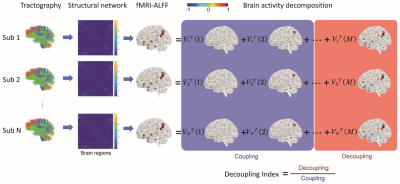 |
Decoupling of brain activity from connectome in multiple sclerosis and neuromyelitis optica
Chenfei Ye1, Jing Huang2, Haiyan Lv3, Jie Lu2, and Ting Ma1,4,5,6
1Department of Electronic and Information Engineering, Harbin Institute of Technology at Shenzhen, Shenzhen, China, 2Department of Radiology, Xuanwu Hospital Capital Medical University, Beijing, China, 3Shenzhen MindsGo Life Technology Co.Ltd, Shenzhen, China, 4Advanced Innovation Center for Human Brain Protection, Capital Medical University, Beijing, China, 5Peng Cheng Laboratory, Shenzhen, China, 6National Clinical Research Center for Geriatric Disorders, Xuanwu Hospital Capital Medical University, Beijing, China
Coupling of brain functional activity with brain structural network (connectome) plays a key role in cognition and movement. In this study, we anticipated that the structural-functional coupling would be altered in multiple sclerosis (MS) and neuromyelitis optica (NMO). By introducing the graph frequency analysis on diffusion and functional MR images, we found that brain activity in patient with both MS and NMO deviated from the underlying structural network, indicating disrupted structural-functional coupling caused by neuronal inflammation and demyelination.
|
|
1372.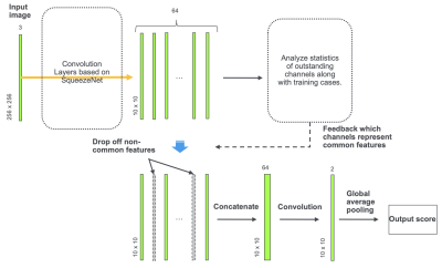 |
Differentiation of Multiple Sclerosis and Neuromyelitis Optica Spectrum Disorders by Convolutional Neural Network
Akifumi Hagiwara1,2, Yujiro Otsuka1,3, Christina Andica1, Shimpei Kato1,4, Kazumasa Yokoyama5, Masaaki Hori1,6, Shohei Fujita1,4, Koji Kamagata1, Ryusuke Irie1,4, Saori Koshino1,4, Tomoko Maekawa1,4, Toshiaki Akashi1, Akihiko Wada1,
Kanako Kunishima Kumamaaru1, Takuya Haruyama1,7, Syo Murata1, Nobutaka Hattori5, and Shigeki Aoki1
1Radiology, Juntendo University School of Medicine, Tokyo, Japan, 2Radiology, UCLA, Los Angeles, CA, United States, 3Milliman Inc., Tokyo, Japan, 4Radiology, Graduate School of Medicine, The University of Tokyo, Tokyo, Japan, 5Neurology, Juntendo University School of Medicine, Tokyo, Japan, 6Radiology, Toho University, Tokyo, Japan, 7Radiological Sciences, Graduate School of Human Health Sciences, Tokyo Metropolitan University, Tokyo, Japan
Multiple sclerosis (MS) and neuromyelitis optica spectrum disorders (NMOSD) are both neuroinflammatory diseases and have overlapping clinical manifestations. We developed a convolutional neural network that differentiates between MS and NMOSD based on multi-dynamic multi-echo sequence that measures R1 and R2 relaxation times and proton density. To avoid overfitting on a small dataset, we aimed to separate features of images into those specific to an image and those common to the group (i.e. MS or NMOSD) based on SqueezeNet. We used only common features for classification. Our model achieved a diagnostic accuracy of 80.7%.
|
|
1373.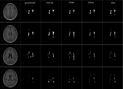 |
Deep learning based automated white matter lesion detection for MS and NMOSD: a retrospective multicenter study
Yajing Zhang1, Weiwei Jiang1, Zhizheng Zhuo2, Yunyun Duan2, and Yaou Liu2
1Philips Healthcare, Suzhou, China, 2Beijing Tiantan Hospital, Capital Medical University, Beijing, China
Multiple sclerosis (MS) and neuromyelitis optical spectrum disorders (NMOSD) are common demyelination diseases in central neural system. With increasingly used MR examinations in clinical practice, the detection and delineation of white matter lesions is helpful to the calculation of total lesion volume, which is of great significance to clinical treatment planning. However, manual delineation is time-consuming to the clinicians and may lead to poor repeatability. In this work, we employed deep learning method to establish a tool for WM lesion delineation of MS and NMOSD routine MRI data from multiple centers.
|
|
1374. |
Structural and functional characteristics in neuromyelitis optica spectrum disorders and multiple sclerosis
Zhizheng Zhuo1, Yunyun Duan1, Xinli Wang1,2, Fenglian Zheng1, Jinli Ding1, Decai Tian3, Xiaoya Chen4, Fuqing Zhou5, Jinhui Wang6, Rongkai Ju7, Yingjie Mei8, Xinghu Zhang3, FuDong Shi2,9, and Yaou Liu1,9
1Department of Radiology, Beijing Tiantan Hospital, Beijing, China, 2Department of Neurology, Tianjin Medical University General Hospital, Tianjin, China, 3Department of Neurology, Beijing Tiantan Hospital, Beijing, China, 4Department of Radiology, the First Affiliated Hospital of Chongqing Medical University, Chongqing, China, 5Department of Radiology, The First Affiliated Hospital of Nanchang University, Nanchang, China, 6Center for Studies of Psychological Application, South China Normal University, Guangzhou, China, 7Clinical Application, Philips Healthcare, Beijing, China, 8Clinical Science, Philips Healthcare, Guangzhou, China, 9China National Clinical Research Center for Neurological Diseases, Beijing, China
We investigated structural and functional alterations in neuromyelitis optica spectrum disorders (NMOSD) and multiple sclerosis (MS) and examine their clinical relevance using multimodal MRI techniques.
|
|
 |
1375.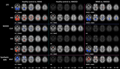 |
Gray Matter Alterations in Multiple Sclerosis and Neuromyelitis Optica Spectrum Disorders Evaluated Using Multimodal Neuroimaging Techniques
Christina Andica1, Akifumi Hagiwara1,2, Shimpei Kato1,3, Kazumasa Yokoyama4, Shohei Fujita1,3, Takuya Haruyama1,5, Koji Kamagata1, Masaaki Hori1,6, Nobutaka Hattori4, and Shigeki Aoki1
1Department of Radiology, Juntendo University Graduate School of Medicine, Tokyo, Japan, 2Department of Radiology, David Geffen School of Medicine at UCLA, Los Angeles, CA, United States, 3Department of Radiology, Graduate School of Medicine, The University of Tokyo, Tokyo, Japan, 4Department of Neurology, Juntendo University School of Medicine, Tokyo, Japan, 5Department of Radiological Sciences, Graduate School of Human Health Sciences, Tokyo Metropolitan University, Tokyo, Japan, 6Department of Radiology, Toho University Omori Medical Center, Tokyo, Japan
Multiple sclerosis (MS) shares similar clinical and imaging characteristics with neuromyelitis optica spectrum disorders (NMOSD), but a correct diagnosis is essential as treatment options differ considerably. We applied diffusion tensor imaging, neurite orientation dispersion and density imaging, and synthetic quantitative MRI to assess the gray matter (GM) of relapsing-remitting MS (RRMS) and NMOSD patients. Our results demonstrated that multimodal neuroimaging techniques of the GM might be useful for differentiating NMOSD from RRMS, where NMOSD spares most of the GM. In contrast, RRMS group demonstrated extensive demyelination, circumscribed axonal loss predominantly in the limbic areas, and broad neuroinflammation in the cerebellum.
|
1376.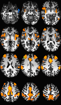 |
Declining frontoparietal connectivity is linked to decreased episodic memory performance in multiple sclerosis
Katherine A Koenig1, Jian Lin1, Daniel Ontaneda2, Kedar Mahajan2, Jenny Feng2, Stephen M. Rao3, Sanghoon Kim1, Stephen J Jones1, and Mark J Lowe1
1Imaging Sciences, The Cleveland Clinic, Cleveland, OH, United States, 2Neurological Institute, The Cleveland Clinic, Cleveland, OH, United States, 3Schey Center for Cognitive Neuroimaging, The Cleveland Clinic, Cleveland, OH, United States
Cognitive dysfunction, often including memory loss, impacts about half of those with Multiple Sclerosis (MS). Our work aims to develop a predictor of future memory decline in MS. Using high resolution MRI, we measured resting state functional connectivity of the frontoparietal network in 77 participants with MS. We found that connectivity was related to episodic memory at baseline, and that the one-year change in connectivity was related to the change in memory performance. This finding suggests that functional connectivity can be developed as a predictor of memory decline in MS.
|
|
1377.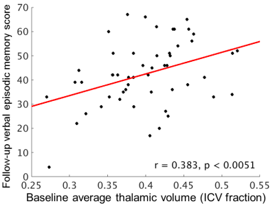 |
Thalamic volume relates to future memory performance in Multiple Sclerosis
Katherine A Koenig1, Jian Lin1, Daniel Ontaneda2, Kedar Mahajan2, Jenny Feng2, Stephen M. Rao3, Sanghoon Kim1, Stephen J Jones1, and Mark J Lowe1
1Imaging Sciences, The Cleveland Clinic, Cleveland, OH, United States, 2Neurological Institute, The Cleveland Clinic, Cleveland, OH, United States, 3Schey Center for Cognitive Neuroimaging, The Cleveland Clinic, Cleveland, OH, United States
Cognitive dysfunction is a common symptom of Multiple Sclerosis (MS), and patients would benefit from a measure that estimates their risk of future decline. Previous work suggests that thalamic volume may have value as a predictive measure. Using 7 tesla MRI, we measured thalamic volume in 79 participants with MS. We found a strong cross-sectional relationship between volume and verbal episodic memory. The longitudinal relationship was significant, but was strongly modulated by disease duration. The current report highlights the need for careful modeling of potential confounding variables.
|
|
1378.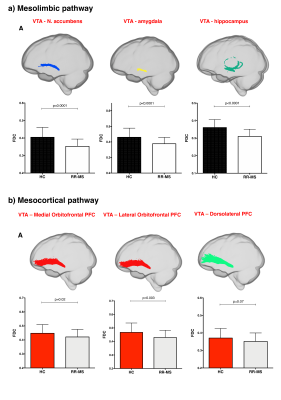 |
Brainstem monoaminergic functional and structural connectivity is altered in multiple sclerosis and contributes to fatigue
Tiziana Carandini1,2, Matteo Mancini1,3,4, Iulia Bogdan1, Charlotte Rae5, Andrew Barritt1, Arjun Sethi6, Neil Harrison7, Waqar Rashid1, Mara Cercignani1, and Marco Bozzali1
1Department of Neuroscience, Brighton and Sussex Medical School, University of Sussex, Brighton, United Kingdom, 2Fondazione IRCCS Ca' Granda Ospedale Maggiore Policlinico, Milan, Italy, 3NeuroPoly Lab, Polytechnique Montreal, Montreal, QC, Canada, 4CUBRIC, Cardiff University, Cardiff, United Kingdom, 5School of Psychology, University of Sussex, Brighton, United Kingdom, 6Psychiatry, Psychology & Neuroscience, King’s College London, London, United Kingdom, 7Department of Psychology and Department of Medicine, Cardiff University, Cardiff, United Kingdom
Brainstem monoaminergic functional and structural connectivity was investigated in a cohort of patients with multiple sclerosis (MS) and healthy controls. RS-fMRI analysis revealed in MS patients reduced functional connectivity between monoaminergic nuclei and central brain networks that are critically involved in the pathophysiology of MS. Functional alterations were associated to structural disconnections between these nuclei and cortical/subcortical efferent targets in MS patients. Axonal loss in the mesocorticolimbic tracts and in the noradrenergic projections to prefrontal cortex was associated with central fatigue in MS patients, whereas brainstem functional connectivity did not correlate with fatigue.
|
|
1379.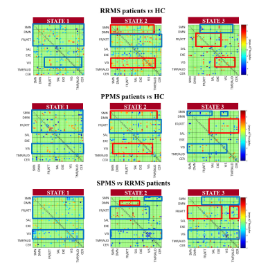 |
Characterizing dynamic functional connectivity in the main clinical phenotypes of multiple sclerosis
Paola Valsasina1, Milagros Hidalgo de la Cruz1, Francesca Sangalli2, Federica Esposito2, Massimo Filippi1,2,3, and Maria A. Rocca1,2
Video Permission Withheld
1Neuroimaging Research Unit, Institute of Experimental Neurology, Division of Neuroscience, IRCCS San Raffaele Scientific Institute, Milan, Italy, 2Neurology Unit, IRCCS San Raffaele Scientific Institute, Milan, Italy, 3Vita-Salute San Raffaele University, Milan, Italy
In this study, we used dynamic functional connectivity (dFC) to characterize time-varying connectivity abnormalities in patients with multiple sclerosis (MS) with the main disease phenotypes. Compared to controls, MS patients presented overall dFC reduction in all networks, along with increased dFC in sensorimotor, default-mode and frontal/attention networks. While progressive MS showed additional dFC decrease vs relapsing-remitting (RR) MS, in benign MS the overall reduction of dFC was accompanied by significantly increased dFC in the sensorimotor, default-mode and frontal/attention networks. Reduced dFC correlated with more severe clinical disability and worse cognitive performance.
|
|
1380.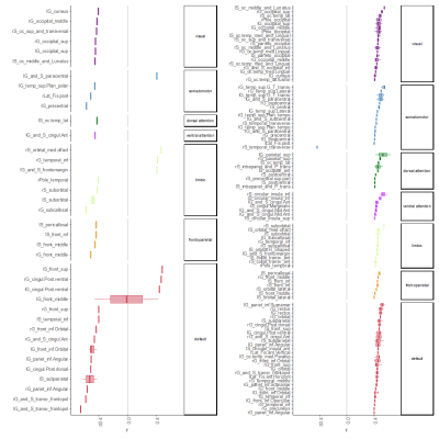 |
Direct and inverse relations in default mode network detected by functional and structural substrate of cognition in mild Relapsing Remitting MS.
Arzu Ceylan Has Silemek1, Lukas Fischer1, Jana Pöttgen1,2, Iris-Katharina Penner3,4, Andreas K. Engel5, Christoph Heesen1,2, Stefan M. Gold1,6, and Jan-Patrick Stellmann1,2,7,8
1Institut für Neuroimmunologie und Multiple Sklerose (INIMS), Universitätsklinikum Hamburg-Eppendorf, Hamburg, Germany, 2Klinik und Poliklinik für Neurologie, Universitätsklinikum Hamburg-Eppendorf, Hamburg, Germany, 3Klinik für Neurologie, Heinrich Heine Universität Düsseldorf, Düsseldorf, Germany, 4Zentrum für Angewandte Neurokognition und Neuropsychologische Forschung, Heinrich-Heine-Universität Düsseldorf, Düsseldorf, Germany, 5Institut für Neurophysiologie und Pathophysiologie, Universitätsklinikum Hamburg-Eppendorf, Hamburg, Germany, 6Charité - Universitätsmedizin Berlin, Freie Universität Berlin, Humboldt Universität zu Berlin, and Berlin Institute of Health (BIH), Klinik für Psychiatrie & Psychotherapie und Medizinische Klinik m.S. Psychosomatik, Campus Benjamin Franklin (CBF), Berlin, Germany, 7CRMBM AMU-CNRS , Marseille, France, Marseille, France, 8CEMEREM, APHM, CHU Timone, Marseille France, Marseille, France
We utilized an approach combining DTI, RS-fMRI and graph-theory to characterize the relation between cognitive profiles and global and local network features in RRMS patients with mild to moderate disability. Closer association of structural network metrics with cognitive abilities were seen compared to standard-MRI outcomes and an interesting pattern of associations with a slight predominance of nodes located in the default mode network (DMN). While structural connectivity always showed a positive correlation with performance, the number of functional connections of nodes was mostly negatively correlated. DMN had inverse and direct relationships with memory, possible indicating adaptive and maladaptive mechanisms.
|
 View the Poster
 Watch the Video
1381.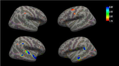 |
3T Surface-based quantitative susceptibility mapping and T1 relaxometry in relapsing-remitting and progressive multiple sclerosis patients
Nicholas John Eichner1, Reza Rahmanzadeh1, Matthias Weigel1, Muhamed Barakovic1, Po-Jui Lu1, Ludwig Kappos1, Pascal Sati2, Daniel S. Reich2, Yi Wang3, Jens Kuhle1, and Cristina Granziera1
1Department of Biomedical Engineering, University of Basel, Basel, Switzerland, 2National Institute of Neurological Disordes and Stroke, Bethesda, MD, United States, 3Cornell University, Ithaca, NY, United States
We assessed the presence and extent of cortical abnormalities in patients with relapsing remitting and progressive multiple sclerosis by using surface-based quantitative susceptibility mapping and T1 relaxometry analysis. Our results showed that QSM and T1 maps acquired at 3T MRI are sensitive to cortical damage in MS patients and reveal more extensive areas of alterations in progressive than in relapsing-remitting MS. Changes in cortical QSM and T1 suggest chronic demyelination and possibly iron accumulation.
|
|
1382.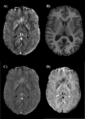 |
Unraveling atrophy, iron and myelin changes in the deep gray matter of multiple sclerosis patients
Giuseppe Pontillo1, Sirio Cocozza1, Serena Monti2, Maria Petracca3, Roberta Lanzillo3, Vincenzo Brescia Morra3, Arturo Brunetti1, and Giuseppe Palma2
1Department of Advances Biomedical Sciences, University of Naples "Federico II", Naples, Italy, 2Institute of Biostructures and Bioimaging, National Research Council, Napoli, Italy, 3Department of Neurosciences and Reproductive and Odontostomatological Sciences, University of Naples "Federico II", Naples, Italy
Alterations of magnetic susceptibility are known to occur in the deep gray matter (DGM) of multiple sclerosis (MS) patients, reflecting a mixture of atrophy, iron and myelin changes. We combined the analysis of quantitative susceptibility mapping (QSM) and R1 relaxometry contrasts in order to obtain iron- and myelin-specific maps, from which we estimated mean iron and myelin concentrations as well as total iron and myelin contents for each DGM structure. We found increased mean iron in the basal ganglia of MS patients compared to healthy controls, probably reflecting atrophy-related concentration, along with actual atrophy-independent iron depletion phenomena in the thalamus.
|
|
1383.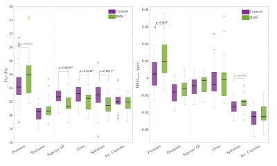 |
QSM and R2* capture physical and cognitive impairment in multiple sclerosis
Vanessa Wiggermann1,2, Shawna Abel3, Irene M Vavasour4, Enedino Hernández-Torres3, Christian Kames1,2, David KB Li4,5, Roger Tam4,5, Shannon H Kolind1,3, Anthony Traboulsee3, and Alexander Rauscher1,2
1Physics and Astronomy, University of British Columbia, Vancouver, BC, Canada, 2Pediatrics, University of British Columbia, Vancouver, BC, Canada, 3Medicine (Neurology), University of British Columbia, Vancouver, BC, Canada, 4Radiology, University of British Columbia, Vancouver, BC, Canada, 5MS/MRI Research (Neurology), University of British Columbia, Vancouver, BC, Canada
Abnormalities seen on clinical MRIs in the brain and spinal cord of multiple sclerosis (MS) patients lack specificity to myelin. Thus, efficacy of new treatments that aim to protect or remyelinate cannot be fully captured. Quantitative MR metrics, such as R2* and quantitative susceptibility mapping (QSM), have demonstrated good sensitivity to myelin and iron-related tissue changes, but the clinical relevance of such changes has not yet been assessed. We showed that R2* provides good group-wise distinction of MS patients and controls, while QSM values reflected clinically meaningful variations in upper and lower motor function as well as cognitive processing speed.
|
|
1384.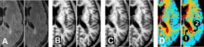 |
Cross-relaxation magnetic resonance imaging for multiple sclerosis
Jung-Jiin Hsu1, Jung-Yu C. Hsu2, and Roland G. Henry1
Video Permission Withheld
1Department of Neurology, University of California San Francisco, San Francisco, CA, United States, 2Department of Cell Biology and Anatomy, National Cheng Kung University, Tainan, Taiwan
Magnetization transfer (MT) is of great interest in clinical and pre-clinical imaging to probe tissue properties that cannot be visualized directly with conventional MRI. Current research and development of MT MRI is based on the chemical exchange mechanism. However, it has long been reported that, instead of chemical exchange, the cross-relaxation mechanism is responsible for MT in biological tissue. In this work, a fast MRI method is developed based on cross relaxation for the measurement of the spin–lattice relaxation rate and the MT rate and is applied to characterize multiple sclerosis lesions.
|
|
1385. |
Trajectories of magnetic susceptibility in the pulvinar provide further evidence for accelerated decline of thalamic iron in multiple sclerosis
Fahad Salman1, Robert Zivadinov1,2, Niels Bergsland3, Michael G. Dwyer1, Bianca Weinstock Guttman4, and Ferdinand Schweser1,5
1Neuroimaging, Buffalo Neuroimaging Analysis Center, Buffalo, NY, United States, 2MRI Clinical and Translational Research Center, Buffalo, NY, United States, 3Neurology, Buffalo Neuroimaging Analysis Center, Buffalo, NY, United States, 4Neurology, Jacobs Comprehensive MS Center for Treatment & Research, Buffalo, NY, United States, 5Neurology, MRI Clinical and Translational Research Center, Buffalo, NY, United States
Several recent cross-sectional studies have observed decreasedmagnetic susceptibility in the thalamus of patients with multiple sclerosis (MS) using Quantitative Susceptibility Mapping (QSM).However, the concavity of the iron concentration trajectory in normal aging renders the interpretation of findings of the previous studies difficult. In the present work, we applied QSM longitudinally in conjunction with a dedicated analysis procedure to obtain optimal longitudinal measurement accuracy. Our longitudinal results confirm previous cross-sectional findings and suggest that thalamic QSM may serve as an imaging marker for disease progression in MS.
|
|
1386.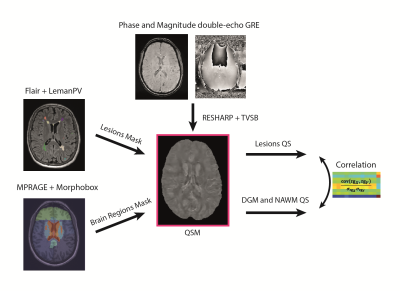 |
Quantitative susceptibility of thalamus, basal ganglia and normal appearing white matter in multiple sclerosis
Gibran Manasseh1, Mário João Fartaria2,3,4, Tom Hilbert2, Jérémy Deverdun5, Meritxell Bach Cuadra6, Philippe Maeder1, Patric Hagmann1, Tobias Kober2, and Vincent Dunet1
1Department of Diagnostic and Interventional Radiology, Lausanne University Hospital and University of Lausanne, Lausanne, Switzerland, 2Department of Diagnostic and Interventional Radiology, Lausanne University Hospital and University of Lausanne; Siemens Healthcare AG; École Polytechnique Fédérale de Lausanne (EPFL), Lausanne, Switzerland, 3Advanced Clinical Imaging Technology, Siemens Healthcare AG, Lausanne, Switzerland, 4Signal Processing Laboratory (LTS 5), École Polytechnique Fédérale de Lausanne (EPFL), Lausanne, Switzerland, 5I2FH, Institut d'Imagerie Fonctionnelle Humaine, Montpellier University Hospital Center, Gui de Chauliac Hospital, Montpellier, France, 6Department of Diagnostic and Interventional Radiology, Lausanne University Hospital and University of Lausanne; École Polytechnique Fédérale de Lausanne (EPFL), Lausanne, Switzerland
Quantitative susceptibility mapping is an emerging MRI technique that may provide additional information on brain tissue with potential applications in multiple sclerosis characterization and monitoring. However, the link between tissue susceptibility and disease evolution is not well known. This study investigates the relationship between basal ganglia, thalamus and normal appearing white matter susceptibility and lesion load, based on a fully automated pipeline for lesion and brain segmentation. Significant correlations were found between lesion load and susceptibility in putamen, thalamus, and white matter, presumably due to myelin loss in basal ganglia and iron loss in normal appearing white matter and thalamus.
|
|
1387.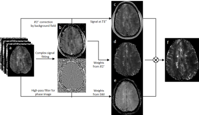 |
Susceptibility Relaxation based Optimization for Visualization of the Central Vein Sign in Multiple Sclerosis Using Multi-Echo Gradient Echo
Jiahao Li1, Weiyuan Huang2, Xianfu Luo2, Thanh D. Nguyen2, Susan A. Gauthier2, Pascal Spincemaille2, and Yi Wang1,2
1Biomedical Engineering, Cornell University, Ithaca, NY, United States, 2Radiology, Weill Cornell Medicine, New York, NY, United States
The central vein sign (CVS) has been suggested as a potential biomarker for multiple sclerosis (MS) lesion detection and differential diagnosis. A major hurdle for clinical investigation of CVS of MS lesion is the lack of high-quality visualization on current MRI. In this work, we propose a Susceptibility Relaxation based Optimization (SRO) method that uses routinely acquired multi-echo gradient echo magnitude and phase data to generate an image with optimal CVS contrast while preserving lesion signal. Preliminary qualitative and quantitative results demonstrate that SRO provides superior MS lesion and CVS detection compared to prior methods.
|
|
1388. |
Clinical disability is associated with normal-appearing white matter MTsat in recently diagnosed relapsing-remitting multiple sclerosis.
Elizabeth N. York1, Rozanna Meijboom1, Nicole White1, Maria Valdes Hernandez1, Michael J. Thrippleton1, Peter Connick1, and Adam Waldman1
1Centre for Clinical Brain Sciences, University of Edinburgh, Edinburgh, United Kingdom
Magnetization transfer saturation (MTsat) is a quantitative measure of white matter (WM) integrity with inherent correction for B1 inhomogeneities and T1 relaxation. MTsat is sensitive to pathological changes in axonal myelin in multiple sclerosis (MS) and may predict clinical disability. MTsat within normal-appearing WM and WM hyperintensities was measured in 76 patients with recently diagnosed relapsing-remitting MS. After accounting for age, MTsat in normal-appearing WM, but not WM hyperintensities, predicted clinical disability (as assessed by Multiple Sclerosis Functional Composite z-scores) in MS when central tendency and standard deviation were considered. This suggests MTsat is sensitive to early MS pathology.
|
|
1389.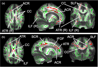 |
White matter magnetic resonance diffusion measures in Multiple Sclerosis with overactive bladder
Xixi Yang1,2,3,4, Martina D Liechti1,2, Baris Kanber3,5, Carole H Sudre5,6,7, Gloria Castellazzi3,8, Jiaying Zhang9,10,11, Marios C Yiannakas3, Gwen Gonzales2, Ferran Prados Carrasco3,5,12, Ahmed T Toosy3, Claudia A. M. G Wheeler-Kingshott3,13,14, and Jalesh N Panicker1,2
1Department of Brain Repair & Rehabilitation, UCL Queen Square Institute of Neurology, London, United Kingdom, 2Department of Uro-Neurology, National Hospital for Neurology and Neurosurgery, London, United Kingdom, 3NMR Research Unit, UCL Queen Square MS Centre, London, United Kingdom, 4Department of Neurology, Xuan Wu Hospital of Capital Medical University, Beijing, China, 5Centre for Medical Image Computing, UCL Department of Medical Physics and Bioengineering, London, United Kingdom, 6School of Biomedical Engineering and Imaging Sciences, King’s College London, London, United Kingdom, 7Dementia Research Centre, UCL Queen Square Institute of Neurology, London, United Kingdom, 8Department of Electrical, Computer and Biomedical Engineering, University of Pavia, Pavia, Italy, 9Department of Perinatal Imaging & Health, School of Biomedical Engineering & Imaging Sciences, King’s College London, London, United Kingdom, 10King’s Health Partners, St Thomas’ Hospital, London, United Kingdom, 11Department of Computer Science and Centre for Medical Image Computing, University College London, London, United Kingdom, 12Universitat Oberta de Catalunya, Barcelona, Spain, 13Department of Brain and Behavioral Sciences, University of Pavia, Pavia, Italy, 14Brain MRI 3T Center, IRCCS Mondino Foundation, Pavia, Italy
White matter (WM) abnormalities subtending overactive bladder (OAB) symptoms in multiple sclerosis (MS) is poorly understood. MS patients with and without OAB symptoms were recruited to investigate the association between WM changes and OAB symptoms in MS. Lower FA were found in MS with OAB compared with MS without LUTS (p=0.072). An inverse correlation was found between FA and OAB severity (p=0.021) in MS. OAB symptoms are associated with WM anisotropy changes in MS as demonstrated by diffusion-weighted imging (DWI).
|
|
1390.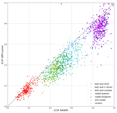 |
A comparison of seven white matter diffusion microstructure models to study intralesional damage in Multiple Sclerosis
Muhamed Barakovic1,2, Reza Rahmanzadeh1,2, Po-Jui Lu1,2,3, Pietro Maggi4,5, Pascal Sati6, Daniel Reich6, Francesco La Rosa7,8, Meritxell Bach Cuadra7,9, Simona Schiavi10, Alessandro Daducci10, Jens Kuhle1, Matthias Weigel1,2,11, Ludwig Kappos1,
and Cristina Granziera1,2
1Translational Imaging in Neurology (ThINk) Basel, Department of Medicine and Biomedical Engineering, University Hospital Basel and University of Basel, Neurologic Clinic and Policlinic, Basel, Switzerland, 2Departments of Medicine, Clinical Research and Biomedical Engineering, University Hospital Basel and University of Basel, Basel, Switzerland, 3Digital Technology and Innovation Siemens Healthineers, Princeton, NJ, United States, 4Department of Neurology, Saint-Luc University Hospital,Université Catholique de Louvain, Bruxelles, Belgium, 5Department of Neurology, Lausanne University and University Hospital, Lausanne, Switzerland, 6Translational Neuroradiology Section, Division of Neuroimmunology and Neurovirology, National Institute of Neurological Disorders and Stroke, National Institutes of Health, Bethesda, MD, United States, 7Signal Processing Laboratory (LTSS), Ecole polytechnique fédérale de Lausanne (EPFL), Lausanne, Switzerland, 8Radiology Department, Center for Biomedical Imaging, Lausanne University and University Hospital, Lausanne, Switzerland, Basel, Switzerland, 9Radiology Department, Center for Biomedical Imaging, Lausanne University and University Hospital Lausanne, Lausanne, Switzerland, 10Department of Computer Science, University of Verona, Verona, Italy, 11Radiological Physics, Department of Radiology, University Hospital Basel, Basel, Switzerland
In this study, we implemented seven microstructure models based on diffusion-weighted MRI data and investigated their sensitivity in assessing between-lesion differences in axonal content in multiple sclerosis lesions. The result of the analysis showed that all the models selected report lower intracellular volume fraction in white matter lesions compared to normal-appearing white matter. Few of the methods highlighted high sensitivity; however, they also show a different spectrum of ranges in terms of intracellular volume fraction. Whether one of the methods reflect a reliable assessment will be addressed in further studies using postmortem material.
|
|
1391.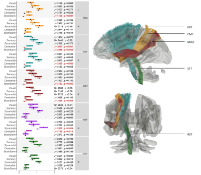 |
Automated atlas-based mapping of white matter tract damage to multiple sclerosis symptoms
Veronica Ravano1,2,3, Michaela Andelova4, Mazen Fouad A-Wali Mahdi1, Reto Meuli2, Tomas Uher4, Jan Krasensky5, Manuela Vaneckova5, Dana Horakova4, Tobias Kober1,2,6, and Jonas Richiardi1,2
1Advanced Clinical Imaging Technology, Siemens Healthcare, Lausanne, Switzerland, 2Department of Radiology, Lausanne University Hospital and University of Lausanne, Lausanne, Switzerland, 3Medical Imaging Processing, Ecole Polytechnique Fédérale de Lausanne (EPFL), Lausanne, Switzerland, 4Department of Neurology and Center of Clinical Neuroscience First Faculty of Medicine, Charles University and General University Hospital in Prague, Prague, Czech Republic, 5MR unit, Department of Radiology First Facutly of Medicine, Charles University and General University Hospital in Prague, Prague, Czech Republic, 6LTS5, Ecole Polytechnique Fédérale de Lausanne (EPFL), Lausanne, Switzerland In multiple sclerosis, the standard radiological metrics correlate poorly with clinical disability (‘clinico-radiological paradox’). To help filling this gap, we propose to map neurological impairments to white matter tract damage resulting from lesions. Because diffusion imaging is typically not part of multiple sclerosis clinical workups, quantitative tract damage metrics were extracted using a tractography atlas and an automated lesion segmentation algorithm. We were able to successfully identify which functional system (EDSS sub-score) was affected by damage on given tracts. These findings suggest the usefulness of using our fully automated atlas-based approach to study mechanisms of neurological diseases. |
|
1392. |
Optimization and evaluation of multi-compartment diffusion MRI using the spherical mean technique for practical multiple sclerosis imaging
Sean P Devan1,2, Xiaoyu Jiang1,3, Francesca Bagnato4, and Junzhong Xu1,3,5,6
1Institute of Imaging Science, Vanderbilt University Medical Center, Nashville, TN, United States, 2Chemical and Physical Biology Program, Vanderbilt University, Nashville, TN, United States, 3Department of Radiology and Radiological Sciences, Vanderbilt University Medical Center, Nashville, TN, United States, 4Neuroimaging Unit/Neuroimmunology Division, Department of Neurology, Vanderbilt University Medical Center, Nashville, TN, United States, 5Department of Biomedical Engineering, Vanderbilt University, Nashville, TN, United States, 6Department of Physics and Astronomy, Vanderbilt University, Nashville, TN, United States
Multi-compartment diffusion imaging using the spherical mean technique (SMT) is a recently developed method that provides more specific information for probing axonal properties in e.g., multiple sclerosis (MS) patients. However, the accuracy and precision of SMT have not been comprehensively investigated to date. We used Cramér-Rao Lower Bound (CRLB) analysis and MS patient data to optimize an SMT acquisition protocol for practical MS imaging. Second, we used computer simulations to comprehensively evaluate the accuracy and precision of SMT for reporting axonal information under various pathologic conditions. The results support better implementation and interpretation of SMT imaging for MS patients.
|
|
1393.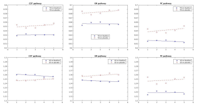 |
Diffusion Tensor Imaging on Three Pathways: Effect of Ibudilast in Progressive Multiple Sclerosis
Xuemei Huang1, Ken E. Sakaie1, Jian Lin1, Robert J. Fox1, and Mark J. Lowe1
1Imaging Institute, U-15, Cleveland Clinic, Cleveland, OH, United States
Diffusion Tensor Imaging (DTI) analysis was performed in the multi-site phase II trial of ibudilast in progressive multiple sclerosis. This study extends that analysis to three pathways to examine DTI measures in 31 progressive MS patients on treatment and 31 patients on placebo. Probabilistic tracking maps were generated. Mean pathway diffusion measures: longitudinal diffusivity (LD) and transverse diffusivity (TD) are reported. We report here that diffusion measures don’t significantly change over time in the treatment group but we observe a significant change over time in the placebo group.
|
|
1394.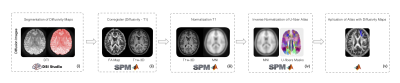 |
Evaluation of different diffusivity and cognitive measures in subcortical regions in healthy subjects and RRMS patients.
Cristian Montalba1,2,3, Mariana Zurita1,4, Tomás Labbé1,5, Marcelo Andia1,2, Miguel Guevara6, Jean-François Mangin7, Juan Pablo Cruz2, Ethel Ciampi8,9, Claudia Cárcamo5,8, Pamela Guevara6, and Sergio Uribe1,2,3
1Biomedical Imaging Center, Pontificia Universidad Católica de Chile, Santiago, Chile, 2Radiology Department, School of Medicine, Pontificia Universidad Católica de Chile, Santiago, Chile, 3Millennium Nucleus for Cardiovascular Magnetic Resonance, Santiago, Chile, 4Institute of Cognitive Neuroscience, University College London, London, UK., London, United Kingdom, 5Interdisciplinary Center of Neurosciences, Pontificia Universidad Católica de Chile, Santiago, Chile, 6Faculty of Engineering, Universidad de Concepción, Concepción, Chile, Concepción, Chile, 7UNATI, Neurospin, CEA,, Université Paris-Saclay, Gif-sur-Yvette, France, 8Neurology Department, School of Medicine, Pontificia Universidad Católica de Chile, Santiago, Chile, 9Neurology Service, Hospital Dr. Sótero del Río, Santiago, Chile, Santiago, Chile
We evaluated different diffusivity measurements of subcortical regions that can be related with different test scores in FAS, SDMT and PASAT test. For this purpose, we evaluated the FA, MD, RD and AD diffusivity maps of U-fibers with the test scores in healthy subjects and RRMS patients. In our sample, FAS test scores are significant linear related with Postcentral gyrus with RD and MD maps in RRMS patients. PASAT test scores are significant linear related with Precentral gyrus with FA and RD maps in healthy subjects. There is no linear relationship between SDMT test score and diffusivity maps.
|
|
1395.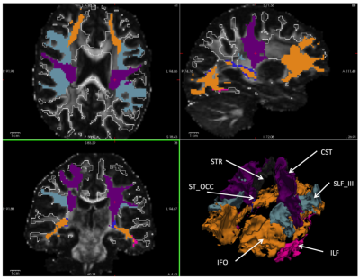 |
Quantitative MR diffusion parameter in normal appearing white matter (NAWM) – a comparison of multiple sclerosis patients and healthy controls
Daniel Güllmar1, Renat Sibgatulin1, Stefan Ropele2, and Jürgen R Reichenbach1,3
1Medical Physics Group / IDIR, Jena University Hospital, Jena, Germany, 2Department of Neurology, Medical University of Graz, Graz, Austria, 3Michael-Stifel-Center for Data driven sciences, Friedrich-Schiller-University Jena, Jena, Germany
Quantitative MR diffusion imaging in multiple sclerosis is a promising tool for deciphering the mechanisms and processes during the disease progression. We employed new MR diffusion acquisition techniques (like SMS) as well as advanced post-processing (like NODDI, SMT) in order to investigate the differences in normal appearing white matter of MS patients in contrast to healthy volunteers.
|
|
1396.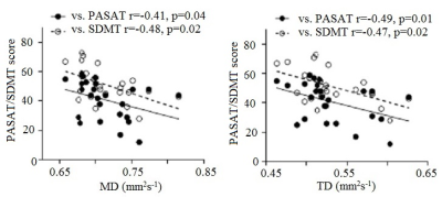 |
Diffusion tractography evaluation of specificity of PASAT and SDMT to cognitive pathway in multiple sclerosis
Pallab K Bhattacharyya1,2, Robert Fox3, Jian Lin1, Hong Li4, Ken Sakaie1, and Mark Lowe1
1Imaging Institute, Cleveland Clinic, Cleveland, OH, United States, 2Radiology, Cleveland Clinic Lerner College of Medicine, CLEVELAND, OH, United States, 3Neurological Institute, Cleveland Clinic, Cleveland, OH, United States, 4Quantitative Health Sciences, Cleveland Clinic, Cleveland, OH, United States
Paced auditory serial addition test (PASAT) and symbol digit modalities test (SDMT) are two most commonly used tests to evaluate cognitive performance in MS. The specificity of the two tests to cognitive network in brain was evaluated with diffusion tensor imaging and correlating transverse diffusivity (TD) along different functionally relevant pathways with PASAT/SDMT scores. While SDMT showed association with TD along all pathways (and whole brain white matter), PASAT was associated with only frontoparietal pathway, establishing its specificity to cognitive network.
|
1397. |
Gray matter relaxation rates show different correlation patterns for cognitive impairment and physical disability in Multiple Sclerosis.
Maria Teresa Cassiano1, Roberta Lanzillo2, Bruno Alfano3, Teresa Costabile2, Marco Comerci3, Anna Prinster3, Marcello Moccia2, Rosario Megna3, Vincenzo Brescia Morra2, Arturo Brunetti1, and Mario Quarantelli3
1Department of Advanced Biomedical Sciences, University "Federico II", Naples, Italy, 2Department of Neurosciences, Reproductive Science and Odontostomatology, University "Federico II", Naples, Italy, 3Biostructure and Bioimaging Institute, National Research Council, Naples, Italy To assess voxelwise in gray matter the correlation between relaxation rates and both physical and cognitive impairment in MS, R1 and R2 relaxation rate maps from 241 Relapsing-Remitting MS patients were assessed voxelwise for correlation with EDSS and the percentage of impaired cognitive test. Inverse correlation between EDSS and R2 were detected throughout the brain, while inverse correlations with R1 were mostly limited to perirolandic and supramarginal cortices. Cognitive impairment correlated negatively with R1, and to a lesser extent with R2, in the limbic system and dorsolateral prefrontal cortices. |
|
1398.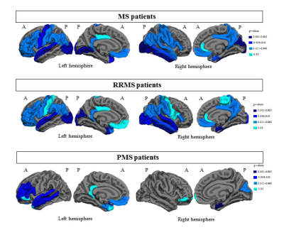 |
Longitudinal progression of cortical thinning differs across MS phenotypes and is clinically relevant: a multicentre study
Paola Valsasina1, Maria A. Rocca1,2, Milagros Hidalgo de la Cruz1, Claudio Gobbi3, Antonio Gallo4, Chiara Zecca3, Alvino Bisecco4, and Massimo Filippi1,2,5
Video Permission Withheld
1Neuroimaging Research Unit, Institute of Experimental Neurology, Division of Neuroscience, IRCCS San Raffaele Scientific Institute, Milan, Italy, 2Neurology Unit, IRCCS San Raffaele Scientific Institute, Milan, Italy, 3Department of Neurology, Neurocenter of Southern Switzerland, Civic Hospital, Lugano, Switzerland, 4Department of Advanced Medical and Surgical Sciences, and 3T MRI Center, University of Campania “Luigi Vanvitelli”, Naples, Italy, 5Vita-Salute San Raffaele University, Milan, Italy
In this study, we used cortical thickness analysis to investigate the evolution over time of cortical grey matter atrophy in a multicenter cohort of patients with multiple sclerosis (MS) acquired at 3 European sites. We detected a different susceptibility to cortical damage across the different clinical phenotypes of MS, with the involvement of temporal, parietal and occipital regions in relapsing-remitting MS, and an additional frontal involvement in progressive MS. Different annualized cortical thinning rates were found across MS clinical phenotypes. Cortical thinning in frontal and temporal areas was crucial for identifying patients with more severe disability at follow-up.
|
|
1399. |
Changes of cortical gray matter myelin and microstructure in multiple sclerosis (MS) assessed with quantitative ihMT and MT
Fanny Munsch1, Gopal Varma1, Manuel Taso1, Shahamat Tauhid2, Olivier M. Girard3, Guillaume Duhamel3, Rob Bakshi4, and David Alsop1
1Radiology, Division of MRI Research, Beth Israel Deaconess Medical Center, Harvard Medical School, Boston, MA, United States, 2Brigham and Women's Hospital, Harvard Medical School, Boston, MA, United States, 3CRMBM, Aix Marseille Univ, CNRS, Marseille, France, 4Neurology, Brigham and Women's Hospital, Harvard Medical School, Boston, MA, United States
Inhomogeneous magnetization transfer (ihMT) and magnetization transfer (MT) have differential sensitivity to myelin and can be used to characterize cortical gray matter. We compared cortical surface-based analyses of ihMT and MT between multiple sclerosis (MS) patients and age-matched healthy volunteers. IhMT at mid-thickness decreased less than MT in most cortical regions of MS patients. Integrated signal across cortical depth showed less spatial variability and clear decrease of ihMT but less than MT. As ihMT is understood to be more specific to myelin than MT, these findings suggest loss of cortical nonmyelinated cells and structures exceed cortical myelin loss in MS.
|
|
1400. |
"Comparative analysis of 3D DIR and 3D FLAIR in cases of acute optic neuritis"
Vivek Shriram Murumkar1, Jitender Saini1, Nethravati Manjunath2, Dhritiman Chakraborty3, and Rakesh Gupta4
xxyy-video
1Neuroimaging and interventional radiology, national institute of mental health and neuroscineces (NIMHANS) bangalore, india, Bangalore, India, Bangalore, India, 2Neurology, national institute of mental health and neuroscineces (NIMHANS) bangalore, india, Bangalore, India, Bangalore, India, 3Neuroanaesthesia, national institute of mental health and neuroscineces (NIMHANS) bangalore, india, Bangalore, India, Bangalore, India, 4radiology, Fortis Hospital, Gurugram, India
Optic neuritis is presenting feature in 15-20% cases of MS and 50% cases of MS have optic neuritis during course of illness. Signals from CSF and intraorbital fat are main hurdles in optic nerve imaging, 3D FLAIR and 3D DIR are used to study optic nerve pathologies as they suppress fat and CSF signals. DIR nulls signal from NAWM providing inherent contrast. 3D DIR and 3D FLAIR was done in 31 case of acute optic neuritis and 30 controls. We found higher contrast rates, sensitivity and negative predictive value for the 3D DIR than 3D FLAIR in acute optic neuritis
|
|
1401.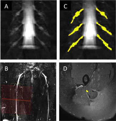 |
A pilot in vivo investigation of peripheral nerve damage in multiple sclerosis using magnetisation transfer ratio
Marios C. Yiannakas1, Marco Battiston1, Francesco Grussu1,2, Ratthaporn Boonsuth1, Rebecca S. Samson1, Torben Schneider3, Masami Yoneyama4, Ferran Prados1,5,6, Carmen Tur1, Sara Collorone1, Rosanna Cortese1, Olga Ciccarelli1, and Claudia A.M. Gandini Wheeler-Kingshott1,7,8
1Queen Square MS Centre, Department of Neuroinflammation, UCL Queen Square Institute of Neurology, Faculty of Brain Sciences, University College London, London, United Kingdom, 2Centre for Medical Image Computing, Department of Computer Science, University College London, London, United Kingdom, 3Philips Healthcare, Guildford, Surrey, United Kingdom, 4Philips Japan, Minatoku, Tokyo, Japan, 5Centre for Medical Image Computing, Medical Physics and Biomedical Engineering, University College London, London, United Kingdom, 6Universitat Oberta de Catalunya, Barcelona, Spain, 7Brain MRI 3T Research Centre, IRCCS Mondino Foundation, Pavia, Italy, 8Department of Brain and Behavioural Sciences, University of Pavia, Pavia, Italy
Evidence from histopathological studies has demonstrated the involvement of the peripheral nervous system in multiple sclerosis (MS), specifically alteration in myelin content and, to a lesser extent, axonal degeneration. However, evidence from objective investigations in vivo is lacking. In this pilot study, the lumbar plexus and the sciatic nerve are investigated in people with MS using a previously optimised magnetisation transfer ratio (MTR) protocol developed in healthy controls. Results demonstrate reduced MTR values in both anatomical regions studied, consistent with histopathological data, and as such highlighting a need for further investigations in a larger sample population.
|
|
1402.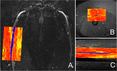 |
Reduced field-of-view multi-shell diffusion-weighted imaging of the sciatic nerve: Application to multiple sclerosis
Marios C. Yiannakas1, Francesco Grussu1,2, Marco Battiston1, Ratthaporn Boonsuth1, Rebecca S. Samson1, Torben Schneider3, Masami Yoneyama4, Ferran Prados1,5,6, Carmen Tur1, Sara Collorone1, Rosanna Cortese1, Olga Ciccarelli1, and Claudia A. M. Gandini Wheeler-Kingshott1,7,8
1Queen Square MS Centre, Department of Neuroinflammation, UCL Queen Square Institute of Neurology, Faculty of Brain Sciences, University College London, London, United Kingdom, 2Centre for Medical Image Computing, Department of Computer Science, University College London, London, United Kingdom, 3Philips Healthcare, Guildford, Surrey, United Kingdom, 4Philips Japan, Minatoku, Tokyo, Japan, 5Centre for Medical Image Computing, Medical Physics and Biomedical Engineering, University College London, London, United Kingdom, 6Universitat Oberta de Catalunya, Barcelona, Spain, 7Brain MRI 3T Research Centre, IRCCS Mondino Foundation, Pavia, Italy, 8Department of Brain and Behavioural Sciences, University of Pavia, Pavia, Italy
Ex vivo investigations have demonstrated the involvement of the peripheral nervous system in multiple sclerosis (MS). However, the use of advanced imaging methods to study individual peripheral nerves in vivo, such as the sciatic nerve, has been hindered by a number of technical challenges. In this pilot in vivo study, we explore the feasibility of acquiring multi-shell diffusion-weighted imaging (DWI) metrics in the sciatic nerve of people with relapsing-remitting MS (RRMS) using reduced field-of-view echo planar imaging. DWI metrics in people with RRMS display changes in comparison to healthy controls, suggesting that the structural integrity of the nerve is compromised.
|
|
1403.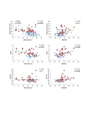 |
Cervical cord myelin abnormality is associated with clinical disability in multiple sclerosis
Irene Margaret Vavasour1,2, Lisa Eunyoung Lee3, Adam V Dvorak2,4, Shawna Abel3, Poljanka Johnson3, Stephen Ristow3, Cornelia Laule1,2,4,5, Roger Tam1,6, David KB Li1,3, Nathalie Ackermans3, Alice Schabas3, Jillian Chan3, Ana-Luiza Sayao3,
Virginia Devonshire3, Robert Carruthers3, Anthony Traboulsee3, and Shannon H Kolind1,2,3,4
1Radiology, University of British Columbia, Vancouver, BC, Canada, 2International Collaboration on Repair Discoveries (ICORD), University of British Columbia, Vancouver, BC, Canada, 3Medicine (Neurology), University of British Columbia, Vancouver, BC, Canada, 4Physics and Astronomy, University of British Columbia, Vancouver, BC, Canada, 5Pathology and Laboratory Medicine, University of British Columbia, Vancouver, BC, Canada, 6School of Biomedical Engineering, University of British Columbia, Vancouver, BC, Canada
Damage to the spinal cord is common in multiple sclerosis (MS) and is an important contributor to physical disability. While spinal cord cross sectional area (CSA) is correlated with disability, CSA is a non-specific measure of tissue damage. The addition of cervical cord myelin water imaging, which measures myelin-related abnormalities, to cord area resulted in better correlations with MS clinical disability than cord CSA alone. In particular, myelin abnormality + CSA was best correlated with 9-hole peg test which requires more fine motor skills and therefore could be strongly influenced by damage to white matter.
|
|
1404.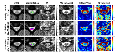 |
Evaluation of DTI Indices in the Cervical Spinal Cord as Markers of Sensorimotor Impairment in MS Patients with Mild Disability
Kristin P. O'Grady1,2, Kurt G. Schilling2, Mereze Visagie2, Sanjana Satish2, Shekinah Malone3, Atlee Witt2, Anna Combes2, Richard Dylan Lawless2,4, Colin D. McKnight1, Francesca R. Bagnato5, and Seth A. Smith1,2,4
1Department of Radiology and Radiological Sciences, Vanderbilt University Medical Center, Nashville, TN, United States, 2Vanderbilt University Institute of Imaging Science, Vanderbilt University Medical Center, Nashville, TN, United States, 3Meharry School of Medicine, Meharry Medical College, Nashville, TN, United States, 4Department of Biomedical Engineering, Vanderbilt University, Nashville, TN, United States, 5Department of Neurology, Vanderbilt University Medical Center, Nashville, TN, United States
Spinal cord pathology is integral to disease symptoms and progression in multiple sclerosis (MS), but imaging methods developed and optimized for studying the spinal cord in vivo with clinically relevant scan times are lacking. Here, we applied a clinically feasible diffusion tensor imaging (DTI) sequence to examine relationships between imaging measures of microstructural damage in the spinal cord and lower extremity functional deficits in low-disability, relapsing-remitting MS patients. Our results show significant correlations between gray matter radial diffusivity and measures of sensation and motor function.
|
|
1405.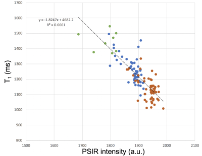 |
Phase Sensitive Inversion Recovery (PSIR) imaging as surrogate for T1 mapping: applications to the spinal cord of multiple sclerosis patients
Nico Papinutto1, Shuiting Cheng1, Mahir Khan1, Jung-Jiin Hsu1, and Roland G Henry1
1UCSF Weill Institute for Neurosciences, Department of Neurology, University of California San Francisco, San Francisco, CA, United States
Acquiring images on a phantom and 54 multiple sclerosis patients, we explored the feasibility of T1 mapping using a fast 2D inversion recovery method based on a TrueFISP sequence. We also explored the relationship between T1 values and the intensity of 2D PSIR images. The fast T1 mapping method was shown to be precise, and PSIR intensities shown to have a strong correlation with T1. PSIR has been extensively used to segment in-vivo gray and white matter tissues in the spinal cord, and this study suggests that it can simultaneously provide microscopic information related to the longitudinal relaxation time T1.
|
|
1406.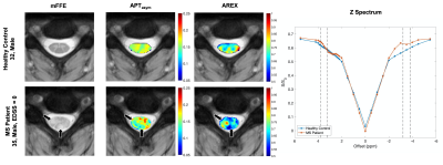 |
The Effect of AREX Correction on APT CEST MRI of Lesions within the Spinal Cord of MS Patients
Richard D Lawless1,2, Kristin O'Grady1,3, Mereze Visagie1, Anna Combes1, Haley Feiler1, Francesca Bagnato4, and Seth A. Smith1,2,3
1Vanderbilt University Institute of Imaging Sciences, Vanderbilt University Medical Center, Nashville, TN, United States, 2Department of Biomedical Engineering, Vanderbilt University, Nashville, TN, United States, 3Department of Radiology and Radiological Sciences, Vanderbilt University Medical Center, Nashville, TN, United States, 4Department of Neurology, Vanderbilt University Medical Center, Nashville, TN, United States
Spinal cord damage in multiple sclerosis often leads to the formation of inflammatory lesions. APT CEST has been proposed as an MRI biomarker capable of detecting the underlying biochemical changes associated with lesion formation, however, CEST findings are susceptible to confounding influences such as the macromolecular magnetization transfer contribution and changes to longitudinal relaxation. AREX is a proposed method which corrects for these contributors and may improve measurement of the CEST component in vivo. In this study we sought to compare MT- and T1-corrected AREX to an uncorrected CEST quantification method in SC lesions of MS patients.
|
|
1407.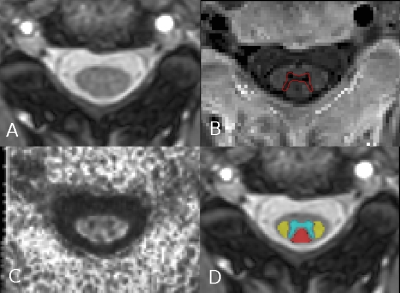 |
Clinical relevance of multiparametric MRI assessment of spinal cord damage in multiple sclerosis
Elisabetta Pagani1, Raffaello Bonacchi1,2, Alessandro Meani1, Laura Cacciaguerra1,2, Ermelinda De Meo1,2, Paolo Preziosa1,2, Maria A. Rocca1,2, and Massimo Filippi1,2,3
Video Permission Withheld
1Neuroimaging Research Unit, Institute of Experimental Neurology, Division of Neuroscience, IRCCS San Raffaele Scientific Institute, Milan, Italy, 2Neurology Unit, IRCCS San Raffaele Scientific Institute, Milan, Italy, 3Vita-Salute San Raffaele University, Milan, Italy
Cervical spinal cord involvement is common in multiple sclerosis (MS). We performed a comprehensive multiparametric MRI study to explore pathophysiological substrates of damage and to identify the most accurate imaging predictors of disability and disease course. We found that the processes contributing to disability differ according to the stage of the disease. In relapsing-remitting MS patients, lesions and microstructural damage to cervical spinal cord tracts have a prominent role, whereas in progressive MS patients, cervical cord grey matter atrophy becomes clinically meaningful.
|
|
1408.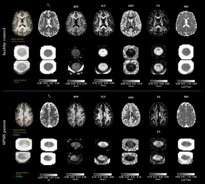 |
Microstructure changes in secondary progressive multiple sclerosis measured using advanced quantitative MRI of the brain and spine
Marco Battiston1, Carmen Tur1,2, Marios C. Yiannakas1, Francesco Grussu1,3, Torben Schneider4, Daniele Martinelli5, Baris Kanber6, Ferran Prados1,6,7, Patrizia Pajak1, Olga Ciccarelli1, Rebecca S. Samson1, and Claudia A.M. Gandini Wheeler-Kingshott1,8,9
1NMR Research Unit, UCL Queen Square MS Centre, Queen Square Institute of Neurology, Faculty of Brain Sciences, University College London, London, United Kingdom, 2Neurology Department, , Luton and Dunstable University Hospital, Luton, United Kingdom, 3Centre for Medical Image Computing, Department of Computer Science, University College London, London, United Kingdom, 4Philips Healthcare, Guilford, United Kingdom, 5IRCCS Mondino Foundation, Pavia, Italy, 6Centre for Medical Image Computing, Medical Physics and Biomedical Engineering, University College London, London, United Kingdom, 7Universitat Oberta de Catalunya, Barcelona, Spain, 8Department of Brain and Behavioural Sciences, University of Pavia, Pavia, Italy, 9Brain MRI 3T Center, IRCCS Mondino Foundation, Pavia, Italy
Multiple Sclerosis (MS) is a complex disease whose mechanisms of progression still remain unclear. The use of quantitative MRI techniques in a multi-modal fashion, i.e. acquisition of more than one contrast type, can help to non-invasively measure changes in the tissue at a microstructural level during the disease course. Moreover, evidence of spinal cord involvement and its role in the diagnosis and prognosis is mounting. Here, we aim to address both needs by developing a multi-modal qMRI protocol for joint investigation of brain and spinal cord microstructure, and we report findings from a small cohort of people with MS.
|
|
1409.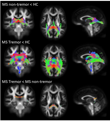 |
Reduced fibre-specific measures in multiple sclerosis patients with cerebellar dysfunction
Sanuji Gajamange1, Frederique Boonstra2, Gustavo Noffs1,3, Thushara Perera1,4, Vilija Jokubaitis2, Adam Vogel1,4,5, Andrew Evans3,4, Helmut Butzkueven2, Anneke van der Walt2, and Scott Kolbe2
1The University of Melbourne, Melbourne, Australia, 2Monash University, Melbourne, Australia, 3Royal Melbourne Hospital, Melbourne, Australia, 4The Bionics Institute, Melbourne, Australia, 5University of Tübingen, Tübingen, Germany
Cerebellar dysfunction is a common feature of multiple sclerosis (MS), leading to disabling symptoms such as tremor. In MS, brain atrophy is the most accepted correlate of neurodegeneration; however cerebellar atrophy does not seem to correlate with the degree of cerebellar dysfunction. Here we explore axonal degeneration, a key driver of disability, with a fibre-specific marker based on diffusion-weighted MRI – fibre density and fibre bundle cross-section. We found that loss of cerebellar fibre density and fibre cross-section was associated with increased clinical cerebellar dysfunction. Fibre-specific measures could provide a useful marker of cerebellar dysfunction in MS.
|
|
1410.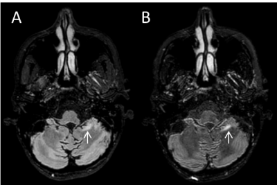 |
Optimized patient-specific refocusing control in turbo spin echo FLAIR for improved detection of infratentorial lesions in multiple sclerosis
Refaat E Gabr1, Luning Wang2, John A Lincoln3, and Ponnada A Narayana1
1Diagnostic and Interventional Imaging, University of Texas Health Science Center at Houston, Houston, TX, United States, 2Philips healthcare, Gainesville, FL, United States, 3Neurology, University of Texas Health Science Center at Houston, Houston, TX, United States
Detection of infratentorial lesions is important for diagnosis of multiple sclerosis. However, infratentorial lesions are hard to detect on conventional FLAIR protocols. In this work, we optimized the refocusing flip angle in the turbo spin echo readout of 3D FLAIR to enhance infratentorial lesion contrast in individual patients. T1, T2, and proton density measured in the brain stem and cortical gray matter were used to calculate the refocusing angle that maximizes the contrast-to-noise ratio. The patient-specific approach was assessed in 8 MS patients. Improved infratentorial lesion contrast was achieved in most of the cases.
|
|
1411.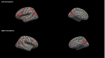 |
Pediatric-Onset Multiple Sclerosis: Imaging with Focus on Posterior Fossa Involvement
Laleh Eskandarian1,2, Bahadır Konuskan3, Hacer Dasgin2, Ismail Solmaz3, Rahsan Gocmen4, Banu Anlar4, and Kader Karli Oguz2,4
1Neuroscience Department, Bilkent University, Ankara, Turkey, 2National Magnetic Resonance Research Center (UMRAM), Bilkent University, Ankara, Turkey, 3Faculty of Medicine, Pediatric Neurology, Hacettepe University, Ankara, Turkey, 4Faculty of Medicine, Department of Radiology, Hacettepe University, Ankara, Turkey
Pediatric-onset-Multiple-Sclerosis (POMS) has been reported to have larger and higher number of T2 lesions as with more frequent posterior fossa involvement compared to adults. We aimed to analyze POMS patients morphological, microstructural alterations and their correlates with clinical characteristics with emphasis on posterior system. SUIT,Freesurfer and TBSS were used for spatialized parcellation, and analysis. Cerebellum except paramedian structures and cerebral cortices showed volume reductions, cerebral white matter and midbrain showed reduced FA and increased MD,AD,RD. Total and periventricular lesion loads, thalamic volumes showed correlations with special clinical metrics. Multiparametric brain MRI may reveal information on tissue changes and clinical correlates.
|
1412.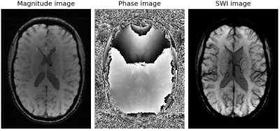 |
CNN-based classification of multiple sclerosis using BOLD venographic imaging (SWI) data
Alina Lopatina1, Renat Sibgatulin1, Stefan Ropele2, Jürgen R Reichenbach1,3, and Daniel Güllmar1
1Medical Physics Group / IDIR, Jena University Hospital, Jena, Germany, 2Department of Neurology, Medical University of Graz, Graz, Austria, 3Michael Stifel Center for Data-driven Sciences, Friedrich-Schiller-University Jena, Jena, Germany
Convolutional neural network (CNN) was proposed to identify multiple sclerosis patients and healthy subjects while susceptibility-weighted imaging (SWI) was used for MRI scans preprocessing in order to disclose important features hidden in brain venograms. Using only one two-dimensional slice for each subject makes the proposed algorithm easy-applied and useful for clinical practice.
|
|
1413.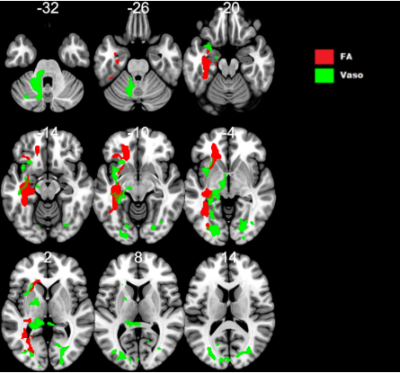 |
Cerebral vasoreactivity reveals white matter tracts alteration in multiple sclerosis patients with motor symptoms
François-Louis Collemiche1, Céline Charroud2, Aude Metzger3, Xavier Ayrignac4, Pierre Labauge4, Nicolas Menjot de Champfleur4, Emmanuelle Le Bars4, and Jérémy Deverdun4
1Neuroradiology, Hospital Gui de Chauliac, MONTPELLIER, France, 2Center for Mind/Brain Science, Rovereto, Italy, 3Hospital Pierre Wertheimer, Lyon, France, 4Hospital Gui de Chauliac, MONTPELLIER, France
One hypothesis in physiopathological mechanisms of multiple sclerosis (MS) involves astrocyte metabolism disruption as a consequence of axonal dysfunction, leading to cerebral vasoreactivity alterations (CVR). The aim was to explore a possible link between axonal tracts alterations and CVR disruption. 35 MS patients underwent MR. We selected motor clinical symptoms, and compared cerebral vasoreactivity and DTI maps. Both poCVR and DTI maps showed vasoreactivity alterations in white matter regions. Maps outlined the Inferior Fronto-Occipital Fasciculus in patients with “difficulties using upper limbs”. Vasoreactivity could be a new biomarker for assessing the evolution of white matter tracts’ alterations in MS follow-up.
|
|
1414.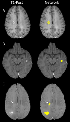 |
Segmentation of contrast enhancing lesions in multiple sclerosis using deep learning and a large cohort study
Refaat E Gabr1, Ivan Coronado1, and Ponnada A Narayana1
1Diagnostic and Interventional Imaging, University of Texas Health Science Center at Houston, Houston, TX, United States
Multiple sclerosis (MS), a demyelinating disease of the central nervous system, affects more than two million people worldwide. Contrast enhancing lesions are thought to reflect active disease state and play a key role in MS management. Deep learning (DL) based on convolutional neural networks has reached state-of-art performance on semantic segmentation tasks. Using annotated images for 398 MS patients, we evaluated DL performance on segmentation of enhancing lesions. Our approach yielded Dice similarity coefficient of 0.78, true positive rate of 0.91, and false positive rate of 0.28 for test data. Network performance was excellent for enhancement volumes ≥70 µl.
|
|
1415.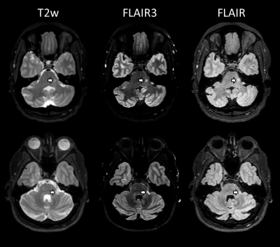 |
Combined 3D FLAIR and T2-weighted (FLAIR3) improves the detection of infratentorial lesions in multiple sclerosis
Refaat E Gabr1, John A Lincoln2, Arash Kamali1, Xiaojun Sun1, Khader M Hasan1, and Ponnada A Narayana1
1Diagnostic and Interventional Imaging, University of Texas Health Science Center at Houston, Houston, TX, United States, 2Neurology, University of Texas Health Science Center at Houston, Houston, TX, United States
Infratentorial lesions play an important role in the diagnosis of multiple sclerosis (MS), but are challenging to detect on conventional FLAIR protocols. Synthetic FLAIR3 images were generated by combining FLAIR and T2-weighted images to improve lesion contrast while maintaining CSF suppression. We evaluated FLAIR3 for detecting infratentorial lesions in 10 patients with MS. Our results show 11-fold increase in the lesion contrast-to-noise ratio and 163% increase in the number of detected lesions using FLAIR3 as compared to FLAIR.
|
|
1416.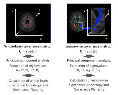 |
Shape and Spatial Pattern Analysis through Covariance-based Estimations of MS lesions: the SSPACE-MS study
Carmen Tur1,2, Francesco Grussu1,3, Ferran Prados1,4,5, Baris Kanber4, Thalis Charalambous1,6, Declan T. Chard1,7, Olga Ciccarelli1,7, and Claudia A.M. Gandini Wheeler-Kingshott1,8,9
1UCL Queen Square Institute of Neurology, University College London, London, United Kingdom, 2Neurology, Luton and Dunstable University Hospital, Luton, United Kingdom, 3Centre for Medical Image Computing, Department of Computer Science, University College London, London, United Kingdom, 4Centre for Medical Image Computing, Medical Physics and Biomedical Engineering department, University College London, London, United Kingdom, 5e-Health center, Universitat Oberta de Catalunya, Barcelona, Spain, 6Department of Medicine, St. George’s University of London, London, United Kingdom, 7University College London Hospitals Biomedical Research Centre, National Institute for Health Research, London, United Kingdom, 8Department of Brain and Behavioural Sciences, University of Pavia, Pavia, Italy, 9Brain MRI 3T Research Centre, IRCCS Mondino Foundation, Pavia, Italy
Lesion load is the main predictor of future disability in multiple sclerosis (MS) but its predictive power is limited possibly because key aspects of lesions, such as their spatial distribution, morphology or pathological substrate are not being considered. Here we demonstrate the relevance of shape and spatial features of white matter lesions in the development of disability in MS through a novel approach. This new methodological framework (SSPACE-MS) is based on the analysis of the covariance matrix defined by the spatial position of lesional voxels. It can be applied longitudinally and is easily implementable in clinical practice.
|
|
1417.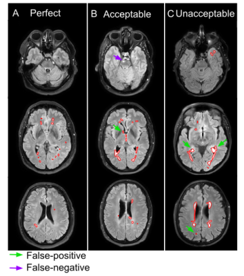 |
Clinical Evaluation of a Fully Convolutional Neural Network for Automatic MS Lesion Segmentation on MRI
Amalie Monberg Hindsholm1, Claes Nøhr Ladefoged1, Flemming Littrup Andersen1, Stig Præstekjær Cramer1, Liselotte Højgaard1, and Ulrich Lindberg1
1Clinical Physiology, Nuclear Medicine and PET, Rigshospitalet University, Copenhagen, Denmark
Automatic segmentation of MRI-visible multiple sclerosis (MS) lesions could potentially reduce assessment time and inter- and intra-rater variability. Recently, automatic methods using deep convolutional neural networks (CNN) have obtained great results in image segmentation. This work implements a state-of-the-art 2D CNN-based segmentation method from literature and extends and recalibrates it to a local MS dataset of 91 patients. A clinical evaluation is performed on an independent MS dataset of 53 patients, where 94% of predicted segmentation masks were deemed valuable for clinical use.
|
|
1418. |
Measurement of white matter atrophy in multiple sclerosis using diffusion weighted imaging
Elisabetta Pagani1, Loredana Storelli1, Paolo Preziosa1,2, Federica Esposito2, Laura Cacciaguerra1,2, Massimo Filippi1,2,3, and Maria A. Rocca1,2
Video Permission Withheld
1Neuroimaging Research Unit, Institute of Experimental Neurology, Division of Neuroscience, IRCCS San Raffaele Scientific Institute, Milan, Italy, 2Neurology Unit, IRCCS San Raffaele Scientific Institute, Milan, Italy, 3Vita-Salute San Raffaele University, Milan, Italy
Gray matter is more relevant than white matter (WM) atrophy in explaining clinical disability and cognitive impairment in multiple sclerosis (MS). However, WM is a complex structure whose fiber orientation should be considered when investigating its morphology. In a group of MS patients and healthy controls, we applied constrained spherical deconvolution and fixel-based morphometry to estimate the distribution of WM fiber bundles and their cross-section area as a measure of atrophy. We found that the application of this approach in MS improved when accounting for the presence of lesions and that atrophy was specific of WM fiber bundles.
|
|
1419.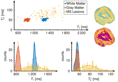 |
Detection of multiple sclerosis lesion with T1 and T2* MR Fingerprinting
Ingo Hermann1,2, Ralf Schmidt1, Benedikt Rieger1, Eloy Martinez-Heras3, Elisabeth Solona3, Sara Llufriu3, Achim Gass4, Lothar R. Schad1, and Frank G. Zöllner1
1Computer Assisted Clinical Medicine, Medical Faculty Mannheim, University Heidelberg, Mannheim, Germany, 2Magnetic Resonance Systems Lab, Department of Imaging Physics, Delft University of Technology, Delft, Netherlands, 3Center of Neuroimmunology, Laboratory of Advanced Imaging in Neuroimmunological Diseases, Hospital Clinic Barcelona, Institut d'Investigacions Biomèdiques August Pi i Sunyer (IDIBAPS) and Universitat de Barcelona, Barcelona, Spain, 4Department of Neurology, Medical Faculty Mannheim, University Heidelberg, Mannheim, Germany
We evaluated an MRF-EPI sequence for simultaneous T1 and T2* quantification in 44 patients with multiple sclerosis (MS) lesions at two scanners. We found significantly increased T1 and T2* times in MS lesions with differences up to 100 %. MS lesions resulted in broad distribution in the T1, T2* space compared with dense, separated clusters for white and gray matter.
|
|
1420.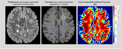 |
Myelin water imaging of Multiple Sclerosis Lesions
Caroline Köhler1, Hannes Wahl1, Paul Kuntke1, Tjalf Ziemssen2, Jennifer Linn1, and Hagen H. Kitzler1
1Institut und Poliklinik für Diagnostische und Interventionelle Neuroradiologie, University Hospital Carl Gustav Carus, Dresden, Germany, 2Neurology, University Hospital Carl Gustav Carus, Dresden, Germany
Myelin water imaging allows the assessment of relative myelination by estimating the myelin water fraction (MWF). This work investigated if the degree of MWF loss in multiple sclerosis is associated with T1 hypointense and isointense lesions. Within n = 1668 FLAIR lesions of n = 63 MS patients, 79% had a hypointense correlate, and 21% of the lesions had an isointense appearance on non-enhanced T1-weighted images. Significant differences were found for hypointense (MWFmean = 0.126) and isointense (MWFmean = 0.185) lesions. These results show varying degrees of myelin reduction.
|
|
1421.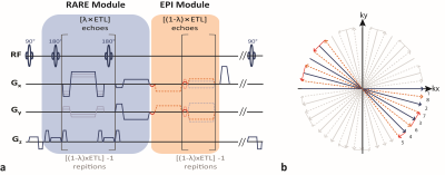 |
Simultaneous T2 and T2* Mapping of Multiple Sclerosis Lesions with a Radially Sampled RARE-EPI Hybrid
Carl Herrmann1, Eckart Grönerwäller1, Antje Els1, Joseph Kuchling2,3,4, Friedemann Paul2,3,4, and Thoralf Niendorf1,2
1Berlin Ultrahigh Field Facility (B.U.F.F.) Max Delbrück Center For Molecular Medicine in the Helmoltz Association, Berlin, Germany, 2Experimental und Clinical Research Center (ECRC), Charité-Campus Berlin-Buch, Berlin, Germany, 3NeuroCure Clinical Research Center, Charité – Universitätsmedizin, Berlin, Germany, 4Department of Neurology, Charité – Universitätsmedizin, Berlin, Germany Poster Permission Withheld
MRI examinations commonly involve a series of multiple imaging contrasts and MR-metrics, resulting in long examination times and propensity to motion and slice misregistration. Dual or multi-contrast techniques offer substantial scan time reduction. Recognizing this opportunity this work presents a dual contrast RARE-EPI hybrid, that provides T2 (RARE module) and T2* (EPI module) contrast and facilitates simultaneous T2 and T2* mapping in a single radially (under)sampled scan (2-in-1 RARE-EPI). The applicability of 2-in-1 RARE-EPI is demonstrated in an in vivo feasibility study involving patients suffering from multiple sclerosis and healthy controls and benchmarked versus conventional T2 and T2* weighted/mapping techniques.
|
|
1422. |
Monitoring the treatment effect of Tecfidera on MS lesion susceptibility with quantitative susceptibility mapping
Xianfu Luo1,2, Elizabeth Sweeney3, Ulrike W. Kaunzner4, Weiyuan Huang1, David Pitt5, Yi Wang1, Susan Gauthier4, and Thanh D. Nguyen1
1Department of Radiology, Weill Medical College of Cornell University, New York, NY, United States, 2Department of Radiology, Northern Jiangsu People's Hospital, Yangzhou, China, 3Department of Healthcare Policy and Research, Weill Medical College of Cornell University, New York, NY, United States, 4Department of Neurology, Weill Medical College of Cornell University, New York, NY, United States, 5Yale University, New Haven, CT, United States
Quantitative susceptibility mapping (QSM) has emerged as a sensitive MRI tool for in vivo monitoring inflammation in MS brain. Low-grade inflammation in chronic active lesions have been shown to contain iron-enriched, activated microglia and macrophages on histopathology and have been linked to greater tissue damage. QSM can provide accurate in vivo quantification of iron-laden inflammatory cells. It might be used to assess treatment response.
|
|
1423. |
Delayed conscious access assessed by structural connectivity and task-based fMRI using visual backward masking paradigm in Multiple Sclerosis.
Arzu Ceylan Has Silemek1, Jean-Philippe Ranjeva2,3, Bertrand Audoin2,3, Christoph Heesen1,4, Stefan M. Gold1,5, and Jan-Patrick Stellmann2,3
1Institut für Neuroimmunologie und Multiple Sklerose (INIMS), Universitätsklinikum Hamburg-Eppendorf (UKE), Hamburg, Germany, 2CRMBM AMU-CNRS , Marseille, France, Marseille, France, 3CEMEREM, APHM, CHU Timone, Marseille France, Marseille, France, 4Klinik und Poliklinik für Neurologie, Universitätsklinikum Hamburg-Eppendorf, Hamburg, Germany, 5Charité - Universitätsmedizin Berlin, Freie Universität Berlin, Humboldt Universität zu Berlin, and Berlin Institute of Health (BIH), Klinik für Psychiatrie & Psychotherapie und Medizinische Klinik m.S. Psychosomatik, Campus Benjamin Franklin (CBF), Berlin, Germany
Visual backward masking paradigm (VB) distinguishes between conscious and unconscious processes and evaluates fundamental brain functions and cognition. Delayed consciousness was shown as related with white matter damage in early Multiple Sclerosis (MS). However, it is unknown, if regional brain activity and structural connectivity are related to delayed consciousness in MS. We investigated the impaired access to consciousness in MS[25(MS)/37(controls)] using task-based-fMRI based on the VB and its relation with structural connectivity. Lower activation was detected in unconscious-processing in MS vs controls. Strong relationship between VB-performance and lower structural connectivity in ventral-attention/visual/default-mode networks supports the sensitivity of VB in MS.
|
|
1424. |
Network alterations at rest in multiple sclerosis patients with tremor
Frederique Boonstra1, Gustavo Noffs2,3, Thushara Perera2,4, Vilija Jokubaitis5, Adam Vogel2,4,6, Andrew Evans3,4, Helmut Butzkueven5, Anneke van der Walt5, and Scott Kolbe5
1Neuroscience, Monash University, Melbourne, Australia, 2University of Melbourne, Melbourne, Australia, 3Royal Melbourne Hospital, Melbourne, Australia, 4Bionics Institute, Melbourne, Australia, 5Monash University, Melbourne, Australia, 6University of Tübingen, Tübingen, Germany
Almost half of patients with multiple sclerosis (MS) experience tremor, which significantly worsens disability. Pathophysiology studies of MS tremor have highlighted the importance of the cerebello-thalamo tract. This study aimed to use resting-state fMRI to identify brain networks that are dysfunctional in MS tremor. We found significantly higher connectivity within the motor network in tremor patients compared to controls, and the mean activation within the motor network was negatively correlated to tremor. Resting-state fMRI could provide useful markers for studying the pathophysiology of tremor in MS.
|
|
1425. |
Impact of Scanner Heterogeneity on a Multi-Center Trial
Ken Sakaie1, Paola Raska2, Nancy Obuchowski2, Kunio Nakamura3, Mark J. Lowe1, and Robert J. Fox4
1Imaging Institute, The Cleveland Clinic, Cleveland, OH, United States, 2Quantitative Health Sciences, The Cleveland Clinic, Cleveland, OH, United States, 3Lerner Research Institute, The Cleveland Clinic, Cleveland, OH, United States, 4Neurological Institute, The Cleveland Clinic, Cleveland, OH, United States
In a clinical trial, it may be necessary to incorporate multiple centers to get a sufficiently large sample size. Trials that use imaging may suffer from heterogeneity among centers in terms of scanner platform. In the SPRINT-MS trial, diffusion tensor imaging (DTI) measures indicated a larger treatment effect than brain parenchymal fraction (BPF). However, the effect measured by DTI was not statistically significant. In this study, we examine the hypothesis that heterogeneity in scanner type contributed to variance in DTI measures, reducing the statistical power of the measurement.
|
1426.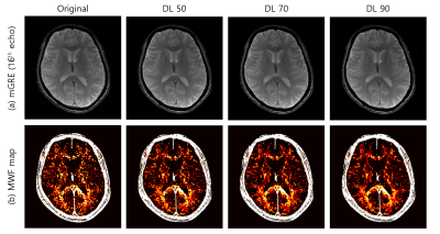 |
Feasibility of myelin water fraction mapping with denoised 2D multi-echo GRE images based on deep learned reconstruction
Ho-Joon Lee1, Yeonah Kang1, Marc Lebel2, Jae Eun Song3, and Sung-Min Gho4
Video Permission Withheld
1Department of Radiology, Haeundae Paik Hospital, Busan, Republic of Korea, 2MR Collaboration and Development, GE Healthcare, Calagary, AB, Canada, 3Department of Electrical and Electronic Engineering, Yonsei University, Seoul, Republic of Korea, 4MR Collaboration and Development, GE Healthcare, Seoul, Republic of Korea
With advances in deep learning, feasibility has been investigated for myelin water fraction (MWF) reconstruction showing promising results, enabling fast reconstruction, however whether images denoised with deep learned reconstruction(DL) will improve MWF map quality has not been investigated. After denoising of multi-echo GRE magnitude images with a DL algorithm, data was fitted to a three-component magnitude model. Use of DL, shows better results as compared to conventionally generated maps (i.e. decreased NRMSE and mean fitting errors (WM), increased PSNR and SSIM). Gibb's ringing artifact was removed remarkably.
|
|
1427.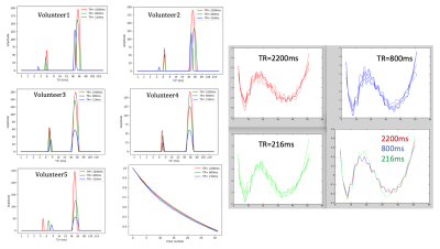 |
Steady state effects on the myelin water pool: evidence for a distinct T1 compartment?
Ana-Maria Oros-Peusquens1, Dennis Thomas1, Alexander L. MacKay2, and N. Jon Shah1
1Research Centre Juelich, Juelich, Germany, 2University of British Columbia, Vancouver, BC, Canada
The influence of steady state on myelin water fraction derived from mGRE data was investigated, with the aim of estimating myelin water T1. Myelin and tissue water were identified by their T2* properties following an NNLS analysis. PCA denoising reduced the variability (SD) of MWF by nearly a factor of 2. The residuals of the NNLS fit were found to be small (below 0.5%) but highly significant, and showed no dependence on TR. The derived T1 of myelin water was highly variable and shorter than that of tissue water. These findings are consistent with a reduced exchange between pools.
|
|
1428.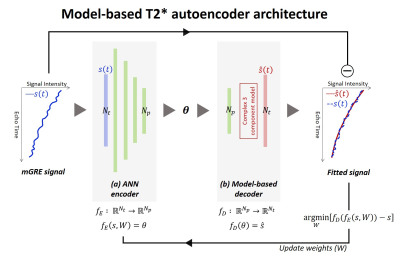 |
Model-based T2* autoencoder for Myelin Water Fraction Mapping
Jae Eun Song1 and Dong-Hyun Kim1
1Electrical and Electronic Engineering, Yonsei University, Seoul, Republic of Korea
In this study, we propose a model-based unsupervised learning algorithm to estimate three-component exponential model parameters. The proposed method, model-based T2* autoencoder, is composed of ANN encoder and three-component model decoder. Incorporation of ANN encoder and three-component model decoder provides noise robust estimation of MWF and physically meaningful information at deep latent space.
|
|
1429.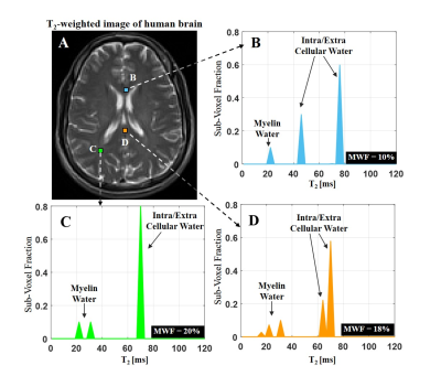 |
A novel multicomponent T2 analysis for identification of sub-voxel compartments and quantification of myelin water fraction
Noam Omer1, Tamar Blumenfeld-Katzir1, Natalie Bnaiahu1, Meirav Galun2, and Noam Ben-Eliezer1,3,4
1Department of Biomedical Engineering, Tel Aviv University, Tel Aviv, Israel, 2Department of Computer Science and Applied Mathematics, Weizmann Institute of Science, Rehovot, Israel, 3Center for Advanced Imaging Innovation and Research (CAI2R), New-York University Langone Medical Center, New York, New York, NY, United States, 4Sagol School of Neuroscience, Tel-Aviv University, Tel Aviv, Israel
Multicomponent T2 analysis (mcT2) can be highly valuable for probing tissue microstructure. However, it remains challenging due to its ill-conditioned nature, and due to inherent contamination of multi spin-echo signals by stimulated echoes. We present a novel mcT2 algorithm that tackles the high-dimensionality of this problem, using correlations between local and global features of the anatomy in question. The accuracy of this tool is demonstrated on phantoms and in vivo. Our results suggest that the method can accurately identify microscopic compartments, operate at realistic scan times, and be used estimate to estimate myelin content in vivo.
|
|
1430.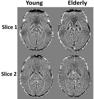 |
Association of age, sex, and myelin content with regional iron deposition in the human brain
Nikkita Khattar1, Richard G. Spencer1, and Mustapha Bouhrara1
1NIA, NIH, Baltimore, MD, United States
Iron is known to play a central role in neuronal viability and pathophysiology. Although susceptibility weighted imaging (SWI) is highly sensitive to iron, few studies have investigated the effect of age and sex on iron deposition using SWI. Further, the potential association between iron deposition and myelination is unclear. It has been suggested that myelin breakdown releases iron, which further acts to potentiate myelin loss through oxidative mechanisms. Here, we investigate the association between local iron deposition and age, sex, and myelination, measured using an advanced myelin content imaging method, on a cohort of cognitively unimpaired participants.
|
|
1431.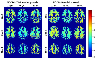 |
Age-dependence of g-ratio values for subcortical white matter structures in the human brain
Richard Wonjoong Kim1, Nikkita Khattar1, Wenshu Qian1, Joseph Alisch1, Richard G. Spencer1, and Mustapha Bouhrara1
1NIA, NIH, Baltimore, MD, United States
Emerging evidence indicates that myelination and axonal abnormalities could lead to alterations of brain connectivity, contributing to a myriad of neurological disorders. Measurement of myelin to axonal volume, as defined by the g-ratio, has been shown to represent a sensitive and specific metric of neurodevelopment and neurodegeneration. However, only a single study to date has sought to evaluate age-related differences in g-ratio in the adult human brain. Here, we introduce and compare two novel approaches to g-ratio mapping. Both methods revealed a quadratic relationship between g-ratio and age in a large cohort of cognitively unimpaired participants.
|
|
1432.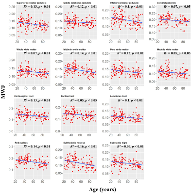 |
Age-related differences in myelin content of the human brainstem
Mustapha Bouhrara1, Luis E. Cortina1, Abinand C. Rejimon1, Nikkita Khattar1, and Richard G. Spencer1
1NIA, NIH, Baltimore, MD, United States
Very few MR investigations have been conducted to assess age-dependent myelination differences in the brainstem, in spite of the brainstem's central role in regulating vital functions. This is likely due to the small structural scale of the brainstem, necessitating accurate high spatial resolution imaging for quantitative studies. Here, we used our recently developed approach to myelin water fraction (MWF) mapping and found a decrease in myelination with age in different brainstem regions, with several regions exhibiting a quadratic association. We believe that this study is the first investigation of MWF changes with normative aging in the brainstem.
|
|
1433.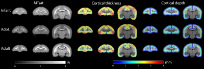 |
Characterisation of MTsat for assessment of intracortical myelination in the developing marmoset brain
Stephen J Sawiak1, M Arsalan Khan2, Gemma Cockcroft3, Edward T Bullmore4, and Angela C Roberts3
1Translational Neuroimaging Laboratory, PDN, University of Cambridge, Cambridge, United Kingdom, 2University of Cambridge, Cambridge, United Kingdom, 3Physiology, Development and Neuroscience, University of Cambridge, Cambridge, United Kingdom, 4Department of Psychiatry, University of Cambridge, Cambridge, United Kingdom
Changes in myelination are prominent throughout brain development. Here, we assess MTsat as a measure of intracortical myelination in the developing marmoset brain with 38 subjects from infancy to adulthood. We find that the measure is consistent between brains and that across the cortex as a whole, adults differ in apparent myelination compared with infants and early adolescents particularly within central and deeper cortical layers. Regional analysis with this metric will facilitate differentiation between morphological and microstructural developmental changes that cannot be elucidated with conventional structural imaging.
|
|
1434.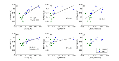 |
Association of Myelination in Internal Capsule with Iron Deposition in the Basal Ganglia in Macaque monkeys
Wenwen Yu1, Run Pu2, Zhe Wu2, Hongjian He2, Yuequan Shi3, Zuofu Zhou3, Zheng Wang4, and Jianhui Zhong2,5
1Institute of Neuroscience, CAS Center for Excellence in Brain Science and Intelligence Technology, State Key Laboratory of Neuroscience, CAS Key Laboratory of Primate Neurobiology, Chinese Academy of Sciences, Shanghai, China, 2Center for Brain Imaging Science and Technology, Key Laboratory for Biomedical Engineering of Ministry of Education, College of Biomedical Engineering and Instrumental Science, Zhejiang University, Hangzhou, China, 3Department of Radiology, Fujian Provincial Maternity and Children’s Hospital of Fujian Medical University, Fuzhou, China, 4Institute of Neuroscience, Chinese Academy of Sciences, Shanghai, China, 5Department of Imaging Sciences, University of Rochester, Rochester, NY, United States
A tight association between iron and myelin content has been demonstrated in the local brain regions. However, it remains unknown whether such relationship exists between adjacent brain regions. Using 8 young and 8 older macaque monkeys, we integrated MRI-based QSM and MWF imaging to examine the relation between iron deposition in components of BG and the myelin content of BG-connecting IC. Our results reveal that the iron of BG is positively correlated with the myelin of IC at different aging stages, and demonstrate that iron in BG also affects the myelin content of the anatomically distinguished yet connected WM structures.
|
|
1435.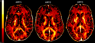 |
Myelin water imaging of Amyoplasia: Comparison to a control atlas
Bretta Russell-Schulz1, Irene M Vavasour1,2,3, Hanwen Liu2,4, Cornelia Laule2,3,4,5, Erin L MacMillan1,6,7, Alex L MacKay1,3,4, Carolyn Sparrey2,8, and Bonita Sawatzky2,9
1UBC MRI Research Centre, University of British Columbia, Vancouver, BC, Canada, 2International Collaboration on Repair and Discoveries (ICORD), University of British Columbia, Vancouver, BC, Canada, 3Radiology, University of British Columbia, Vancouver, BC, Canada, 4Physics & Astronomy, University of British Columbia, Vancouver, BC, Canada, 5Pathology & Laboratory Medicine, University of British Columbia, Vancouver, BC, Canada, 6SFU ImageTech Lab, Simon Fraser University, Surrey, BC, Canada, 7Philips Canada, Marham, ON, Canada, 8School of Mechatronic Systems Engineering, Simon Fraser University, Surrey, BC, Canada, 9Orthopaedics, University of British Columbia, Vancouver, BC, Canada Adults living with Amyoplasia report lower physical activity but no educational or cognitive impairment. Myelin water fraction (MWF) is a quantitative MRI metric useful for examining white matter myelin content. We expect brain MWF to be similar or higher between Amyoplasia and controls in most of the brain but lower in areas associated with physical ability. In 3 adults with Amyoplasia MWF maps were created using a 32-echo T2 GRASE compared to an open-source healthy control atlas. Small reductions in corticospinal tract myelin were observed, while the remainder of the brain examined was similar to the control atlas. |
|
1436.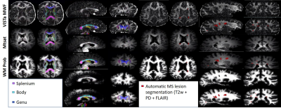 |
Assessing MRI-Based Myelin Measures in Multiple Sclerosis and Healthy Brain White Matter
Ahmed M. Elkady1, Zhe Wu1,2, Dumitru Fetco1, Ilana R. Leppert1, Douglas L. Arnold1, Sridar Narayanan1, and David A. Rudko1,3
1McConnell Brain Imaging Centre, Montreal Neurological Institute and Hospital, Department of Neurology and Neurosurgery, McGill University, Montreal, QC, Canada, 2Techna Institute, University Health Network, Toronto, ON, Canada, 3Department of Biomedical Engineering, McGill University, Montreal, QC, Canada
We comparatively evaluated non-corrected Magnetization Saturation (MTSat), B1-corrected MTsat, wave-CAIPI direct Visualization of Short myelin Transverse component (wave-CAIPI ViSTa), and multi-atlas probabilistic tissue classification in multiple sclerosis (MS) lesions and white matter tracts of healthy and MS subjects. The relationship between ViSTa myelin water fraction (MWF) and MTSat myelin volume fraction (MVF) was altered in MS normal appearing white matter (NAWM) compared NAWM of controls. This may reflect specific alterations in the macromolecular and myelin water pool fractions observed in demyelinating tissue. ViSTa MWF was more sensitive in detecting pathological differences between the MS and control groups.
|
|
1437.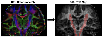 |
Tract-Specific Myelin Content Mapping of the Corticospinal Tract Predicts Impairment in Patients with Multiple Sclerosis
Richard Dortch1 and Francesca Bagnato2,3
1Barrow Neurological Institute, Phoenix, AZ, United States, 2Department of Neurology, Vanderbilt University Medical Center, Nashville, TN, United States, 3Neuroimaging Unit/Neuroimmunology Division, Vanderbilt University Medical Center, Nashville, TN, United States
Quantitative magnetization transfer using the selective inversion recovery method yields indices [macromolecular-to-free water pool-size-ratio (PSR) and longitudinal spin-lattice relaxation rate (R1f)] that report on myelin content in the brain. In this study, SIR indices were estimated in the corticospinal tract (CST) of patients with multiple sclerosis for comparison to clinical disability measures [Expanded Disability Status Scale (EDSS) and Timed 25-Foot Walk T25FW)]. Significant correlations were observed between SIR indices (PSR/R1f) and disability scores (EDSS/T25FW). The strongest correlation was between PSR and EDSS, suggesting that measurements of PSR in the CST may report on myelin damage and/or repair in multiple sclerosis.
|
|
1438.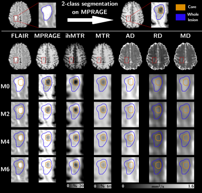 |
Longitudinal follow-up of MS active lesions with 3D inhomogeneous Magnetization Transfer (ihMT)
Lucas Soustelle1,2, Soraya Gherib1,2, Samira Mchinda1,2, Sylviane Confort-Gouny1,2, Arnaud Le Troter1,2, Maxime Guye1,2, Jean-Philippe Ranjeva1,2, Patrick Viout1,2, Lauriane Pini1,2, Claire Costes1,2, Adil Maarouf1,2,3, Bertrand Audoin1,2,3, Audrey Rico1,2,3,
Clémence Boutière1,2,3, Fanelly Pariollaud1,2, Françoise Reuter1,2, Victor Nunes Dourado de Carvalho1,2, Véronique Gimenez1,2, Andreea Hertanu1,2, Gopal Varma4, David C Alsop4, Jean Pelletier1,2,3, Guillaume Duhamel1,2, and Olivier M Girard1,2
1Aix-Marseille Univ, CNRS, CRMBM, Marseille, France, 2APHM, Hôpital Universitaire Timone, CEMEREM, Marseille, France, 3APHM, Hôpital Universitaire Timone, Service de neurologie, Marseille, France, 4Division of MR Research, Radiology, Beth Israel Deaconess Medical Center, Harvard Medical School, Boston, MA, United States
Assessment of lesion evolution in Multiple Sclerosis (MS) is critical to monitor the disease progression but remains challenging in clinical practice. Inhomogeneous magnetization transfer (ihMT) is a promising technique that has demonstrated sensitivity to demyelination in preclinical and clinical studies. In this work, a 3D ihMT technique was integrated as part of a multiparametric MRI protocol and applied to active MS lesion follow up over a period of six months. Variations of ihMT ratio were compared to those of conventional magnetization transfer imaging and diffusion tensor imaging often used in clinical research to assess demyelination.
|
|
1439.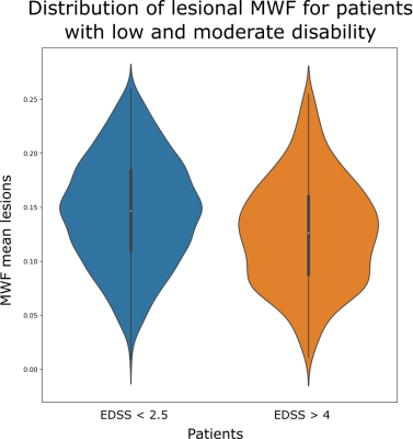 |
Multi-component relaxation in relapsing remitting multiple sclerosis: Variability of myelin water fraction estimates of individual lesions
Caroline Köhler1, Hannes Wahl1, Tjalf Ziemssen2, Paul Kuntke1, Jennifer Linn1, and Hagen H. Kitzler1
1Institut und Poliklinik für Diagnostische und Interventionelle Neuroradiologie, University Hospital Carl Gustav Carus, Dresden, Germany, 2Neurology, University Hospital Carl Gustav Carus, Dresden, Germany
Multiple Sclerosis lesions are pathologically heterogeneous. The Multi-component relaxation method (mcDESPOT) which allows the determination of relative myelination by estimating the myelin water fraction (MWF) could be valuable to capture the high variability of disease development. In a pilot study n=112 RRMS patients got MRI examination and MWF values were determined of white matter (WM) and individual lesions. The MWF of WM was 0.230 for healthy controls and 0.224 for RRMS patients. 2787 WM FLAIR lesions were examined. Patients with a higher degree of disability of EDSS ≥ 4 significantly have lower MWF values than patients with EDSS < 2.5.
|