
Digital Poster Session
Neuro: Aging Brains and Dementia
Neuro
1440 -1455 Aging Brains and Dementia - Aging Brains
1456 -1467 Aging Brains and Dementia - Alzheimer's-Type Dementia
1468 -1481 Aging Brains and Dementia - Dementia: Clinical Applications
1482 -1494 Aging Brains and Dementia - Dementia: Emerging Methods
1440.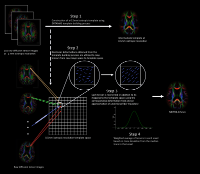 |
Development and evaluation of a high spatial resolution diffusion tensor template of the older adult human brain
Mohammad Rakeen Niaz1, Yingjuan Wu1, Abdur Raquib Ridwan1, Xiaoxiao Qi1, Shengwei Zhang1, David A. Bennett2, and Konstantinos Arfanakis1,2
1Biomedical Engineering, Illinois Institute of Technology, Chicago, IL, United States, 2Rush Alzheimer's Disease Center, Rush University Medical Center, Chicago, IL, United States
A high-resolution older adult brain template containing full diffusion tensor (DT) information has not been constructed. Available DT templates of low spatial resolution lack fine details, and most are not representative of the older adult brain. Both factors limit spatial normalization accuracy in studies of aging. This work a) introduced a novel approach for constructing a DT template with high spatial resolution based on principles of super-resolution, and using this technique, b) developed and quantitatively evaluated a DT template of the older adult brain with high spatial resolution using high-quality data from 202 non-demented older adults.
|
|
1441.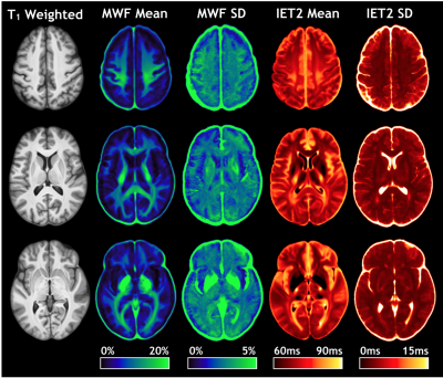 |
Uncharted Waters: Insights From A 100-Subject Myelin Water Imaging Atlas
Taylor Swift-LaPointe1, Irene M. Vavasour2,3, Lisa Eunyoung Lee4, Shawna Abel4, Bretta Russell-Schulz2, Carina Graf1,3, Anika Wurl1, Hanwen Liu1,3, Cornelia Laule1,2,3,5, David K.B. Li2,4, Anthony Traboulsee4, Roger Tam2,6, Lara A. Boyd7,
Alex L. MacKay1,2, Shannon H. Kolind1,2,3,4, and Adam V. Dvorak1,3
1Physics and Astronomy, University of British Columbia, Vancouver, BC, Canada, 2Radiology, University of British Columbia, Vancouver, BC, Canada, 3International Collaboration on Repair Discoveries (ICORD), Vancouver, BC, Canada, 4Medicine (Neurology), University of British Columbia, Vancouver, BC, Canada, 5Pathology and Laboratory Medicine, University of British Columbia, Vancouver, BC, Canada, 6Biomedical Engineering, University of British Columbia, Vancouver, BC, Canada, 7Department of Physical Therapy, University of British Columbia, Vancouver, BC, Canada
Data from 100 healthy volunteers (58F/42M, mean age 41 years, range 20-78 years) scanned at 3.0T were used to create a structural template. Voxel-wise mean myelin water fraction (MWF) and intra/extracellular T2 (IET2) atlases were created, and 19 regions of interest were obtained using the joint label fusion framework. Pearson correlations with both age and sex were calculated for MWF and IET2. Mean MWF and IET2 demonstrated clear ranking between brain structures for different age groups, indicating that relative metric values are generally consistent between ROIs. There were no significant correlations between MWF or IET2 and sex.
|
|
1442.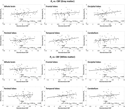 |
Evidence of association between regional cerebral blood flow deficits and demyelination in normal aging
Mustapha Bouhrara1, Joseph E. Alisch1, Nikkita Khattar1, Abinand C. Rejimon1, Luis E. Cortina1, and Richard G. Spencer1
1NIA, NIH, Baltimore, MD, United States
Little is known about the relationship between white matter (WM) perfusion and microstructure across cognitively normal or impaired subjects. WM maintenance through oligodendrocyte metabolism is an energy-intensive process, so that myelin homeostasis is particularly sensitive to hypoxia, ischemia, or hypoperfusion. In addition to substrate delivery, adequate cerebral blood flow (CBF) is crucial for removal of metabolic byproducts. We investigated associations between CBF deficits and myelin loss in multiple brain regions in a cohort of cognitively unimpaired participants across a wide age range. We show significant correlations between CBF deficits and myelin loss in critical brain structures.
|
|
1443.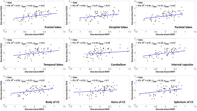 |
Effect of myelin integrity on rapid speed gait in normal aging
Mustapha Bouhrara1, Nikkita Khattar1, Luis E. Cortina1, Abinand C. Rejimon1, Elango Palchamy1, and Richard G. Spencer1
1NIA, NIH, Baltimore, MD, United States
Microstructural white matter (WM) degeneration has been shown to have a direct impact on mobility performance. However, the association between speed gait and myelin changes is less clear. This study examines the effect of myelin degeneration measured using myelin water fraction (MWF), a specific proxy of myelin content, and relaxation rates, sensitive but non-specific measures of myelin, on rapid gait speed (RGS), which represents an integrated metric of physiologic function. Our study is conducted on a large age-range cohort of cognitively unimpaired participants. We show statistically significant correlations between MWF loss and RGS decline in several WM structures.
|
|
1444.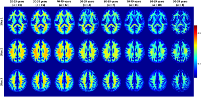 |
Brain aging and sexual dimorphism in myelination investigated using magnetic resonance myelin water fraction imaging
Mustapha Bouhrara1, Abinand C. Rejimon1, Luis E. Cortina1, Nikkita Khattar1, and Richard G. Spencer1
1NIA, NIH, Baltimore, MD, United States
Changes in brain myelination with age and sex have been the subject of intense MRI study. However, most studies have used nonspecific methods to probe myelin, including DTI and relaxation times. We investigated age- and sex-related differences in brain myelin water fraction (MWF), a surrogate of myelin content, in a large cohort of unimpaired participants, spanning a wide age range. We observed a quadratic relationship between MWF and age in all brain regions investigated, suggesting that myelination continues until middle age followed by a decrease through older age. Sexual dimorphism in this process was observed in several brain regions.
|
|
1445.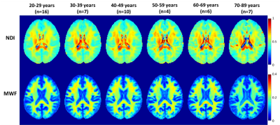 |
Neurite density and myelination differences during normal aging assessed using multicomponent diffusion and relaxometry imaging
Wenshu Qian1, Nikkita Khattar1, Abinand C. Rejimon1, Mustapha Bouhrara 1, and Richard G. Spencer1
1National Institute on Aging, Baltimore, MD, United States
While sensitive to microstructural changes, conventional quantitative MR techniques, such as diffusion tensor imaging, are not specific to underlying physiological mechanisms. Advanced multi-shell diffusion and multicomponent relaxometry analyses have been shown to provide more specific insights regarding microstructural differences with age and disease. In this study, we combined our multicomponent relaxometry method for myelin mapping and the neurite orientation dispersion and density imaging (NODDI) method to investigate neurite myelination and density in a cohort of cognitively unimpaired participants. Quadratic relationships were observed between neurite density and myelination, and age, in critical brain regions.
|
|
1446.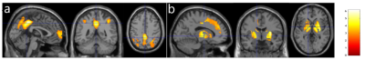 |
Age-associated changes in cerebral blood flow-related measures using arterial spin labeling
Zheng Li1, Zongpai Zhang1, Wenna Duan1, Lissa Riley2, Adam K. Anderson2, Eve DeRosa2, and Weiying Dai1
1Computer Science, State University of New York at Binghamton, Binghamton, NY, United States, 2Human Development, Cornell University, Ithaca, NY, United States
Disrupted CBF-related measures in clinical diseases has highlighted the importance of reliable baseline data from normal controls. We characterized CBF-related measures in normal aging by acquiring PCASL images in 11 young and 12 elderly subjects. We found that the elderly exhibited longer ATT in the frontal, parietal, temporal, and subcortical regions, reduced CBF in the prefrontal and parietal regions and increased CBF in the subcortical regions, and increased LFF in the superior frontal and paracentral region, compared to the young. This work sets the stage for a larger scale use of PCASL to investigate CBF-related alterations in neurological diseases.
|
|
1447.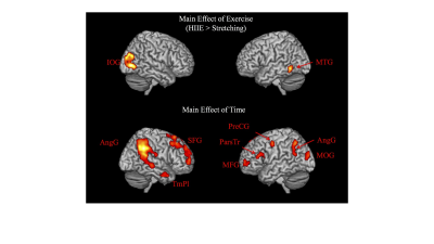 |
Neural correlates of associative new word learning enhancement following high-intensity interval exercise in healthy older adults.
Marie-Pier McSween1,2, Megan L. Isaacs1,2, David A. Copland1,2, Jeff S. Coombes3, Amy D. Rodriguez4, Kirk I. Erickson5, and Katie L. McMahon6
1The University of Queensland School of Health and Rehabilitation Sciences, Brisbane, Australia, 2University of Queensland Centre for Clinical Research, Brisbane, Australia, 3The University of Queensland School of Human Movement and Nutrition Sciences, Brisbane, Australia, 4Centre for Visual and Neurocognitive Rehabilitation, Atlanta, GA, United States, 5University of Pittsburgh, Pittsburgh, PA, United States, 6Queensland University of Technology School of Clinical Sciences, Brisbane, Australia Poster Permission Withheld
High-intensity interval exercise (HIIE) can benefit word learning in young adults, however this is yet to be investigated in older adults and the neural mechanisms responsible have not been directly examined. Twenty-six older adults participated in this study and engaged in stretching or HIIE prior to performing an in-scanner (fMRI) associative new word learning task. Results showed increased activation after HIIE, in the left middle temporal gyrus, and right inferior occipital gyrus in addition to better word recognition accuracy, suggesting benefits of HIIE on word learning that may be attributed to increased activation in key language and visual processing areas.
|
|
1448.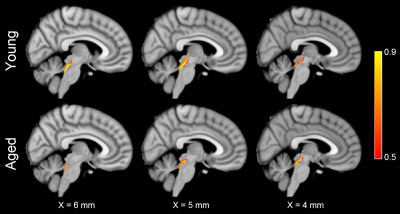 |
Assessing the impact of age-related locus coeruleus degeneration on cognitive decline
Jason Langley1, Sana Hussain2, Justino J Flores3, Daniel E Huddleston4, Ilana Bennett3, and Xiaoping Hu1,2
1Center for Advanced Neuroimaging, University of California Riverside, Riverside, CA, United States, 2Bioengineering, University of California Riverside, Riverside, CA, United States, 3Psychology, University of California Riverside, Riverside, CA, United States, 4Neurology, Emory University, Atlanta, GA, United States
Characterization of age-related alterations in microstructure of locus coeruleus and its projections will aid in the development of new biomarkers and may provide insight in the development of novel interventions to arrest progression of Alzheimer's disease or Parkinson's disease. Imaging locus coeruleus with diffusion-weighted images is difficult due to its small stature (locus coeruleus is 1.5 mm in diameter and 15 mm long) and location in the brain stem. In this abstract, we utilize a high resolution diffusion-weighted protocol to examine age-related microstructural changes in locus coeruleus and examine its effect on age-related cognitive decline.
|
|
1449.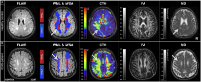 |
Similar mechanisms and locations of white matter lesions in elderly healthy controls and asymptomatic carotid artery stenosis patients
Stephan Kaczmarz1,2, Jens Göttler1,2,3, Andreas Hock4, Claus Zimmer1, Fahmeed Hyder2, and Christine Preibisch1,5
1School of Medicine, Department of Neuroradiology, Technical University of Munich, Munich, Germany, 2MRRC, Yale University, New Haven, CT, United States, 3School of Medicine, Department of Radiology, Technical University of Munich, Munich, Germany, 4Philips Healthcare, Hamburg, Germany, 5School of Medicine, Clinic of Neurology, Technical University of Munich, Munich, Germany
White matter lesions (WML) are detected as hyperintensities on clinically frequently used T2-weighted FLAIR-images and can manifest as cognitive decline. Though most elderly subjects show WML, the pathogenesis is still unclear. Potential causes are small vessel damages and myelin-sheath deformations. We present data from 30 healthy elderly and 29 carotid stenosis patients measuring WML, individual watershed areas (iWSA), capillary transit-time heterogeneity (CTH) and DTI-based structural parameters. We hypothesize increased lesion load inside iWSAs and capillary dysfunction with structural damages in WML. Our results confirm those hypotheses and furthermore, indicate similar WML formation mechanisms in healthy aging and carotid artery stenosis.
|
|
1450.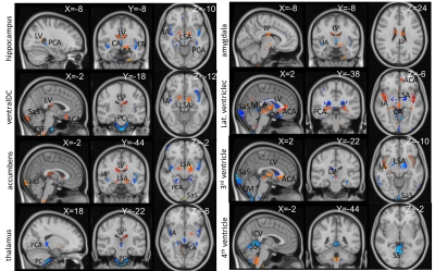 |
Relationship of regional volume changes and cardiac pulsatility in the brain
Tae Kim1, Annie Cohen1, and James T Becker1
1University of Pittsburgh, Pittsburgh, PA, United States
Brain atrophy is highly associated with cardiac pulsatility in the brain. The reduction of brain regional volumes was correlated with increased pulsatility of its supply arteries and a decrease of sinus and CSF. The regional volume reduction was affected by the pulsatility of different vessels.
|
|
1451.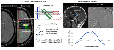 |
INVESTIGATING THE LINK BETWEEN CEREBROVASCULAR AND NEURO HEALTH IN COGNITIVE DECLINE
Kathryn Broadhouse1, Natalie Winks1, and Jim Lagopoulos1
1University of the Sunshine Coast, Sunshine Coast, Australia Current markers of early neurodegeneration include β-amyloid plaque accumulation and Tau-mediated neuronal injury; both of which are thought to emerge prior to significant cognitive deficits and measurable brain atrophy. Unfortunately, the role of these biomarkers within the cascade of pathophysiological processes of AD remains poorly understood. Impairment in the glymphatic clearance system has garnered attention and is thought to represent a pathway to decline. Here we demonstrate that quantification of glymphatic clearance measures are feasible in healthy controls. Combining these measures with cognitive performance scores and diagnosis will provide insight into the role of the glymphatics system with decline. |
|
1452.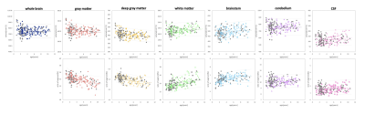 |
Age-related changes in brain morphology based on a large-scale MRI database of non-human primates
Mayu Iida1,2, Junichi Hata2,3,4, Yawara Haga1,4, Akiko Uematsu4,5, Fumiko Seki2,4, Daisuke Yoshimaru2,3,4, Kei Hagiya4, Hirotaka James Okano3,4, Hideyuki Okano4, and Takako Shirakawa1
1Department of Radiological Sciences, Human Health Sciences, Tokyo Metropolitan University Graduate School, Tokyo, Japan, 2Live Imaging Center, Central Institute for Experimental Animals, Kanagawa, Japan, 3Division of Regenerative Medicine, Jikei University School of Medicine, Tokyo, Japan, 4Laboratory for Marmoset Neural Architecture, Center for Brain Science, RIKEN, Saitama, Japan, 5Center of Evolutionary Cognitive Sciences, Tokyo University Graduate School, Tokyo, Japan Poster Permission Withheld
MRI data of 204 large common marmoset colonies were obtained and analyzed regarding brain volume. Marmosets have a brain structure and nerve paths are similar to those in humans compared to other experimental animals. The volume from puberty to old age in six regions (gray matter, deep gray matter, white matter, brainstem, cerebellum, and CSF) could be evaluated. From the viewpoint of volume and variability when performing brain image analysis, it was suggested that marmosets are experimental animals that can evaluate the effects of individual differences at the same level as humans or with slightly less variation than humans.
|
|
1453.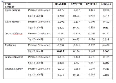 |
Blood-brain barrier integrity and cognitive function in a healthy aging population
SAMUEL BARNES1, SHILPY CHOWDHURY2, JENNIFER GARCIA-CANO3, NICOLE M GATTO4, and GRACE J LEE3
1RADIOLOGY, LOMA LINDA UNIVERSITY HEALTH, Loma Linda, CA, United States, 2LOMA LINDA UNIVERSITY, LOMA LINDA, CA, United States, 3PSYCHOLOGY, LOMA LINDA UNIVERSITY SCHOOL OF BEHAVIORAL HEALTH, LOMA LINDA, CA, United States, 4School of Community and Global Health, Claremont Graduate University, CLAREMONT, CA, United States
Dynamic-contrast enhanced (DCE) MRI can detect changes in the blood brain barrier integrity which can be associated with cognitive changes in aging populations. 40 participants from a large cohort underwent contrast MRI and battery of neuropsychological tests. DCE Ktrans values showed a negative correlation with verbal learning and memory testing (RAVLTIR and RAVLTSD). Blood brain barrier disruption, defined by higher Ktrans values, is associated with worse performance on verbal learning, memory and category fluency tests.
|
|
1454. |
Association of PC-MRI measured Cerebroarterial Pulsatility with Cognitive Impairment and White Matter Hyperintensity in Aged Subjects
Soroush Heidari Pahlavian1,2, Xinhui Wang 2, Samantha Ma1,2, John Ringman2, Helena Chui2, Danny J.J. Wang1,2, and Lirong Yan1,2
1USC Stevens Neuroimaging and Informatics Institute, Keck School of Medicine, University of Southern California, Los Angeles, CA, United States, 2Department of Neurology, University of Southern California, Los Angeles, CA, United States
Elevated cerebroarterial pulsations due to arterial stiffness can impart abnormal forces to downstream capillary/tissue leading to microvascular damage, which are thought to be a contributing factor in the pathogenesis of neurodegenerative disorders such as Alzheimer’s disease and small vessel disease. In this study, we assessed the utility of two-dimensional (2D) phase-contrast MRI (PC-MRI) in quantifying cerebroarterial pulsations and evaluated the associations of pulsatile and non-pulsatile hemodynamic measures with cognitive performance and white matter hyperintensity (WMH). Our results indicated significant associations of PC-MRI derived pulsatility metrics with cognitive performance and WMH.
|
|
1455.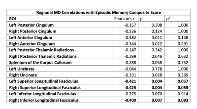 |
Mean diffusivity in white matter correlates negatively with episodic memory performance in aging, non-demented adults with Down Syndrome
Austin Bazydlo1, Sigan Hartley1, Minjie Wu2, Patrick Lao3, Douglas C Dean, III1, Matthew Zammit1, Sterling Johnson1, Dana Tudorascu2, Annie Cohen2, Karly Cody1, Charles Laymon2, William Klunk2, Shahid Zaman4, Benjamin Handen2,
Andrew Alexander1, and Bradley Christian1
1University of Wisconsin-Madison, Madison, WI, United States, 2University of Pittsburgh Medical Center, Pittsburgh, PA, United States, 3Columbia University, New York, NY, United States, 4University of Cambridge, Cambridge, United Kingdom
People with Down Syndrome (DS) are at an increased risk of developing Alzheimer’s Disease (AD), due to an overproduction of β-amyloid resulting from triplication of the amyloid precursor protein (APP) gene located on chromosome 21. The effect of white matter degradation on early AD-related cognitive decline in the DS population is not known. In this work, we present results from a study of non-demented adults with DS showing correlations between increased mean diffusivity (MD), derived using diffusion tensor imaging (DTI), within several white matter regions of interest and poorer performance on measures of episodic memory.
|
 View the Poster
 Watch the Video
 |
1456. |
Longitudinal trajectory of fibre-specific degeneration in Alzheimer’s disease and mild cognitive impairment
Remika Mito1,2, Robert Smith1, Thijs Dhollander1, Christopher Rowe3,4, Victor Villemagne3,4, Amy Brodtmann1,3, and Alan Connelly1,2
1Florey Institute of Neuroscience and Mental Health, Melbourne, Australia, 2Florey Department of Neuroscience and Mental Health, University of Melbourne, Melbourne, Australia, 3Department of Medicine, Austin Health, University of Melbourne, Melbourne, Australia, 4Department of Molecular Imaging & Therapy, Centre for PET, Austin Health, Heidelberg, Australia
Alzheimer’s disease (AD) is characterised by degeneration of specific white matter tracts, however the longitudinal trajectory of tract-specific decline has not yet been well described. In this work, we utilise a longitudinal fixel-based analysis framework to investigate specific fibre tracts that exhibit accelerated decline in AD and mild cognitive impairment (MCI). Whole-brain analysis revealed that select fixels exhibit accelerated rates of decline in AD compared to healthy elderly controls, while MCI patients did not exhibit accelerated decline in any fibre structures. Tract-of-interest analysis revealed group differences in tract trajectories over time.
|
|
1457. |
Pathway-specific polygenic scores for Alzheimer’s disease are associated with multi-modal structural brain imaging markers in young adults
Judith Harrison1, Xavier Caseras2, Sonya Foley1, Emily Baker2, David Linden3, Peter Holmans2, Derek Jones1, and Valentina Escott-Price2
1CUBRIC, CUBRIC, Cardiff University, Cardiff, United Kingdom, 2MRC Centre for Neuropsychiatric Genetics and Genomics, Cardiff University, Cardiff, United Kingdom, 3School for Mental Health and Neuroscience, Maastricht University, Maastricht, Netherlands
This study explores the relationship between structural brain measurements and novel pathway-specific polygenic scores for Alzheimer’s Disease (AD). We observed associations between the pathway-polygenic profiles, cortical thinning, and changes in white matter signal particularly in the fornix, which have been shown to be markers of Alzheimer’s degeneration, in our sample of healthy young adults. Surprisingly, despite the contribution of APOE to AD risk, the signal in the white matter was stronger when APOE was excluded from the polygenic scores.
|
1458. |
White Matter Integrity and Cortical Structure – Dependence on Apolipoprotein E Homozygous ε4 Allele Status
Harald Kugel1, Jonathan Repple2, Janik Goltermann2, Ronny Redlich2, Katharina Dohm2, Claas Kaehler2,3, Dominik Grotegerd2, Katharina Foerster2, Susanne Meinert2, Verena Enneking2, Tim Hahn2, Andreas Jansen4, Axel Krug4, Igor
Nenadic4, Simon Schmitt4, Frederike Stein4, Tina Meller4, Dilara Yueksel4, Elena Fischer4, Marcella Rietschel5, Stephanie Witt5, Andreas J Forstner6,7, Markus Noethen6, Andreas Johnen8, Tilo Kircher4, Bernhard T Baune2, Udo
Dannlowski2, and Nils Opel2
1Institute of Clinical Radiology, University of Muenster, Muenster, Germany, 2Department of Psychiatry, University of Muenster, Muenster, Germany, 3Department of Mathematics and Computer Science, University of Muenster, Muenster, Germany, 4Department of Psychiatry, University of Marburg, Marburg, Germany, 5Department of Genetic Epidemiology in Psychiatry, University of Heidelberg, Mannheim, Germany, 6Institute of Human Genetics, University of Bonn, Bonn, Germany, 7Centre for Human Genetics, University of Marburg, Marburg, Germany, 8Department of Neurology, University of Muenster, Muenster, Germany
Apoliprotein E (APOE) genotype status, a predictor of Alzheimer's disease, has been associated with brain structural alterations. Supplementing previous research that primarily focused on hippocampal morphometry, we found a widespread effect of APOE allele status on cortical surface area and white matter microstructure. There is a significant cortical surface area decrease in 57 out of 68 cortical brain regions in APOE ε4 homozygous carriers. Furthermore, APOE ε4 homozygous carriers showed significant and widespread reductions in fractional anisotropy in corpus callosum, right internal and external capsule, left corona radiata and right fornix.
|
|
1459. |
Cognitive Status in Patients with Alzheimer's Disease Using Quantitative Susceptibility Mapping
Yangyingqiu Liu1, Junyi Dong1, Yanwei Miao1, Ailian Liu1, Qingwei Song1, and Lizhi Xie2
1Department of Radiology, First Affiliated Hospital of Dalian Medical University, Dalian, China, 2GE healthcare, Beijing, China
Blood oxygen level of the cerebral vein is quantitatively assessed by magnetic sensitivity value (MSV)using quantitative susceptibility mapping (QSM), and its’correlation to cognitive scores is also analyzed in patients with Alzheimer's disease (AD).The results show that increased MSV value of cerebral vein in AD patients may affect cognitive scores.
|
|
1460.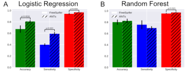 |
Cortical Thickness Estimates to Classify Early Onset Alzheimer’s Disease versus Frontotemporal Dementia
Arun Venkataraman1, Zachary Christensen2, and Jianhui Zhong3
1Physics, University of Rochester, Rochester, NY, United States, 2Translational Biomedical Sciences, University of Rochester, Rochester, NY, United States, 3Imaging Sciences, University of Rochester, Rochester, NY, United States
Frontotemporal Dementia (FTD) and Early-Onset Alzheimer's Dementia (EOAD) can occur in the same age group and have overlapping symptoms, often complicating diagnosis. In this study, we focused on trying to understand cortical thickness as a biomarker of dementia type. More specifically, we wanted to classify EOAD vs FTD using only data from T1w MRI. We found that classification of FTD vs EOAD using FreeSurfer and ANTs cortical thickness data is highly dependent on the type of classifier used. In addition, it seems that ANTs is better suited than FreeSurfer independent of classification model.
|
|
1461.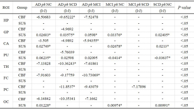 |
Study of the cerebral blood flow and Iron Deposition metabolism in Alzheimer’s disease using ASL-MRI and QSM-MRI
Dongxue Li1, Yuancheng Liu1, Zhenliang Xiong1, Xianchun Zeng1, Lisha Nie2, Pu-Yeh Wu2, and Rongpin Wang1
1Department of Radiology, Guizhou Provincial People's Hospital, Guiyang, China, 2GE Healthcare, MR Research, Beijing, China
The CBF and iron deposition evaluation are commonly used in alzheimer's disease. we analyzed the changes and correlations of CBF and iron deposition in different stages of AD. ASL and QSM identified several abnormal brain regions of interest, which were sensitive to tissue alterations in each stage. This study found QSM was detected in the early stage of AD, which is expected to be an effective tool for early AD diagnosis and timely intervention. Additionally, the iron deposition and CBF in the globus pallidum and putamen flow negatively correlated in the AD group that may be relevant to pathologic changes.
|
|
1462.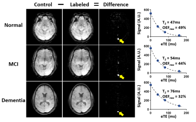 |
Brain oxygen extraction fraction in cognitive impairment: a potential tool to differentiate between Alzheimer’s and vascular dementia
Dengrong Jiang1, Zixuan Lin1, Peiying Liu1, Sandeepa Sur1, Cuimei Xu1, George Pottanat1, Kaisha Hazel1, Sevil Yasar2, Paul Rosenberg3, Marilyn Albert4, and Hanzhang Lu1
1Department of Radiology, Johns Hopkins University School of Medicine, Baltimore, MD, United States, 2Department of Medicine, Johns Hopkins School of Medicine, Baltimore, MD, United States, 3Department of Psychiatry and Behavioral Sciences, Johns Hopkins School of Medicine, Baltimore, MD, United States, 4Department of Neurology, Johns Hopkins School of Medicine, Baltimore, MD, United States
Alzheimer’s disease (AD) and vascular dementia (VD) are the two most common types of cognitive impairment. However, there is still a lack of effective tools for their differential diagnosis. In this work, we demonstrated that OEF is differentially (in opposite direction) affected by AD and vascular pathology. Among patients with cognitive impairment, patients with low OEF are associated with more AD pathology and less vascular pathology; and the opposite can be said for patients with high OEF. These findings suggest that OEF may provide valuable information in differentiating AD and VD.
|
|
1463.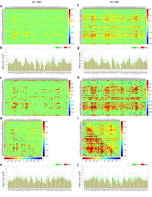 |
Functional connectivity of white matter as a biomarker of cognitive decline in Alzheimer’s disease
Yurui Gao1,2, Anirban Sengupta1, Muwei Li1,3, Zhongliang Zu1,3, Baxter P Rogers1,3, Adam W Anderson1,2,3, Zhaohua Ding1,4, and John C Gore1,2,3
1Vanderbilt University Institute of Imaging Science, Vanderbilt University Medical Center, Nashville, TN, United States, 2Biomedical Engineering, Vanderbilt University, Nashville, TN, United States, 3Radiology and Radiological Sciences, Vanderbilt University Medical Center, Nashville, TN, United States, 4Electrical Engineering and Computer Science, Vanderbilt University, Nashville, TN, United States
Pathological alterations of white matter (WM) have been reported during the development of Alzheimer’s disease (AD). This study extended our previous rsfMRI analyses of WM tract BOLD correlations to evaluate WM functional connectivity (FC) in 383 subjects at different stages of cognitive impairment and found that WM FCs 1) decline regionally in late AD groups relative to a control group, 2) are related to cognitive behavioral scores, and 3) are well-performing features for distinguishing AD patients from controls. These findings suggest the potential of WM FC, which has been overlooked, as a novel neuroimaging biomarker to assess AD progression.
|
|
1464.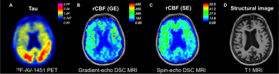 |
Perfusion Imaging of Patients with Alzheimer’s Disease by Using highly-accelerated Spin and Gradient Echo (SAGE) DSC-MRI
Yi-Fen Yen1, Mary Kate Manhard1, Annie G. Bryant2, Rachel E. Bennett2, Kimberly A. Stephens1, David H. Salat1,3, Keith A. Johnson2, Bradley T. Hyman2, Kawin Setsompop1, and Susie Huang1
1Athinoula A. Martinos Center for Biomedical Imaging, Department of Radiology, Massachusetts General Hospital, Charlestown, MA, United States, 2Department of Neurology, Massachusetts General Hospital, Boston, MA, United States, 3VA Boston Healthcare System, Neuroimaging Research for Veterans Center, Boston, MA, United States
We have identified reduced cerebral blood flow, abnormally long mean transit time, and large capillary transit time heterogeneity in seven individuals with mild cognitive impairment or Alzheimer’s disease as compared to healthy subjects by using a highly accelerated dynamic susceptibility enhanced (DSC) MRI technique. This perfusion imaging technique provides whole brain coverage using simultaneous multi-slice acquisition and collects spin and gradient echo in one dynamic scan. The spin-echo and gradient-echo DSC-MRI acquisition enables probing and potentially distinguishing micro- and macro-vascular contributions to perfusion in older adults using a single injection of gadolinium.
|
|
1465.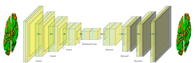 |
Identifying Alzheimer's Disease Brain Atrophy Subtypes by Deep Learning
Xingjuan Li1, Jurgen Fripp1, Samantha Burnham2, Vincent Doré2, and Pierrick Bourgeat1
1Australian eHealth Research Center, CSIRO, Brisbane, Australia, 2Australian eHealth Research Center, CSIRO, Melbourne, Australia
Understanding the heterogeneity in atrophy changes from Alzheimer's disease (AD) can be an important factor to develop effective drugs and improve patients' outcomes [7]. In this study, we propose a deep learning approach to identify AD subtypes based on T1w magnetic resonance images (MRI). Experimental results show that the proposed method can accurately identify four AD subtypes exhibiting a distinctive cortical atrophy pattern.
|
|
1466.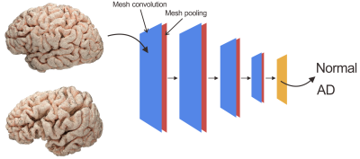 |
A preliminary study of geometric deep learning in brain morphometry: with application to Alzheimer’s disease
Huaiqiang Sun1, Lin Zou1, Haoyang Xing1, Min Wu1, Lei Chen2, Qing Li3, and Qiyong Gong1
1Huaxi MR Research Center (HMRRC), West China Hospital of Sichuan University, Chengdu, China, 2Department of Neurology, West China Hospital of Sichuan University, Chengdu, China, 3MR Collaborations, Siemens Healthcare Ltd., Shanghai, China
A preliminary study that apply geometric deep learning to brain morphometry analysis of Alzheimer’s disease, the results show promising prediction accuracy suggesting geometric deep learning opens new prespectives for brain morphometry analysis of neurological and psychiatric disorders.
|
|
1467.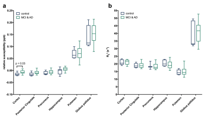 |
Quantitative susceptibility and R2* mapping in mild cognitive impairment and early Alzheimer’s disease
Catherine A Morgan1,2, Zichang Dong3, David Lythgoe4, Lynette Tippett1,2, and Tracy Melzer5,6
1School of Psychology and Centre for Brain Research, University of Auckland, Auckland, New Zealand, 2Brain Research New Zealand, Auckland, New Zealand, 3Department of Physics, University of Auckland, Auckland, New Zealand, 4Institute of Psychiatry, Psychology & Neuroscience, King's College London, London, United Kingdom, 5University of Otago, Christchurch, New Zealand, 6Brain Reserach New Zealand, Christchurch, New Zealand
There is a growing body of work (post-mortem and in vivo) to support the link between iron and the pathophysiology of neurodegenerative disorders. While much of the current literature has focused on later stages of diseases such as Parkinson’s and Alzheimer’s disease (AD), how iron may contribute to early pathology is less well understood. We employed QSM and R2* mapping in a group of mild cognitive impairment and early AD subjects along with age-matched controls. Our pilot results suggest that magnetic susceptibility is higher in the cortex of the cognitively impaired group and is negatively correlated with cognition.
|
1468.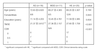 |
Diffusion Kurtosis Imaging of Basal Ganglia and Thalamus: Compare between Alzheimer’s Disease, Major Depressive Disorder and Healthy Subjects
Siriwan Piyapittayanan1, Sahutchadech Tangisarapap1, Chanon Ngamsombat1, Orasa Chawalparit1, Weerasak Muangpaisan2, and Suwit Charoensak3
1Radiology, Faculty of Medicine Siriraj Hospital, Bangkok, Thailand, 2Geriatric Medicine, Faculty of Medicine Siriraj Hospital, Bangkok, Thailand, 3Psychiatry, Faculty of Medicine Siriraj Hospital, Bangkok, Thailand
The brain circuit pathways of major depressive disorder (MDD) and Alzheimer’s disease (AD) could overlap. The authors hypothesized that AD and MDD subjects would have different diffusion parameters compared to healthy elderly controls (HE). Forty-seven age-matched subjects (AD=19, MDD=11, HE=20) were enrolled and performed MRI included structural brain imaging and diffusion kurtosis imaging (DKI) and then diffusional parameters were compared to differentiate between the three groups. Caudate nucleus and thalamus revealed significant different of diffusion parameters between AD and HE, and between AD and MDD but no significant different between MDD and HE. Diffusion imaging can demonstrate microstructural change in AD.
|
|
1469. |
fMRI complexity is associated with tau-PET and cognitive decline in Alzheimer’s disease
Kay Jann1, Hosung Kim1, Danny JJ Wang1, and for the Alzheimer's Disease Neuroimaging Initiative2
1USC Stevens Neuroimaging and Informatics Institute, Keck School of Medicine at USC, Los Angeles, CA, United States, 2http://adni.loni.usc.edu/wp-content/uploads/how_to_apply/ADNI_Acknowledgement_List.pdf, Los Angeles, CA, United States
Recently entropy measures have been explored as indices of the complexity of rs-fMRI time-series which decrease in aging and dementia. Here, we compared multi-scale entropy (MSE) of rs-fMRI with tau PET measures of neurofibrillary tangles in a cohort of 50 aged subjects in the ADRC database, through correlational and machine learning approaches. We show that the complexity of BOLD signals provides an index of the information processing capacity of regional neuron populations, and is associated with tau-related neuronal injury and cognitive decline in the AD processes.
|
|
1470. |
In-vivo classification of arteriolar sclerosis and small vessel atherosclerosis in aging, and prediction of cognitive decline
Nazanin Makkinejad1, Arnold M. Evia2, Ashish A. Tamhane2, David A. Bennett2, Julie A. Schneider2, and Konstantinos Arfanakis1,2
1Biomedical Engineering, Illinois Institute of Technology, Chicago, IL, United States, 2Rush Alzheimer's Disease Center, Rush University Medical Center, Chicago, IL, United States
Arteriolar sclerosis and small vessel atherosclerosis are two age-related neuropathologies that are common in older adults and have been linked to cognitive decline and dementia. Definitive diagnosis of either pathology is only possible at autopsy. In this work, a novel MRI-based classifier was developed for predicting the presence of arteriolar sclerosis or small vessel atherosclerosis in-vivo. The classifier was first developed based on ex-vivo MRI and pathology data from a large community-based cohort of older adults and was then translated in-vivo. The performance of the classifier was assessed both ex-vivo and in-vivo.
|
|
1471.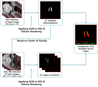 |
Comparison of locus coeruleus volume by using a high spatial-resolution MRI template among AD, MCI, and healthy controls
Sung-Jong Eun1, Sang-Young Kim2, Young Noh3, and Eung-Yeop Kim4
1Health IT Research Center, Gil Medical Center, Gachon University College of Medicine, Incheon, Korea, Republic of, 2Center for Parkinson's Disease and Dementia, Neuroscience Research Institute, Gachon University, Incheon, Korea, Republic of, 3Department of Neurology, Gil Medical Center, Gachon University of College of Medicine, Incheon, Korea, Republic of, 4Department of Radiology, Gil Medical Center, Gachon University of College of Medicine, Incheon, Korea, Republic of
Locus coeruleus (LC) is involved in regulating working memory, learning, attention, and arousal/wakefulness in the brain and accumulating evidence suggest that the LC is the initial brain region that the earliest pathological changes occur in Alzheimer’s disease (AD). We employed neuromelanin-sensitive MRI to detect the changes of LC volumes in AD. Automatic segmentation of the LC revealed profound reductions in LC volumes in AD dementia as compared to prodromal AD and/or healthy controls. Our finding suggests that volumetric reduction of the LC would be a non-invasive biomarker for detecting early pathological changes in AD.
|
|
1472.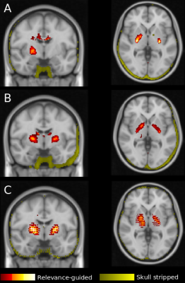 |
Relevance-guided Deep Learning for Feature Identification in R2* Maps in Alzheimer’s Disease Classification
Christian Tinauer1, Stefan Heber1, Lukas Pirpamer1, Anna Damulina1, Martin Soellradl1, Maximilian Sackl1, Edith Hofer1, Marisa Koini1, Reinhold Schmidt1, Stefan Ropele1, and Christian Langkammer1
1Medical University of Graz, Graz, Austria
Using R2* maps we separated Alzheimer's patients (n=119) from healthy controls (n=131) by using a deep neural network and found that the preprocessing steps might introduce unwanted features to be used by the classifier. We systematically investigated the influence of registration and brain extraction on the learned features using a relevance map generator attached to the classification network. The results were compared to our relevance-guided training method. While the resulting classification accuracy on the testset was similar for all training configurations, the relevance-guided method identified anatomical regions, which are known to have higher R2* values.
|
|
1473. |
Dynamic longitudinal evolving patterns of functional cortical networks in progression of Alzheimer's disease
Wenliang Fan1, Jia Liu1, Jiazheng Wang2, and Jing Wang1
1Radiology, Union Hospital, Tongji Medical College, Huazhong University of Science and Technology, Wuhan, China, 2Philips Healthcare, Beijing, China
The dynamic longitudinal change of brain in progression of AD is still unknown. We analyzed the brain networks of AD patients at different stages. We found in global network properties, most differences only existed between healthy people and patients, and few were discovered between patients at different stages. However, nearly all subnetwork properties showed significant differences between patients at different stages. Moreover, we found two different functional evolving patterns of cortical networks in progression of AD, named ‘Temperature inversion’ and “Monotonous decline”, but not the same monotonous decline trend as the external functional assessment observed in disease progression.
|
|
1474. |
A novel MRI biomarker for the characterization of early and later forms of Alzheimer’s disease
Marianna Inglese1, Haonan Lu2,3, Matthew Grech-Sollars1,4, and Eric O Aboagye1
1Surgery and Cancer, Imperial College, London, United Kingdom, 2Cancer Imaging Centre, Imperial College, London, United Kingdom, 3Ovarian Cancer Action Research Centre, Imperial College, London, United Kingdom, 4Department of Imaging, Imperial College Healthcare NHS Trust, London, United Kingdom
Alzheimer’s disease(AD) is the most common form of dementia. In the past few decades, great advances have been made in the understanding of its pathophysiology due to the use of biomarkers. Imaging biomarkers, either based on structural brain changes and metabolic alterations alone, or in conjunction with cerebrospinal fluid(CSF) biomarkers, can be informative of the ongoing pathological processes occurring in a patient with AD. In our method, we propose a novel MRI biomarker that is based on the extraction of structural features from a T1-w MRI scan, and it provides biological characterization of early and later forms of AD.
|
|
1475.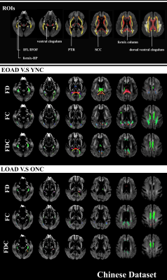 |
Distinct fibre-specific white matter reductions pattern in early- and late-onset patients
Shuyue Wang1, Xiao Luo1, Kaicheng Li1, Qingze Zeng1, Yeerfan Jiaerken1, Hui Hong1, Yong Zhang2, Peiyu Huang1, and Minming Zhang1
1Radiology, The 2nd Affiliated Hospital of Zhejiang University, School of Medicine, Hangzhou, China, 2MR Research, GE Healthcare, Shanghai, China Poster Permission Withheld For the first time, we applied fixel-based analysis (FBA) to investigate fibre tract-specific WM changes in early-onset Alzheimer’s disease (EOAD) and late-onset AD (LOAD). To test the repeatability of the results, we repeated the analysis in two independent cohort. We demonstrated that EOAD in two cohorts had more widespread WM tracts loss than LOAD. The different FD extended from the limbic system to long posterior projection regions. Further, we found that FD, rather than FC, had a higher sensitivity in detecting WM loss of EOAD, and vice versa in LOAD. Results may provide clinical clues for early diagnosis of EOAD. |
|
1476.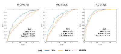 |
Source Based Brain Structural Features extraction and Classification on Alzheimer’s Disease
Zhao Qing1, Feng Chen1, Jilei Zhang2, and Bing Zhang1
1Nanjing Drum Tower Hospital, Nanjing, China, 2Clinical science, Philips Health Care, Shanghai, Shanghai, China Poster Permission Withheld
We used independent components analysis to analysis the inter-subject variation of the voxel-based morphometry results of 446 subjects of Alzheimer's spectrum. 20 components which reflect the inter subject volume variation were extracted and used as features in SVM. We observed better performance in SVM classification by these features than those using atlas-based method. These findings might be helpful for classification of AD, MCI and NC populations, which provide assistance to the early diagnosis and intervention of Alzheimer’s disease.
|
|
1477.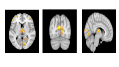 |
The Evaluation of Cerebral Blood Flow Deficits in Parkinson’s Disease Dementia using ASL-MRI at 3T
Sena Tunçer1, Dilek Betul Arslan1, Tanya Ipek Deniz2, Oznur Aslan 3, Ani Kiçik4,5, Kardelen Eryürek4,6, Emel Erdoğdu4,7, Zerrin Yildirim8, Zeynep Tüfekcioglu8, Basar Bilgic8, Hasmet Hanagasi8, Hakan Gürvit8, Tamer Demiralp4,9,
and Esin Ozturk-Isık1
1Institute of Biomedical Engineering, Bogazici University, Istanbul, Turkey, 2Department of Electrical and Electronic Engineering, Bogazici University, Istanbul, Turkey, 3Department of Biomedical Engineering, Namik Kemal University, Tekirdağ, Turkey, 4Neuroimaging Unit, Hulusi Behcet Life Sciences Research Center, Istanbul University, Istanbul, Turkey, 5Department of Physiology, Faculty of Medicine, Demiroglu Bilim University, Istanbul, Turkey, 6Department of Neuroscience, Aziz Sancar Institute of Experimental Medicine, Istanbul University, Istanbul, Turkey, 7Department of Psychology, Faculty of Arts and Sciences, Isik University, Istanbul, Turkey, 8Behavioral Neurology and Movement Disorders Unit, Department of Neurology, Istanbul Faculty of Medicine, Istanbul University, Istanbul, Turkey, 9Department of Physiology, Istanbul Faculty of Medicine, Istanbul University, Istanbul, Turkey
This study aims to find cerebral blood flow (CBF) deficits in Parkinson’s disease dementia (PDD). ASL-MRI was utilized to obtain CBF values in PD patients with different stages of cognitive decline at 3T. The CBF values were compared using a whole brain voxel-based analysis. The findings of this study revealed that the PDD group had significant CBF reductions in posterior cerebral areas, mostly in visual cortices, but also in medial parietal areas, and cortical and thalamic components of dorsolateral prefrontal fronto-striatal circuit as compared to both PD group with mild cognitive impairment (PD-MCI) and the cognitively normal PD group (PD-CN).
|
|
1478.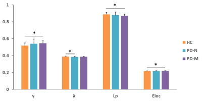 |
Topologically Convergent and Divergent Morphological Grey Matter Networks in Parkinson’s Disease with and without Mild Cognitive Impairment
Xueling Suo1,2, Du Lei1,2,3, Wenbin Li1,2,3, Jing Yang1,2, Nannan Li4, Lan Cheng4, Rong Peng4, and Qiyong Gong1,2
1Huaxi MR Research Center (HMRRC), Department of Radiology, West China Hospital of Sichuan University, Chengdu, China, 2Psychoradiology Research Unit of Chinese Academy of Medical Sciences (2018RU011), West China Hospital of Sichuan University, Chengdu, China, 3Department of Psychiatry and Behavioral Neuroscience, University of Cincinnati, Cincinnati, OH, United States, 4Department of Neurology, West China Hospital of Sichuan University, Chengdu, China
To use graph theory approaches and high resolution T1-weighted structural MRI to explore the brain grey matter (GM) morphological network in patients with Parkinson's disease (PD) with and without mild cognitive impairment (MCI). The individual morphological brain networks were constructed by estimating interregional similarity in the distribution of regional GM volume of 116 brain regions. The lower path length were found in both patients relative to healthy controls. Altered morphological connection primarily in default mode network were common deficits, while different connectivity characteristic in widespread regions involving temporal and subcortical regions, and cerebellum were observed between the patient groups.
|
|
1479.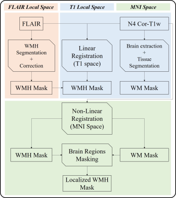 |
CDR stage of MCI in Parkinson’s disease is associated with whiter matter hyperintensities characteristics: an initial comparison study
Hanyu Wei1, Le He1, Rongsong Zhou2, Xuesong Li3, Shuo Chen1, Yu Ma2, and Rui Li1
1Center for Biomedical Imaging Research, Department of Biomedical Engineering, Tsinghua University, Beijing, China, 2Department of Neurosurgery, Tsinghua University Yuquan Hospital, Beijing, China, 3School of Computer Science and Technology, Beijing Institute of Technology, Beijing, China
The aim of this study was to investigate the white matter hyperintensities (WMH) on FLAIR MRI in patients of Parkinson’s disease (PD) with questionable mild cognitive impairment (PD-Q) and without MCI (PD-N). Automatic segmentation and artificial localization of WMH were done to accurately describe WMH characteristics. By the Clinical Dementia Rating (CDR) staging dementia in PD patients, the WMH showed more in the region of bilateral cornu occipital in PD-Q than PD-N. This finding may initially improve understanding about the association between WMH and PD cognitive dysfunction.
|
|
1480. |
Multivariate Structural and Diffusion MRI Measures Correspond to Mild Cognitive Impairment in Parkinson’s disease
Virendra R Mishra1, Jessica Caldwell1, Karthik R Sreenivasan1, Xiaowei Zhuang1, Zhengshi Yang1, Dietmar Cordes1,2, Jeffrey Cummings1,3, Zoltan Mari1, Aaron Ritter1, and Irene Litvan4
1Cleveland Clinic Lou Ruvo Center for Brain Health, Las Vegas, NV, United States, 2University of Colorado, Boulder, Boulder, CO, United States, 3Department of Brain Health, University of Nevada, Las Vegas, Las Vegas, NV, United States, 4University of California, San Diego, San Diego, CA, United States
Mild Cognitive Impairment (MCI) affects approximately 30% of individuals with Parkinson’s disease (PD). However, no reliable imaging methodology currently exists to identify PD-MCI within PD. We hypothesized that multivariate analysis of sophisticated diffusion-MRI (dMRI) voxelwise measures, volumetric measures, and dMRI-derived graph-theoretical network measures will provide a set of imaging measures that are both sensitive and specific to PD-MCI. Our preliminary analysis with 22 PD-MCI and 15 PD-non-MCI participants suggest that beyond single tensor dMRI voxelwise measures, network measures, and structural MRI-derived measurements might provide both sensitive and specific measures for further multivariate analysis to identify imaging markers corresponding to PD-MCI.
|
|
1481. |
Longitudinal assessment of brain iron deposition in Alzheimer’s disease
Christian Langkammer1, Anna Damulina1, Lukas Pirpamer1, Martin Soellradl1, Edith Hofer1,2, Christian Tinauer1, Maximilian Sackl1, Christian Enzinger1,3, Stefan Ropele1, and Reinhold Schmidt1
1Department of Neurology, Medical University of Graz, Graz, Austria, 2Institute for Medical Informatics, Statistics and Documentation, Medical University of Graz, Graz, Austria, 3Department of Radiology, Medical University of Graz, Graz, Austria
Using R2* relaxation rate mapping we found higher iron concentrations in the deep grey matter and neocortical regions in 101 patients with Alzheimer’s disease compared to 101 age-matched community-dwelling individuals. AD patients had significantly higher R2* in the temporal and occipital lobes and caudate nuclei. Independently of brain volume loss, changes in cortical iron levels over time were associated with cognitive decline in participants with Alzheimer's disease.
|
1482. |
Quantitative T1 mapping of the substantia nigra using phase-sensitive inversion recovery: a healthy volunteer study at 3.0 T
Yasuhiro Fujiwara1, Tetsuyoshi Hirai2, Tomohiro Ueda2, Hiroyuki Kumazoe2, and Shigeki Ito3
1Department of Medical Image Sciences, Faculty of Life Sciences, Kumamoto University, Kumamoto, Japan, 2Department of Radiology, National Hospital Organization Omuta National Hospital, Omuta, Japan, 3Department of Medical Radiation Sciences, Faculty of Life Sciences, Kumamoto University, Kumamoto, Japan
We estimated the T1 values of the SN in healthy subjects and clarified its correlation with the SN characteristics obtained in neuromelanin (NM) images to identify an imaging biomarker for early diagnosis of PD. In healthy adults, the area and T1 value of the SN, measured quantitatively from PSIR images, has different characteristics from those obtained from NM images, and may help assess SN degeneration.
|
|
1483. |
Quantitative measurement of age-related change of myelin content in a large sample of Chinese population
Junye Yao1, Qiqi Tong1, Ting Gong1, Qiuping Ding1, Yi Sun2, Peipeng Liang3, Kuncheng Li4, Hongjian He1, and Jianhui Zhong1,5
1Center for Brain Imaging Science and Technology, College of Biomedical Engineering and Instrumental Science, Zhejiang University, Hangzhou, China, 2MR Collaboration, Siemens Healthcare Ltd, Shanghai, China, 3School of Psychology, Capital Normal University, Beijing, China, 4Department of radiology, Xuanwu Hospital Capital Medical University, Beijing, China, 5Department of Imaging Sciences, University of Rochester, Rochester, NY, United States
Magnetic resonance structural images of 902 Chinese subjects were used to study the change of myelin content with the age based on the T1-weighted/T2-weighted ratio. Significant quadratic age effects were observed in white matter tracts and subcortical areas. The gender difference was reflected in the results that the myelin content levels are higher and begin to decline earlier in women. The inverted U-shape myelin-vs-age curve accords with the existing research literature. However, the age of the observed peaks may have been delayed by iron deposition in basal ganglia.
|
|
1484. |
Age and location related hemodynamic changes in the carotid artery of healthy adults assessed by 4D flow MRI
Guiling Zhang1 and Wenzhen Zhu1
1Tongji hospital, Wuhan, China
Atherosclerosis is the predominant risk factor of ischemic stroke. Age and location are intrinsic factors in the development of carotid artery plaque. Therefore, the relationship between hemodynamic changes and location/age in healthy subjects can help understand how the hemodynamic states influence carotid incidents. Our study found velocity/WSS/PG decreased with age. And proximal ICA, the most common point arising atherosclerotic plaque, displayed the lowest velocity/WSS/PG. It may imply this state of hemodynamics are more likely to cause atherosclerotic plaques. The multi-parameter analysis of 4D flow MRI may offer suggestions for choosing age and location matched control cohorts in future study.
|
|
1485.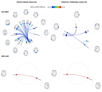 |
FUNCTIONAL DISCONNECT PRECEDES STRUCTURAL ATROPHY IN CONFIRMED MULTI-DOMAIN AMNESTIC MILD COGNITIVE IMPAIRMENT
Kathryn Broadhouse1, Natalie Winks1, and Mathew Summers1
1University of the Sunshine Coast, Sunshine Coast, Australia
Recent work suggests that aberrant functional neurocircuitries arise prior to significant structural atrophy and clinically cognitive deficits in dementia. Using a rigorous, longitudinally confirmed multi-domain amnestic MCI cohort we show that functional decoupling of networks implicated in memory and executive function, occur prior to measurable MTL atrophy in this population. Moreover, we show that decreased functional connectivity in these networks is associated with poorer cognitive performance. Further longitudinal studies investigating the neuronal underpinnings of disease progression will provide insight into potential functional biomarkers and establish significance of functional decoupling within the sequential model of dynamic biomarkers of Alzheimer’s pathological cascade.
|
|
1486.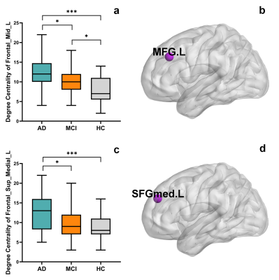 |
Progressive Increased Degree Centrality in Alzheimer’s Disease and Mild Cognitive Impairment: A Resting-state fMRI Study
Yuan Luo1, Chunchao Ma2, Xianchang Zhang3, Xiuwei Fu4, and Hongyan Ni5
1Department of Radiology, First Central Clinical College, Tianjin Medical University, Tianjin, China, 2Department of Neurology, Tianjin First Central Hospital, Tianjin, China, 3MR Collaboration, Siemens Healthcare Ltd., Beijing, China, 4Department of Radiology, Tianjin Medical University General Hospital, Tianjin, China, 5Department of Radiology, Tianjin First Central Hospital, Tianjin, China
Brain network alterations remain poorly understood in mild cognitive impairment (MCI). This functional magnetic resonance imaging (fMRI) study aimed to investigate the degree centrality alterations in patients with Alzheimer's disease (AD) and MCI using graph theory analyses based on a population-specific template, as well as associations with neuropsychological test scores. The degree centrality of the left middle frontal gyrus and left medial superior frontal gyrus increased as patients’ cognitive function and daily activities declined. These findings suggest that abnormalities in the network topology progress in the prodromal and clinical stages of AD and are associated with clinical manifestations.
|
|
1487.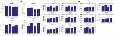 |
Role of Diffusion MRI-Derived Free Water Fraction in Predicting Progression to Alzheimer’s Disease Dementia from Mild Cognitive Impairment
Virendra R Mishra1, Karthik R Sreenivasan1, Xiaowei Zhuang1, Zhengshi Yang1, Dietmar Cordes1,2, Jeffrey Cummings1,3, Jessica Caldwell1, and Aaron Ritter1
1Cleveland Clinic Lou Ruvo Center for Brain Health, Las Vegas, NV, United States, 2University of Colorado, Boulder, Boulder, CO, United States, 3Department of Brain Health, University of Nevada, Las Vegas, Las Vegas, NV, United States
We hypothesized that diffusion-MRI (dMRI)-derived free-water fraction (fiso) will show a significantly lower and a faster change over time in mild cognitive impaired (MCI) participants who progressed to Alzheimer’s disease (AD) dementia in various cortical, subcortical, and white-matter fiber tracts compared to those who did not progress to AD dementia. Utilizing five β-Amyloid (Aβ) positive (+)/ApoE-4 carriers MCI participants who progressed to AD dementia within one-year, and thirteen Aβ+/ApoE-4 carriers MCI participants who did not progress to AD dementia within one-year, we showed that although the relationship between various cortical/subcortical volume and fiso is complex, it was distinct between groups.
|
|
1488.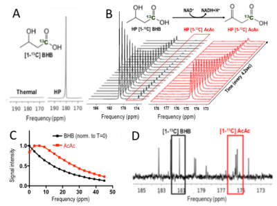 |
Imaging cerebral ketone metabolism with hyperpolarized 13C magnetic resonance spectroscopy
Lydia M Le Page1,2, Soo Hyun Shin3, Kai Qiao1,2, and Myriam M Chaumeil1,2
1Physical Therapy and Rehabilitation Science, University of California, San Francisco, San Francisco, CA, United States, 2Radiology and Biomedical Imaging, University of California, San Francisco, San Francisco, CA, United States, 3Bioengineering, University of California, Berkeley, Berkeley, CA, United States
As ketogenic diets have been shown to elicit cognitive improvements in Alzheimer’s disease (AD) patients, non-invasive methods allowing for in vivo monitoring of brain ketone metabolism are critically needed to understand and monitor these observations. We hypothesized that 13C magnetic resonance spectroscopy of hyperpolarized (HP) [1-13C] beta-hydroxybutyrate (BHB) could be used to monitor response to ketogenic diets in health and AD. Here, we characterized HP [1-13C] BHB and validated an AD mouse model for application of our HP probe. We also carried out the first proof-of-concept acquisition of data showing in vivo metabolism of HP BHB in the mouse brain.
|
|
1489.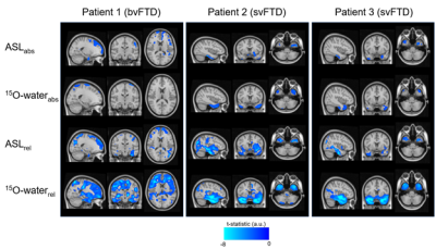 |
Sensitivity of ASL MRI for Detecting Perfusion Abnormalities in Frontotemporal Dementia: Preliminary Comparison with 15O-water PET
Tracy Ssali1,2, Lucas Narciso1,2, Justin Hicks1,2, Matthias Günther3, Frank Prato1,2, Udunna Anazodo1,2, Elizabeth Finger4, and Keith St Lawrence1,2
1Lawson Health Research Institute, London, ON, Canada, 2Department of Medical Biophysics, Western University, London, ON, Canada, 3Fraunhofer Institute for Medical Image Computing MEVIS, Bremen, Germany, 4Department of Clinical Neurological Sciences, Western University, London, ON, Canada
The ability of arterial spin labeling (ASL) to detect perfusion abnormalities in clinical populations, such as frontotemporal dementia, can be limited by poor signal to noise and transit-time artefacts. Recent advances in ASL imaging protocols should enable detection of more subtle perfusion abnormalities. This study presents a head-to-head comparison of regional hypoperfusion detected by ASL and PET with radiolabeled water (15O-water) - the gold standard for measuring CBF in humans. While 15O-water PET data showed greater sensitivity, as identified by larger and focal clusters on the t-maps, similar areas of hypoperfusion were identified by ASL, particularly with relative CBF.
|
|
1490.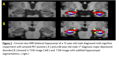 |
MRI subfield hippocampal volumetric analysis combined with amyloid PET result: Comparison between mild cognitive impairment, major depressive disorder and healthy elderly subjects.
Doonyaporn Wongsawaeng1, Chanon Ngamsombat1, Tanyaluck Thientunyakit1, Weerasak Muangpaisan2, Suwit Charoensuk3, Panida Charnchaowanish1, and Orasa Chawalparit1
1Department of Radiology, Faculty of Medicine Siriraj Hospital, Mahidol University, Bangkok, Thailand, Bangkok, Thailand, 2Department of Geriatric Medicine, Faculty of Medicine Siriraj Hospital, Mahidol University, Bangkok, Thailand, Bangkok, Thailand, 3Department of Psychiatry, Faculty of Medicine Siriraj Hospital, Mahidol University, Bangkok, Thailand, Bangkok, Thailand
Hippocampus is a part of limbic system involving both neurodegenerative disease and emotional regulation circuit. This study aimed to use MRI automated subfield hippocampal volumetric analysis to differentiate mild cognitive impairment (MCI) from major depressive disorder (MDD) and age-matched healthy elderly (HE) subjects. We found a trend of relative smaller size in several subfield hippocampal regions in MCI than MDD patients. Even though, there were no statistical significance. These may raise possibility of MRI volumetry to be the tool for discriminating early neurodegenerative disease from major depressive disorder.
|
|
1491.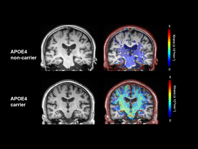 |
Greater BBB permeability change in APOE4 carrier in cognitively normal and mild cognitive impaired subjects: a new imaging phenotype for APOE4
Won-Jin Moon1, Ilheon Ha1, Changmok Lim1, Yaehoon Kim1, Yeolsil Moon2, and Seol-Heui Han2
1Radiology, Konkuk University Medical Center, Seoul, Republic of Korea, 2Neurology, Konkuk University Medical Center, Seoul, Republic of Korea
In normal and cogntively impaired subjects, BBB permeability of hippocampus is increased in APOE4 carrier group than in APOE4 non-carrier group. After adjusting for education years, and medial temporal lobar atrophy, the only valuable predicting factor for predicting cognitive function was BBB permeability of hippocampus. Our study indicates that BBB permeability imaging can be a distinct imaging phenotype of APOE4 mutation as well as an early imaging marker for clinical decline in cognitive impaired subjects.
|
|
1492.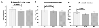 |
Aberrant cerebral blood flow in patients with subjective cognitive decline: a whole-brain atlas based analysis
Hongyuan Ding 1, Yi Zhu 2, Han Wu 3, Ling Zhang 1, Yaxin Gao 4, Qian Zhong 4, Qiumin Zhou 2, Ming Qi 1, Long Qian 5, Weiqiang Dou5, and Tong Wang2
1Radiology department, The First Affiliated Hospital of Nanjing Medical University, Nanjing, China, 2Rehabilitation Department, The First Affiliated Hospital of Nanjing Medical University, Nanjing, China, 3Rehabilitation Department, Nanjing Drum Tower Hospital, The Affiliated Hospital of the Medical School at Nanjing University, Nanjing, China, 4Nanjing Medical University, Nanjing, China, 5MR Research China, GE Healthcare, Beijing, China
In this study, the whole brain cerebral blood perfusion (CBF) values have been respectively investigated for patients with subjective cognitive decline (SCD) and mild cognitive impairment (MCI) and healthy controls (HCs). Significantly lower CBF values for the left superior frontal gyrus, left middle frontal gyrus, and left caudate nucleus regions have been shown in SCD patients than HCs. Additionally, the CBFs at these regions also showed strong correlations with multiple clinical scales. Therefore, CBF can be considered an effective tool in the early detection of SCD patients.
|
|
1493.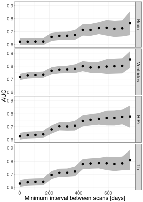 |
Establishing and evaluating reference atrophy ranges from consecutive image pairs in brain MR volumetry
Jonas Richiardi1,2, Bénédicte Maréchal1,2,3, Ricardo Corredor2, Mazen Mahdi2, Reto Meuli1, and Tobias Kober1,2,3
1Department of Radiology, Lausanne University Hospital and University of Lausanne, Lausanne, Switzerland, 2Advanced Clinical Imaging Technology, Siemens Healthcare, Lausanne, Switzerland, 3LTS5, Ecole Polytechnique Fédérale de Lausanne (EPFL), Lausanne, Switzerland
Estimating clinical significance of changes in regional brain volumetry using only two consecutive images is difficult because algorithmic measurement error dominates actual biological changes in short-term (less than a year) follow-up imaging. Here, we evaluate an approach to compute reference ranges from image pairs, using empirical Bayesian regularization. With over 6400 image pairs, we evaluate the impact of regularization strength and time between images. Regularization is essential; optimal regularization amount depends on brain region. Increased time between pairs of images improves clinical discrimination in dementia. We recommend a minimum of eight months to one year to obtain discriminative atrophy estimates.
|
|
1494.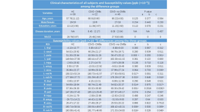 |
Quantitative susceptibility mapping of brain iron deposition in patients with cerebral small-vessel disease with cerebral microbleeds
Lingfei Guo1,2, Liangdong Zhou1, Thanh D. Nguyen1, Weiyuan Huang1, Junghun Cho1, and Yi Wang1
1Weill Cornell Medicine, Cornell University, New York, NY, United States, 2Shandong Medical Imaging Research Institute Affiliated to Shandong University, Jinan, China
Quantitative susceptibility mapping (QSM) was used to evaluate and compare the characteristics of iron deposition in gray matter nuclei of the brain between patients with cerebral small-vessel disease and cerebral microbleeds (CSVD-CMBs) and those with CSVD and no CMBs(CSVD–no CMBs). The susceptibility values of the bilateral caudate nucleus, putamen, red nucleus, and substantia nigra were significantly higher in patients with CSVD-CMBs than in those with CSVD–no CMBs and in healthy controls. The change in the susceptibility values of these regions may be an imaging marker of cognitive decline in patients with CSVD.
|