
Digital Poster Session
Neuro: Neurodegeneration
Neuro
1495 -1508 Neurodegeneration - Neurodegeneration: Parkinson's Disease 1
1509 -1522 Neurodegeneration - Neurodegeneration: Parkinson's Disease 2
1523 -1537 Neurodegeneration - Typical & Atypical Parkinson's Diseases
1538 -1552 Neurodegeneration - Neurodegeneration: From HIV to CJD & Many Things in Between
1553 -1566 Neurodegeneration - Neurodegeneration: Small Vessel Disease & More
1567 -1579 Neurodegeneration - Neurodegeneration: Motor Neuron Disease & More
1580 -1592 Neurodegeneration - Clinical Epilepsy & TBI
1495.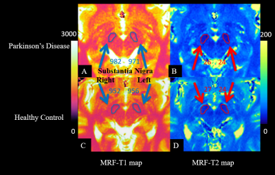 |
Magnetic Resonance Fingerprinting May Differentiate Parkinson’s Disease From Healthy Controls
Yan Bai1, Rushi Chen1, Rui Zhang1, Wei Wei1, Ying Wang1, Mathias Nittka2, Gregor Koerzdoerfer2, Xianchang Zhang3, and Meiyun Wang1
1Department of Medical Imaging, Henan Provincial People's Hospital, Zhengzhou, China, 2MR Pre-development, Siemens Healthcare, Erlangen, Germany, 3MR Collaboration, Siemens Healthcare Ltd, Beijing, China
Magnetic resonance parametric mapping techniques such as T1 and T2 relaxation time mapping have been used to capture the potential Parkinson’s disease (PD)-related changes in the substantia nigra (SN). However, the findings in different studies were inconsistent. This study utilized a novel technique magnetic resonance fingerprinting (MRF) to obtain T1 and T2 values on thirty patients with PD and thirty matched healthy controls. Comparison results found that T1 values of the left and right SN in the PD patients were significantly higher than in healthy controls. T1 values in the SN acquired by MRF may differentiate PD from healthy controls.
|
|
1496. |
Different patterns of perfusion changes in tremor-dominant Parkinson’s disease and essential tremor using 3D arterial spin labeling imaging
Yong Zhang1, Jian Wang2, Chang-Peng Wang2, Li-Rong Jin2, and Bing Wu3
1GE Healthcare, Shanghai, China, 2Zhongshan Hospital, Shanghai, China, 3GE Healthcare, Beijing, China
This preliminary study aimed to identify potential markers in diagnosis of tremor disorders such as tremor-dominant Parkinson’s disease (PDT) and essential tremor (ET). A novel 3D pulsed-continuous arterial spin labeling technique (3D pCASL) was used to provide whole brain quantitative perfusion measurement, followed by voxel-wise comparison to evaluate regional CBF characteristics in patients with PDT, ET and age- and gender- matched healthy controls. PDT patients showed decreased CBF in the caudate and precuneus when compared to ET patients. The altered metabolic patterns of PDT and ET can help to understand different pathophysiological mechanism of tremor disorders.
|
|
1497. |
Fixel-based analysis on white matter changes in patients with Parkinson's disease, progressive supranuclear palsy and multiple system atrophy
Nguyen Thanh Thao1, Chih-Chien Tsai2, Yao-Liang Chen3, Jur-Shan Cheng4, Chin-Song Lu5, Yi-Hsin Weng5, Sung-han Lin4, Po-Yuan Chen4, and Jiun-Jie Wang6
1Department of Radiology, Hue University of Medicine and Pharmacy, Hue University, Hue, Vietnam, 2Healthy Aging Research Center, Chang-Gung University, TaoYuan, Taiwan, 3Department of Diagnostic Radiology, Chang Gung Memorial Hospital, Keelung Branch, Keelung, Taiwan, 4Chang-Gung University, TaoYuan, Taiwan, 5Division of Movement Disorders, Department of Neurology, Chang Gung Memorial Hospital, Linkou Branch, TaoYuan, Taiwan, 6Department of Medical Imaging and Radiological Sciences, Chang-Gung University, TaoYuan, Taiwan
White matter degeneration have been attributed to the motor and non-motor symptoms of Parkinson’s disease and atypical parkinsonism. Our study shows different pattern of white matter changes in multiple system atrophy and progressive supranuclear palsy compared to Parkinson’s disease. Furthermore, different affected areas of white matter changes were found among atypical parkinsonism. The involved regions are consistent with the understanding of the pathogenesis of the diseases. Our result proves that fixel based analysis is a robust technique to study white matter degeneration in PD and atypical parkinsonism.
|
|
1498. |
Perfusion-Based Biomarkers of Mild Cognitive Impairment in Parkinson’s disease with different MAPT haplotypes using Arterial Spin Labeling MRI
Dilek Betul Arslan1, Hakan Ibrahim Gurvit2, Ozan Genc1, Ani Kicik3,4, Kardelen Eryurek3,5, Sevim Cengiz1, Emel Erdogdu3,6, Zerrin Yildirim2, Zeynep Tufekcioglu2, Aziz Mufit Ulug1,7, Basar Bilgic2, Hasmet Hanagasi2, Erdem Tuzun5,
Tamer Demiralp3,8, and Esin Ozturk-Isik1
1Institute of Biomedical Engineering, Bogazici University, Istanbul, Turkey, 2Behavioral Neurology and Movement Disorders Unit, Department of Neurology, Istanbul Faculty of Medicine, Istanbul University, Istanbul, Turkey, 3Neuroimaging Unit, Hulusi Behcet Life Sciences Research Center, Istanbul University, Istanbul, Turkey, 4Department of Physiology, Faculty of Medicine, Demiroglu Bilim University, Istanbul, Turkey, 5Department of Neuroscience, Aziz Sancar Institute of Experimental Medicine, Istanbul University, Istanbul, Turkey, 6Department of Psychology, Faculty of Arts and Sciences, Isik University, Istanbul, Turkey, 7CorTechs Labs, San Diego, CA, United States, 8Department of Physiology, Istanbul Faculty of Medicine, Istanbul University, Istanbul, Turkey
The main purpose of this study was to define possible brain perfusion deficits in risky gene carriers in Parkinson’s disease (PD) using arterial spin
|
|
1499.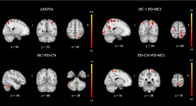 |
The Cerebral Blood Flow Changes in Parkinson’s Disease with Mild Cognitive Impairment Using Arterial Spin Labeling MRI
Dilek Betul Arslan1, Hakan Ibrahim Gurvit2, Ozan Genc1, Ani Kicik3,4, Kardelen Eryurek3,5, Sevim Cengiz1, Emel Erdogdu3,6, Zerrin Yildirim2, Zeynep Tufekcioglu2, Aziz Mufit Ulug1,7, Basar Bilgic2, Hasmet Hanagasi2, Tamer Demiralp3,8,
and Esin Ozturk-Isik1
1Biomedical Engineering Institution, Bogazici University, Istanbul, Turkey, 2Behavioral Neurology and Movement Disorders Unit, Department of Neurology, Istanbul Faculty of Medicine, Istanbul University, Istanbul, Turkey, 3Neuroimaging Unit, Hulusi Behcet Life Sciences Research Center, Istanbul University, Istanbul, Turkey, 4Department of Physiology, Faculty of Medicine, Demiroglu Bilim University, Istanbul, Turkey, 5Department of Neuroscience, Aziz Sancar Institute of Experimental Medicine, Istanbul University, Istanbul, Turkey, 6Department of Psychology, Faculty of Arts and Sciences, Isik University, Istanbul, Turkey, 7CorTechs Labs, San Diego, CA, United States, 8Department of Physiology, Istanbul Faculty of Medicine, Istanbul University, Istanbul, Turkey
The main aim of this study is to define possible brain perfusion-based signatures based on voxelwise comparison of cerebral blood flow (CBF) maps of Parkinson’s disease (PD) with mild cognitive impairment, cognitively normal PD and healthy controls. CBF maps were calculated by fitting a general kinetic curve model with Look-Locker readout for each pixel of arterial spin
|
|
1500.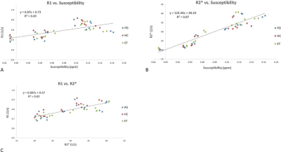 |
Quantitative assessment of abnormal susceptibility, R1 and R2* changes in the deep gray matter nuclei in Parkinson’s disease and essential tremor
Kiarash Ghassaban1,2, Sean Kumar Sethi1,2, David Utriainen3, Zenghui Cheng4, Pei Huang5, Yan Li4, Rongbio Tang 4, Paul Kokeny3, Kiran Kumar Yerramsetty6, Vinay Kumar Palutla6, Shengdi Chen 5, Fuhua Yan4, and Ewart Mark Haacke1,2,4
1Radiology, Wayne State University, Detroit, MI, United States, 2Biomedical Engineering, Wayne State University, Detroit, MI, United States, 3SpinTech, Bingham Farms, MI, United States, 4Radiology, Ruijin Hospital, Shanghai Jiao Tong University School of Medicine, Shanghai, China, 5Neurology, Ruijin Hospital, Shanghai Jiao Tong University School of Medicine, Shanghai, China, 6MR Medical Imaging Innovations, Telangana, India
This work proposes two investigated problems. The first is the separation of confounding tissue properties of deep gray matter using R1, R2*, and QSM from a 3D GRE protocol known as STAGE by sampling a distribution of low and high iron regions across subjects, including very high iron regions in aceruloplasminemia. The second is investigating if we see any differences in these structures in Parkinson’s Disease, essential tremor, and healthy control subjects. These problems are investigated to show that susceptibility changes and R1 are linked, and that water and iron related changes are observable when comparing controls versus Parkinson’s Disease
|
|
1501.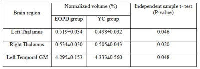 |
Exploration of structural brain volume changes in patients with early- and middle-late-onset Parkinson's disease using the MPRAGE sequence
Ruichen Zhao1, Chunyan Zhang1, Jinxia Zhu2, Bénédicte Maréchal3, Chen Chen1, Hong Lu4, and Jingliang Cheng1
1Department of MRI, The First Affiliated Hospital of Zhengzhou University, Zhengzhou, China, 2MR Collaboration, Siemens Healthcare Ltd., Beijing, China, 3Advanced Clinical Imaging Technology, Siemens Healthcare AG;Department of Radiology, Lausanne University Hospital and University of Lausanne;LTS5, École Polytechnique Fédérale de Lausanne (EPFL), Lausanne, Switzerland, 4Department of Neurology, The First Affiliated Hospital of Zhengzhou University, Zhengzhou, China
In this study, the structural brain volume changes in early-onset (EOPD) and middle-late-onset Parkinson’s disease (M-LOPD) patients were evaluated. We found different patterns of volume changes in these patients. The results showed that some brain regions might represent a potential imaging marker for the early diagnosis of EOPD and could explain different clinical characteristics. MPRAGE-based morphometry may be a suitable method to provide a reference for EOPD diagnoses in clinical practice.
|
|
1502.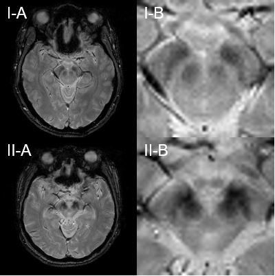 |
Localization of the iron deposits along myelinated fibers within the substantia nigra of progressive supranuclear palsy on brain MRI
Hansol Lee1, Sun-Yong Baek2, Eun-Joo Kim3, Gi Yeong Huh4, Jae-Hyeok Lee5, and HyungJoon Cho1
1Department of Biomedical Engineering, Ulsan National Institute of Science and Technology, Ulsan, Korea, Republic of, 2Department of Anatomy, Pusan National University School of Medicine, Yangsan, Korea, Republic of, 3Department of Neurology, Pusan National University Hospital, Busan, Korea, Republic of, 4Department of Forensic Medicine, Pusan National University School of Medicine, Yangsan, Korea, Republic of, 5Department of Neurology, Research Institute for Convergence of Biomedical Science and Technology, Pusan National University Yangsan Hospital, Yangsan, Korea, Republic of
The purpose of this study was to determine the morphology change in the substantia nigra of progressive supranuclear palsy using MRI with histopathological validation. MR experiments for progressive supranuclear palsy brains were operated using 3T in vivo and 7T postmortem imaging systems. Perls’ Prussian blue staining, Luxol fast blue staining, and LA-ICP-MS for 2D iron mapping confirmed the large amount of iron deposits along the myelinated fibers within substantia nigra of PSP brain. The iron deposits along the myelinated fibers could be the potential source causing the blurred boundary between red nucleus and substantia nigra in in vivo MRI.
|
|
1503. |
Functional compensation on disrupted cingulate structural network in Parkinson’s disease
Cheng Zhou1 and Minming Zhang1
1Zhejiang university, Hangzhou, China
Parkinson’s disease (PD) is the second most common neurodegenerative disease and its related brain changes appear not to be localized in isolate region but rather in the networks. We desire to exploring the large-scale structural and functional networks change which are of great importance for understanding the mechanism of disease. Moreover, it deserves to further answer the unsolved question that whether a functional compensation resulting from structural network disruption and whether such effect would be lasting or changed longitudinally.
|
|
1504.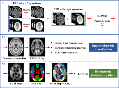 |
Disrupted interhemispheric coordination with unaffected lateralization of global eigenvector centrality characterizes hemiparkinsonism.
Jingjing Wu1, Tao Guo1, Cheng Zhou1, Ting Gao 2, Xiaoujun Guan 1, Peiyu Huang1, Xiaojun Xu1, and Minming Zhang1
1Department of Radiology, The Second Affiliated Hospital, Zhejiang University School of Medicine, Hangzhou, China, 2Department of Neurology, The Second Affiliated Hospital, Zhejiang University School of Medicine, Hangzhou, China
The motor dysfunctions always affect hemi-body first in Parkinson's disease (PD). However, the interhemispheric relationships in patients with only unilateral motor impairment were barely known to date. In this study, 43 unilateral-symptomatic PD patients (UPD, Hoehn-Yahr staging scale, H-Y: 1-1.5), and 54 NC were recruited. We aimed to investigate the interhemispheric coordination and hemispheric dominance pattern for further understanding the pathogenesis of PD. We found that the disrupted interhemispheric coordination in bilateral sensorimotor regions may have significant implications for elucidating the mechanisms underlying the hemiparkinsonism and enabling the individual diagnosis and assessment of early PD.
|
|
1505.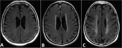 |
Differential clinical associations of Periventricular and Deep White Matter Hyperintensities on FLAIR
Han Yu1, Jingyun Chen1, Henry Rusinek2, and Yulin Ge3
1Department of Neurology, New York University School of Medicine, New York, NY, United States, 2Department of Neurology and Radiology, New York University School of Medicine, New York, NY, United States, 3Department of Radiology, New York University School of Medicine, New York, NY, United States
In this study, we examined the differences of two subtypes of white matter hyperintensities (WMHs), periventricular WMHs (PVWMH) and deep WMHs (DWMH) on MRI, as they associate with cognitive dysfunction and dementia, and other clinical assessments of the elderly. A robust computational method (Bilateral Distance classification) was implemented to quantify PVWMH and DWMH. Clinical associations revealed by the algorithm are consistent with the literature findings based on subjective classification methods that the two types of WMHs have differential clinical associations and may have different pathological etiologies and roles in cognitive impairment and dementia.
|
|
1506.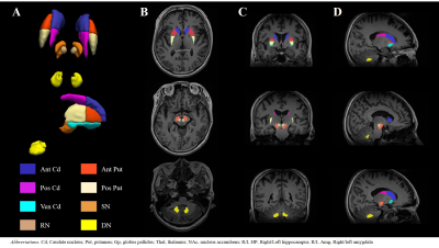 |
Investigation of iron-sensitive MRI biomarkers for non-motor symptoms in early stage Parkinson’s disease
Seulki Yoo1,2, MinKyeong Kim3,4, Doyeon Kim5, Jin Whan Cho3,4, Ji Sun Kim3,4, Jong Hyun Ahan3,4, Jun Kyu Mun3,4, Jinyoung Youn3,4, and Seung-Kyun Lee1,2
1Department of Biomedical Engineering, Sungkyunkwan University, Suwon, Korea, Republic of, 2Center for Neuroscience Imaging Research, Institute for Basic Science, Suwon, Korea, Republic of, 3Department of Neurology, Samsung Medical Center, Seoul, Korea, Republic of, 4Neuroscience Center, Samsung Medical Center, Seoul, Korea, Republic of, 5Department of Biomedical Engineering, Gachon University, Incheon, Korea, Republic of
Deep brain iron accumulation in Parkinson’s disease (PD) has been much studied in MRI but its involvement in clinical non-motor symptoms has been inconclusive. In this work we investigated the correlation between the deep brain iron contents and a wide array of non-motor symptoms in drug naive early PD patients using QSM and R2* mapping at 3T. We found that many non-motor symptoms are significantly correlated with R2* in the extra-striatal system, in particular the thalamus and red nucleus.
|
|
1507. |
Insula structural changes and behavioral disinhibition in Parkinson’s Disease
Megan Aumann1,2, Kathleen Larson3, Elise Bradley4, David Zald2,5, Ipek Oguz3, and Daniel O Claassen6
1Neurology, Vanderbilt University, Nashville, TN, United States, 2Psychology, Vanderbilt University, Nashville, TN, United States, 3Biomedical Engineering, Vanderbilt University, Nashville, TN, United States, 4Neuropsychiatry, Vanderbilt University Medical Center, Nashville, TN, United States, 5Psychiatry, Vanderbilt University Medical Center, Nashville, TN, United States, 6Neurology, Vanderbilt University Medical Center, Nashville, TN, United States
Parkinson’s Disease (PD) patients who take dopaminergic therapy are at risk for developing impulsive-compulsive behaviors, and these behaviors localize to the mesocorticolimbic regions. Using a caregiver-reported values from the Frontal Systems Behavioral Scale (FrSBe), we assessed disinhibition, apathy, and dysexecutive symptoms in 72 PD patients. All participants completed brain MRI, and we measured cortical thickness in frontal regions, assessing the relationship between cortical thickness and FrSBE scores. We find that thickness in the insula is directly related to disinhibited behaviors. These results provide new insights into how cortical changes and behavioral symptoms are linked in PD.
|
|
1508.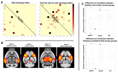 |
Characterisation of white matter integrity and functional connectivity in ApoE4 and ApoE3 mice
Jiayi Zhang1, Ling Yun Yeow1, Joanne Huifen Koh2, Isaac Huen1, Pei Huang1, Tuck Wah Soong2, Boon Seng Wong2,3, Kuan Jin Lee1, and Bhanu Prakash KN1
1SBIC, A*STAR, Singapore, Singapore, 2Department of Physiology, National University of Singapore, Singapore, Singapore, 3Health and Social Sciences Cluster, Singapore Institute of Technology, Singapore, Singapore
Cholesterol-transporter ApoE4 is involved in lipid metabolism and is associated with neurodegenerative diseases. Studies show, functional connectivity(FC) in ApoE4 mice is significantly deviating from ApoE3 and Wild-Type(WT). Underlining cause of these differences are less explored. Since myelin is mostly comprised of cholesterol, we investigated if a change in white matter integrity(WMI) could have contributed to the FC changes. FC and WMI in ApoE4 are distinctively different from ApoE3 and WT as shown by our results. Our results within the auditory cortex show that an increase in FA in ApoE4 is associated with a decrease of FC in the region.
|
 View the Poster
 Watch the Video
1509. |
Comparison of T1/T2 values of white matter and deep gray matter between healthy controls and Parkinson’s disease patients using plug-and-play MRF
Koji Fujimoto1, Martijn A. Cloos2, Atsushi Shima3, Thuy Dinh Ha Duy1, Nobukatsu Sawamoto4, Ryosuke Takahashi3, Tadashi Isa1,5, and Tomohisa Okada1
1Human Brain Research Center, Graduate School of Medicine, Kyoto University, Kyoto, Japan, 2Center for Advanced Imaging Innovation and Research (CAI2R) and Bernard and Irene Schwartz Center for Biomedical Imaging, Department of Radiology, New York University School of Medicine, New York, NY, United States, 3Department of Neurology, Kyoto University Graduate School of Medicine, Kyoto University, Kyoto, Japan, 4Department of Human Health Sciences, Kyoto University Graduate School of Medicine, Kyoto University, Kyoto, Japan, 5Department of Neuroscience, Kyoto University Graduate School of Medicine, Kyoto University, Kyoto, Japan
To investigate changes in T1 and T2 of deep gray matter in Parkinson’s disease (PD) patients at 7T, healthy volunteers (N=104, age range 20-77) and PD patients (N=42, age range 50-72) were scanned using a 1ch-Tx/32ch-Rx coil and a Plug-and-Play MR Fingeprinting (prototype) sequence. ROIs were drawn in six regions (left and right putamen, globus pallidus, caudate head, thalamus, frontal white matter (WM)) and a linear and quadratic curve fitting was performed. T2 value of the putamen was larger in PD than the age-matched subgroup of HC, but was not significant.
|
|
1510. |
Quantitative Evaluation of Impaired Neuroenergetics in Parkinson’s Disease and the Treatment Effects of Ursodeoxycholic Acid
Xiao-Hong Zhu1, Byeong-Yuel Lee1, Lisa Coles2, Abhishek G Sathe2, Paul Tuite3, Jim Cloyd2, Walter Low4, Clifford J. Steer5, Chi Chen6, and Wei Chen1
1CMRR, Department of Radiology, University of Minnesota, Minneapolis, MN, United States, 2Department of Experimental and Clinical Pharmacology, University of Minnesota, Minneapolis, MN, United States, 3Department of Neurology, University of Minnesota, Minneapolis, MN, United States, 4Department of Neurosurgery, University of Minnesota, Minneapolis, MN, United States, 5Departments of Medicine and Genetics, Cell Biology and Development, University of Minnesota, Minneapolis, MN, United States, 6Department of Food Science and Nutrition, University of Minnesota, Minneapolis, MN, United States Poster Permission Withheld
Abnormal energy metabolism due to mitochondrial dysfunction is thought to be a major contributor to the progression of Parkinson’s disease (PD). We employed 31P MRS-MT technique at 7T to quantify key bioenergetic parameters in the occipital lobe of people with PD (PWPs). Significantly lower intracellular ATP concentrations together with elevated ATPase activity was found in PWPs; suggesting that augmented ATPase enzymatic activity may represent a compensatory mechanism to bioenergetic deficits that occur in PD. The FDA-approved drug, ursodeoxycholic acid (UDCA), shown to have energy-enhancing properties was evaluated for its effect on improving neuroenergetics in PWPs using the 31P MRS-MT approach.
|
|
1511. |
Susceptibility Map-Weighted Imaging and Neuromelanin-Sensitive MRI in Parkinson’s Disease
Septian Hartono1,2, Isabel Hui Min Chew3, Weiling Lee3, Amanda May Yeng Choo1, Celeste Yan Teng Chen1, Leon Qi Rong Ooi4, Lirong Yin3, Kuan Jin Lee5, Jongho Lee6, Ching-Yu Cheng7, Eng King Tan1,2, and Ling Ling Chan2,3
1National Neuroscience Institute, Singapore, Singapore, 2Duke-NUS Medical School, Singapore, Singapore, 3Singapore General Hospital, Singapore, Singapore, 4National University of Singapore, Singapore, Singapore, 5Singapore BioImaging Consortium, Singapore, Singapore, 6Seoul National University, Seoul, Republic of Korea, 7Singapore Eye Research Institute, Singapore, Singapore
Nigrosome-1 imaging and neuromelanin contrast have been identified as good radiological biomarkers of dopaminergic nigral degeneration in Parkinson's disease (PD) pathology. We evaluated the sensitivity of quantitative Susceptibility-Mapping Weighted Imaging (SMWI) derived from Quantitative Susceptibility Mapping (QSM) and neuromelanin sensitive (NMS) imaging in differentiating a case control cohort of PD patients. Region-of-interest analysis of the substantia nigra on both QSM/SMWI and NMS offered excellent differentiation of PD and healthy controls. However, QSM/SMWI offered more robust disease classification compared to NMS and might be preferred for use in the clinical setting.
|
|
1512. |
ViSTa Myelin Water Imaging in Parkinson’s Disease
Septian Hartono1,2, Leon Qi Rong Ooi3, Amanda May Yeng Choo1, Celeste Yan Teng Chen1, Amanda Jieying Lee4, Weiling Lee4, Pik Hsien Chai4, Kuan Jin Lee5, Jongho Lee6, Eng King Tan1,2, and Ling Ling Chan2,4
1National Neuroscience Institute, Singapore, Singapore, 2Duke-NUS Medical School, Singapore, Singapore, 3National University of Singapore, Singapore, Singapore, 4Singapore General Hospital, Singapore, Singapore, 5Singapore BioImaging Consortium, Singapore, Singapore, 6Seoul National University, Seoul, Republic of Korea
There is increasing evidence that myelin can be directly involved in Parkinson's disease (PD). We investigated the utility of ViSTa myelin water imaging (MWI) to characterize changes in myelination in PD. Slight decrease of global white matter myelin water fraction (MWF) was observed in PD patients. MWF was also associated with cognitive status, while no such association was found between DTI metrics and cognitive status. These indicated that MWF may potentially be a more specific biomarker for dysmyelination in the brain.
|
|
1513.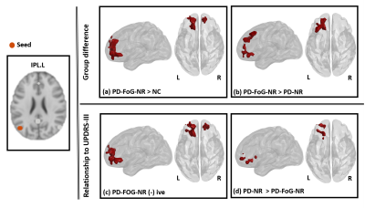 |
Default mode network connectivity differences in levodopa responsive subtypes of Parkinson’s disease patients with freezing of gait.
Karthik R Sreenivasan1, Xiaowei Zhuang1, Jason Longhurst1, Zhengshi Yang1, Dietmar Cordes1, Aaron Ritter1, Jessica Caldwell1, Jeffrey L Cummings1, Zoltan Mari1, Irene Litvan2, Brent Bluett3, and Virendra Mishra1
1Cleveland Clinic Lou Ruvo Center for Brain Health, Las Vegas, NV, United States, 2University of California San Diego, La Jolla, CA, United States, 3Stanford University, Palo Alto, CA, United States
Dopaminergic deficiency can cause altered deactivation of the default mode network (DMN) connectivity, which subsequently impacts executive task performance resulting in freezing of gait (FOG). While the majority of Parkinson’s disease (PD) patients with FOG (PD-FOG) are responsive to levodopa, about 36% of PD patients are levodopa-resistant. Our results show increased DMN connectivity in levodopa-resistant PD-FOG group. Furthermore, we found that altered network connectivity in the levodopa-resistant group was correlated differently with neuropsychological measures. To the best of our knowledge this is the first study to investigate the functional connectivity differences in levodopa-resistant subtypes in PD-FOG.
|
|
1514.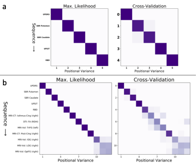 |
Data-driven model of Parkinson’s disease progression performs precision staging with magnetic resonance imaging biomarkers
Neil P Oxtoby1, Leon M Aksman2, Louise-Ann Leyland3, Rimona S Weil3, and Daniel C Alexander1
1Department of Computer Science, University College London, London, United Kingdom, 2Department of Medical Physics and Biomedical Engineering, University College London, London, United Kingdom, 3Department of Neurodegenerative Diseases, University College London, London, United Kingdom
We estimate a data-driven signature of de novo Parkinson's disease progression as a sequence of disease events. We show that clinical decline in classic markers precedes grey-matter and white-matter neurodegeneration estimated from T1-weighted MRI and diffusion-weighted MRI. Using only cross-sectional data from the PPMI data set, we show model utility for fine-grained staging/stratification of patients, which holds promise for future clinical applications.
|
|
1515.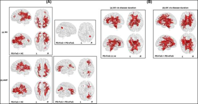 |
Beyond Single Tensor Diffusion Metrics to Quantify White Matter Disorganization in Parkinson’s disease With Freezing of Gait
Virendra R Mishra1, Jason Longhurst1, Jessica Caldwell1, Aaron Ritter1, Karthik R Sreenivasan1, Xiaowei Zhuang1, Zhengshi Yang1, Zoltan Mari1, Dietmar Cordes1,2, Jeffrey Cummings1,3, Irene Litvan4, and Brent Bluett5
1Cleveland Clinic Lou Ruvo Center for Brain Health, Las Vegas, NV, United States, 2University of Colorado, Boulder, Boulder, CO, United States, 3Department of Brain Health, University of Nevada, Las Vegas, Las Vegas, NV, United States, 4University of California, San Diego, San Diego, CA, United States, 5Stanford University, Stanford, CA, United States
Freezing-of-gait (FoG) which is one of the main causes of falls in Parkinson’s disease (PD), results in significant morbidity and mortality. Currently, there are no robust methods of elucidating the neural mechanisms underlying this disabling aspect of PD. Utilizing a well-characterized cohort of PD-patients with-FoG (PD-FoG), PD-patients without-FoG (PD-nFoG), and healthy controls, we showed that diffusion kurtosis imaging and free-water corrected single-tensor diffusion MRI (dMRI)-derived measures identified significant differences in dMRI-derived measures between PD-FoG and PD-nFoG. Our study indicate that these beyond single-tensor dMRI models may identify robust and generalizable dMRI-derived measures to elucidate the neural mechanisms underlying PD-FoG.
|
|
1516.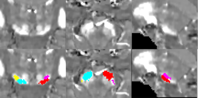 |
Investigating Iron deposition in the Substantia Nigra of Early Parkinson’s Disease and Idiopathic REM Sleep Behavior Disorder using QSM and R2*
Rahul Gaurav1,2, Romain Valabregue1, Nadya Pyatigorskaya1, Emma Biondetti1, Graziella Mangone3, Claire Ewenczyk4, Matthew Hutchison5, Isabelle Arnulf6, Jean-Christophe Corvol4, Marie Vidailhet4, Mathieu D. Santin1, and Stephane Lehericy1
1CENIR - Center for Neuroimaging Research, ICM - Brain and Spine Institute, Paris, France, 2Move'IT - Movement Investigations and Therapeutics, ICM - Brain and Spine Institute, Paris, France, 3Clinical Investigation Center (CIC-9503), INSERM - French National Institute of Medical Research and Health, Paris, France, 4Department of Neurology, Pitie-Salpetriere Hospital, Paris, France, 5Biogen Inc., Cambridge, MA, United States, 6Sleep Disorders Unit, Pitie-Salpetriere Hospital, Paris, France
Parkinson’s disease (PD) and idiopathic rapid eye movement sleep behavior disorder (iRBD) demonstrate neurodegenerative changes in the substantia nigra (SN) associated with an increase in iron deposition in PD patients. We aimed to quantify iron overload in the SN in early stage PD and iRBD patients using QSM-based and neuromelanin (NM)-based automated segmentation for QSM and R2* maps. We observed an increase in iron deposition in both early PD and iRBD patients with respect to healthy volunteers (HV).
|
|
1517.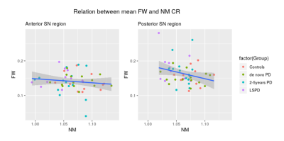 |
Substantia nigra changes in Parkinson’s Disease: correlation between Neuromelanin contrast and Free Water fraction
Joana M Grilo1, Marc Golub1, Sofia Reimão2,3, Rafael Neto Henriques4, Ana Fouto1, Patrícia Pita Lobo3, Margherita Fabbri3, Joaquim J Ferreira3,5, and Rita G Nunes1
1Department of Bioengineering, ISR-Lisbon/LARSyS, Lisbon, Portugal, 2Neurological Imaging Department, Hospital de Santa Maria - CHLN, Lisbon, Portugal, 3Instituto de Medicina Molecular, Faculty of Medicine, University of Lisbon, Lisbon, Portugal, 4Champalimaud Neuroscience Programme, Champalimaud Centre for the Unknown, Lisbon, Portugal, 5CNS – Campus Neurológico Sénior, Torres Vedras, Portugal
Parkinson’s Disease is characterized by the degeneration of neuromelanin (NM)-containing neurons in the Substantia Nigra (SN). MRI techniques have emerged aiming to evaluate PD disease progression. NM-MRI (current gold-standard), depicts a reduction in signal intensity at the posterior SN. Recently, free water (FW) fraction maps obtained from DWI, have shown an increase of FW in the SN in PD. This work’s goal was to evaluate how the FW and NM signal relate. A negative correlation between FW and NM was observed in the SN posterior region, suggesting that both metrics can potentially be used as imaging biomarkers.
|
|
1518. |
In-vivo Visualization of Locus Coeruleus using MTC-STAGE Imaging
Yu Liu1, Jun Chen Li1,2, Yongsheng Chen3, Naying He1, Zhijia Jin1, Weibo Chen4, Fuhua Yan1, and Ewart Mark Haacke1,5,6
1Radiology, Ruijin Hospital, Shanghai Jiao Tong University School of Medicine, Shanghai, China, 2Radiology, Changshu Hospital Affiliated to Nanjing University of Chinese Medicine, Changshu, China, 3Neurology, Wayne State University, Detroit, MI, United States, 4Philips Healthcare, Shanghai, China, 5Radiology, Wayne State University, Detroit, MI, United States, 6Biomedical Engineering, Wayne State University, Detroit, MI, United States Poster Permission Withheld
The locus coeruleus (LC) is mainly responsible for the synthesis of noradrenaline in the brain. Pathological alterations of the LC are involved in many neurodegenerative diseases. In this work, we use the tissue properties (spin density and T1 value) of the LC extracted from an MTC-STAGE (strategically acquired gradient echo) susceptibility weighted imaging protocol. Choosing the right flip angle and resolution can provide optimal visualization of the LC. We found that a short echo scan, with a flip angle of 25-30o and a resolution of 0.67 x 0.67 x 1.34mm3 provides the best visualization of the LC.
|
|
1519. |
Investigating Functional Connectivity of Substantia Nigra pars compacta in Parkinson’s Disease
Apoorva Safai1, Shweta Prasad2,3, Jitender Saini4, Pramod Pal2, and Madhura Ingalhalikar5
1Symbiosis Center of Medical Image Analysis, Symbiosis International University, PUNE, India, 2Department of Neurology, National Institute of Mental Health and Neurosciences, Bangalore, India, 3Department of Clinical Neurosciences, National Institute of Mental Health and Neurosciences, Bangalore, India, 4Department of Neuroimaging & Interventional Radiology, National Institute of Mental Health and Neurosciences, Bangalore, India, 5Symbiosis Center of Medical Image Analysis, Symbiosis International University, Pune, India
Parkinson’s disease (PD) is characterized by loss of dopaminergic neurons in Substantia Nigra pars compacta (SNc). SNc to whole brain resting state functional connectivity (rsFC) was compared between healthy controls (HC) and patients with PD to study the functional network of SNc in PD, and its association with disease progression was evaluated using a neuromelanin sensitive MRI based probabilistic atlas of SNc. Putamen, cerebellum and insular cortex connectivity with SNc was significantly reduced in PD as compared to HC. Widespread frontal, occipital regions, SMA and cerebellum were associated with duration and severity of PD.
|
|
1520.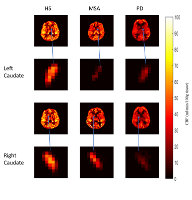 |
Brain Perfusion in Parkinson’s Disease and Multiple System Atrophy using Arterial Spin Labelling MRI
Roshni Kedia1, Archana Vadiraj Malagi1, Jitender Saini2, and Amit Mehndiratta3
1Centre for Biomedical Engineering, Indian Institute of Technology Delhi, New Delhi, India, 2Department of Neuroimaging & Interventional Radiology, National Institute of Mental Health and Neuro Sciences, Bengaluru, India, 3Indian Institute of Technology Delhi, New Delhi, India
Absolute perfusion varies in different brain regions for Parkinson’s disease (PD) and Multiple System Atrophy (MSA). These were studied quantitatively using ASL-MRI and the mean perfusion for each brain region was compared. Significant decrease in perfusion was noted for PD compared to healthy subjects for left and right caudate, anterior and posterior cingulate gyrus and occipital fusiform gyrus. For MSA, right caudate showed significant decrease in perfusion compared to healthy subjects.
|
|
1521.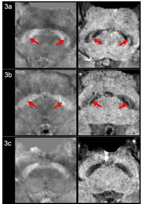 |
Quantifying Nigrosome-1 Loss in the Substantia Nigra on Susceptibility-Map Weighted Images in Essential Tremor and Parkinson’s Disease
Septian Hartono1,2, Isabel Hui Min Chew3, Amanda Jieying Lee3, Joey Xin Yi Oh3, Leon Qi Rong Ooi4, Yao-Chia Shih3, Jongho Lee5, Zheyu Xu1,2, Eng King Tan1,2, and Ling Ling Chan2,3
1Department of Neurology, National Neuroscience Institute, Singapore, Singapore, 2Duke-NUS Medical School, Singapore, Singapore, 3Department of Diagnostic Radiology, Singapore General Hospital, Singapore, Singapore, 4Department of Electrical & Computer Engineering, National University of Singapore, Singapore, Singapore, 5Department of Electrical & Computer Engineering, Seoul National University, Seoul, Korea, Republic of Poster Permission Withheld
Our study is the first to investigate the value of susceptibility map-weighted imaging in quantifying Nigrosome-1 loss in the substantia nigra to distinguish between tremor-dominant Parkinson’s disease (TDPD) and Essential Tremor (ET). Region-of-interest (ROI) masks of the Substantia Nigra and midbrain background were manually drawn on SMWI images of 50 subjects comprising 18 healthy controls, 25 ET and 7 TDPD patients. The SN in TDPD patients contained significantly more voxels more hypointense than the background than in ET patients. This could be explained by greater Nigrosome-1 loss and iron deposition in TDPD patients early in the disease.
|
|
1522.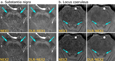 |
Evaluation of the Substantia Nigra and Locus Coeruleus by Neuromelanin-Sensitive MR Imaging with Deep Learning Based Noise Reduction
Sonoko Oshima1, Yasutaka Fushimi1, Satoshi Nakajima1, Yusuke Yokota1, Sayo Otani1, Azusa Sakurama1, Krishna Pandu Wicaksono1, Yuichiro Sano2, Ryo Matsuda2, Masahito Nambu2, Koji Fujimoto3, Hitomi Numamoto4, Kanae Kawai Miyake4,
Tsuneo Saga4, and Kaori Togashi1
1Department of Diagnostic Radiology and Nuclear Medicine, Graduate School of Medicine, Kyoto University, Kyoto, Japan, 2MRI Systems Division, Canon Medical Systems Corporation, Otawara, Japan, 3Human Brain Research Center, Graduate School of Medicine, Kyoto University, Kyoto, Japan, 4Department of Advanced Medical Imaging Research, Graduate School of Medicine, Kyoto University, Kyoto, Japan
We assessed neuromelanin-sensitive MR images with number of excitations of 1 or 2 with and without deep learning reconstruction (DLR) denoising method about visualization of the substantia nigra (SN) and locus coeruleus (LC) in 19 patients. The results of visual assessment were better in images with DLR. Contrast ratios of SN did not change after application of DLR, whereas contrast ratios of LC were decreased and hyperintense SN areas became larger. Neuromelanin imaging with DLR has a potential to reduce scan time without spoiling image quality, but further studies are needed for interpreting the signal contrast of SN and LC.
|
 |
1523.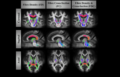 |
Fibre-specific white matter degeneration in the patients with Progressive supranuclear palsy
Po-Yuan Chen1, Yi-Ming Wu2, Yi-Hsin Weng3, and Jiun-Jie Wang1
1Chang Gung University, Taoyuan, Taiwan, 2Chang Gung Memorial Hospital, Linkou, Taoyuan, Taiwan, 3Chang Gung Memorial Hospital, Taoyuan, Taoyuan, Taiwan
Progressive supranuclear palsy (PSP) is an atypical Parkinsonism but with a faster progressive course. Previous studies indicated that PSP patients showed not only gray matter volume decrease but also white matter tract degeneration. We use fixel based analysis to examine the difference in the fibre bundle (FD), fibre-bundle cross-section (FC) and combination of fibre density and bundle cross-sectional area (FDC) between patients with PSP and healthy controls. The results show that significant degeneration of white matter in PSP patients. The major advantage to this study is providing a fixel-based comparison that indicate more directly interpretable measures of structural integrity.
|
1524.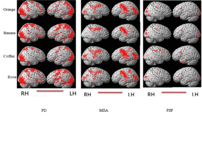 |
Multi-modality evaluation of Hyposmia in patients with Parkinson’s disease and atypical Parkinsonism
A Ankeeta1, Shefali Chaudhary1, S Senthil Kumaran1, Priyanka Bhat2, and Vinay Goyal2
1NMR and MRI facility, All India Institute of Medical Sciences, New Delhi, India, 2Neurology, All India Institute of Medical Sciences, New Delhi, India
The pattern of Hyposmia hemodynamic response, functional connectivity and ERP response was investigated in patients with Parkinson's disease, Multiple System Atrophy (MSA) and Progressive Supranuclear Palsy (PSP). Results revealed the presence of significant olfactory loss correlated with differential pattern in the olfactory pathway including frontal region and temporal areas. MRI, BOLD and EEG, can be used to detect early biomarker(s) for the identification of Parkinson and atypical Parkinsonism patients on the basis of hyposmia.
|
|
1525.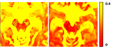 |
Free-water imaging in Substantia Nigra in Parkinson’s disease: A Neuromelanin Sensitive MRI Atlas Based Study
Apurva Shah1, Jacob Antony Alapatt2, Shweta Prasad3, Jitender Saini4, Pramod Pal3, Ragini Verma2, and Madhura Ingalhalikar1
1Symbiosis Center for Medical Image Analysis, Symbiosis International University, Pune, India, 2Department of Radiology, University of Pennsylvania, Philadelphia, PA, United States, 3Department of Neurology, National Institute of Mental Health and Neurosciences, Bengaluru, India, 4Department of Radiology, National Institute of Mental Health and Neurosciences, Bengaluru, India
Parkinson’s disease is characterized by degeneration of dopaminergic neurons in the substantia nigra pars compacta (SNc). To study the micro-structural and free-water (FW) changes using diffusion-MRI in the SNc it is critical to extract SNc accurately. Our work employs a neuromelanin sensitive MRI based atlas to delineate the SNc and demonstrates significant FW and FW eliminated microstructural alterations in a large cohort of PD (with and without psychosis) and its association with PD severity, indicative of novel diagnostic and progression markers of PD which however demonstrate no role in genesis of psychosis in PD.
|
|
1526.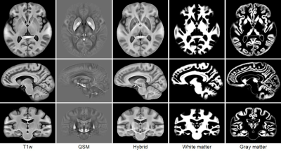 |
Investigation of subcortical brain structures in patients with Parkinson's disease using a quantitative susceptibility mapping atlas
Boliang Yu1, Ling Li2, Xueling Liu2, Naying He3, Hongjiang Wei4, Chuantao Zuo2, Fuhua Yan3, and Yuyao Zhang1
1School of Information Science and Technology, ShanghaiTech University, Shanghai, China, 2PET Center, Huashan Hospital, Fudan University, Shanghai, China, 3Department of Radiology, Ruijin Hospital, Shanghai Jiaotong University School of Medicine, Shanghai, China, 4Institute for Medical Imaging Technology, School of Biomedical Engineering, Shanghai Jiao Tong University, Shanghai, China
A limited number of atlases have been constructed using quantitative susceptibility mapping (QSM) images from subjects with Parkinson’s disease (PD), and given disease-specific subcortical structures. In this work, we generated three standard-space templates i.e. the hybrid, QSM and T1w atlas, which kept good image quality to observe brain white, gray matter, and deep-brain nuclei. Based on the atlases, we achieved the manual annotation of a few brain subcortical structures, e.g. globus pallidus, substantia nigra, subthalamic nucleus and thalamus. The results gave the position and shape of subcortical nuclei which could be meaningful for the research and surgical treatment of PD.
|
|
1527.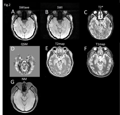 |
Visualization of Nigrosome-1 with Improved Contrast-to-noise Ratio and Correlation with Signals on Neuromelanin-sensitive MRI
Tzu-Wei Lee1, Chao-Wei Tso1, Kuan Chen1, Cheng-Yu Chen2, and Hua-Shan Liu1
1School of Biomedical Engineering, College of Biomedical Engineering, Taipei Medical University, Taipei City, Taiwan, 2Department of Radiology, School of Medicine, Taipei Medical University, Taipei City, Taiwan
A comparative study of different techniques for delineation of the nigrosome-1 in substantia nigra (SN) is needed to define the most sensitive imaging biomarker for SN-related diseases. This study was conducted to assess the potential of multiecho susceptibility-weighted imaging (SWI) in the delineation of the nigrosome-1. We also evaluated the relationship between neuromelanin (NM) and relaxation times of SN. We found that multiecho SWI can improve the contrast-to-noise ratio (CNR). The older subjects exhibited increased CNR values. The correlation between NM-MRI and T2* value suggested that magnetization-transfer effect may be related to the presence of melanin-iron complex in the nigrosome-1.
|
|
1528.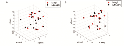 |
Can short echo time magnitude image of quantitative susceptibility mapping resembles neuromelanin-sensitive MRI image of the substantia nigra?
Xueling Liu1, Liqin Yang1, Yuxin Li1, Daoying Geng1, Pu-Yeh Wu2, and Yong Zhang2
1Department of Radiology, Huashan Hospital, Fudan University, Shanghai, China, 2GE Healthcare China, Beijing, Shanghai, China
Loss of melanized dopaminergic neurons(1) and iron deposition(2) in substantia nigra (SN) were pathological hallmarks of Parkinson’s disease (PD). Susceptibility images from QSM could detect iron deposition(3, 4) while neuromelanin-sensitive MRI (NM-MRI) could reflect change of melanized dopaminergic neurons(5, 6) in SN. In this study, we found highly spatial similarity of SNhyperintense on Mag1 images from QSM and on NM-MRI images. PD-patients could be differentiated from old HCs on Mag1 images as similar as that on NM-MRI images. Combined with Mag1 and susceptibility images, QSM could provide a promising imaging biomarker for iron deposition and NM deficiency in PD simultaneously.
|
|
1529.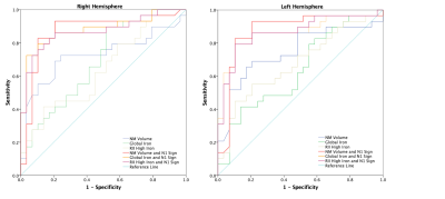 |
Assessing the Diagnostic Power of Parkinson’s Disease Biomarkers: Nigrosome-1 Sign, Neuromelanin and Iron Quantification
Zhijia Jin1, Zenghui Cheng1, Naying He1, Pei Huang2, Sean K. Sethi3,4, Mojtaba Jokar3, Weibo Chen5, Shengdi Chen2, Fuhua Yan1, and E. Mark Haacke1,3,4,6
1Department of Radiology, Ruijin Hospital, Shanghai Jiao Tong University School of Medicine, Shanghai, China, 2Department of Neurology, Ruijin Hospital, Shanghai Jiao Tong University School of Medicine, Shanghai, China, 3Magnetic Resonance Innovations, Inc., Bingham Farms, MI, United States, 4Department of Radiology, Wayne State University, Detroit, MI, United States, 5Philips Healthcare, Shanghai, China, 6Department of Biomedical Engineering, Wayne State University, Detroit, MI, United States
Twenty-nine Parkinson’s disease (PD) patients and 29 age- and sex-matched healthy controls (HCs) were scanned using a single 3D gradient echo magnetization transfer sequence to evaluate neuromelanin volume, global and regional iron content, and the appearance of nigrosome-1 territory (the “N1 sign”) in the substantia nigra. Iron increase and neuromelanin volume reduction were found in PD patients compared to HCs. 21/29 and 4/29 of PD patients showed bilateral and unilateral loss of the N1 sign, respectively. Combining the N1 sign with neuromelanin volume, global iron content and regional iron content respectively improved diagnostic performance to differentiate PD patients from HCs.
|
|
1530. |
Prospective Longitudinal Diffusion Tensor Tractography Evidence of Nigrostriatal Degeneration in Early Parkinson’s Disease
Arthur Yong1, Amanda Lee2, Septian Hartono2,3, Isabel Chew2, Samuel Ng3, Xinyi Choi3, Wilson Jia Wei Wong2, Linda Soo Lee Lim4, Eng King Tan1,3, Louis Tan1,3, and Ling Ling Chan1,2
1Duke-NUS Graduate Medical School, Singapore, Singapore, 2Singapore General Hospital, Singapore, Singapore, 3National Neuroscience Institute, Singapore, Singapore, 4National Heart Centre Singapore, Singapore, Singapore
Parkinson's disease (PD) is characterized by progressive dopaminergic neuronal loss in the substantia nigra (SN) and dopaminergic deafferentation in the striatal nuclei. Diffusion tensor tractography (DTT) has been used to document cross-sectional changes in the nigrostriatal pathway (NSP) in PD. Our prospective longitudinal study using DTT revealed significant interval NSP degeneration over a two-year period compared to controls, besides cross-sectional changes in the NSP congruent with current literature. Our results suggested that demyelination may be the dominant factor in NSP degeneration in PD. DTT may be a useful objective biomarker of disease progression in the early stages of PD.
|
|
1531. |
Patterns of regional cortical thinning in cognitively impaired patients with Parkinson’s disease
Shefali Chaudhary1, S Senthil Kumaran1, Vinay Goyal2, GS Kaloiya3, M Kalaivani4, NR Jagannathan1, Rajesh Sagar5, Nalin Mehta6, and Achal Srivastava2
1Department of NMR & MRI Facility, All India Institute of Medical Sciences, New Delhi, India, 2Department of Neurology, All India Institute of Medical Sciences, New Delhi, India, 3National Drug Dependence Treatment Centre, All India Institute of Medical Sciences, New Delhi, India, 4Department of Biostatistics, All India Institute of Medical Sciences, New Delhi, India, 5Department of Psychiatry, All India Institute of Medical Sciences, New Delhi, India, 6Department of Physiology, All India Institute of Medical Sciences, New Delhi, India
Cognitive impairment (CI) affects 20-40% Parkinson’s disease (PD) patients. Cortical thickness (CT), measuring the shortest distance between brain surface and inner edge of cortical gray matter may relate to CI in PD. In this study, we evaluated CT alteration in cognitively impaired PD patients (PD-CI) in comparison to cognitively normal (CN) healthy controls (HC) and PD patients (PD-CN) using 3DT1 MR data. Extended cortical thinning in frontal, temporal, parietal, occipital regions in PD-CI and significant positive association with global cognition MoCA score may signify cognition linked Lewy pathology and may be a promising tool to characterize cognition in PD.
|
|
1532. |
In-vivo characterization of the biochemical properties of the locus coeruleus and substantia nigra in healthy controls and Parkinson’s disease
Catarina Rua1, Claire O'Callaghan2, Ron Ye3,4, Luca Passamonti3, P Simon Jones3, Guy B Williams1, James B Rowe3,4, and Christopher T Rodgers1
1Wolfson Brain Imaging Centre, Department of Clinical Neurosciences, University of Cambridge, Cambridge, United Kingdom, 2Behavioural and Clinical Neuroscience Institute and Department of Psychology, University of Cambridge, Cambridge, United Kingdom, 3Department of Clinical Neurosciences, University of Cambridge, Cambridge, United Kingdom, 4Medical Research Council Cognition and Brain Sciences Unit, University of Cambridge, Cambridge, United Kingdom
In Parkinson’s disease, there is severe loss of dopaminergic projection neurons of the substantia nigra (SN) and locus coeruleus (LC). Histology shows that damage is non-uniform and occurs in stages; i.e. there is preferential degeneration of neurons first in the rostral portion of the SN and only later on in the LC. In this study, we measured the biochemical properties of the SN and LC with T2* and Magnetization Transfer imaging in patients with Parkinson’s disease and two groups of healthy controls.
|
|
1533.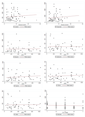 |
Identification of potential saliva bio-markers in early and advanced Parkinson’s disease using high resolution NMR
Sadhana Kumari1, S.Senthil Kumaran1, Vinay Goyal2, Achal Srivastava2, SadaNand Dwivedi3, and N.R. Jagannathan1
1NMR & MRI Facility, All India Institute of Medical Sciences, New Delhi, India, 2Neurology, All India Institute of Medical Sciences, New Delhi, India, 3Biostatistics, All India Institute of Medical Sciences, New Delhi, India
NMR-based metabolomics of saliva was studied in patients with Parkinson’s disease (PD) in early and advanced stages in comparison with that of healthy controls (HC). Higher levels of histidine, TMAO, propionate, GABA, valine, isoleucine, alanine, and fucose were observed in early PD in comparison with HC. Higher propionate and acetoin concentrations were observed in both early and advanced PD groups as compared to the HC group. An association of a few metabolites with the disease duration, LEDD and H&Y stage of PD patients was observed. Gut microflora system, ketone body and energy metabolisms may be impaired in patients with PD.
|
|
1534.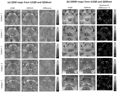 |
A Preliminary attempt to Visualize Nigrosome 1 in the Subtantia Nigra for Parkinson’s Disease at 3T: An efficient SMWI imaging with QSMnet
Minju Jo1 and Se-Hong Oh2,3
1Laboratory for Imaging Science and Technology, Department of Electrical and Computer Engineering, Seoul National University, Seoul, Republic of Korea, 2Department of Biomedical Engineering, Hankuk University of Foreign Studies, Yongin, Republic of Korea, 3Imaing Institute, Cleveland Clinic Foundation, Cleveland, OH, United States
We have described an efficient approach for SMWI visualizing SN and nigrosome 1 on clinical field strength (). QSMnet provides a similar SMWI image to that obtained with the conventional iterative QSM algorithm (such as iLSQR) but improves QSM processing speed by avoiding iterative computation. Since QSM reconstruction is the most time-consuming step of SMWI processing, QSMnet can help to achieve an improved SMWI processing speed. The application of QSMnet will be helpful when processing a massive amount of data or may contribute to the development of a scanner embedded real-time reconstruction of SWMI.
|
|
1535.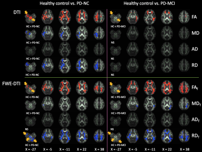 |
White Matter Plasticity in Newly Diagnosed Parkinson’s Disease With/Without Mild Cognitive Impairment
Christina Andica1, Koji Kamagata1, Yuya Saito1,2, Wataru Uchida1,2, Akifumi Hagiwara1, Shohei Fujita1,3, Syo Murata1, Masaaki Hori1,4, and Shigeki Aoki1
1Department of Radiology, Juntendo University Graduate School of Medicine, Tokyo, Japan, 2Department of Radiological Sciences, Graduate School of Human Health Sciences, Tokyo Metropolitan University, Tokyo, Japan, 3Department of Radiology, Graduate School of Medicine, The University of Tokyo, Graduate School of Medicine, Tokyo, Japan, 4Department of Radiology, Toho University Omori Medical Center, Tokyo, Japan
We evaluated the white matter (WM) of patients with newly diagnosed Parkinson’s disease (PD) with normal cognition (PD-NC) and mild cognitive impairment (PD-MCI) using diffusion tensor imaging (DTI) and free-water elimination DTI. Increased fractional anisotropy and decreased mean diffusivity and radial diffusivity in patients with PD suggested compensatory neural circuit reorganization. Changes were less extensive in WM areas previously considered vulnerable to MCI in the PD-MCI group than in the PD-NC group. Longitudinal analyses indicated neural compensation in the PD-NC group after 1 year. Overall, extensive and long compensatory mechanisms may be associated with preserved cognitive function in PD.
|
|
1536.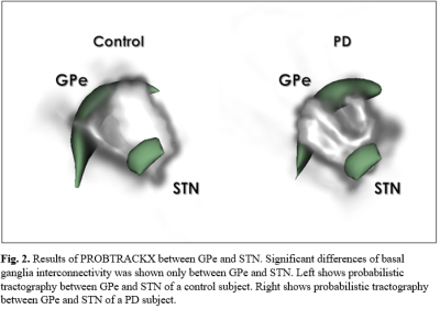 |
Comparison of interconnected basal ganglia probabilistic tractography between Parkinson's disease patients and controls
Jae-Hyuk Shim1 and Hyeon-Man Baek1
1Gachon University, Incheon, Republic of Korea
Basal ganglia structures, globus pallidus internal, globus pallidus external, subthalamic nucleus, substantia nigra, red nucleus and striatum were automatically segmented on 7T diffusion weighted images of controls and Parkinson's disease patients. Connectivity between each basal ganglia structure was observed using probabilistic tractography generated with FSL's diffusion tools such as BEDPOSTX and PROBTRACKX. Basal ganglia tractography was compared between controls and Parkinson's disease patients to observe the possible changes that could occur in tractography due to Parkinson's disease.
|
|
1537.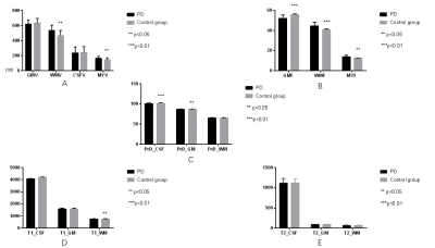 |
Investigation of the Global Volumetry and Relaxometry of the Brain in Parkinson's Disease using Synthetic MRI
Na Lu1,2, Chunmei Li1, Shuhua Li3, Pu-Yeh Wu4, Wen Su3, Haibo Chen3, and Min Chen1,2
1Department of Radiology, Beijing Hospital, National Center of Gerontology, Beijing, China, 2Graduate School of Peking Union Medical College, Beijing, China, 3Department of Neurology, Beijing Hospital, National Center of Gerontology, Beijing, China, 4GE Healthcare, MR Research China, Beijing, China
This study revealed both the brain volumetric and relaxometric characteristics from synthetic MRI technique in Parkinson's disease (PD). Significantly differences were found in fraction of gray matter (GM), white matter (WM) and myelin. We also observed significantly differences in WM T1 value, GM and CSF proton density value. Hence Synthetic MRI might be a quantitative tool for clinical diagnosis of PD.
|
1538.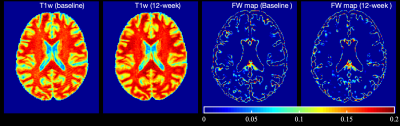 |
Relationship between Free Water and Neuroinflammation/Neurodegeneration Markers in HIV Before and After Combination Antiretroviral Therapy
Md Nasir Uddin1, Abrar Faiyaz2, Yuchuan Zhuang2, Madalina Tivarus3,4, Jianhui Zhong4,5, Maxime Descoteaux6, and Giovanni Schifitto4,7
1Department of Neurology, University of Rochester, Rochester, NY, United States, 2Electrical & Computer Engineering, University of Rochester, Rochester, NY, United States, 3Radiology, University of Rochester, Rochester, NY, United States, 4Imaging Sciences, University of Rochester, Rochester, NY, United States, 5Physics, University of Rochester, Rochester, NY, United States, 6Computer Science, University of Sherbrooke, Sherbrooke, QC, Canada, 7Neurology, University of Rochester, Rochester, NY, United States
Free water (FW) index, a measure of extracellular non-flowing water in the brain parenchyma, can be sensitive to neuroinflammation. We examine the relationship between the FW index and putative markers of neuroinflammation in cART-naïve participants before and after 12 weeks of the treatment. We found that FW index correlated with neuroinflammation markers in HIV+ participants for some GM and WM structures at baseline while this correlation diminished after 12 weeks of cART treatment in some WM structures for NfL.
|
|
1539. |
Quantitative Assessment of Pathological Brain Changes in HIV using MP2RAGE
Antonio Jimenez Gonzalez*1,2,3, Mário João Fartaria*1,2,3, Pietro Maggi4, Tobias Kober1,2,3, Jean-Philippe Thiran2,3, Karl Egger5, Renaud Du Pasquier4, François Lazeyras6, Frédéric Assal7, Alexandra Calmy8, Matthias Cavassini9, and Cristina Granziera10,11
1Advanced Clinical Imaging Technology, Siemens Healthcare, Lausanne, Switzerland, 2Department of Radiology, Lausanne University Hospital and University of Lausanne, Lausanne, Switzerland, 3LTS5, École Polytechnique Fédérale de Lausanne (EPFL), Lausanne, Switzerland, 4Departement of Neurology, Lausanne University Hospital and University of Lausanne, Lausanne, Switzerland, 5Department of Neuroradiology, Faculty of Medicine, University of Freiburg, Freiburg, Germany, 6Department of Radiology and Medical Informatics, CIBM, Geneva University Hospital, Geneva, Switzerland, 7Cognitive Neurology Unit, Department of Neurology, University Hospitals of Geneva, Geneva, Switzerland, 8Division of Infectious Diseases, University Hospital Geneva, Geneva, Switzerland, 9Department of Infectious Diseases, Lausanne University Hospital and University of Lausanne, Lausanne, Switzerland, 10Neurologic Clinic and Policlinic, Departments of Medicine, Clinical Research and Biomedical Engineering, University Hospital Basel and University of Basel, Basel, Switzerland, 11Translational Imaging in Neurology (ThINk) Basel, Department of Medicine and Biomedical Engineering, University Hospital Basel and University of Basel, Basel, Switzerland
The clinical landscape of HIV has evolved from a fatal disease to a manageable condition, giving rise to secondary complications associated with the chronic infection still present under treatment. HIV penetrates the brain very early after infection. Here, we investigated the mechanism behind structural brain changes observed in 92 aviremic HIV patients compared to 125 seronegative controls using quantitative MRI. Changes in cortical and subcortical structures and T1 relaxation times were observed. We thus speculate that the differential pattern in HIV patients reflects biological mechanisms underlying different stages of brain infection, namely acute inflammation and neuronal loss.
|
|
1540. |
Cerebral Blood Flow and Cerebrovascular Reactivity in Acute and Chronic HIV-Infection Treated by Combination Antiretroviral Therapies
Kyle Murray1, Md. Nasir Uddin2, Madalina Tivarus3, Arun Venkataraman1, Yuchuan Zhuang4, Xing Qiu5, Lu Wang5, Meera Singh6, Jianhui Zhong1,3, Sanjay Maggirwar7, and Giovanni Schifitto2,3
1Physics and Astronomy, University of Rochester, Webster, NY, United States, 2Neurology, University of Rochester, Rochester, NY, United States, 3Imaging Sciences, University of Rochester, Rochester, NY, United States, 4Electrical and Computer Engineering, University of Rochester, Rochester, NY, United States, 5Biostatistics and Computational Biology, University of Rochester, Rochester, NY, United States, 6Microbiology and Immunology, University of Rochester, Rochester, NY, United States, 7Microbiology, Immunology and Tropical Medicine, The George Washington University, Washington DC, DC, United States
Combination antiretroviral therapy (cART) maintains virologic control in HIV patients, but may lead to neurotoxicity. By using neuroimaging and cellular microparticle quantification, we explore the effects cART may have in both acute and chronic HIV-infection. We find that cART treatment does reduce microparticle levels associated with neuroinflammation to those of controls. Further, microparticle levels and neuroimaging results strengthen assumptions about immune dysfunction in HIV infection. We demonstrate that cerebral blood flow and cerebrovascular reactivity can be used in conjunction with quantitative microparticle levels to study the effects of neuroinflammation and cART treatment in both acute and chronic HIV infection.
|
|
|
1541. |
Temporal progression patterns of brain atrophy in CBS and PSP determined using Subtype and Stage Inference
Yuya Saito1,2, Koji Kamagata2, Christina Andica2, Wataru Uchida1,2, Syo Murata2, Akifumi Hagiwara2, Toshiaki Akashi2, Akihiko Wada2, Masaaki Hori3, and Shigeki Aoki2
1Graduate School of Human Health Sciences, Tokyo Metropolitan University, Tokyo, Japan, 2Department of Radiology, Graduate School of Medicine, Juntendo University, Tokyo, Japan, 3Department of Radiology, Toho University Omori Medical Center, Tokyo, Japan
Corticobasal syndrome (CBS) and progressive supranuclear palsy (PSP) are two classic clinical syndromes derived from 4 microtubule-binding domain-repeat tau pathology. However, their clinical diagnosis remains challenging due overlaps in their motor symptoms. While majority of previous studies have assessed brain volumes using cross-sectional data, the present study utilizes Subtype and Stage Inference (SuStaIn) for brain volumes based on cross-sectional brain structural magnetic resonance imaging to identify the differences in temporal brain atrophy progression patterns between CBS and PSP. Our results suggested the utility of SuStaIn for estimating brain atrophy progression patterns in and discriminating between patients with CBS and PSP.
|
1542.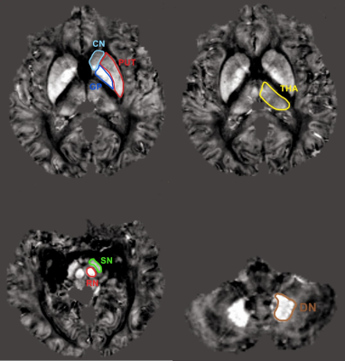 |
Different iron deposition patterns in hemodialysis patients with and without restless legs syndrome on MRI-QSM
Hao Wang1, Zhenchang Wang1, and Zhili on Xie2
1Department of Radiology, Beijing Friendship Hospital, Capital Medical University, Beijing, China, 2GE Healthecare, MR Research China, Beijing, Beijing, China, Beijing, China
Based on gradient echo (GRE) magnetic resonance phase data, quantitative susceptibility mapping (QSM) is a novel technology which allows the noninvasive assessment of magnetic tissue susceptibility distribution in hemodialysis (HD) patients. And iron deficiency in gray matter nuclei has been reported to lead to idiopathic restless legs syndrome (RLS) symptoms. In this study, we investigated the differences of iron deposition patterns between HD-RLS and HD-nRLS patients scanned at 3T. Compared with HD-nRLS patients, HD-RLS patients demonstrated reduced susceptibility in caudate nucleus and puteman. Hence, QSM can be used for HD-RLS diagnosis and intervention.
|
|
1543.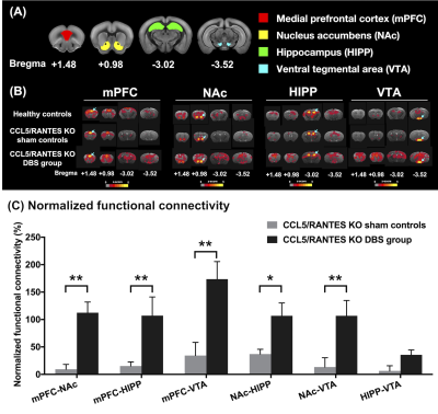 |
Nucleus Accumbens Deep Brain Stimulation Ameliorated Cognitive Impairment in Metabolic Syndrome Animal Model
Ting-Chieh Chen1, Yu-Chun Lo2, Szu-Yi Chou2, Ssu-Ju Li1, Ting-Chun Lin1, Ching-Wen Chang1, Yin-Chieh Liu1, Hsin-Tzu Lu1, and You-Yin Chen1,2
1Biomedical Engineering, National Yang-Ming University, Taipei, Taiwan, 2Ph.D. Program for Neural Regenerative Medicine, Taipei Medical University, Taipei, Taiwan
Cognitive dysfunctions were demonstrated to be associated with the metabolic syndrome (MetS), which could be treated with deep brain stimulation (DBS) of nucleus accumbens (NAc) by altering brain circuitss and facilitating synapse plasticity. However, NAc-DBS for memory-related cognitive function has yet to be investigated. Diffusion MRI, resting-state functional MRI, and behavioral test were applied in this study. We found restoration of the microstructure, increased functional connectivity, and an improvement in cognitive behaviors after NAc-DBS in the MetS models, C-C motif ligand 5/Regulated-on-Activation-Normal-T-cell-Expressed-and-Secreted knockout mice.
|
|
1544.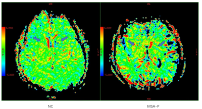 |
Investigation of amide proton transfer imaging in Multiple system atrophy at 3.0 Tesla
Na Lu1,2, Shuhua Li3, Chunmei Li1, Pu-Yeh Wu4, Wen Su3, Haibo Chen3, Piu Chan3, and Min Chen1,2
1Department of Radiology, Beijing Hospital, National Center of Gerontology, Beijing, China, 2Graduate School of Peking Union Medical College, Beijing, China, 3Department of Neurology, Beijing Hospital, National Center of Gerontology, Beijing, China, 4GE Healthcare, MR Research China, Beijing, China
Multiple system atrophy (MSA) is in great need of diagnosis in its early stage. Our study aims to evaluate the feasibility of using amide proton transfer (APT) imaging in detection of multiple system atrophy (MSA) at 3.0 Tesla. We found that APT MTRasym values were significantly higher in MSA patients than in normal controls at red nucleus, substantia nigra, thalamus and putamen. We also found that APT MTRasym values were significantly higher in probable MSA than in possible MSA patients at red nucleus, caudate and putamen. Hence, CEST may be valuable in diagnosing and predicting the progression of MSA.
|
|
1545.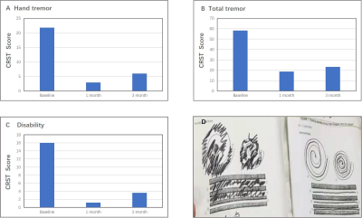 |
A pilot study of MR–guided focused ultrasound thalamotomy for refractory essential tremor
jianfeng he1, yongqin xiong1, rui zong1, dekang zhang1, xin zhou2, longsheng pan1, and xin lou1
1Chinese PLA General Hospital, Beijing, China, 2Wuhan Institute of Physics and Mathematics, Chinese Academy of Sciences, Wuhan, China
Essential tremor is the most common movement disorder and is often refractory to medical treatment, Deep brain stimulation in the thalamus has proved the efficiency for these patients. However, this treatment has risks associated with an open neurosurgical procedure. MR-guided focused ultrasound has been developed as a non-invasive means of generating precisely placed focal lesions. We examined its application to the management of refractory essential tremor. Satisfactory results were found that the mean reduction in tremor score of the treated hand was 86.7% at 1 month and 72.5% at 3 months, what’s more,no adverse events lasted beyond 3 months.
|
|
1546.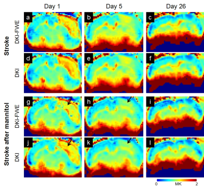 |
Exploration of mannitol-treated dehydration in acute stroke using diffusion kurtosis imaging with free water elimination
Chia-Wen Chiang1, Ezequiel Farrher2, Kuan-Hung Cho1, Shih-Yen Lin1,3, Kuo-Jen Wu4, Yun Wang4, Teh-Chen Wang5, Yi-Ping Chao6, Yeun-Chung Chang7, Chang-Hoon Choi2, and Li-Wei Kuo1,8
1Institute of Biomedical Engineering and Nanomedicine, National Health Research Institutes, Miaoli, Taiwan, 2Institute of Neuroscience and Medicine – 4, Medical Imaging Physics, Forschungszentrum Jülich, Jülich, Germany, 3Department of Computer Science, National Chiao Tung University, Hsinchu, Taiwan, 4Center for Neuropsychiatric Research, National Health Research Institutes, Miaoli, Taiwan, 5Department of Medical Imaging, Taipei City Hospital, Taipei, Taiwan, 6Department of Computer Science and Information Engineering, Chang Gung University, Taoyuan, Taiwan, 7Department of Medical Imaging, National Taiwan University Hospital, Taipei, Taiwan, 8Institute of Medical Device and Imaging, National Taiwan University College of Medicine, Taipei, Taiwan
Brain swelling typically occurs in acute stroke.1 Mannitol, as a hyperosmolar agent, enables to effectively treat the increased intraocular pressure and cerebral edema in brain injury.2,3 Diffusion kurtosis imaging with free water elimination (DKI-FWE)4 has been recently reported the ability to assess diffusion indices by separating free water compartment in simulations and healthy volunteers. The purpose of the study was to examine the effect of mannitol infusion using a stroke rat model at acute and chronic stages assessed by DKI-FWE and DKI. Our preliminary results revealed that mean kurtosis (MK) was sensitive to reflect mannitol-treated dehydration in acute stroke rat.
|
|
1547. |
Structural and resting state network alterations in Spinocerebellar Ataxia Type 2 in comparison with healthy controls
Pankaj Pankaj1, S Senthil Kumaran1, Snigdha Agrawal2, Achal Kumar Srivastava3, and Ramesh Kumar Agrawal2
1Department of NMR & MRI Facility, All India Institute of Medical Sciences, New Delhi, India, 2School of Computer & Systems Sciences, Jawaharlal Nehru University, New Delhi, India, 3Department of Neurology, All India Institute of Medical Sciences, New Delhi, India
Spinocerebellar ataxia type 2 (SCA2) is a progressive disorder with an early onset (10-15 years). On resting state functional connectivity and volume morphometrics, we observed reduced midbrain, sensory-motor and prefrontal cortex functional connectivity with cerebellum and atrophy in inferior parietal lobule, middle occipital gyrus, fusiform gyrus, posterior cingulate, precentral gyrus, parahippocampal gyrus, superior temporal gyrus, postcentral gyrus, fusiform gyrus, middle frontal gyrus, middle temporal gyrus, inferior frontal gyrus, middle temporal gyrus, pyramis, uvula, culmen, inferior semi-lunar lobule in SCA2, with respect to healthy controls. Atrophy and alterations in the rsfMRI connectivity suggest deficits in motor and cognition in SCA2 patients.
|
|
1548. |
Grey matter volume alterations in trigeminal neuralgia: A systematic review and meta-analysis of voxel-based morphometry studies
Yu Tang1, Maohua Wang2, Ting Zheng1, Fengying Yuan1, Fugang Han1, and Guangxiang Chen1
1Department of Radiology, The Affiliated Hospital of Southwest Medical University, Luzhou, China, 2Department of Anesthesiology, The Affiliated Hospital of Southwest Medical University, Luzhou, China
In recent decades, a growing number of structural neuroimaging studies of grey matter (GM) in trigeminal neuralgia (TN) have reported inconsistent alterations. We carried out a systematic review and meta-analysis to identify consistent and replicable GM volume abnormalities in TN patients. Our findings provide a thorough profile of GM volume alterations in TN patients and constitute robust evidence that aberrant GM volumes in the brain regions regulating and moderating sensory-motor and affective processing may play an important role in the pathophysiology of TN.
|
|
1549. |
Brain Iron Deposition and Cognitive Impairment in Idiopathic RBD: A Quantitative Susceptibility Mapping Study
Chao Chai1, Huiying Wang1, Tong Zhang2, Jinxia Zhu3, Xianchang Zhang3, E Mark Haacke4, Shuang Xia1, and Wen Shen1
1Department of Radiology, Tianjin First Central Hospital, Tianjin Medical Imaging Institute, Tianjin, China, 2School of Graduates, Tianjin Medical Univeristy, Tianjin, China, 3MR Collaboration, Siemens Healthcare Ltd., Beijing, China, 4Department of Radiology, Wayne State University, Detroit, MI, United States
Iron metabolism is a research focus in α‑synucleinopathies. Excessive iron deposition may damage neurons and induce cognitive impairment. However, research on brain iron deposition in patients with idiopathic rapid eye movement sleep behavior disorder (iRBD) is lacking. Using quantitative susceptibility mapping (QSM), the present study showed that iRBD patients have greater brain iron deposition in the substantia nigra and dentate nucleus than healthy controls (HCs). Additionally, brain iron deposition in the striatum and cerebellum was associated with cognitive impairment, suggesting the potential of QSM as an auxiliary biomarker for neurodegeneration and the early evaluation of cognitive decline in iRBD patients.
|
|
1550.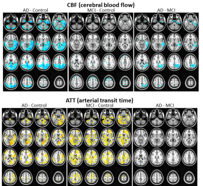 |
Revealing reduced CBF and prolonged ATT in prodromal AD using a 3D pCASL with Hadamard encoded multiple PLDs
Yang Wang1, Alexander Cohen1, Guanyu Chen2, Veena Nair3, Piero Antuono4, Malgorzata Franczak4, Vivek Prabhakaran3, Barbara Bendlin5, Shi-Jiang Li2, and the Alzheimer’s Disease Connectome Project6
1Radiology, Medical College of Wisconsin, Milwaukee, WI, United States, 2Biophysics, Medical College of Wisconsin, Milwaukee, WI, United States, 3Radiology, University of Wisconsin School of Medicine and Public Health, Madison, WI, United States, 4Neurology, Medical College of Wisconsin, Milwaukee, WI, United States, 5Medicine, University of Wisconsin School of Medicine and Public Health, Madison, WI, United States, 6Medical College of Wisconsin, Milwaukee, WI, United States
Measured using an advanced 3D pCASL with Hadamard encoded multiple PLDs, patients with MCI showed different patterns of reduced CBF and prolonged ATT in comparison with both AD patients and healthy controls, where CBF and ATT changes highly correlated with severity of disease as assessed by neuropsychological test scores. These findings raised the speculation of underlying vascular abnormality in prodromal AD. Our results also suggested that ATT could serve as useful hemodynamic measure of itself, may be of diagnostic utility for prodromal AD or vascular dementia.
|
|
1551.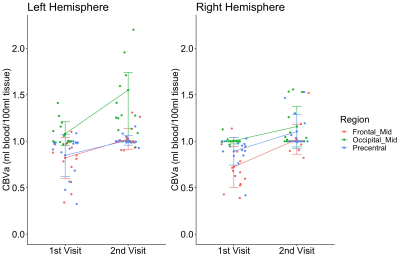 |
Increases in Arteriolar Cerebral Blood Volume in Huntington’s Disease Measured with Inflow-based Vascular-space-occupancy (iVASO) MRI at 7T
Chunming Gu1,2,3, Martin Kronenbuerger4,5, Di Cao1,2,3, Adrian G. Paez1,2, Xinyuan Miao1,2, Xirui Hou1,3, Jee Bang5,6, Kia E. Ultz5, Wenzhen Duan6,7, Russell L. Margolis5,6, Peter C. M. van Zijl1,2, Christopher A. Ross5,6,7,8, and Jun Hua1,2
1The Russell H. Morgan Department of Radiology and Radiological Sciences, Johns Hopkins University School of Medicine, Baltimore, MD, United States, 2F.M. Kirby Research Center for Functional Brain Imaging, Kennedy Krieger Institute, Baltimore, MD, United States, 3Department of Biomedical Engineering, Johns Hopkins University, Baltimore, MD, United States, 4Department of Neurology, University of Greifswald, Greifswald, Germany, 5Department of Neurology, Johns Hopkins University School of Medicine, Baltimore, MD, United States, 6Department of Psychiatry and Behavioral Sciences, Johns Hopkins University School of Medicine, Baltimore, MD, United States, 7The Solomon H. Snyder Department of Neuroscience, Johns Hopkins University School of Medicine, Baltimore, MD, United States, 8Department of Pharmacology and Molecular Sciences, Johns Hopkins University School of Medicine, Baltimore, MD, United States
Significantly elevated arteriolar cerebral blood volume (CBVa) in premanifest Huntington’s Disease (HD) patients has been reported previously. In this study, inflow-based vascular-space-occupancy (iVASO) MRI at 7 Tesla was used to measure CBVa in HD patients longitudinally. We found significant longitudinal increases in CBVa in premanifest HD patients in several brain regions primarily related to motor, visual and cognitive functions, which suggests CBVa as a potential candidate biomarker for HD especially in the premanifest stage.
|
|
1552.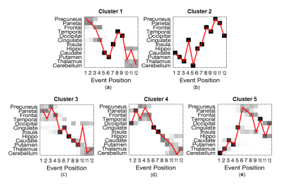 |
Strain-specific disease progression patterns of sporadic Creutzfeldt-Jakob disease revealed by Subtype and Stage Inference model
Riccardo Pascuzzo1, Alexandra L. Young2,3, Neil P. Oxtoby2, Janis Blevins4, Gianmarco Castelli1, Pierluigi Gambetti5, Brian S. Appleby4, Daniel C. Alexander2, and Alberto Bizzi1
1Neuroradiology Unit, Fondazione IRCCS Istituto Neurologico Carlo Besta, Milan, Italy, 2Centre for Medical Image Computing, Department of Computer Science, University College London, London, United Kingdom, 3Department of Neuroimaging, Institute of Psychiatry, Psychology and Neuroscience, King′s College London, London, United Kingdom, 4National Prion Disease Pathology Surveillance Center, Case Western Reserve University, School of Medicine, Cleveland, OH, United States, 5Department of Pathology, Case Western Reserve University, School of Medicine, Cleveland, OH, United States
Transmission studies in animal models have identified four strains of sporadic Creutzfeldt-Jakob disease (sCJD). Using a data-driven approach, we aim to identify subgroups of sCJD patients with distinct diffusion-weighted MRI (DWI) abnormality patterns, and test their association with disease strains. We used an unsupervised machine-learning algorithm named Subtype and Stage Inference, that identified 5 clusters of patients each having a distinct pattern of DWI abnormality progression: one had initial involvement of the parieto-frontal cortex; two started with subcortical regions (striatum, thalamus and cerebellum); and two had cortical and limbic regions affected early. Data-driven subgroups were significantly associated with sCJD strains.
|
1553.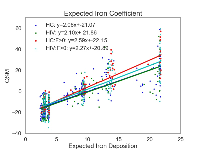 |
Brain Iron Imaging Markers in the Presence of White Matter Hyperintensities
Kyle Murray1, Md. Nasir Uddin2, Jianhui Zhong3, and Giovanni Schifitto2
1Physics and Astronomy, University of Rochester, Webster, NY, United States, 2Neurology, University of Rochester, Rochester, NY, United States, 3Imaging Sciences, University of Rochester, Rochester, NY, United States
HIV-infected individuals are at increased risk of developing white matter hyperintensities (WMHs), which can lead to increased iron deposition in deep gray matter structures. In this abstract, we evaluate three region of interest (ROI) brain iron metrics and introduce a novel population-based whole-brain iron metric, the expected iron coefficient (EIC), derived from quantitative susceptibility mapping (QSM) in the context of HIV and mild WMH burden. While the ROI metrics did not show any significant differences between cohorts, the EIC was able to detect iron related differences in a cohort with WMH burden, due to increased statistical power.
|
|
1554. |
Diffusion kurtosis imaging for characterizing the microstructural changes of hippocampus in mild cognitive impairment patients with cerebral small vascular disease.
Liu Dongtao1, Li Kun2, Bu Qiao2, Pan Zhenyu2, Feng Xiang3, Shi Qinglei3, and Zhou Lichun1
1Department of Neurology, Beijing Chaoyang Hospital, Capital Medical University, Beijing, China, 2Department of Radiology, Beijing Chaoyang Hospital, Capital Medical University, Beijing, China, 3MR Scientific Marketing, Diagnosis Imaging, Siemens Healthcare Ltd, Beijing, China
We investigated the early microstructural alterations in hippocampus in MCI patients with cSVD by DKI. Our study found that MCI patients with cSVD show more seriously white matter hyperintensity, and showed significantly increased MD and RD values, and decreased MK, AK, RK, FA and KFA values in left hippocampus. In left hippocampus, values of FA, MK, RK, and KFA showed significantly positive correlations with MoCA score, while MD and RD values were negatively correlated with MoCA score. This may be due to the loss of neuron cell bodies, synapses and dendrites. DKI technique may be feasible to probe the microstructural changes of hippocampus in MCI patients with cSVD.
|
|
1555. |
Synthetic MRI reveals leukoaraiosis is associated with cerebral small vessel diseases rather than cerebral atherosclerosis
jingdong Yang1, Juan Huang1, Yan Song1, and Pu Yeh Wu2
1Radiology, Beijing Hospital, Beijing, China, 2GE Healthcare, Beijing, China
We explored the feasibility of applying Synthetic MRI to quantify leukoaraiosis and further investigated the association of leukoaraiosis with small vascular cerebral diseases and large vessel atherosclerosis in individuals with acute cerebral vascular symptom. The results show that LA could be evaluated by the volume and relaxation time from synthetic MRI and it was associated with lacune and cerebral microbleeds rather than large cerebral atherosclerosis. This finding suggests that quantitative information provided by Synthetic MRI can help to evaluate leukoaraiosis, and leukoaraiosis may be a type of cerebral small vascular diseases instead of intracranial or extracranial cerebral atherosclerosis.
|
|
1556.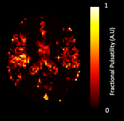 |
Physiological correlates of brain pulsatility in cerebrovascular small vessel disease using data-driven high temporal resolution fMRI
Paula L Croal1,2, Kevin J Ray1, Karolina Wartolowska3, Amy Lawson3, Alastair Webb3, and Peter Jezzard1
1Wellcome Centre for Integrative Neuroimaging, University of Oxford, Oxford, United Kingdom, 2Institute of Biomedical Engineering, University of Oxford, Oxford, United Kingdom, 3Centre for the Prevention of Stroke and Dementia, University of Oxford, Oxford, United Kingdom
Small vessel disease (SVD) is associated with stroke and dementia, however pathophysiological mechanisms are poorly understood, and effective treatments are lacking. Here, we determine the association between systemic arterial pulsatility and tissue-level cerebral pulsatility in patients with SVD, and its modulation by anti-hypertensive medication, using a novel analysis technique. We observe a significant association between systemic pulsatility and regional cerebral pulsatility, at baseline and on antihypertensive medication. The association was strongest in periventricular white matter most commonly affected by SVD. This regional dependence suggests that pulsatility is a pathophysiological factor underlying tissue damage in SVD, providing a potential treatment target.
|
|
1557.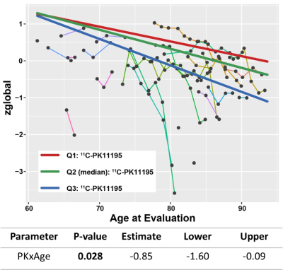 |
Neuro-inflammation is associated with WMH burden at baseline and predicts longitudinal cognitive decline in cerebral small vessel disease
Chunwei Ying1, Andria L. Ford2, Michael M. Binkley2, Yasheng Chen2, Peter Kang2, Jon Christensen1, Qing Wang1, Lisa Cash1, Jason Hassenstab2, Jin-Moo Lee1,2, Tammie L. S. Benzinger1,3, and Hongyu An1
1Mallinckrodt Institute of Radiology, Washington University School of Medicine, St. Louis, MO, United States, 2Department of Neurology, Washington University School of Medicine, St. Louis, MO, United States, 3Department of Neurological Surgery, Washington University School of Medicine, St. Louis, MO, United States
Neuro-inflammation has been suggested as an important pathogenesis factor for cerebral small vessel disease, but direct evidence in human is lacking. In this study, we found that neuro-inflammation, measured by 11C-PK11195 uptake, was associated with white matter hyperintensities burden at baseline. More importantly, it predicted cognitive decline in a longitudinal follow-up study.
|
|
1558.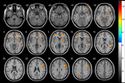 |
Increased Cerebral Blood flow was Correlated with Neurocognitive Dysfunction in Chronic Hemodialysis Patients: A Arterial Spin Labeling Study
Chao Chai1, Huiying Wang1, Tong Zhang2, Jinping Li3, Yingying Han3, Jinxia Zhu4, Xianchang Zhang4, Shuang Xia1, and Wen Shen1
1Department of Radiology, Tianjin First Central Hospital, Tianjin Medical Imaging Institute, Tianjin, China, 2School of Graduates, Tianjin Medical Univeristy, Tianjin, China, 3Department of Hemodialysis, Tianjin First Central Hospital, Tianjin, China, 4MR Collaboration, Siemens Healthcare Ltd., Beijing, China
Cerebral blood flow (CBF) can be significantly reduced during a single hemodialysis session. However, few studies have investigated the effect of long-term hemodialysis on CBF and its correlation with neuropsychological tests. This study used pulsed arterial spin labeling to show that hemodialysis patients had significantly increased CBF in some cerebral regions. Increased CBF of the right opercular and triangular part of inferior frontal gyrus was negatively correlated with Mini Mental State Examination (MMSE) scores, and after adjusting for hemoglobin and hematocrit levels, these correlations became stronger. Dialysis duration, hemoglobin, hematocrit, and serum phosphorus were clinical risk factors for increased CBF.
|
|
1559.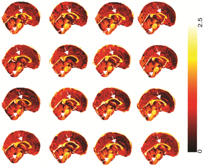 |
Higher levels of myelination were observed in chronic fatigue syndrome patients relative to healthy controls
KIRAN THAPALIYA1,2, Sonya Marshall-Gradisnik1, Don Staines1, Sandeep Bhuta3, Timothy Ireland3, and Leighton Barnden1
1Menzies Health Institute Queensland, Griffith University, Gold Coast, Australia, 2Centre for Advanced Imaging, The University of Queensland, Brisbane, Australia, 3Gold Coast University Hospital, Gold coast, Australia
Myalgic Encephalomyelitis (ME)/Chronic fatigue syndrome (CFS) patients suffer from a variety of physical and neurological complaints indicating the central nervous system plays a role in CFS pathophysiology. Studies based on genetic, immune system, psychiatric, and brain volume abnormalities and white matter hyperintensities have been investigated to identify the pathomechanism of this disease. However, assessment of myelination in brain regions between ME/CFS patients and healthy controls has not been investigated. In this study, we showed elevated myelination in white matter regions and grey matter regions in ME/CFS patients relative to normal controls.
|
|
1560.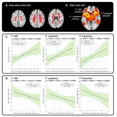 |
Delayed cerebrovascular response within deep white matter may alter the cortical-subcortical connections in small vessel disease patients
Yi-Tien Li1,2, Yi-Wen Chen1, and David Yen-Ting Chen1,3
1Department of Radiology, Taipei Medical University - Shuang Ho Hospital, New Taipei, Taiwan, 2Institute of Biomedical Engineering, National Taiwan University, Taipei, Taiwan, 3Department of Radiology, Stanford University, Palo Alto, CA, United States
We applied cross-correlation method on breath-holding task to estimate the cerebrovascular response (CVR) strength and latency within deep white matter. The CVR latency showed significant negative correlation with working memory (WM) 1-back accuracy (r = -0.546, p = 0.009). Further, the significant negative correlation between the average CVR latency within deep white matter and the functional connectivity in specific regions of WM activation area, including left anterior insular cortex and bilateral putamen was found. The result suggested that the changes of cortical-subcortical connections in cerebral small vessel disease can be reflected by the delayed CVR latency within deep white matter.
|
|
1561.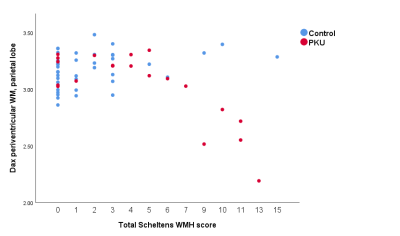 |
Diffusion Kurtosis Imaging Detects Subclinical White Matter Abnormalities in Phenylketonuria
Thomas Welton1, Sarah C Hellewell1, Michel Tchan2, and Stuart M Grieve1
1University of Sydney, Sydney, Australia, 2Department of Genetic Medicine, Westmead Hospital, Sydney, Australia
Diffusion kurtosis MRI (DKI) was used to measure changes in white matter structure in 20 patients with phenylketonuria and 43 controls. We found significant differences primarily in the periventricular parietal white matter in various DKI metrics. These scores were related to phenylalanine levels measured in the 3 years prior to MRI and with Scheltens score, reflecting white matter hyperintensities. DKI may be sensitive to pathology invisible to clinical MRI, and we propose a simple metric in the parietal lobe which robustly captures this effect.
|
|
1562.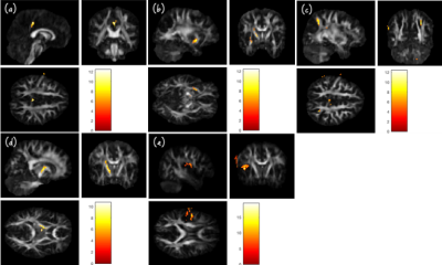 |
Detecting brain structural abnormality associated with breast cancer and chemotherapy using GQI
Wei Chuang1, Vincent Chin-Hung Chen2,3, Yuan-Hsiung Tsai4, and Jun-Cheng Weng1,3,5
1Department of Medical Imaging and Radiological Sciences, Chang Gung University, Taoyuan, Taiwan, 2School of Medicine, Chang Gung University, Taoyuan, Taiwan, 3Department of Psychiatry, Chang Gung Memorial Hospital, Chiayi, Taiwan, 4Department of Diagnostic Radiology, Chang Gung Memorial Hospital, Chiayi, Taiwan, 5Medical Imaging Research Center, Institute for Radiological Research, Chang Gung University and Chang Gung Memorial Hospital at Linkou, Taoyuan, Taiwan
Breast neoplasms are the most common cancer among women. Whether received adjuvant chemotherapy or not, lots of them presented cognitive impairment which decreased their quality of life. Therefore, we attempted to use generalized q-sampling imaging (GQI) to detect the white matter alteration between groups that may due to post-traumatic stress disorder (PTSD), post-traumatic growth (PTG) and chemotherapy. Our results showed the differences in the brain regions associated with cognition, emotion, and execution, such as angular gyrus, cingulate gyrus and so on. It indicted traumatic stressor or chemotoxicity can cause myelin injuries in these regions.
|
|
1563.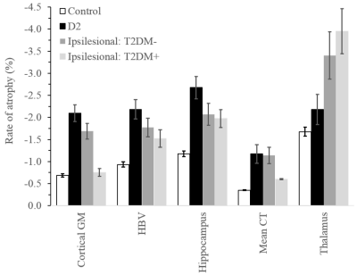 |
Comparison of brain atrophy in patients with type 2 diabetes from the Diabetes and Dementia (D2) study and stroke patients from the CANVAS study
Mohamed Salah Khlif1, Carolina Restrepo1, Emilio Werden1, Laura Bird1, Sheila K. Patel1,2, Rebecca Singleton1, Jeffrey D. Zajac2,3, Richard MacIsaac4, Elif I. Ekinci2,3, Louise M. Burrell2,5, and Amy Brodtmann1,2,6
1The Florey Institute of Neuroscience and Mental Health, University of Melbourne, Melbourne, Australia, 2Department of Medicine, University of Melbourne, Austin Health, Heidelberg, Australia, 3Austin Health Endocrine Centre, Heidelberg, Australia, 4Department of Endocrinology & Diabetes, St Vincent's Hospital, Melbourne, Australia, 5Department of Cardiology, Austin Health, Heidelberg, Australia, 6Department of Neurology, Austin Health, Heidelberg, Australia
Type 2 diabetes and stroke are associated with reduced brain volumes and independently present a high risk for cognitive impairment or dementia. The interaction between diabetes and stroke and its effects on brain atrophy are unknown. We used linear mixed-effect modelling to estimate rates of atrophy over two years based on FreeSurfer segmentation of MR images of healthy participants and patients with type 2 diabetes and/or stroke. Cerebral atrophy, defined by volume reductions in specific regions of interest, was accelerated the most in the group with only type 2 diabetes. Thalamus was affected more by stroke.
|
|
1564.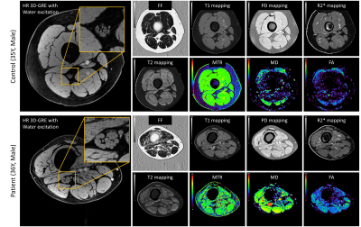 |
Proximal Nerve Quantitative MRI in Charcot-Marie-Tooth (CMT) Diseases
Yongsheng Chen1, E. Mark Haacke2, Yang Xuan2, Melody Hackett1, Sadaf Saba3, Bo Hu1, Daniel Moiseev1, and Jun Li1,3,4,5
1Department of Neurology, Wayne State University, Detroit, MI, United States, 2Department of Radiology, Wayne State University, Detroit, MI, United States, 3Center for Molecular Medicine & Genetics, Wayne State University, Detroit, MI, United States, 4Department of Biochemistry, Microbiology and Immunology, Wayne State University, Detroit, MI, United States, 5John D. Dingell VA Medical Center, Detroit, MI, United States
To test the hypothesis that quantitative MRI (qMRI) detects proximal nerve dysmyelination and axonal degeneration in Charcot-Marie-Tooth (CMT) diseases. Nine qMRI indices were collected, including whole muscle mean fat fraction (wmmFF), nerve fascicular cross-sectional area (fCSA), magnetization transfer ratio (MTR), T1, proton density (PD), R2, R2*, mean diffusivity (MD) and fractional anisotropy (FA). Compared to controls, patients with CMT had significantly elevated fCSA, T1, PD, T2* and MD for both divisions of sciatic nerves and wmmFF, elevated T2 for the tibial division but not the peroneal division, and decreased MTR for both divisions of sciatic nerves.
|
|
1565.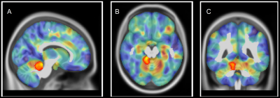 |
STRUCTURAL PLASTICITY IN THE BRAIN OF CMT1A PATIENTS? A COMBINED VBM AND TBSS STUDY
Sirio Cocozza1, Giuseppe Pontillo1, Raffaele Dubbioso1, Stefano Tozza1, Daniele Severi1, Lucio Santoro1, Andrea Elefante1, Fiore Manganelli1, Arturo Brunetti1, and Mario Quarantelli2
1University "Federico II", Naples, Italy, 2National Research Council, Naples, Italy
We investigated the presence of gray matter (GM) and white matter (GM) structural modifications in a homogeneous group of genetically defined CMT1A patients, by means of VBM and TBSS analyses, respectively. We found increased GM volume in CMT1A patients compared to HC encompassing the right paravermian portions of the cerebellar lobules III, IV and V, showing an inverse correlation with electrophysiological measures. These structural changes may reflect compensatory mechanisms in response to CMT1A peripheral nerve pathology, providing new insights into the comprehension of CNS physiopathology and its role in the development of clinical disability in this condition.
|
|
1566.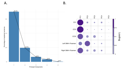 |
Drumming training induces myelin remodelling in Huntington’s disease: a diffusion MRI and quantitative magnetization transfer study
Chiara Casella1, Jose Bourbon-Teles1, Derek K Jones1, Greg Daniel Parker1, Sonya Bells2, Anne Rosser3, Elizabeth Coulthard4, and Claudia Metzler-Baddeley1
1Department of Psychology, Cardiff University Brain Research Imaging Centre, Cardiff, United Kingdom, 2Department of Neurosciences and Mental Health, The Hospital for Sick Children, Toronto, ON, Canada, 3Department of Biosciences, Cardiff University, Cardiff, United Kingdom, 4Department of Clinical Neurosciences, University of Bristol, Bristol, United Kingdom
Huntington’s disease is a neurodegenerative disorder leading to debilitating cognitive and motor symptoms. It has been proposed that impaired myelination contributes to HD pathogenesis. As well, evidence shows that myelin formation underlies the learning of new motor skills. Here we demonstrate that two months of drumming and rhythm exercises result in an increase in a proxy MRI measure of myelin in patients with early HD relative to healthy controls. This suggests that tailored behavioural stimulation has the potential to result in neural benefits in early HD that could be exploited for future therapeutics aiming to delay disease progression.
|
1567.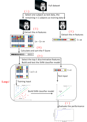 |
Identification of amyotrophic lateral sclerosis based on diffusion tensor imaging and support vector machine
Nao-Xin Huang1, Tian-Xiu Zou1, Zhongshuai Zhang2, and Hua-Jun Chen1
1Fujian Medical University Union Hospital, Fuzhou, China, 2SIEMENS Healthcare, Shanghai, China
White matter (WM) impairments have been well documented in amyotrophic lateral sclerosis (ALS). This study tested the potential of diffusion measurements in WM for identifying ALS based on the support vector machine (SVM). In the optimized SVM model, the FA values from the motor areas (including the bilateral precentral gyrus and the corticospinal tract) and extra-motor areas (including right postcentral gyrus, left superior/inferior longitudinal fasciculus) contributed mostly to classification. Our study suggests the feasibility of ALS diagnosis based on SVM analysis and diffusion measurements of WM. Future study with larger cohort is needed to validate the generality of our results.
|
|
1568.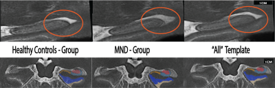 |
A population-specific framework for the morphological analysis of Motor Neuron Disease using ultra-high field MRI
Thomas B Shaw1, Saskia Bollmann1, Frederik Steyn1, Christine Guo2, Amir Fazlollahi3, Jurgen Fripp3, Olivier Salvado3,4, Steffen Bollmann1, and Markus Barth1
1The University of Queensland, Brisbane, Australia, 2QIMR Berghofer Medical Research Intstitute, Brisbane, Australia, 3CSIRO Health and Biosecurity, Brisbane, Australia, 4CSIRO Data61, Sydney, Australia
Imaging biomarkers for Motor Neuron Disease (MND) using MRI presents challenges due to neurodegeneration leading to heterogeneous brain morphology, low SNR, and movement artefacts. Here, we present optimised methods for imaging and processing MND patient data using UHF MRI with submillimetre resolution, and compare hippocampal subfields between patients and healthy controls. We used automatic measures to examine the shape, volume, and morphology of hippocampal subfields and showed specific changes in MND patients that are more sensitive than previous research. This work may serve as a framework for more sensitive computational models for biological characterisation of MND in vivo.
|
|
1569. |
Serial assessment of magnetic susceptibility in the primary motor cortex in limb-onset Amyotrophic Lateral Sclerosis
Anjan Bhattarai1,2, Zhaolin Chen2, Phillip G. D. Ward2, Paul Talman3, Susan Mathers4, Thanh Phan5, Caron Chapman4, James Howe4, Sarah Lee4, Yennie Lie4, Gary F Egan2, and Phyllis Chua1,4
1Department of Psychiatry, Monash University, Clayton, Australia, 2Monash Biomedical Imaging, Monash University, Clayton, Australia, 3Department of Neuroscience, Barwon Health, Geelong, Australia, 4Statewide Progressive Neurological Services, Calvary Health Care Bethlehem, South Caulfield, Australia, 5Department of Neuroscience, Monash Health, Clayton, Australia
We performed in-vivo measurements of the magnetic susceptibility in the motor cortex in individuals with Amyotrophic Lateral Sclerosis (ALS) at baseline and six-month follow-up, and healthy controls at baseline using Quantitative Susceptibility Mapping (QSM). The results show significant susceptibility difference between individuals with ALS compared to healthy controls. There was a trend towards more pronounced susceptibility changes in lumbar onset compared to healthy controls than cervical onset ALS compared to healthy controls. These findings may lead to the development of a sensitive neuroimaging biomarker that can provide meaningful insights into pathophysiologic changes in ALS subtypes.
|
|
1570.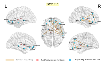 |
Disrupted grey matter network morphology in amyotrophic lateral sclerosis
Jing Yang1, Du Lei1,2, Xueling Suo1, and Qiyong Gong1,3
1Huaxi MR Research Center (HMRRC), Department of Radiology, West China Hospital of Sichuan University, Chengdu, China, 2University of Cincinnati, Cincinnati, OH, United States, 3Psychoradiology Research Unit of Chinese Academy of Medical Sciences (2018RU011), Chengdu, China
The present psychoradiological study demonstrated significant alterations both in global and nodal topological properties of single-subject brain morphological networks in a relatively large population of patients with ALS relative to HCs. This was achieved by combining graph theoretical analysis with a method of describing patterns of intercortical morphological similarities in individual participants using structural MRI data. We found that ALS showed a weaker small world organized brain network in global properties and decreased nodal centralities mainly in prefrontal-limbic network relative to HCs. Further, the nodal efficiency of right inferior frontal gyrus was positively correlated to ALSFRS in ALS group.
|
|
1571.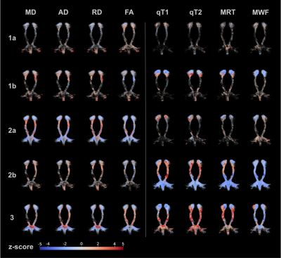 |
Spatial Distribution Patterns of Secondary White Matter Tract Degeneration in Amyotrophic Lateral Sclerosis
Hagen H Kitzler1, Paul Kuntke1, Carolin Schwamborn1, Hannes Wahl1, Rene Guenther2, Andreas Herrmann3, Sean C Deoni4, and Jennifer Linn1
1Neuroradiology, Technische Universität Dresden, Dresden, Germany, 2Neurology, Technische Universität Dresden, Dresden, Germany, 3Neurology, Universitätsmedizin Rostock, Rostock, Germany, 4Alpert Medical School, Brown University, Providence, RI, United States
Amyotrophic lateral sclerosis (ALS) is a rapidly progressing neurodegenerative disease of primary cortical neuronal pathology and central nervous system multisystem involvement including related white matter. Based on previous findings of early myelination changes we aimed to investigate the spatial distribution of changes of markers of axonal (diffusion) and myelin (relaxation) integrity of the related motor corticospinal tract (CST). We found different pattern with minor, predominantly diffusion or predominantly relaxation, and extensive ubiquitary alteration. However, heterogeneous distribution of changes suggest a non-uniform secondary CST degeneration and limited prognostic value of ALS disease course.
|
|
1572.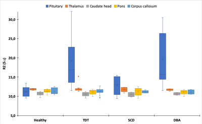 |
R2-based brain iron measurements in patients with iron overload – a retrospective analysis of selected brain regions
Rosalie Victoria McDonough1, Roland Fischer2,3, Regine Grosse4, Thomas Lindner1, Roberta Ward5, Zhiyue Jerry Wang6, Marcela Weyhmiller3, Jens Fiehler1, and Jin Yamamura2
1Department of Diagnostic and Interventional Neuroradiology, University Medical Center Hamburg-Eppendorf, Hamburg, Germany, 2Department of Diagnostic and Interventional Radiology and Nuclear Medicine, University Medical Center Hamburg-Eppendorf, Hamburg, Germany, 3UCSF Benioff Children’s Hospital Oakland, Oakland, CA, United States, 4Department of Pediatric Hematology and Oncology, University Medical Center Hamburg-Eppendorf, Hamburg, Germany, 5Imperial College, London, United Kingdom, 6Department of Radiology, University of Texas Southwestern Medical Center, Dallas, TX, United States
This study investigates the measurement of iron overload in patients using R2 MRI in selected brain regions.
|
|
1573.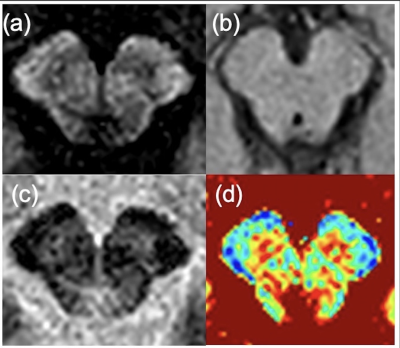 |
Method for quantitative evaluation of the substantia nigra using phase-sensitive inversion recovery in 1.5-T magnetic resonance imaging
Tomohisa Doi1, Yasuhiro Fujiwara2, and Hirotoshi Maruyama3
1Graduate School of Health Sciences, Kumamoto University, Kumamoto, Japan, 2Department of Medical Image Sciences, Faculty of Life Sciences, Kumamoto University, Kumamoto, Japan, 3Department of Radiology, National Hospital Organization Kumamoto Saisyun Medical Center, Kumamoto, Japan
Degeneration of the substantia nigra cannot be evaluated using neuromelanin imaging at 1.5 T. Therefore, an imaging method to quantitatively evaluate the substantia nigra with 1.5-T magnetic resonance imaging is required. This study aimed to investigate whether the substantia nigra can be quantitatively evaluated by acquiring a T1 map using three-dimensional phase-sensitive inversion recovery (PSIR) at 1.5 T. T1 mapping using the PSIR sequence enables quantitative evaluation of the substantia nigra independent of the imaging parameters.
|
|
1574.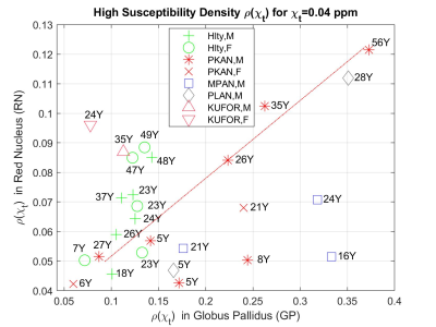 |
QSM Attributes of Neurodegeneration with Brain Iron Accumulation Disorders Subtypes in Basal Ganglia
Alpay Ozcan1, Ozge Uygun2, Fuat Kaan Aras1, Murat Gultekin3, Zuhal Yapici2, and Alp Dincer4
1Acibadem Mehmet Ali Aydinlar Univ., Istanbul, Turkey, 2Neurology, Istanbul University, Istanbul, Turkey, 3Neurology, Erciyes University, Kayseri, Turkey, 4Radiology, Acibadem Mehmet Ali Aydinlar Univ., Istanbul, Turkey QSM has the potential of describing objectively iron accumulation in brain regions such as basal ganglia which is relevant in neurodegeneration with brain iron accumulation (NBIA). Herein, for accurately assessing iron accumulation, a new metric, high susceptibility density, is introduced against mean value pitfalls which may be hindered by negative susceptibility within ROIs. When analyzing iron accumulation in basal ganglia of 16 patients with subtypes PKAN dispersion, KUFOR lower and PLAN higher accumulation, MPAN separation was obeserved in the Globus Pallidus-Red Nucleus space compared to healthy volunteers. |
|
1575.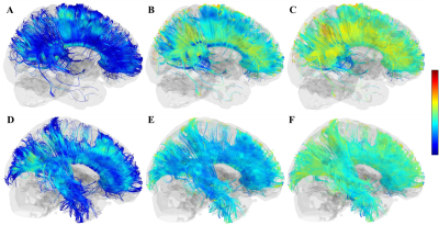 |
The white matter impairment of Gordon-Holmes syndromes: a tracking based analysis
Lei Wei1, KeLiang Chen2, and He Wang1,3
1Institute of Science and Technology for Brain-Inspired Intelligence, Fudan University, Shanghai, China, shanghai, China, 2Department of Neurology and Institute of Neurology, Huashan Hospital, Shanghai Medical College, Fudan University, Shanghai, China, ShangHai, China, 3Human Phenome Institute, Fudan University, Shanghai, China, Shanghai, China The Gordon-Holmes syndrome is an rare condiion caused by mutation of genes, few study has reported the finding of GHS by neuroimaging, but several studies has found the white matter abnormal in GHS brain. According the tracking based analysis. The different quantitative diffusion parameters suggest the white matter abnormal from different aspects. We firstly iinvestigated the white matter abnormal of GHS in the distribution of each diffusion property. |
|
1576.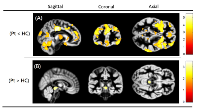 |
Gray Matter Changes of Pain-matrix Network in Patients with Acute Carbon Monoxide Intoxication
Ming-Chung Chou1, Jei-Yuan Li2, and Ping-Hong Lai3
1Department of Medical Imaging and Radiological Sciences, Kaohsiung Medical University, Kaohsiung, Taiwan, 2Department of Neurology, E-Da Hospital, Kaohsiung, Taiwan, 3Department of Radiology, Kaohsiung Veterans General Hospital, Kaohsiung, Taiwan Poster Permission Withheld
This study performed a voxel-based morphometry to investigate global gray matter changes in patients with acute CO intoxication. The results show significantly decreased gray matter volumes mostly in the pain-matrix regions, and significantly increased gray matter volumes in the periaqueductal gray (pain-modulating center). In the pain-matrix regions, the GM volumes were significantly negatively correlated with duration of coma, suggesting that longer duration of coma may lead to more headache symptoms in patients with acute CO intoxication.
|
|
1577.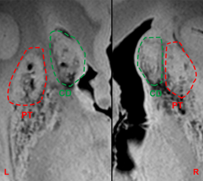 |
Ex-vivo Imaging of Intracerebral Fetal Neural Transplants in a Huntington Patient at 7T: Graft Survival and Interconnectivity 11 year post-OP
David Lohr1, Philipp Capetian2, Bernhard Landwehrmeyer3, and Laura Maria Schreiber1
Video Permission Withheld
1Chair of Cellular and Molecular Imaging, Comprehensive Heart Failure Center (CHFC), University Hospital Würzburg, Würzburg, Germany, 2Department of Neurology, University Hospital Würzburg, Würzburg, Germany, 3Department of Neurology, University Hospital Ulm, Ulm, Germany
The brain of a Huntington Disease (HD) patient who received neural fetal tissue transplants 11 years before her death, was analyzed ex-vivo in a 7T MRI scanner with a total scan time of 25 hours and 8 minutes. Signs of engrafted tissue could be found on all sites which received a stereotactic implantation (caudate nucleus, putamen) with a tendency to overgrowth for the right hemisphere. Deterministic fiber tracking based on DTI images clearly demonstrated interconnectivity of individual grafts to numerous brain regions.
|
|
1578.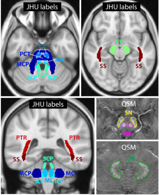 |
Does quantitative ultra-high-field magnetic resonance imaging have added value in the characterization of Friedreich’s ataxia?
Sina Straub1, Stephanie Mangesius2,3, Julian Emmerich1,4, Elisabetta Indelicato5, Wolfgang Nachbauer5, Katja S. Degenhardt1,4, Mark E. Ladd1,4,6, Sylvia Boesch5, and Elke R. Gizewski2
1Medical Physics in Radiology, German Cancer Research Center (DKFZ), Heidelberg, Germany, 2Department of Neuroradiology, Medical University of Innsbruck, Innsbruck, Austria, 3Neuroimaging Core Facility, Medical University of Innsbruck, Innsbruck, Austria, 4Faculty of Physics and Astronomy, Heidelberg University, Heidelberg, Germany, 5Department of Neurology, Medical University of Innsbruck, Innsbruck, Austria, 6Faculty of Medicine, Heidelberg University, Heidelberg, Germany
Friedreich’s ataxia is a rare disease involving degenerative processes within white matter fiber tracts, spinal nerves, and the cerebellum such as the atrophy of the dentate nuclei. A correlation of patients’ clinical status and white matter atrophy has been shown in MR volumetry studies. This ultra-high-field study assesses the degeneration of fiber tracts throughout the brainstem as well as of the dentate nuclei, the red nuclei, and the substantia nigra in Friedreich’s ataxia with quantitative MR parameters – susceptibility, diffusion anisotropy, and R2 and R1 relaxometry. Statistically significant differences between patients and controls and between disease characteristics were found.
|
|
1579.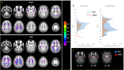 |
Lesion distribution and substrate in Type 1 Myotonic Dystrophy: comparison with Multiple Sclerosis
Sara Leddy1, Laura Serra2, Davide Esposito3, Camilla Vizzotto4, Gabriella Silvestri5, Antonio Petrucci6, Giovanni Meola7, Mara Cercignani2,8, and Marco Bozzali2,8
1Brighton and Sussex University Hospitals Trust, Brighton, United Kingdom, 2Neuroimaging Laboratory, Santa Lucia Foundation, Rome, Italy, 3Department of Vascular and Endovascular Surgery, University of Florence, Florence, Italy, 4University of, Rome, Italy, 5Department of Geriatrics, Orthopedics and Neuroscience, Catholic University of Sacred Heart Fondazione Policlinico Universitario A. Gemelli IRCCS, Rome, Italy, 6UOC Neurologia e Neurofisiopatologia, AO San Camillo Forlanini, Rome, Italy, 7Department of Neurorehabilitation Sciences, Casa di Cura Policlinico, Milan, Italy, 8Department of Neuroscience, University of Sussex, Brighton, United Kingdom
This study compares the lesion distribution and substrate (by means of quantitative MRI) between patients with type 1 Myotonic Dystrophy (DM1) and patients with Multiple Sclerosis (MS). The main differences in anatomical distribution are the prevalence of anterior temporal lobe lesions in the former group, in the absence of cerebellum and brainstem lesions. MRI markers of myelination were not different between the normal appearing white matter of DM1 and healthy controls. By contrast they were reduced in lesions, but larger than in MS lesions.
|
1580.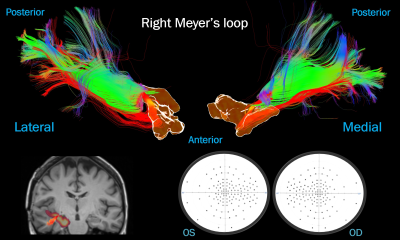 |
Linking priors-assisted Meyer’s loop tractography with visual field deficits in patients undergoing temporal lobe epilepsy surgery
Dmitri Shastin1,2, Sanchita Bhatia2, Chantal M. W. Tax1, Greg Parker1, Stefan Schwartz2, Khalid Hamandi1,2, William Gray2,3, Derek Jones1, and Maxime Chamberland1
1School of Psychology, Cardiff University Brain Research Imaging Centre, Cardiff, United Kingdom, 2School of Medicine, Cardiff University Brain Research Imaging Centre, Cardiff, United Kingdom, 3BRAIN Biomedical Research Unit, Cardiff, United Kingdom
Pre-operative reconstruction of the Meyer’s loop (ML) using diffusion MRI has a clinical utility when planning temporal lobe resection in order to avoid post-operative visual field deficit. Due to its complex anatomy, precise reconstruction of the ML is challenging. Previous literature has suggested that state-of-the-art hardware and tractography using oriented priors better approximates reconstruction to the reported histological prosections. This pilot work evaluates the ability of these improvements to predict visual field deficit in surgical patients. We report a good association in three out of four cases and suggest that simplistic metrics may not necessarily correlate with function.
|
|
1581.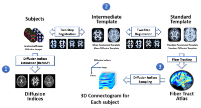 |
White matter degeneration in Papez circuit is associated with memory function in patients with mesial temporal lobe epilepsy
HSI-YUAN HU1, Yao-Chia Shih2, Chang-Le Chen1, Horng-Huei Liou3, Yu-Ling Chang4, Yung-Chin Hsu5, and Wen-Yih Isaac Tseng6
1Institute of Medical Device and Imaging, National Taiwan University College of Medicine, Taipei, Taiwan, Taipei, Taiwan, 2Department of Diagnostic Radiology, Singapore General Hospital, Singapore, Singapore, Singapore, Singapore, 3Department of Neurology, National Taiwan University Hospital and College of Medicine, Taipei, Taiwan, Taipei, Taiwan, 4Department of Psychology, College of Science, National Taiwan University, Taipei, Taiwan, Taipei, Taiwan, 5AcroViz Technology, Inc., Taipei, Taiwan, Taipei, Taiwan, 6Molecular Imaging Center, National Taiwan University, Taipei, Taiwan, Taipei, Taiwan
Previous studies did not characterize side-specific associations between structural integrity of the Papez circuit and memory function in patients with mesial temporal lobe epilepsy (MTLE). Here, we used diffusion spectrum imaging (DSI) and T1-weighted MRI to calculate the white matter integrity and gray matter volume, respectively. The structural metrics were correlated with visual and verbal memory function scores assessed by neuropsychological test. The results showed distinct brain-behavior associations in two subtypes of MTLE.
|
|
1582.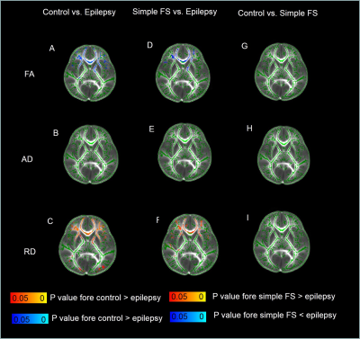 |
Diffusion Tensor Metrics Abnormalities in Developing brain: A Comparative Study Between Epilepsy and Simple Febrile Seizure aged 6-60 months
Abdelkareem Salimeen Mustafa1, Xianjun Li1, Miaomiao Wang1, Congcong Liu1, Martha Singh1, Mengxuan Li 1, Xiaocheng Wei2, and Jian Yang1,3
1First Affiliated Hospital of Xi’an Jiaotong University, Xi’an, China, Xi’an, China, 2MR Research China, GE Healthcare, China, Beijing, China,, Beijing, China, 3The Key Laboratory of Biomedical Information Engineering, Ministry of Education, Department of Biomedical Engineering, School of Life Science and Technology, Xi’an Jiaotong University, Xi’an, Shaanxi 710049, People’s Republic of China,, Xi’an, China
Epilepsy is a common neurological disorder spread worldwide. Simple febrile seizures (simple FS) is convulsive disorder associated with fever. However, little known about diffusion changes during the developing brain. The study aimed was to assess the diffusion changes in epilepsy and simple FS at aged of 6 to 60 months, using diffusion tensor imaging (DTI). Through inter-group comparisons, fraction anisotropy (FA) decreased, and radial diffusivity (RD) increased in epileptic children compared to simple FS and control. These results suggested DTI is sensitive method in diagnosis delayed myelination in epilepsy, while simple FS have good prognosis due to different pathophysiological mechanism.
|
|
1583.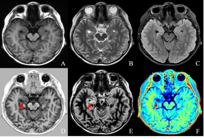 |
Conventional and synthetic MRI in children with drug-resistant epilepsy: a comparative study
XIAONA Zhang1, QUANXIN YANG1, and XIAOCHEN WEI2,3
1The Second affiliated hospital of Xi'an JiaoTong Uuniversity, Xi'an, China, 2GE Healthcare, MR Research China, Beijing, China, 3GE Healthcare, MR Research China, Xi'an, China
The current study aims to assess whether multi-contrast qualitative and quantitative relaxation imaging derived from the synthetic MRI can enhance detection of epileptogenic lesions in children. It was concluded that the synthetic MRI has the potential to be more sensitive in detecting lesions than conventional neuroimaging. Studies with more patients are needed to further demonstrate the relative advantages of the synthetic MRI over conventional MRI, and compare the synthetic MRI with 3-dimentional high resolution MRI.
|
|
1584.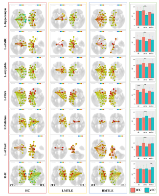 |
Altered cerebral functional connectivity asymmetry in unilateral mesial temporal lobe epilepsy
Xu Zhao1, Zhiqiang Zhou2, and Wenzhen Zhu1
1Radiology department, Tongji Hospital, Tongji Medical College, Huazhong University of Science and Technology, Wuhan, China, 2Anesthesiology department, Tongji Hospital, Tongji Medical College, Huazhong University of Science and Technology, Wuhan, China
The human brain is structurally and functionally asymmetrical. The asymmetry of mesial temporal lobe epilepsy (MTLE) were not clarified yet. This study analyzed the functional asymmetry of MTLE by comparing the functional connectivity (FC) of the left to the right hemisphere directly. The results showed reduction of asymmetrical areas in MTLE and a different asymmetrical features in left and right MTLE. The reduced FC asymmetry in MTLE may play an important role in the cognitive impairment of MTLE. The different asymmetrical features in left and right MTLE may hold the potential for differentiating left from right MTLE.
|
|
1585.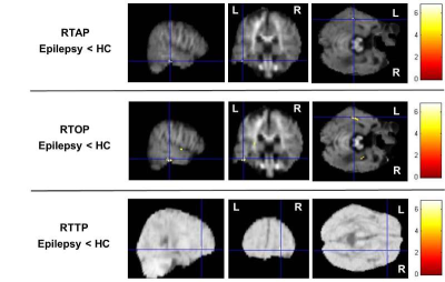 |
Detection of brain microstructural alterations in epilepsy using DTI and mean apparent propagator (MAP)-MRI
Weike Zeng1, Mengzhu Wang2, Xu Yan3, and Guang Yang4
1Deptpartment of Radiology, SUN YAT-SEN Memorial Hospital, SUN YAT-SEN University, Guangzhou, China, 2MR Scientific Marketing, Siemens Healthcare, Guangzhou, China, 3MR Scientific Marketing, Siemens Healthineers, Shanghai, China, 4Shanghai Key Laboratory of Magnetic Resonance, East China Normal University, Shanghai, China
We investigated the clinical feasibility of characterizing brain tissue microstructure in epilepsy with mean apparent propagator (MAP)-MRI, and compared with diffusion tensor imaging (DTI) using whole-brain voxel-based analyses. Results demonstrated that the MAP-MRI microstructural parameters could potentially provide more sensitive clinical biomarkers than conventional DTI techniques due to increased pathophysiological specificity in microstructural changes detection.
|
|
1586.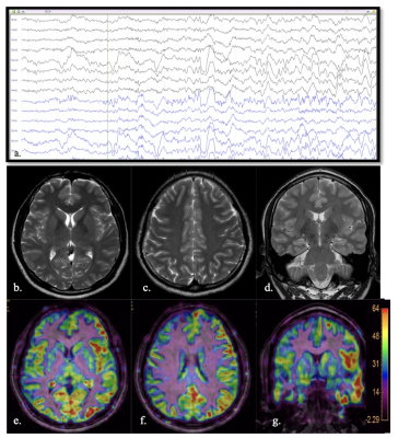 |
Yield of peri-ictal arterial spin-labelling MRI perfusion in refractory epilepsy
Jitender Saini1, Sarbesh Tiwari2, Sanjib Sinha3, Ravindranadh Chowdary M3, and Raghavendra K3
1Nueroimaging and Interventional Radiology, National Institute of Mental Health and, Bangalore, India, 2Diagnostic and Interventional Radiology, All India Institute of Medical Sciences,, Jodhpur, India, 3Neurology, National Institute of Mental Health and, Bangalore, India
Arterial Spin Labelling (ASL), a non-invasive MR based perfusion technique, has emerged as an excellent method for quantifying brain perfusion and its potential in detecting perfusion changes in drug resistant epilepsy. Our study also demonstrates the perfusion changes in the epileptogenic zone with good concordance with structural MRI and video-EEG findings.
|
|
1587.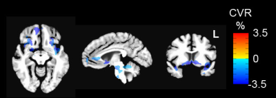 |
BOLD Responses to Breath-by-Breath O2-CO2 Exchange Ratio Under Breath Hold Challenge in Mild Traumatic Brain Injury Patients
Suk-tak Chan1, Cora Ordway1, Ronald J Calvanio2, Ferdinando S Buonanno2, and Kenneth K Kwong1
1Athinoula A. Martinos Center for Biomedical Imaging, Massachusetts General Hospital, Boston, MA, United States, 2Neurology, Massachusetts General Hospital, Boston, MA, United States
We used fMRI to map BOLD responses to breath-by-breath O2-CO2 exchange ratio (bER) under breath hold protocol for patients with persistent symptoms from mild traumatic brain injury (mTBI). Comparing to controls, reduced cerebrovascular reactivity (CVR) to bER was reported for patients in insula, anterior cingulate, nucleus accumbens and ventral tegmental area, basically regions which overlapped with the dopamine pathway. The mean resting bER decreased with increased symptom severity indicated by burden scores. Our findings are consistent with impaired dopamine pathway reported for TBI patients and show the success of using bER to evaluate CVR to breath hold.
|
|
1588.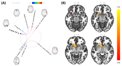 |
Changes in resting state connectivity and brain metabolites during a season of collegiate basketball: A pilot study
Candace C Fleischer1,2, Jeremy L Smith1, Maame Owusu-Ansah1, Selin Ekici1, Dongsuk Sung2, Ojaswa Prasad3, Brandon Mines4, and Jason W Allen1
1Department of Radiology and Imaging Sciences, Emory University School of Medicine, Atlanta, GA, United States, 2Department of Biomedical Engineering, Georgia Institute of Technology and Emory University, Atlanta, GA, United States, 3Department of Medicine, Philadelphia College of Osteopathic Medicine, Suwanee, GA, United States, 4Department of Sports Medicine, Emory University School of Medicine, Atlanta, GA, United States Poster Permission Withheld
Sports-related traumatic brain injuries are difficult to diagnose and prognose due to a lack of standardized metrics. Furthermore, there has been limited research on the effects of repeated sub-concussive and sub-clinical injuries over time. In this study, we characterized changes in resting state connectivity and brain metabolites over a season of collegiate basketball in athletes without a diagnosed concussion. We observed significant changes in resting state connectivity and brain metabolites as a function of game time played. No changes in plasma inflammatory markers were observed over time, suggesting that brain changes were not driven by systemic inflammation.
|
|
1589.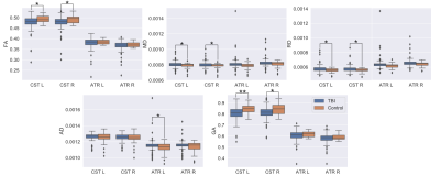 |
White Matter Structural Changes after Multiple Concussions in Adolescents and Young Adults
Arun Venkataraman1, Steven P. Meyers2, and Jianhui Zhong3
1Physics, University of Rochester, Rochester, NY, United States, 2Radiology, University of Rochester, Rochester, NY, United States, 3Imaging Sciences, University of Rochester, Rochester, NY, United States
Diffusion MRI (dMRI)-based studies in Traumatic Brain Injury have elucidated local and global WM alterations after injury. However, no studies have quantified how repeated concussions affect WM microstucture and coherence. We, therefore, sought to udnerstand how the number of previous concussions impact diffusion metrics. We found that, compared to the control group, there were significant decreases in FA and increases in MD and RD in the corticospinal tract. With respect to the number of concussions, we found that FA actually increased and was related with higher uniformity of the fibers. We believe this is related to aberrant remyelination.
|
|
1590.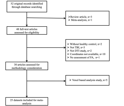 |
White matter alterations in mild traumatic brain injury: A tract-based spatial meta-analysis exploring the effects of trauma type
SuMing Zhang1, Xinyu Hu1, Xuan Bu1, and Xiaoqi Huang1
1Huaxi MR Research Center (HMRRC), Department of Radiology, West China Hospital of Sichuan University, ChengDu City, China
Many tract-based spatial statistics (TBSS) studies in mild traumatic brain injury (mTBI) have been inconsistent. Meanwhile, there is evidence that trauma type may have an effect on white matter (WM) microstructure in mTBI. We performed a tract-based spatial meta-analysis of mTBI and examined the potential effects of trauma type on regional WM microstructure. Our findings identified the left anterior thalamic radiation and right superior longitudinal fasciculus were the most convergent circuitry affected in mTBI and indicated distinct patterns of anatomical connectivity abnormalities in accident related and sport related mTBI, which highlighted the potential importance of trauma specific alterations in mTBI.
|
|
1591.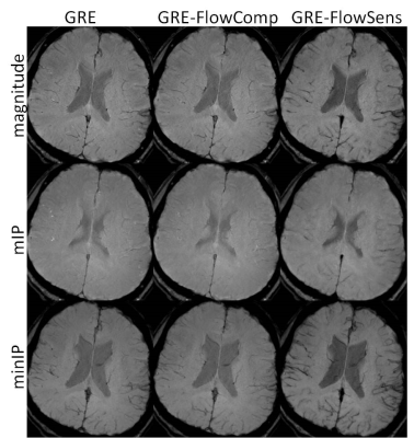 |
Feasibility of Dark Blood Susceptibility-Weighted Imaging
Wen-Tung Wang1, Dzung Pham1, and John A Butman1,2
Video Permission Withheld
1Center for Neuroscience and Regenerative Medicine, NIH/USU, Bethesda, MD, United States, 2Radiology and Imaging Sciences, NIH, Bethesda, MD, United States
First-order flow compensation has been widely used in MRI. While it works for visualization of vessels and CSF, the residual flow effects can impact the vessels visualization in some applications, such as minimum intensity projection, which is often used in detecting cerebral microhemorrhages on SWI images. When applying minIP, slight offset of spatial registration, and residual flow dephasing from acceleration and pulsatility can manifest as segments of hypointensities, compounding the difficulties in microhemorrhage detection. By including flow sensitization gradients in SWI sequence to suppress flow signal, dark vessels are delineated at correct spatial locations.
|
|
1592. |
FDG-PET imaging shows mesiotemporal hypometabolism in MRI-negative temporal lobe epilepsy
Ben A Duffy1, Julia Pia Simon1, Yan Li2, Arthur W Toga1, Wolfgang G Muhlhofer3, Robert C Knowlton2, and Hosung Kim1
1USC Stevens Neuroimaging and Informatics Institute, University of Southern California, Los Angeles, CA, United States, 2UCSF Weill Institute for Neurosciences, San Francisco, CA, United States, 3University of Alabama at Birmingham, Birmingham, AL, United States
The pathophysiology of MRI-negative temporal lobe epilepsy is not well understood. Here, we used a surface-based approach for investigating metabolic changes in the subregions of mesiotemporal structures for MRI-negative TLE patients. We found significant hypometabolism in the anterior to the intermediate hippocampus, the intermediate entorhinal cortex and the centro-medial and laterobasal amygdala. In patients with poor outcome, metabolism tended to be more interhemispherically symmetric in all three regions compared to those with good outcome, which suggests a more complicated seizure origin in this subtype of the disorder.
|