
Digital Poster Session
Neuro: Neurodegeneration
Neuro
1593 -1605 Neurovascular - Neurovascular: Intracranial Vessel Wall MRI
1606 -1619 Neurovascular - Neurovascular: Stroke Imaging Clinical
1620 -1635 Neurovascular - Neurovascular: MRA, Reactivity & Flow 1
1636 -1650 Neurovascular - Neurovascular: Systemic Disease-Related Stroke & Others
1651 -1665 Neurovascular - Neurovascular: MRA, Reactivity & Flow 2
1666 -1675 Neurovascular - Encephalopathies & Systemic Diseases
1676 -1690 Neurovascular - Blood-Brain Barrier Imaging/Perivascular Spaces
 |
1593.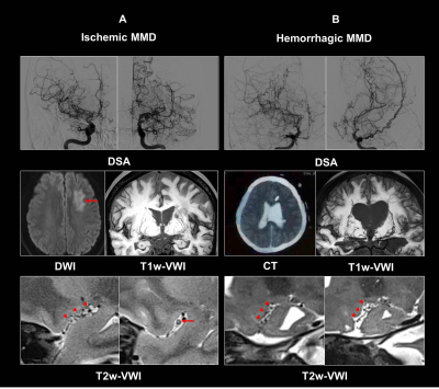 |
Association between Morphologies of Middle Cerebral Artery Occlusion on Vessel Wall Imaging and Cerebrovascular Events in Moyamoya Patients
Fang Wu1, Cong Han2, Zhaoyang Fan3, and Qi Yang4
1Radiology, Xuanwu hospital, Capital Medical University, Beijing, China, 2Department of Neurosurgery, 307 Hospital of Chinese People's Liberation Army, Beijing, China, 3Biomedical Imaging Research Institute, Cedars Sinai Medical Center, Los Angeles, CA, United States, 4Radiology, Xuanwu Hospital, Capital Medical University, Beijing, China
In this study, we used whole-brain vessel wall imaging (VWI) to characterize morphologies of middle cerebral artery (MCA) occlusion in patients with MMD, and explore their relationship to clinical findings. We found that plugged MCA was associated with ischemic stroke, however, vanishing MCA correlated with intraventricular hemorrhage. We also found that hemispheres with vanishing MCA manifested a higher degree of lenticulostriate artery (LSA) dilation and proliferation. The results suggest that there are two types of MCA occlusion in MMD, correlating with different stroke types and extent of perforator proliferation. Morphological analysis of occluded MCA using VWI may help understand the stroke mechanism of MMD.
|
 |
1594.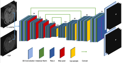 |
Intracranial aneurysms (IA) segmentation from 3D TOF-MRA and black-blood MRI (BB-MRI) using a deep convolutional neural network
Miaoqi Zhang1, Shuo Han2, Aaron Carass2, Fei Peng3, Aihua Liu3, Jerry L. Prince2, and Rui Li1
1Center for Biomedical Imaging Research, Department of Biomedical Engineering, Tsinghua University, Beijing, China, 2The Johns Hopkins University, Baltimore, MD, United States, 3Department of Interventional Neuroradiology, Beijing Neurosurgical Institute and Beijing Tiantan Hospital, Capital Medical University, Beijing, China
In this work, we proposed an automatic segmentation algorithm of intracranial aneurysms from dual input 3D TOF-MRA and black-blood MRI (BB-MRI) using a deep convolutional neural network to study its clinical potential for assisting intracranial aneurysm detection. The positioning of an intracranial aneurysm can benefit from the TOF-MRA, and the BB-MRI image can be used to accurately trace its boundary and measure its size. The average Dice coefficients are 0.69 and 0.73 for the TOF-MRA and the BB-MRI images, respectively.
|
1595. |
3T intracranial vessel wall MRI reveals that arterial wall thickness increases with reducing hematocrit in patients with sickle cell disease
Shuai Yuan1, Larry Davis1, Petrice Cogswell2, Spencer Waddle3, Lori Jordan1, and Manus Donahue4
1Vanderbilt University Medical Center, Nashville, TN, United States, 2Mayo clinic, Rochester, MN, United States, 3Vanderbilt University, Nashville, TN, United States, 4Vanderbilt University Medical Center Department of Radiology, Nashville, TN, United States
Sickle cell disease leads to increased risk of intracranial vasculopathy and stroke. Therefore, sensitive radiological indicators of cerebrovascular disease severity are needed to aid in treatment planning. 3T vessel wall imaging MRI studies demonstrated increased basilar artery wall thickness in sickle cell disease patients and inverse relationship between hematocrit and basilar wall thickness likely due to increased blood flow velocities and wall stress. Evaluation of internal carotid artery is difficult possibly due to variable CSF signal suppression in these regions.
|
|
1596. |
Optimized Intracranial Vessel Wall Imaging Framework – First clinical results
Konstanze Viktoria Anna Valerie Guggenberger1, Patrick Vogel1, Nils Venhoff2, Ute Ludwig3, Marc Schmalzing4, Esther Raithel5, Axel Joachim Krafft3, Thomas Ness6, Jost Hillenkamp7, Horst Urbach8, Stephan Meckel8, and Thorsten Bley1
1Department of Diagnostic and Interventional Radiology, University Hospital Wuerzburg, Wuerzburg, Germany, 2Clinic for Rheumatology and Clinical Immunology, University Hospital Freiburg, Freiburg, Germany, 3Department of Radiology, Medical Physics, University Hospital Freiburg, Freiburg, Germany, 4Rheumatology/Clinical Immunology Department, Internal Medicine II, University Hospital Wuerzburg, Wuerzburg, Germany, 5Siemens Healthcare, Erlangen, Germany, 6University Eye Center, University Hospital Freiburg, Freiburg, Germany, 7Department of Ophthalmology, University Hospital Wuerzburg, Wuerzburg, Germany, 8Department of Neuroradiology, University Hospital Freiburg, Freiburg, Germany
The extent of intracranial large artery involvement and its assessment is still part of on-going research. Our study group has developed a whole-brain T1-weighted dark blood CS-SPACE sequence suitable for intracranial vessel wall imaging in large-artery vasculitis combined with a dedicated post-processing tool. 53 patients were analyzed in a prospective blinded two university medical center trial. First clinical results demonstrate the technique`s suitability for clinical application. Mural thickening and contrast-enhancement as well as luminal changes are readily visible. However, certain confounders, especially atherosclerotic vessel wall lesions as well as vasa vasorum still propose major challenges for diagnosing intradural vasculitic affection.
|
|
1597.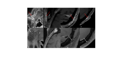 |
Optimized T1-weighted dark blood CS-SPACE - assessment of superficial extracranial arteries
Konstanze Viktoria Anna Valerie Guggenberger1, Patrick Vogel1, Nils Venhoff2, Ute Ludwig3, Marc Schmalzing4, Esther Raithel5, Axel Joachim Krafft3, Thomas Ness6, Jost Hillenkamp7, Horst Urbach8, Stephan Meckel8, and Thorsten Bley1
1Department of Diagnostic and Interventional Radiology, University Hospital Wuerzburg, Wuerzburg, Germany, 2Clinic for Rheumatology and Clinical Immunology, University Hospital Freiburg, Freiburg, Germany, 3Department of Radiology, Medical Physics, University Hospital Freiburg, Freiburg, Germany, 4Rheumatology/Clinical Immunology Department, Internal Medicine II, University Hospital Wuerzburg, Wuerzburg, Germany, 5Siemens Healthcare, Erlangen, Germany, 6University Eye Center, University Hospital Freiburg, Freiburg, Germany, 7Department of Ophthalmology, University Hospital Wuerzburg, Wuerzburg, Germany, 8Department of Neuroradiology, University Hospital Freiburg, Freiburg, Germany Superficial extracranial arteries are predilection sites for disease manifestation in large vessel vasculitides. We evaluated the suitability of a whole-brain T1-weighted dark blood CS-SPACE sequence, initially developed for intracranial vessel wall imaging, for visualization of vasculitic changes of superficial extracranial arteries. 53 patients were analyzed in a prospective blinded two university medical center trial. First clinical results demonstrate the technique`s applicability for clinical application. Mural thickening and contrast-enhancement of superficial extracranial arteries are clearly visible. The optimized VWI protocol offers potential for assessment of intra- and extracranial disease extent in case of suspected large vessel vasculitis in one single examination. |
|
1598.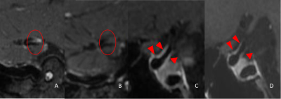 |
High-resolution MRI of primary and secondary central nervous system vasculitis
Shuai Han1, Xinyi Wang2, and Weiqiang Dou3
1Shandong First Medical University & Shandong Academy of Medical Sciences, Jinan, China, 2The First Affiliated Hospital of Shandong First Medical University, Jinan, China, 3GE Healthcare, MR Research, Beijing, China
The main goal of this study was to evaluate the treatment effect of primary angiitis of the central nervous system (PACNS) and secondary central nervous system vasculitis(CNSV) with high-resolution magnetic resonance vascular wall imaging. We measured 4 patients with PACNS and 10 patients with secondary CNSV using high resolution CUBE MRI. We found that high-resolution magnetic resonance vascular wall imaging can assess the treatment effect of PACNS and secondary CNSV. Therefore, CUBE MRI can be considered an effective tool in the evaluation of PACNS and secondary CNSV.
|
|
1599.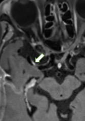 |
Association between type 2 diabetes mellitus and characteristics of intracranial atherosclerotic plaque: a high-resolution MRI study
Sheng Jiao1, Juan Huang2, Yan Song2, Yuhui Chen3, Chengwen Liu4, Jinxia Zhu4, Jintao Zhang2, and Min Chen2
1Radiology, Beijing hospital, Beijing, China, 2Radiology, Beijing Hospital, Beijing, China, 3Neurology, Beijing Hospital, Beijing, China, 4MR Collaboration, Siemens Healthcare Ltd., Beijing, China
In this study we used high-resolution MRI to image intracranial atherosclerotic plaque more precisely, and then characteristics of plaque were obtained to compare with the blood glycemic (BG) level in Type 2 Diabetes mellitus (T2DM). Results demonstrated that patients with poor BG control had heavier intracranial plaque burden and more vulnerability than the patients without DM. While compared with non-DM, patients with good BG control showed no significant different in plaque burden. It suggested that BG level is an independent risk factor for the vulnerability of the intracranial atherosclerotic plaque.
|
|
1600.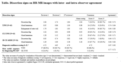 |
Diagnostic feasibility of high resolution vessel wall imaging for isolated posterior inferior cerebellar artery dissection
Miran Han1, Woo Sang Jung1, and Jin Wook Choi1
1Radiology, Ajou University Medical Center, Suwon, Republic of Korea
We evaluate the feasibility of HR-VWI for diagnosing PICA dissection. Our results demonstrated that HR-VWI could effectively diagnose arterial dissection even in PICA, small diameter vessel. The dissection flap with outer wall enlargement on T2W HR-VWI was most confident sign for diagnosing PICAD. Isolated PICAD is not a rare cause of ischemic stroke in posterior circulation. Therefore, in case of PICA territory infarction of uncertain origin, differentiating PICAD using HR-VWI is necessary for proper treatment of patients, and T2WI should be included in HR-VWI.
|
|
1601.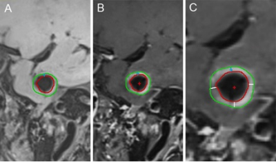 |
CAWE and higher WEI on VW-MRI were associated with symptomatic unruptured intracranial aneurysms
Qichang Fu1, Yi Zhang1, Sheng Guan2, Chengcheng Zhu3, and Jingliang Cheng1
1Department of Magnetic Resonance, The First Affiliated Hospital of Zhengzhou University, Zhengzhou, China, 2Department of Interventional Neuroradiology, The First Affiliated Hospital of Zhengzhou University, Zhengzhou, China, 3Department of Radiology and Biomedical Imaging, University of California, San Francisco, San Francisco, CA, United States
This study aimed to demonstrate the feasibility of aneurysmal wall enhancement (AWE) and wall enhancement index (WEI) in the identification of symptomatic and asymptomatic unruptured intracranial aneurysms by using vascular wall magnetic resonance imaging (VW-MRI) in a large cohort of Chinese patients with unruptured intracranial aneurysms (UIAs). VW-MRI were obtained at MAGNETOM Skyra/Verio/Prisma 3T MR scanner (Siemens Healthcare, Erlangen, Germany) in these patients. We found CAWE and WEI>0.91 were more frequently identified in symptomatic UIAs.
|
|
1602.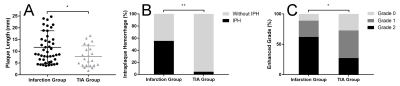 |
Combination of Plaque Characteristics and Hemodynamics Predicts Neurological Impairment in Infarction and TIA Patients
Song Liu1, Ruowei Tang2, Yu Luo3, Song Jin4, and Shuang Xia5
1Radiology Department, Tianjin Huanhu Hospital, Tianjin, China, 2Radiology Department, Beijing Friendship Hospital, Capital Medical University, Beijing, China, 3Radiology Department, Shanghai Fourth People's Hospital, Shanghai, China, 4Tianjin Huanhu Hospital, Tianjin, China, 5Tianjin First Central Hospital, Tianjin, China
Ischemic stroke is the second frequent cause of death worldwide. Some patients have transient symptoms of neurological defect without parenchymal damage, irreversible cerebral tissue injury happens in someone else. How to discriminate TIA from acute stroke needs further study. This study uses muti-modal MRI scaning to investigate the difference of plaque characteristics in Middle Cerebral Artery (MCA) between patients with infarction and transient ischemic attack (TIA), and to explore the predictive model for infarction region and neurological impairment.
|
|
1603. |
3D-CUBE T1 vessel wall imaging and Susceptibility Weighted Imaging in the diagnosis of cerebral venous sinus thrombosis: a comparison study
Bing Zhao1, Jianshe Zhao1, Weiqiang Dou2, Chao Zhang3, Meijia Zhu3, and Xinyi Wang3
1Department of Radiology, Qilu Children's Hospital of Shandong University, Jinan, China, 2GE Healthcare, MRI Research China, Beijing, China, 3Shandong Province Qianfoshan Hospital, Jinan, China
This study was aimed at investigating the clinical value of CUBE MRI for high resolution imaging and susceptibility weighted imaging (SWI) in the diagnosis of cerebral venous sinus thrombosis (CVST). The sensitivity, specificity, positive predictive value, and negative predictive value for 3D-CUBE T1 and SWI were calculated respectively. In this study, the diagnosis results of T1 weighted CUBE were better than that of SWI in comparison with the referential diagnosis in the detection of CVST. We therefore demonstrated that the T1 weighted CUBE MRI provided more effective usefulness than SWI in the diagnosis efficacy of the CVST.
|
|
1604. |
Clinical Feasibility of High-Resolution Intracranial Vessel Wall MRI with Compressed Sensing
Chae Jung Park1, Jihoon Cha1, Sung Soo Ahn1, Hyun Seok Choi1, and Seung-Koo Lee1
1Department of Radiology, Severance hospital, Seoul, Republic of Korea
We investigated the clinical feasibility of compressed sensing (CS) in the intracranial vessel wall magnetic resonance imaging (VW-MRI) by applying CS to 3D post-contrast T1-weighted images (T1C). CS enabled larger scan coverage with nearly same scan time, while achieving comparable image quality, normal wall and lesion wall delineation. Furthermore, most T1C with CS provided acceptable quality with regard to overall image quality (84.7%), normal wall delineation (83.3%), and lesion wall delineation (97.8%), which was similar to T1C without CS. Therefore, CS is clinically feasible when applied to the post-contrast intracranial VW-MRI.
|
|
1605.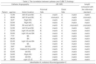 |
The feasibility of High resolution MRI in Length Measurement of Cerebral Arterial Thrombosis : a correlation study with Catheter Angiography
Chao Zhang1, Xinyi Wang1, Weiqiang Dou2, and Bing Zhao3
1The First Affiliated Hospital of Shandong First Medical University, Jinan, China, 2GE Healthcare, MR Research China, Beijing, China, 3Qilu Children's Hospital of Shandong University, Jinan, China
This study aimed to investigate the feasibility of CUBE MRI for high resolution imaging in the length measurement of intraluminal thrombi for acute stroke patients. The T1 weighted CUBE images displayed the segments proximal and distal to the thrombus with sufficient quality for clinical diagnosis, and showed the length of it as high signal or iso-signal filling in the lumen. As a result, about 88.9% of the thrombus lengths assessed by CUBE T1 were consistent with the reference DSA. We therefore, demonstrated that the T1 weighted CUBE MRI can effectively evaluate the length of intraluminal thrombi.
|
 View the Poster
1606. |
Quantitation of penumbra volumes in acute ischemic stroke using susceptibility- weighted imaging and mapping
Xiudi Lu1, Linglei Meng2, Yongmin Zhou3, Shaoshi Wang2, Miller Fawaz4, Meiyun Wang5, E. Mark Haacke4, Chao Chai6, Meizhu Zheng7, Jinxia Zhu8, Shuang Xia6, and Yu Luo3
1The First Teaching Hospital of Tianjin University of Traditional Chinese Medicine, Tianjin, China, 2Shanghai Fourth People’s Hospital, Shanghai, China, 3Translational Research Institute of Brain and Brain-Like Intelligence, Shanghai Fourth People’s Hospital Affiliated to Tongji University School of Medicine, Shanghai, China, 4Wayne State University, Detroit, MI, United States, 5Zhengzhou University People’s Hospital, Zhengzhou, China, 6Tianjin First Central Hospital, Tianjin, China, 7Third Central Hospital of Tianjin, Tianjin, China, 8Siemens Healthcare Ltd, Beijing, China
This study aimed to measure ischemic penumbra volumes using susceptibility-weighted imaging and mapping (SWIM). We compared the diagnostic accuracy between SWIvolume-diffusion-weighted imaging (DWI) and SWIASPECTS-DWI mismatches and analyzed the relationships among the SWIvolume-DWI, SWIASPECTS-DWI, and PWI-DWI mismatches and the different parameters and NIHSS. We found that quantitative SWIvolume-DWI mismatch volumes were more accurate than those of the SWIASPECTS-DWI mismatch in being able to evaluate ischemic penumbra. The asymmetrical prominent cortical vein volumes using SWIM showed hypoperfused regions and Tmax >6s volumes, based on perfusion-weighted imaging (PWI). The quantitative SWIvolume-DWI mismatch was highly consistent with the PWI-DWI mismatch.
|
|
1607.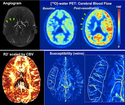 |
Comparison of oxygenation MRI methods with cerebrovascular reactivity in Moyamoya disease using simultaneous [15O]-water PET/MRI
Audrey P Fan1, David Y.T. Chen2, David D. Shin3, Moss Y. Zhao1, Mohammad M. Khalighi1, Jun-Hyung Park1, Bin Shen1, Dawn Holley1, Kim Halbert1, Gary K. Steinberg4, and Greg Zaharchuk1
1Radiology, Stanford University, Stanford, CA, United States, 2Radiology, Taipei Medical University, Taipei, Taiwan, 3GE Healthcare, Menlo Park, CA, United States, 4Neurosurgery, Stanford University, Stanford, CA, United States
In this [15O]-water PET/MRI study, we compared two non-invasive MRI techniques to measure brain oxygen extraction fraction (OEF) in 10 patients with Moyamoya disease, a steno-occlusive disease of arteries at the base of the brain. Relative OEF from magnetic susceptibility in veins and tissue R2' inversely correlated with [15O]-water PET baseline cerebral blood flow and cerebrovascular reactivity (after vasodilation with acetazolamide). Susceptibility-based OEF in veins was abnormally elevated by 20.4% in regions with severe stenosis compared to healthy tissue. R2' maps showed a smaller OEF elevation (9.6%) that may be confounded by other physiological changes (e.g. blood volume) in disease.
|
|
1608.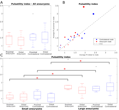 |
Investigation of the Windkessel Effect in Intracranial Aneurysms with Dual VENC 4D Flow MRI
Maria Aristova1, Kelly Jarvis1, Michael Pan1, Matthew Potts2, Michael Hurley2, Babak Jahromi2, Ali Shaibani1, Sameer Ansari1, and Susanne Schnell1
1Radiology, Northwestern University, Chicago, IL, United States, 2Neurosurgery, Northwestern University, Chicago, IL, United States
The Windkessel effect is hypothesized to be the mechanism of altered cerebral hemodynamics in intracranial aneurysms, via damping of pulsatile flow. 4D Flow MRI with specialized, network-based post-processing workflow provides damping factor (DF), by measuring pulsatility index (PI) ratio between proximal and distal vessels. PI values were higher in subjects with large aneurysms, even for contralateral vessels, suggesting patients with larger aneurysms may have other contributions to hemodynamics that also impact their vasculature outside the lesion. A multivariate linear correlation model shows that smaller aneurysms may have lower DF than contralateral, while larger aneurysms have higher DF ipsilateral than contralateral.
|
|
1609. |
Evaluating cervical artery dissections in young adults - A Comparison Study between High-Resolution MRI and CT Angiography
Xiao Cui1, Xianshun Yuan1, Hui Gu1, Mo Wang2, Yin Dong1, Ximing Wang1, and Xiang Feng3
1Department of Radiology, Shandong Provincial Hospital Affiliated to Shandong University, Jinan, China, 2Department of Vascular Surgery, Shandong Provincial Hospital Affiliated to Shandong University, Jinan, China, 3MR Scientific Marketing, Diagnosis Imaging, Siemens Healthcare Ltd, Beijing, China
This study aimed to compare the diagnostic values between HR-MRI and CTA in young adults with cervical artery dissections. Totally 42 patients with ischemic stroke were recruited, including 20 carotid artery dissections, 12 vertebral artery dissections and 10 non-dissected cervical arteries. Two radiologists separately analyzed all the images. According to the radiologists’ scores for the likelihood of the dissections, positive predictive value, negative predictive value, sensitivity and specificity for both modalities were calculated. We found the higher sensitivity and specificity of HR-MRI. This study supports the value of HR-MRI in non-invasive diagnosis of young adults due to cervical artery dissections.
|
|
1610. |
Noninvasive assessment of revascularization in adult-onset moyamoya disease after bypass surgery using super-selective ASL perfusion
Inpyeong Hwang1, Chul-Ho Sohn1, Won-Sang Cho2, Jeong Eun Kim2, Roh-Eul Yoo1, Koung Mi Kang1, Dong Hyun Yoo1, Tae Jin Yun1, Seung Hong Choi1, and Ji-hoon Kim1
1Department of Radiology, Seoul National University Hospital, Seoul, Korea, Republic of, 2Department of Neurosurgery, Seoul National University Hospital, Seoul, Korea, Republic of
The purpose of this study was to evaluate whether super-selective ASL (SS-ASL) perfusion imaging could precisely visualize the revascularization area after bypass surgery in moyamoya disease, compared to digital subtracted angiography (DSA). Twenty-eight bypassed hemispheres of twenty-six patients in moyamoya disease underwent postoperative six months SS-ASL and DSA. Subjective image analysis of the revascularization area, as well as collateral grading, were performed. The agreement of the revascularization area was excellent (weighted kappa, 0.83), and agreement of collateral grading was good (weighted kappa, 0.722.) SS-ASL could evaluate revascularization territory precisely in the patient of moyamoya disease who underwent bypass surgery.
|
|
1611. |
Ultrashort TE 4D-MRA for Giant Aneurysms Treated with Flow-Diverter Stents: Visualization of Flow in the Stents and Hemodynamic Vascular Flow
Nao Takano1, Michimasa Suzuki1, Yutaka Ikenouchi1, Munetaka Yamamoto2, Kohsuke Teranishi2, Syo Murata1, Nozomi Hamasaki1, Haruyoshi Hoshito1, Christina Andica1, Toshiaki Akashi1, Akihiko Wada1, Hidenori Oishi3, and Shigeki Aoki1
Video Permission Withheld
1Department of Radiology, Juntendo University Hospital, Tokyo, Japan, Tokyo, Japan, 2Department of Neurosurgery, Juntendo University School of Medicine, Tokyo, Japan, Tokyo, Japan, 3Department of Neuroendovascular Therapy, Juntendo University School of Medicine, Tokyo, Japan, Tokyo, Japan
We assessed the usefulness of ultrashort TE (UTE) 4D-MR angiography for giant aneurysms treated with flow-diverter stents. We evaluated the depiction of flow in the stents, and visualization of hemodynamic vascular flow in aneurysms. The newly developed UTE 4D-MRA could visualize flow in the stents at the acceptable quality for diagnosis, and hemodynamic vascular flow in aneurysms were comparable to X-ray DSA. Moreover, UTE 4D-MRA showed intraaneurysmal flow within the coil which was difficult to visualize even with X-ray DSA. Our study demonstrated that UTE 4D-MRA was adequate to visualize the hemodynamic vascular flow in aneurysms, and might be useful.
|
|
1612. |
Non-Contrast Enhanced Ultrashort Echo Time MR Angiography (Silent MRA) for Intracranial Aneurysms Treated with Flow-Diverter Stents and Coils
Nao Takano1, Michimasa Suzuki1, Yutaka Ikenouchi1, Munetaka Yamamoto2, Kohsuke Teranishi2, Syo Murata1, Nozomi Hamasaki1, Haruyoshi Hoshito1, Christina Andica1, Toshiaki Akashi1, Akihiko Wada1, Hidenori Oishi3, and Shigeki Aoki1
Video Permission Withheld
1Department of Radiology, Juntendo University Hospital, Tokyo, Japan, Tokyo, Japan, 2Department of Neurosurgery, Juntendo University School of Medicine, Tokyo, Japan, Tokyo, Japan, 3Department of Neuroendovascular Therapy, Juntendo University School of Medicine, Tokyo, Japan, Tokyo, Japan
We evaluated the usefulness of non-contrast enhanced ultrashort TE MRA (Silent MRA) for intracranial aneurysms treated with flow-diverter (FD) stents and coils. FD stent has a higher metal coverage, thus it is difficult to visualize in-stent flow using TOF-MRA, due to the magnetic susceptibility. In contrast, silent MRA uses ultrashort TE, therefore may visualize in-stent flow. In silent MRA, in-stent flow score was superior to that of TOF-MRA (p<0.05). Moreover, aneurysm occlusion status was in good agreement with X-ray DSA (κ = 0.84). Therefore, silent MRA is useful for the evaluation of intracranial aneurysms treated with FD stents and coils.
|
|
1613.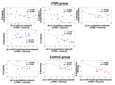 |
Modulating Functional Networks and Language Performance by Repetitive Transcranial Magnetic Stimulation in Post-stroke Aphasia
Bing-Fong Lin1, Po-Yi Tsai2, and Chia-Feng Lu1
1Biomedical imaging and radiological sciences, National Yang-Ming University, Taipei, Taiwan, 2Taipei Veterans General Hospital, Taipei, Taiwan
Low-frequency repetitive transcranial magnetic stimulation (rTMS) provided promising results to facilitate the language recovery in stroke patients with non-fluent aphasia. This study demonstrated a contralesional inhibitory rTMS treatment can modulate the brain functional networks compare to the conventional therapy. The modulated functional networks were further correlated with the improvement of language performance after the rTMS treatment.
|
|
1614.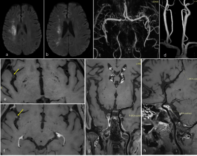 |
Plaque characteristics and implication on mechanism in border-zone infarction caused by middle cerebral artery atherosclerosis
Yu Guo1,2, Jiayu Xiao2, Zhongying Gong3, Wen Shen1, Shuang Xia1, and Zhaoyang Fan2
1Radiology, Tianjin First Central Hospital, tianjin, China, 2BIRI, Cedars-Sinai Medical Center, Los Angeles, CA, United States, 3Neurology, Tianjin First Central Hospital, tianjin, China
This study aimed to investigate atherosclerotic plaque characteristics in middle cerebral artery (MCA) atherosclerotic patients with border-zone (BZ) infarction using magnetic resonance vessel wall imaging (MR-VWI). In 84 retrospectively enrolled ischemic stroke patients caused by MCA atherosclerosis, the characteristics of culprit plaques were quantitatively compared between BZ only infarction and combined border-zone and pial (BZ+PI) infarction groups. BZ+PI infarction was associated with a higher degree of stenosis and higher plaque-wall contrast ratio and enhancement ratio, compared with BZ only infarction, suggesting that embolic pathogenesis may also account for BZ+PI infarction in addition to hemodynamic compromise.
|
|
1615.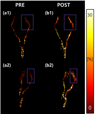 |
Quantitative Evaluation of Neuronal Recovery in stroke patients by Rehabilitation Using Myelin Water Fraction mapping
Junghyeob Kim1, Yejin Jo2, Jae Eun Song1, Hyunjung Kim2, Deogyoung Kim2, and Dong-Hyun Kim1
1Electrical & Electronic Engineering, Yonsei University, Seoul, Republic of Korea, 2Department and Research Institute of Rehabilitation Medicin, Yonsei University College of Medicine, Seoul, Republic of Korea
Stroke is a worldwide disease and requires quantitative evaluation for better patient management. In evaluating rehabilitation of stroke patients, quantitative analysis was performed using the conventional DTI-FA and multi echo GRE Myelin Water Fraction mapping technique
|
|
1616. |
Characteristics of Intracranial Artery Dissection on High-resolution MRI Associated with Recurrent Stroke/Transient Ischemic Attack
Bing Tian1, Zhang Shi2, Xia Tian2, Qi Liu2, and Jianping Lu2
1Radiology, Changhai hospital of Shanghai, Shanghai, China, 2Changhai hospital of Shanghai, Shanghai, China
HR-MRI is helpful for the classification of IAD, could be used to identify the culprit IAD from non-culprit ones.
|
|
1617.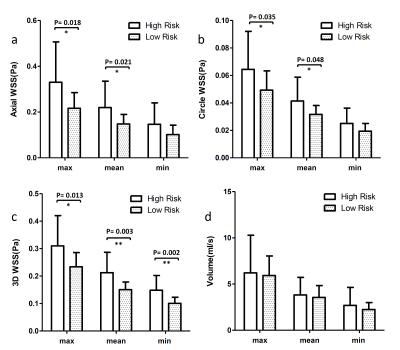 |
Differences of Wall shear stress between patients with high-risk and low-risk plaque in carotid artery: A 4D flow MRI study.
Guiling Zhang1, Zhenxiong Wang1, Weiyin Vivian Liu2, and Wenzhen Zhu1
1Tongji hospital, Wuhan, China, 2MR Research, GE Healthcare, Beijing, China
High risk plaque is the is the predominant risk factor of stroke. Accurate diagnosis and intervention could prevent patients from cerebrovascular events. Axial, circumferential and 3D wall shear stress calculated by 4D flow MRI were utilized to explore the difference between high risk plaque patients and low risk plaque patients. The results demonstrated the WSS were all significant higher in high risk plaque group. Our results may provide crucial clues to therapeutic interventions.
|
|
1618.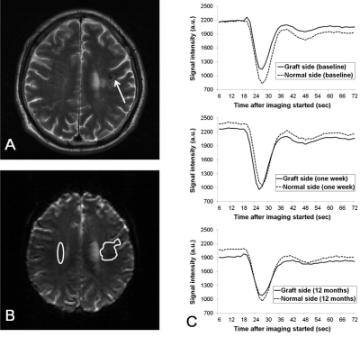 |
Angiogenesis following peripheral blood stem cell therapy: evidence from perfusion MRI of patients with ischemic stroke
Chao-Chun Lin1, Wen-Chau Wu2, Woei-Cherng Shyu3, Yi-Jui Liu4, Hing-Chiu Chang5, Yu-Chien Luo3, Der-Cherng Chen3, Chia-Wei Lin3, Hsiao-Wen Chung6, and Shinn-Zong Lin7
1Department of Radiology, China Medical University Hospital, Taichung, Taiwan, 2Institute of Medical Device and Imaging, National Taiwan University, Taipei, Taiwan, 3China Medical University Hospital, Taichung, Taiwan, 4Department of Automatic Control Engineering, Feng-Chia University, Taichung, Taiwan, 5Department of Diagnostic Radiology, University of Hong Kong, Hong Kong, Hong Kong, 6Graduate Institute of Biomedical Electronics and Bioinformatics, National Taiwan University, Taipei, Taiwan, 7Buddhist Tzu Chi General Hospital, Hualien, Taiwan
Dynamic susceptibility-contrast perfusion-weighted MRI was performed on 15 chronic stroke patients receiving intracerebral implantation of peripheral blood stem cells (PBSC) at baseline and 6 longitudinal stages after therapy to derive relative cerebral blood volume around the implanted graft relative to contralateral white matter (rCBV ratio), with 15 control patients receiving baseline MRI plus two follow-ups. Nine of the 15 patients (60%) in the PBSC group showed significantly increased rCBV ratio than the baselines (1.39±0.60 versus 1.07±0.44; p < 0.01) at one week after implantation only, preceding functional improvements starting at one month as assessed by NIH stroke scale.
|
|
1619.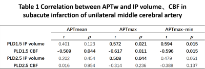 |
Correlation analysis between APTW and blood flow status in the ischemic penumbra of patients with subacute ischemic stroke
Yuhan Jiang1, Yanwei Miao1, Liangjie Lin2, Zhiwei Shen2, Peipei Chang1, Yiwei Che1, Ailian Liu1, Qingwei Song1, and Jiazheng Wang2
1the First Affiliated Hospital of Dalian Medical University, Dalian, China, Dalian, China, 2Philips Healthcare, Beijing, China, Beijing, China
The presence, dynamic evolution, and outcome of the ischemic penumbra (IP) are important for the choice of treatment options and prognosis. At present, there is currently no study to combine the APTw MRI with the blood flow measurements for IP analyses. Purpose of this study was to analyze the association between APT metabolic changes and blood flow status in the IP of patients with subacute infarction in the unilateral middle cerebral artery by APT and ASL techniques. Significant correlations of APTw values to CBF values of PLD1.5.
|
 Watch the Video
1620.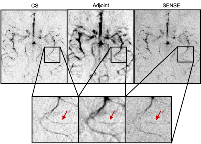 |
Highly Accelerated Time-Encoded Dynamic ASL Angiography
Sophie Schauman1, Joseph G. Woods1,2, Mark Chiew1, and Thomas W. Okell1
1Wellcome Centre for Integrative Neuroimaging, NDCN, University of Oxford, Oxford, United Kingdom, 2Department of Radiology, University of California San Diego, La Jolla, CA, United States
Time-encoded (TEnc) dynamic ASL angiography is a method that provides high SNR, dynamic information about the blood supply to the brain. However, as is commonly the case with all ASL-based methods, multiple repeat encodings are required to fully sample the information required to decode the dynamic angiographic data. Here we apply spatial sparsity and temporal smoothness constraints to reconstruct highly under-sampled TEnc data, and demonstrate that high fidelity, high resolution ASL angiography can be performed in a fraction of the time it takes using conventional methods.
|
|
1621.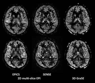 |
Rapid Brain Arterial Spin Labeling (ASL) using pseudo-Continuous ASL Echo Planar Imaging with Compressed SENSE (pCASL-EPICS)
Kosuke Morita1, Masami Yoneyama2, and Takeshi Nakaura3
1Kumamoto University Hospital, Kumamoto, Japan, 2Philips Japan, Tokyo, Japan, 3Graduate School of Medical Sciences, Kumamoto University, Kumamoto, Japan Multi-slice single-shot 2D echo-planar imaging (EPI) readout can be used effectively for pseudo-continuous arterial spin labeling (pCASL) perfusion imaging. In this study we tried to utilize the Compressed SENSE reconstruction for pCASL-EPI without further optimization of EPI sampling scheme. pCASL-EPICS clearly reduces noise-like artifacts and significantly improves the robustness of perfusion images in a short scan time less than 3 minutes compared with conventional pCASL SENSE, without any penalty for scan parameters. This technique may be helpful to further assess the many brain diseases. |
|
1622.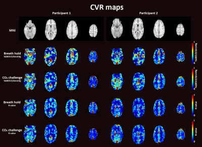 |
Investigating the Optimal Vasoactive Stimulus to Extract Dynamic Cerebrovascular Reactivity Information: Breath holding versus CO2 Challenge
Eva Elisabeth van Grinsven1, Allen A. Champagne2, Marielle Philippens3, and Alex Bhogal4
1Department of Neurology & Neurosurgery, University Medical Center Utrecht Brain Center, Utrecht, Netherlands, 2Center for Neuroscience studies, School of medicine, Queen’s University, Kingston, ON, Canada, 3Department of Radiotherapy, University Medical Center Utrecht, Utrecht, Netherlands, 4Department of Radiology, University Medical Center Utrecht, Utrecht, Netherlands
This study compared multiple BOLD cerebrovascular reactivity (CVR)-metrics during breath holding (BH) versus a targeted CO2 challenge to determine the degree of similarity in extracting dynamic information about hemodynamic reserves. The preliminary data confirm previous research; CVR-maps were spatially comparable and correlated well between the two stimuli. That said, the data suggests reliable information regarding the flow dynamics is best provided by a controlled CO2 stimulus. Thus, even though BH may be easier to implement, it may also be limited in its ability to provide additional information regarding cerebrovascular health (beyond CVR), which could be of value when investigating patients.
|
|
1623.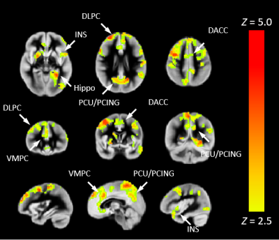 |
Towards non-invasive estimation of "resting-state" CVR: associations with congenital heart disease, cognition, and nasal nitric oxide
Vincent Jerome Schmithorst1, Cecilia Lo2, Philip Adams2, and Ashok Panigrahy1
1Radiology, Children's Hospital of Pittsburgh of UPMC, Pittsburgh, PA, United States, 2University of Pittsburgh, Pittsburgh, PA, United States
Estimation of cerebrovascular reserve/reactivity (CVR) typically necessitates an invasive vasoactive stimulus. We here propose a metric to non-invasively estimate “resting-state” CVR (rCVR), the capacity of the vasculature to respond to resting-state metabolic demand, as the negative ratio of functional connectivity strength (FCS) to regional cerebral blood flow (CBF). Construct validity was demonstrated via prediction of end-tidal CO2 levels (PETCO2). rCVR was lower in older children with congenital heart disease (CHD) in default mode network (DMN), salience network (SN), and central executive network (CEN), and was positively associated with neurocognitive outcome (NIH Toolbox) and nasal nitric oxide (nNO) levels.
|
|
1624.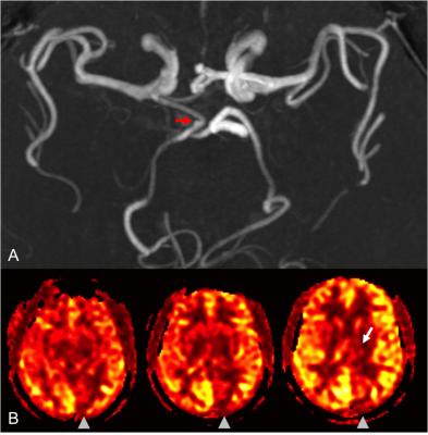 |
Apparent PCA territory hypoperfusion on arterial spin labeling MRI is a common artifact in patients with a unilateral fetal PCA.
Abraham Noorbakhsh1 and Divya Bolar1
1Department of Radiology, University of California, San Diego, San Diego, CA, United States
A retrospective review was performed of 50 patients undergoing MRI with pseudo-continuous arterial spin labeling (pCASL) perfusion imaging and confirmed unilateral fetal PCA. The aim is to determine the frequency of visually-apparent unilateral PCA territory hypoperfusion in patients with a contralateral fetal PCA, but without underlying clinical or imaging pathology to suggest true hypoperfusion. Eight of the fifty cases (16%) had visually-apparent hypoperfusion in the PCA territory contralateral to the fetal PCA. Given these findings, we advise caution when interpreting ASL in patients with a fetal PCA/variant circle of Willis anatomy or suggest using longer post-labeling delay times.
|
|
1625.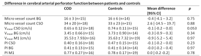 |
The relationship between cerebral arterial perforator function on 7 Tesla MRI and carotid occlusive disease
Tine Arts1, Laurien Onkenhout2, Doeschka Ferro2, Eline Oudeman2, Jaap Kappelle2, Thijs van Osch3, Jaco Zwanenburg1, Jeroen Hendrikse1, and Geert Jan Biessels2
1Radiology, University Medical Center Utrecht, Utrecht, Netherlands, 2Neurology, University Medical Center Utrecht, Utrecht, Netherlands, 3Radiology, Leiden University Medical Center, Leiden, Netherlands
Recently, flow velocity and pulsatility can be measured in cerebral perforating arteries. However, whether this perforator function is independent of upstream large vessel function is unknown. This study therefore investigates cerebral perforator velocity and pulsatility in the centrum semi-ovale (CSO) and basal ganglia (BG) using 7 Tesla MRI in patients with carotid occlusive disease and controls, and in patients inter-hemispherical. Cerebral perforator function was found to be similar in patients and controls, and also similar between hemispheres of patients with unilateral COD. These results show that cerebral arterial perforator function is independent of upstream large vessel disease.
|
|
1626.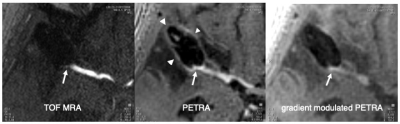 |
Clipped cerebral aneurysm evaluation with gradient modulated PETRA, preliminary study
Minoru Morikawa1, Hideki Ishimaru1, Yohei Ikebe1, Masataka Uetani1, Reiko Ideguchi2, Hiroshi Imai3, and Naoharu Kobayashi4
1Department of Radiology, Nagasaki University Hospital, Nagasaki, Japan, 2Department of Radioisotope Medicine, Atomic Bomb Disease Institute, Nagasaki University, Nagasaki, Japan, 3Siemens Healthcare K.K., Tokyo, Japan, 4Department of Radiology, University of Minnesota Medical School, Minneapolis, MN, United States
Gradient modulated pointwise encoding time reduction with radial acquisition sequence (GM-PETRA) is a new sequence, which can minimize metallic artifact caused by surgical clips. The GM-PETRA samples k-space more quickly by increasing the gradient amplitude after excitation while keeping a relatively low excitation bandwidth. GM-PETRA provides decrease the susceptibility artifact caused by a metallic implant such as surgical clip and can demonstrate the patency of parent artery adjacent to the surgical clip more clearly than conventional PETRA and TOF MRA.
|
|
1627.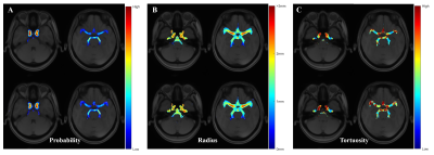 |
Cerebrovascular Atlas: A Novel Framework to Detect Structural Alterations Using MRA
Boyu Zhang1, Bei Wang1, Chengyan Wang2, Ying-Hua Chu3, and He Wang1,2
1Institute of Science and Technology for Brain-Inspired Intelligence, Fudan University, Shanghai, China, 2Human Phenome Institute, Fudan University, Shanghai, China, 3MR Collaboration, Siemens Healthcare Ltd., Shanghai, China
Cerebral vascular alterations are leading risk of several brain diseases. To detect the structural alterations, we proposed the novel framework to automatically generate the cerebrovascular probability atlas and corresponding radius and tortuosity atlas using multistage segmentation and concatenated registration method. 198 hypertension patients and 149 health participants scanned with TOF MRA and T1 sequences were included in our study. The tortuosity in middle cerebral artery and posterior cerebral artery are obviously higher in hypertension group, which may reveal the intrinsic mechanism of cerebrovascular structural changes in hypertension.
|
|
1628.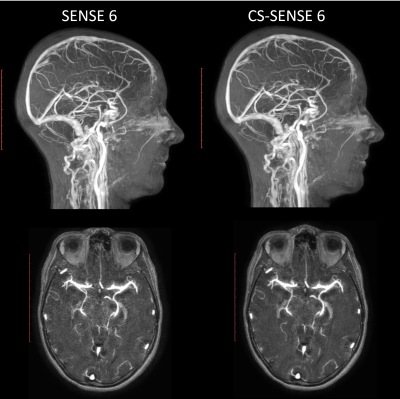 |
Compressed SENSE Accelerated CE-MRA: Comparison of Image Quality and Vessel Detectability with standard SENSE imaging.
Maarten J. Versluis1, Yi Wang2, Karthik Gopalakrishnan3, Burkhard Maedler4, Charles Truwit5, Velmurugan Gnanaprakasam1, Johan van den Brink1, and Liesbeth Geerts1
1BIU MR, Philips Healthcare, Best, Netherlands, 2Clinical Science, Philips Healthcare, Seattle, WA, United States, 3BIU MR, Philips Healthcare, Bangalore, India, 4Clinical Science, Philips Healthcare, Hamburg, Germany, 5Diagnostic Imaging, Philips Healthcare, Cambridge, MA, United States
In this study we compared the image quality of CE-MRA scans using Compressed SENSE and SENSE reconstruction in three subjects. A visual comparison between the number of intracranial vessels showed no differences for both reconstruction techniques. Structural Similarity Index (SSIM) and Mean squared error (MSE) metrics were used to compare retrospectively undersampled CS-SENSE and SENSE acquisitions at a number of reduction factors with the fully sampled dataset. It was found that CS-SENSE reconstructed scans show a higher similarity to the fully sampled acquisitions than using conventional SENSE parallel imaging allowing a 50% increase in acceleration compared to SENSE.
|
|
1629.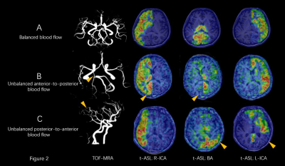 |
Investigation of the relationship between anatomical morphology of circle of Willis and the primary collateral blood flow based on t-ASL
Zhangli Xing1, Yunjing Xue1, Bin Sun1, and Pu-Yeh Wu2
1Fujian Medical University Union Hospital, Fuzhou, China, 2GE Healthcare, MR Research China, Beijing, China
We used 3D-TOF-MRA and territorial-ASL (t-ASL) maps to investigate the relationship between anatomical morphology of circle of Willis (CoW) and the primary collateral blood flow in physiological conditions. We found that the primary collateral blood flow had a direct relationship with anatomical structure of CoW. A1 segment has a role in controlling the blood flow in anterior circulation, while in posterior circulation, P1 segment and PcomA cooperate to dominate the blood flow.
|
|
1630. |
Non-enhanced hybrid arterial spin labeling zTE MRA for intracranial arteries: a feasibility study
Lijuan Wang1, Songan Shang1, Weiqiang Dou2, Jianxun Qu2, Jing Ye1, and Jingtao Wu1
1Northern Jiangsu People’s Hospital, Yangzhou, China, 2GE Healthcare, Beijing, China
In this study, we aimed to investigate the feasibility of a hybrid arterial spin labeling (hASL) zero-echo-time (zTE) magnetic resonance angiography (MRA) for intracranial arteries. Comparing with the conventional zTE MRA (continuous ASL, cASL), the hASL-zTE-MRA showed higher signal homogeneity in the proximal intracranial arteries. Additionally, hASL-zTE-MRA provided more robust performance in depicting cerebrovascular diseases, such as stenosis and arteriovenous malformation (AVM). We therefore demonstrated that the utilization of hASL strategy could potentially improve the image quality of zTE-MRA, and further be applied routinely in the clinic for patients with cerebrovascular diseases.
|
|
1631. |
Zero Echo Time Arterial Spinning Labeling Magnetic Resonance Angiography in the diagnosis of intracranial artery stenosis: a feasibility study
Mingzhen Wu1, Weiqiang Dou2, Chengbing He1, Xiaoqian Huang1, Jixin Luan1, Chuanying Shi1, and Chuanchen Zhang1
1Liaocheng People's Hospital, Liaocheng, China, 2GE Healthcare, MR Research China, Beijing, China
This study aimed to investigate if zero-echo-time arterial-spin-labeling (ZTE ASL) MRA is feasible in the diagnosis of intracranial artery stenosis(ICAD). 25 patients with confirmed clinically stroke or transient ischemic attacks were recruited. For these patients, the correspondingly acquired ZTE-MRA images were compared with those using TOF-MRA technique. DSA imaging was also applied for and considered as a reference standard. For large-medium-sized intracranial arterial segments, ZTE-MRA showed better performance in the image quality and diseased artery detection than TOF-MRA. Therefore, ZTE-MRA can be considered as a promising technique to be further applied in the clinic routinely for patients with ICAD.
|
|
1632. |
Highly Accelerated Compressed Sensing Time-of-Flight MRA for head and neck arterial stenoses: comparison with digital subtraction angiography
Xuan Zhang1, Yue zhou Cao2, Yi Sun3, Michaela Schmidt4, Christoph Forman4, Peter Speier4, and Shanshan Lu1
1Radiology department, The first affiliated hospital of nanjing medical university, Nanjing, China, 2Interventional Radiology department, The first affiliated hospital of nanjing medical university, Nanjing, China, 3MR Collaboration NE Asia, Siemens Healthcare, Shanghai, China, 4Siemens Healthcare GmbH, Erlangen, Germany
As a widely used technique for cerebrovascular disease, the anatomical coverage of TOF-MRA is often limited in order to achieve a compromise between high spatial resolution and acceptable scan time. Compressed sensing (CS) is an effective technique for accelerating 3D acquisitions. In this study, we report the preliminary results of CS TOF-MRA for diagnosing head and neck arterial steno-occlusive disease by taking DSA as the reference standard. CS TOF-MRA provides a relatively large coverage, high resolution, good image quality and comparable diagnostic accuracy to DSA in the assessment of head and neck arterial stenoses within a reasonable acquisition time.
|
|
1633. |
Bright Vessel Appearance on Arterial Spin Labeling MRI: Beyond Localizing Arterial Occlusion
Jinhao Lyu1, Xiangbing Bian1, Liuxian Wang1, Yina Lan1, Xin Zhou2, Lin Ma1, and Xin Lou1
1Department of Radiology, Chinese PLA General Hospital, Beijing, China, 2State Key Laboratory of Magnetic Resonance and Atomic and Molecular Physics, National Center for Magnetic Resonance in Wuhan, Wuhan Institute of Physics and Mathematics, Chinese Academy of Sciences‐Wuhan National Laboratory for Optoelectronics, Wuhan, China
Bright vessel appearance on arterial spin labeling (ASL) had been reported to be a useful marker for localizing arterial occlusion in acute ischemic stroke. But ischemia due to cerebral arterial occlusion did not always show bright vessel appearance on ASL. In the present study, we found that the presence of bright vessel appearance may associate with thrombus age and be able to specifically identify acute cerebral arterial occlusion. The findings may help to optimize current stroke protocols and facilitate the diagnosis of fresh thrombus, thus to support the treatment decision making in stroke patients with large vessel occlusion.
|
|
1634. |
Investigation of collateral circulation compensatory capacity of Willis ring using vessel selective arterial spin labeling
Yuzhu Yan1, Dan Tong1, Xiaochao Liu2, Xiaolei Wang1, Zhuo Wang1, Fan Yang1, and Lizhi Xie3
1radiology, The first hospital of Jilin University, Changchun, China, 2The third people's hospital of Datong, Datong, China, 3GE Healthcare, MR Research China,Beijing, Beijing, China
The Willis ring plays an important role in the collateral circulation especially in patients with internal carotid artery (ICA) stenosis. However conventional time of flight (TOF) MR angiography may only offer limited knowledge of the compensatory blood flow attributed to collateral circulation for patients with stenosis. Vessel selective arterial spin labeling allows the assessment of blood flow of a specific vessel. The purpose of this study was to quantify the influence of various anatomical types on the compensatory capacity of the Willis ring for patients with severe stenosis or occlusion using combined TOF MRA and vessel selective ASL method.
|
|
| 1635. | WITHDRAWN |
1636. |
Evidence for reduced oxygen extraction efficiency in sickle cell anemia patients with cerebral capillary shunting
Meher Juttukonda1,2, Spencer Waddle3, Larry Davis3, Chelsea Lee4, Niral Patel4, Sumit Pruthi3, Adetola Kassim5, Manus Donahue3, and Lori Jordan4
1Athinoula A. Martinos Center for Biomedical Imaging, Massachusetts General Hospital, Charlestown, MA, United States, 2Radiology, Harvard Medical School, Boston, MA, United States, 3Radiology and Radiological Sciences, Vanderbilt University Medical Center, Nashville, TN, United States, 4Pediatrics-Division of Pediatric Neurology, Vanderbilt University Medical Center, Nashville, TN, United States, 5Medicine-Division of Hematology/Oncology, Vanderbilt University Medical Center, Nashville, TN, United States
Venous hyperintense signal in arterial spin labeling (ASL) MRI has been associated with abnormal tissue-capillary water exchange in adults with sickle cell anemia (SCA). We tested the hypothesis that such hyperintense signal is associated with reduced oxygen extraction fraction (OEF) in adults with SCA. Higher categorical scores of shunting were associated with lower OEF in SCA participants with silent infarcts and/or white matter lesions but not in participants without lesions. These findings indicate that venous hyperintense signal in ASL images may reflect impaired abilities of blood to subserve oxygen and may contribute to lesion development in SCA patients.
|
|
1637. |
Anemia, silent stroke, and vascular abnormalities independently contribute to microstructural integrity in Tanzanian sickle cell patients
Hanne A Stotesbury1, Mboka Jacob2, Jamie M. Kawadler1, Dawn E. Saunders1,3, Chris A. Clark1, and Fenella J. Kirkham1
1Imaging and Biophysics, Developmental Nerosciences, UCL Great Ormond St Institute of Child Health, London, United Kingdom, 2Department of Radiology & Imaging, Muhimbili University of Health and Allied Sciences, Dar Es Salaam, Tanzania, 3Department of Radiology, Great Ormond Street Hospital, London, United Kingdom
Reductions in white matter integrity are associated with neurocognitive dysfunction in sickle cell anemia (SCA) patients, but the aetiology is poorly understood. Aiming to explore whether anemia severity, silent cerebral infarction (SCI), and vascular abnormalities may all play a role, we conducted tract-based-spatial statistics in 62 Tanzanian children with SCA. We found anemia severity to be independently associated with increased mean and axial diffusivity, SCI with increased radial diffusivity, and turbulence or vasculopathy with reduced fractional anisotropy. Our findings are consistent with a model of neurological complications in which these pathologies may all contribute to functionally-significant reductions in tissue integrity.
|
|
1638.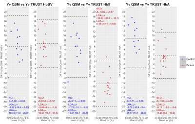 |
Venous Oxygenation in Sickle Cell Patients and Controls using Quantitative Susceptibility Mapping versus T2-relaxation-under-spin-tagging
Hanne A Stotesbury1, Russell Murdoch2, Patrick Hales1, Jamie M. Kawadler1, Melanie Kölbel 1, David Carmichael3, Chris A. Clark1, Fenella Kirkham1, and Karin Shmueli2
1Imaging and Biophysics, Developmental Nerosciences, UCL Great Ormond St Institute of Child Health, London, United Kingdom, 2Department of Medical Physics and Biomedical Engineering, University College London, London, United Kingdom, 3Biomedical Engineering & Imaging Sciences, Kings College London, London, United Kingdom
In 15 homozygous sickle-cell disease patients (SCD; hemoglobin-SS) and 12 healthy controls (HC; 10 Hb-AA, 2 Hb-AS), we compared a quantitative susceptibility mapping (QSM)-based estimate of venous oxygen saturation (Yv) with T2-relaxation-under-spin-tagging (TRUST)-based estimates using bovine-hemoglobin (TRUST-HbBV), hemoglobin-S (TRUST-HbS), or hemoglobin-A (TRUST-HbA) calibrations. Agreement between methods varied, with QSM-Yv estimates in HC and SCD respectively on average 5-6% higher versus TRUST-HbBV, 5% higher and 9% lower versus TRUST-HbS, and 9% higher and 2% lower versus TRUST-HbA. Across all comparisons, the limits of agreement were wide (18-26%) underscoring the need for further studies comparing non-invasive methods with gold-standard jugular vein catheterization.
|
|
1639.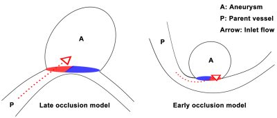 |
Intense Ostium Hemodynamics as the Potential Causes for Delayed Occlusion of Unruptured Sidewall Intracranial Aneurysms by Flow Diverter
Tianhao Su1, Philippe Reymond2, Olivier Brina2, Long Jin1, Karl-Olof Lovblad2, and Maria Isabel Vargas2
1Interventional radiology, Beijing Friendship Hospital, Capital Medical University, Beijing, China, 2Neuroradiology and Neuro-interventional radiology, University Hospitals of Geneva, Geneva, Switzerland Poster Permission Withheld
Flow diverter (FD) has recently been introduced for cerebral aneurysms, especially for the uncoilable sidewall type in the tortuous internal carotid artery (ICA). However, some of treated aneurysms remain patent in clinical practices, probably because of hemodynamic reason. Four-dimensional phase contrast sequences (4D flow MRI) targeting the aneurysm bulge were prospectively performed. The results showed that intense hemodynamic effect at the aneurysmal neck and large ostium might need a longer time for FD induced occlusion. 4D flow MRI before FD procedures has the potential to depict and quantify ostium hemodynamics that could bring new insights in characterizing their treatment responses.
|
|
1640.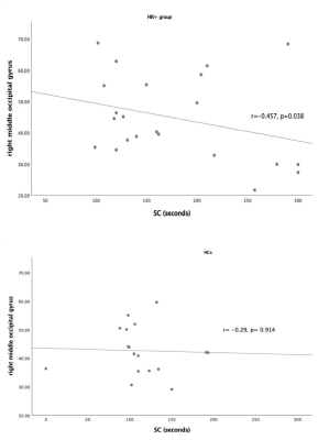 |
Hemodynamic changes underpinning the discrepancy of task performance in HIV+ patients
Danhui Fu1, Guanqiao Jin1, Wenjuan Deng1, Weiyin Vivian Liu2, Long Qian2, and Danke Su1
1GuangXi Medical University Cancer Hospital, Nanning, China, 2MR Research, GE Healthcare, Beijin, China
A bulk of research shows reduced cerebral blood flow is linked to thinning of the cortex and cognitive impairment in dementias and vascular disease while negative relations between functional connectivity and cognition in health are positive in HIV. Our findings of significantly increased CBF in the bilateral middle occipital gyri (MCG) while only left MCG negatively correlated with time for Stroop Color in HIV+ group might underpin that the transition from normal cognition to cognitive impairment in compensation for the abnormal structural and functional alterations. CBF values might reflect hemodynamic differences in HIV+ patients without signs of HIV-associated neurocognitive disorder.
|
|
1641.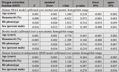 |
Cerebral perfusion and oxygen extraction are similar in sickle cell disease patients with hemoglobin SS and hemoglobin S-thalassemia phenotypes
Spencer L. Waddle1, Ifeanyi Ikwuanusi1, Lori C. Jordan1,2,3, Chelsea A. Lee1,2, Niral J. Patel1,2, Sumit Pruthi1, L. Taylor Davis1, Allison Griffin1, Michael R. Debaun4,5, Adetola A. Kassim5, and Manus J. Donahue1,3,6
1Department of Radiology, Vanderbilt University Medical Center, Nashville, TN, United States, 2Department of Pediatrics, Division of Pediatric Neurology, Vanderbilt University Medical Center, Nashville, TN, United States, 3Department of Neurology, Vanderbilt University Medical Center, Nashville, TN, United States, 4Department of Pediatrics, Vanderbilt-Meharry Center for Excellence in Sickle Cell Disease, Vanderbilt University Medical Center, Nashville, TN, United States, 5Department of Internal Medicine, Division of Hematology/Oncology, Vanderbilt University Medical Center, Nashville, TN, United States, 6Department of Psychiatry, Vanderbilt University Medical Center, Nashville, TN, United States
Sickle cell disease (SCD) comprises multiple sickle phenotypes, yet it is assumed that most phenotypes have similar cerebral hemo-metabolic impact, despite differing hematological characteristics. Here, patients (n=120) with the two most common phenotypes, hemoglobin (Hb)-SS and HbSβ0-thalassemia, were evaluated using anatomical, cerebral blood flow (CBF)-weighted, and oxygen extraction fraction (OEF)-weighted 3T MRI. Results suggest that while CBF depends closely on hematocrit, SCD phenotype does not discriminate either CBF or OEF and anatomical findings of prior infarct and vasculopathy were not significantly different between groups. These findings are consistent with HbSβ0 and HbSS phenotypes having similar impact on cerebral hemo-metabolic dysfunction.
|
|
1642. |
Differential Correlations Between White Matter Microstructure and Perfusion Reveal Microvascular Dysregulation in Sickle Cell Anemia
Soyoung Choi1,2, Chau Vu3, Richard M Leahy4, and John Wood5
1Neuroscience Graduate Program, University of Southern California, LOS ANGELES, CA, United States, 2Children's Hospital Los Angeles, LOS ANGELES, CA, United States, 3Biomedical Engineering, University of Southern California, LOS ANGELES, CA, United States, 4Signal and Image Processing Institute, University of Southern California, Los Angeles, CA, United States, 5Division of Hematology, Children's Hospital Los Angeles, Los Angeles, CA, United States
Patients with chronic anemia has been shown to be susceptible to structural, functional and cognitive impairments. This study explores differences in white matter microstructure (measured by ADC) and cerebrovascular perfusion patterns (measured by MTT) and their relationship to hemoglobin in patients with sickle cell anemia, non-sickle anemia and controls. Larger ADC values and faster MTT is found along the anterior circulation of the brain in sickle-cell anemic patients. Relationships were found between hemoglobin, ADC and MTT in controls that were not observed in sickle-cell patients possibly indicating microvascular dysregulation. Our observations indicate possible microvascular dysregulation in sickle cell anemic patients.
|
|
1643.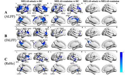 |
Altered Spontaneous Brain Activity in Patients with MELAS at Attack and Remission Stages: Evidence from Resting-state fMRI
Rong Wang1, Yuxin Li1, Jie Lin1, Chong Sun1, Daoying Geng1, and Liqin Yang1
1Huashan Hospital, Shanghai, China
This is the first study that used resting-state fMRI (rs-fMRI) to assess the alterations of spontaneous neuronal activity in patients with mitochondrial encephalomyopathy, lactic acidosis and stroke-like episodes (MELAS) at attack and remission stages, which may provide a new insight into the mechanisms of cerebral cortex damage in MELAS.
|
|
1644.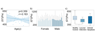 |
Characterization of biomechanical properties of cerebral vessels using magnetic resonance elastography
Shengyuan Ma1, Linjie Wang2, Mo Zhu2, Yanle Hu3, Yiping Du1, Joel R. Garbow4, Curtis Johnson5, Chun-hong Hu2, and Yuan Feng1
1Shanghai Jiao Tong University, Shanghai, China, 2Department of Radiology, The first affiliated hospital of Soochow University, Suzhou, China, 3Mayo Clinic, Phoenix, AZ, United States, 4Washington University in St. Louis, St. Louis, MO, United States, 5University of Delaware, Newark, DE, United States
Measurements of the biomechanical properties of the cerebral vessels could help diagnosis and prognosis of diseases such as lacunar infarction. In this study, shear moduli of the cerebral vessels were measured using both magnetic resonance elastography (MRE) and MR Angiography (MRA). The magnitude images from MRE were registered to MRA images to obtain the shear moduli of the cerebral vessels. Results showed significant correlations between age and gender in terms of the storage modulus. Significant differences between lacunar infarction and healthy control group were also observed in terms of the absolution shear modulus.
|
|
1645. |
Posterior collateral circulation and bypassing perfusion changes after revascularization in Moyamoya disease patients
Chuanying Shi1, Weidong Liu2, Chuanchen Zhang1, Jiheng Hao2, Weiqiang Dou3, Jipeng Wang1, and Jixin Luan1
1Radiology, Liaocheng people's hospital, Liaocheng, China, 2Neurosurgery department, Liaocheng people's hospital, Liaocheng, China, 3MR Research China, GE Healthcare, Beijing, China
In patients with Moyamoya disease, posterior collateral circulation plays an important role in defending ischemic syndrome and good revascularization outcome. The association between bypassing perfusion and posterior collateral circulation remains unknown. This study thus used territory arterial spin labeling to evaluate the territory changes of these vessels. Our results showed that good bypassing perfusion is along with the improvement of posterior collateral perfusion territory. This indicates that good operation outcome was not only associated with poor collateral perfusion, but also related with good collateral vessels functioning as blood channels.
|
|
1646.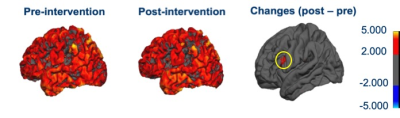 |
Physiotherapy-induced changes in brain in post-stroke rehabilitation : a longitudinal MRI study
Rama Jayasundar1, Dushyant Kumar1, Rajesh Mishra1, Govind Maurya1, Priyanka Bhagat2, Surbhi Kaura2, Senthil Kumaran1, and Padma Srivastava2
1Department of NMR, All India Institute of Medical Sciences, New Delhi, India, 2Department of Neurology, All India Institute of Medical Sciences, New Delhi, India Poster Permission Withheld
This MRI based study has evaluated for the first time, the cortical structural changes effected by physiotherapy in post-stroke (ischemic) recovery. Patients (n=7) with deficits did physiotherapy for one hour daily for six months under bi-weekly supervision of certified physiotherapists. Pre- and post-physiotherapy intervention assessment involved NIHSS and mRS scores and MRI at 3T. The latter included 3D-T1, 3D-FLAIR and fMRI (motor task). Preliminary findings showed individual patients’ positive response to physiotherapy reflected in the significantly reduced NIHSS and increased mRS scores, the recovery of fMRI activation and increase in cortical gray matter volume in specific anatomical locations in brain.
|
|
1647. |
The Capability of Contrast-enhanced T1-weighted MR Imaging for Quantifying Ivy Sign in Moyamoya Disease: compared with FLAIR
Liu-Xian Wang1, Hui Wang2, Jin-Hao Lyu1, Fang-Bin Hao2, Lian Duan2, and Xin Lou1
1The First Medical Center of Chinese PLA General Hospital, Beijing, China, 2The Fifth Medical Center of Chinese PLA General Hospital, Beijing, China
Previous study has reported superiority of contrast-enhanced T1-weighted MR (CEMR) imaging compared with FLAIR for depicting ivy sign in Moyamoya disease (MMD), but no quantitative comparison of both scoring modalities was performed. In this study, we found better consistency between CEMR and DSA, higher ability of CEMR to differentiate severity of MCA stenosis, as well as higher specificity of CEMR compared with FLAIR. Our study proposed that for chronic cerebrovascular disease like MMD, CEMR might be more appropriate in assessing leptomeningeal collaterals.
|
|
1648.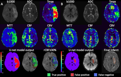 |
Deep learning based Ischemic core prediction from perfusion-weighted imaging in acute ischemic stroke
Yannan Yu1, Yuan Xie1, Thoralf Thamm1, Enhao Gong1, Jiahong Ouyang1, Soren Christensen1, Michael P Marks1, Maarten G Lansberg1, Gregory W Albers1, and Greg Zaharchuk1
1Stanford University, Stanford, CA, United States
Ischemic core of acute ischemic stroke is commonly defined by diffusion-weighted imaging (DWI). CT perfusion, although widely used for acute stroke triaging, is challenging to identify the ischemic core as precise as DWI. In this study, we predicted the DWI lesion from MR perfusion-weighted imaging using U-Net. We found U-net model can predict the ischemic core from perfusion imaging with a better performance compared to clinically-used relative cerebral blood flow map thresholding. In the future study, we will apply the model to patients underwent CT perfusion using transfer learning.
|
|
1649.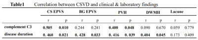 |
MRI features of cerebral small vessel disease in patients with systemic lupus erythematosus
Yiwei Che1, Yanwei Miao1, Yuhan Jiang1, Peipei Chang1, and Lizhi Xie 2
1Department of Radiology, The First Affiliated Hospital of Dalian Medical University, Dalian, China, 2Department of Radiology, GE healthcare, Beijing, China
Systemic lupus erythematosus (SLE) is a diffuse connective tissue disease, which can involve the nervous system, and cerebrovascular damage is the most common. Yet, the relationship between SLE and Cerebral small vessel disease (CSVD) are still not clear. The following study evaluated the MRI findings of enlarged perivascular space (EPVS), white matter hyperintense (WMH) and lacunes of CSVD in patients with SLE. The results showed that CVSD in SLE group was more serious than healthy normal control group, and complement C3 and disease duration were the influencing factors on the development of CSVD in SLE patients.
|
|
1650.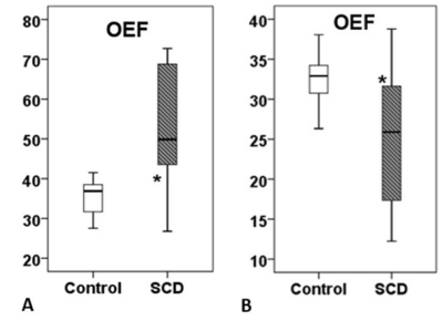 |
Whole-Brain Brain Oxygen Metabolism in Adult Sickle-Cell Patients Using Two Different Quantitative MRI Approaches
Seyed Ali Nabavizadeh1, Sanjeev Chawla1, Pei-Hsin Wu1, Ana Rodriguez-Soto2, Erin Englund2, Michael C Langham1, Farzana Sayani1, Eric Russell1, and Felix W Wehrli1
1University of Pennsylvania, Philadelphia, PA, United States, 2University of California San Diego, La Jolla, CA, United States
Silent cerebral infarcts are the most common neurologic injury in patients with sickle cell disease (SCD). In this study, we compared T2 -relaxation under spin tagging (TRUST) and susceptibility-based oximetry (SBO) techniques in a cohort of SCD patients compared to healthy control subjects. We observed opposite trends in oxygen extraction fraction (OEF) and cerebral metabolic rate of oxygen (CMRO2) in SCD patients compared to controls using TRUST and SBO methods. High OEF measured by TRUST technique lead to very high supraphysiologic CMRO2 values. We conclude that SBO method is more reliable in measuring OEF and CMRO2 in patients with SCD.
|
1651.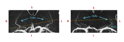 |
Measuring blood velocity pulsations in tortuous lenticulostriate arteries:1D versus 3D velocity-encoded imaging at 7T
Marieke van den Kerkhof1,2, Jacobus F.A. Jansen1,2,3, Robert J. van Oostenbrugge2,4,5, and Walter H. Backes1,2
1Department of Radiology and Nuclear Medicine, Maastricht University Medical Center, Maastricht, Netherlands, 2School for Mental Health and Neuroscience, Maastricht University, Maastricht, Netherlands, 3Department of Electrical Engineering, Eindhoven University of Technology, Eindhoven, Netherlands, 4Department of Neurology, Maastricht University Medical Center, Maastricht, Netherlands, 5Cardiovascular Research Institute Maastricht, Maastricht University, Maastricht, Netherlands
Recently developed 2D phase-contrast sequences showed the feasibility of measuring the small lenticulostriate arteries with 7T MRI. However, these vessels are known to be tortuous, and their tortuosity generally increases with aging and in vascular disease. Therefore, we aimed to measure the blood flow velocity not only in one direction, but in three directions, to capture all three velocity components. We found significant components for the other two, less conventional, velocity directions, which resulted in a lower pulsatility index and peak acceleration when comparing to the 1D measurements.
|
|
1652.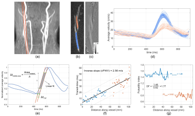 |
One-stop-shop Carotid Pulse Wave Velocity (cPWV) Using a Single-slice 2D Phase-Contrast MRI
Soroush Heidari Pahlavian1,2, Xiaoming Bi3, Samantha Ma1,2, Helena Chui2, Danny J.J. Wang1,2, and Lirong Yan1,2
1USC Stevens Neuroimaging and Informatics Institute, Keck School of Medicine, University of Southern California, Los Angeles, CA, United States, 2Department of Neurology, University of Southern California, Los Angeles, CA, United States, 3Siemens Medical Solutions USA Inc., Los Angeles, CA, United States
The association between increased cerebrovascular risk factors induced arterial stiffness, and age-related neurodegenerative disorders has been demonstrated in multiple studies. In this study, we evaluated the utility of single-slice two–dimensional (2D) phase-contrast MRI (PC-MRI) to assess arterial stiffness by calculating the carotid pulse wave velocity (cPWV) along the common carotid artery (CCA) and internal carotid arteries (ICA) within a single two-minute scan. Our results demonstrated the feasibility of cPWV measurement using single-slice 2D PC-MRI and indicated a positive correlation between cPWV and arterial damping factor. 2D PC-MRI-measured cPWV holds potential as biomarker to quantify age-related alterations in arterial stiffness.
|
|
1653.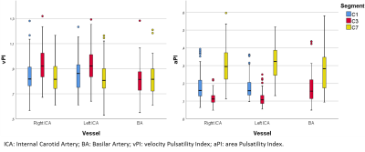 |
Variation in blood-flow pulsatility and arterial distensibility along the internal carotid artery
Rick J van Tuijl1, Ynte M Ruigrok2, Birgitta K Velthuis1, Irene C van der Schaaf1, Gabriël J. E. Rinkel2, and Jaco J.M. Zwanenburg1
1Radiology, UMC Utrecht, Utrecht, Netherlands, 2Neurology and Neurosurgery, Rudolf Magnus Institute of Neuroscience, UMC Utrecht, Utrecht, Netherlands
Velocity pulsatility and area distensibility variation along the internal carotid artery (ICA) trajectory were studied with 2D phase-contrast flow measurements (118 subjects; 3T MRI). Results showed a more complicated behavior than just progressive pulsatility decrease along the ICA towards the circle of Willis. The bony carotid canal appears to constrain the distensibility of the ICA, yielding locally increased velocity pulsatility. Velocity pulsatility was significantly higher in men compared to women and increased with ageing. This age-related increase was strongest just proximal to the circle of Willis, indicating reduced damping of the pulsatility along the ICA with age.
|
|
1654. |
New Predict Parameter of Prognosis on Hyperacute Cerebral Infarction Evaluated by Multi-delay pCASL Imaging
Masafumi HARADA1, Takashi ABE1, Moriaki YAMANAKA1, Tomoki MATSUSHITA1, Yasushi TAKAGI2, Yuishin IZUMI3, Marc R Lebel4, and Mitsuharu Miyoshi5
1Department of Radiology, Tokushima University, Tokushima, Japan, 2Department of Neurosurgery, Tokushima University, Tokushima, Japan, 3Department of Neurology, Tokushima University, Tokushima, Japan, 4GE Healthcare, Calgary, AB, Canada, 5GE Healthcare, Hino, Japan
The purpose of this study was to find a prognostic perfusion parameter to predict the enlargement of acute cerebral infarction. The prognostic parameter of the enlargement ratio (ER) was only mean delay transit time (DT) in all perfusion parameters, and the tendency was different depending on the existence of recanalization or not. DT obtained by multi-delay ASL may become a better prognostic index of acute cerebral infarction than the mismatch area between the infarction core and ischemic penumbra.
|
|
1655.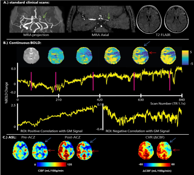 |
Multi-modal assessment of cerebrovascular reactivity by multiband BOLD and multidelay ASL in moyamoya patients using acetazolamide
Pieter T Deckers1, Alex A Bhogal2, Lisa A van der Kleij2, Mathijs BJ Dijsselhof2, Kees PJ Braun3, Jeroen CW Siero2, and Albert Van der Zwan1
1Neurosurgery, UMC Utrecht, Utrecht, Netherlands, 2Radiology, UMC Utrecht, Utrecht, Netherlands, 3Pediatric Neurology, UMC Utrecht, Utrecht, Netherlands Measuring the cerebral blood flow and cerebrovascular reactivity is vital in the preoperative assessment of moyamoya patients. We propose a combination of continuous multiband BOLD-MRI and multidelay ASL-MRI with acetazolamide as a comprehensive multi-modal alternative for the current commonly used PET-CT method. Eight female patients (age 8-41) were scanned at 3T with a ±30 min scan protocol, which is also suitable for patients scanned under general anesthesia. The obtained reactivity maps of the two sequences showed good agreement for severity and location of hemodynamic impairment. This accessible MRI method is directly suitable for routine clinical evaluation of moyamoya patients. |
|
1656.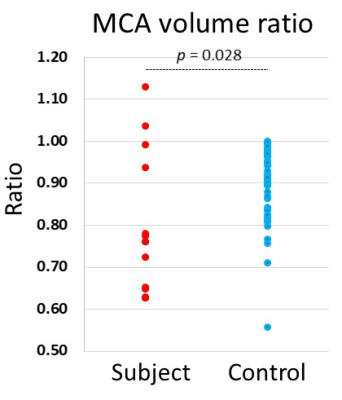 |
Prediction of severe stenosis at the unilateral proximal internal carotid artery by intracranial MR angiography
Kentaro Akazawa1, Masashi Yasuike2, Koji Sakai1, Chisa Bamba1, Jun Tazoe1, Nagara Tamaki1, and Kei Yamada1
1Radiology, Kyoto Prefectural University of Medicine, Kyoto, Japan, 2Radiology, Kyoto Yamashiro General Medical Center, Kizugawa, Japan Poster Permission Withheld
The proximal internal carotid artery (pICA) stenosis is an important cause of ischemic cerebrovascular disease. We evaluated the predicting power to identify unilateral pICA stenosis based on the intracranial 3D time of flight MR angiography (MRA) with multi-slab acquisition used in daily clinical practice by a quantitative assessment of the asymmetry in the middle cerebral artery (MCA). The result revealed that the MCA volume ratio of subjects were significantly lower than controls and the accuracy, sensitivity and specificity to identify subjects were 0.71, 0.71 and 0.88. The quantitative evaluation on intracranial MRA may provide additional information to unilateral pICA stenosis.
|
|
1657. |
Evaluating collateral circulation and its association with clinical outcomes in patients with ischemic stroke using random vessel-encoded ASL
Xiaoyue Ma1,2, Yan Wang1,2, Fengshan Yan1,2, Xianchang Zhang3, Lirong Yan4, and Meiyun Wang1,2
1Radiology, Henan Provincial People's Hospital, Zhengzhou, China, 2Henan Key Laboratory of Neurological Imaging, Zhengzhou, China, 3MR Collaboration, Siemens Healthcare Ltd., Beijing, China, 4Stevens Neuroimaging Informatics Institute, Department of Neurology, Keck School of Medicine, University of Southern California, Los Angeles, CA, United States
This study aimed to explore the clinical value of random vessel-encoded arterial spin labeling (rVE-ASL) in evaluating the collateral status as well as its association with clinical outcomes in patients with ischemic stroke. Results suggested that the rVE-ASL findings were consistent with DSA/CTA findings in characterizing collateral flows and poor collateral flows evaluated by rVE-ASL were associated with poor outcomes. The rVE-ASL could be a potentially useful tool to assess the cerebral collateral circulation in patients with ischemic stroke and predict the clinical outcome.
|
|
1658.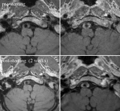 |
Visualizing the Lumen and Wall of Intracranial Artery Stenosis Before and After StentingUsing High Resolution MRI Vessel Wall Imaging
Bing Tian1, Xia Tian2, Zhang Shi2, Qi Liu2, and Jianping Lu2
1Radiology, Changhai hospital, Shanghai, China, 2Changhai hospital of Shanghai, Shanghai, China
HR-MRI provides important insights of assessing both vessel lumen and wall inintracranial artery stenosis disease.
|
|
1659. |
Cerebral perfusion territory changes after artery recanalization in unilateral MCA stenosis disease: a territory arterial spin labeling study
Xinyu Wang1, Xinyi Wang2, and Weiqiang Dou3
1Medical imaging, Shandong First Medical University & Shandong Academy of Medical Sciences, Jinan,Shandong, China, 2Medical imaging, The First Affiliated Hospital of Shandong First Medical University, Jinan,Shandong, China, 3MR Research China, GE Healthcare, Beijing, China
In this study, the main purpose was to explore the clinical value of territory arterial spin labeling (T-ASL) in assessing perfusion changes after unilateral middle cerebral artery (MCA)recanalization. We included 15 patients diagnosed by Digital Substraction Angiography(DSA) with unilateral MCA stenosis, T-ASL examination was performed before and after surgery. Patients showed significantly different reperfusion areas between pre- and post-operations, and some had hyperperfusion displacement in the major cerebral arterial blood supply area. Therefore, T-ASL technique can be used as a valuable method to evaluate the effect of vascular recanalization accurately.
|
|
1660. |
The application of High-Resolution Vessel Wall MRI(HR-VW-MRI) in determining the stability of intracranial MCA and BA plaques
Hongwei Zhou1, Dezheng Kong1, and Tianjing Zhang2
1Jilin University First Hospital, Changchun, China, 2Philips healthcare, Guangzhou, China
The stability of intracranial of the middle cerebral artery(MCA) and basilar artery( BA) plaques related to the stroke events is a crucial issue. However, the discrimination of the plaque’s stability could be rather challenging. Compared with traditional imaging methods such as CTA, MRA or DSA, high-resolution vessel wall MR imaging(HR-VW-MRI) method could demonstrate the abnormality of the vessel wall .It could also potentially evaluate the stability of the intracranial artery. This study aims to compare HR-VW-MRI’s characteristic features of MCA plaque with BA plaque, and to figure out the relationship between plaques’ imaging features with the stroke events
|
|
1661. |
Toward the hemodynamic analysis of Moyamoya disease using ultra-fast DCE-MRI by GRASP
Koji Fujimoto1, Yasutaka Fushimi2, Takeshi Funaki3, Satoshi Nakajima2, Yusuke Yokota2, Sonoko Oshima2, Sayo Otani2, Azusa Sakurama2, Krishna Wicaksono Pandu2, Tomohisa Okada1, and Kaori Togashi2
1Human Brain Research Center, Graduate School of Medicine, Kyoto University, Kyoto, Japan, 2Department of Diagnostic Imaging and Nuclear Medicine, Kyoto University, Kyoto, Japan, 3Department of Neurosurgery, Kyoto University, Kyoto, Japan
One subject with Moyamoya disease was scanned with radial vibe sequence. GRASP reconstruction was performed using different number of spokes/frame. ROI-based analysis performed to compare the maximum slope for each ROI placed in the vessels showed that the faster imaging gave the larger slope, but the blurring at the corner of TIC is seen at the higher temporal resolution. By using images with 5 spokes/frame, voxel-by-voxel analysis could create the arrival map and the maximum slope map.
|
|
1662.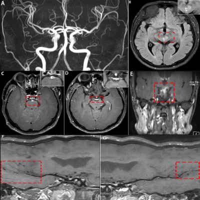 |
Application of high-resolution 3D vessel wall MR(3D VW-MR) imaging in Primary Angiitis of the Central Nervous System
Hong-Wei Zhou1, Dezheng Kong1, and Tianjing Zhang2
1Jilin University First Hospital, Changchun, China, 2Philips healthcare, Guangzhou, China
Primary angiitis of the central nervous system(PACNS) is a kind of rare disease, but the clinical diagnosis is difficult. Recently, high-resolution HR-3D-VW-MRI(3-dimensional vessel wall MR imaging) method has been used to evaluate cerebral vessels because it could directly show the vessel wall as well the lumen; thus it can assist in differentiating various types of vasculopathy. The purpose of our study was to summarize the typical imaging performance of PACNS and evaluate the value of 3D- VW-MRI sequence in demonstrating the detailed information in detection, diagnosis, evaluation, and follow-up for PACNS.
|
|
1663.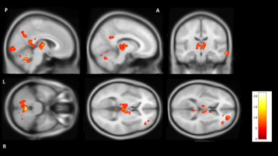 |
Using Arterial Spin Labelling to measure cerebral blood flow during sleep
Balazs Orzsik1, Iris Asllani2, Mara Cercignani2, and Neil Harrison3
1University of Sussex, Brighton, United Kingdom, 2Department of Neuroscience BSMS, University of Sussex, Brighton, United Kingdom, 3CUBRIC, University of Cardiff, Cardiff, United Kingdom
Cerebral blood flow during awake and pharmacologically induced sleep was investigated with arterial spin labeling imaging in healthy young volunteers. Participants were scanned after a good night sleep and in a separate session after 24h sleep deprivation. Participant received 10mg Zolpidem after the sleep deprivation to maximize sleep in the scanner. There was a significant reduction in cerebral blood flow in the sleep condition compared to awake. The most prominent changes were observed in the Thalamus, cerebellum, posterior cingulate gyrus, these areas stayed significant after family wise error correction at a cluster level.
|
|
1664.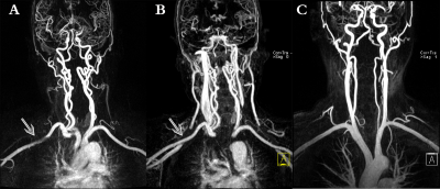 |
Quantitative evaluation of magnetic susceptibility effects in carotid artery CE-MRA with different injected flow rate of contrast medium
Liu Xiaoming1, Kong Xiangchuang1, Lei Ziqiao1, Zhang Xiaoyong2, and Liu Dingxi1
1Departments of Radiology, Union Hospital, Tongji Medical College, Huazhong University of Science and Technology, Wuhan, China, 2MR Collaborations, Siemens Healthcare Ltd, Shenzhen, China
Contrast-enhanced magnetic resonance angiography (CE-MRA) is the main modality used for assessing carotid artery stenosis in the vessel lumen, but limited to the susceptibility artifact, which would result in artifactual stenosis of artery adjacent to the subclavian vein on the injection side of contrast medium. This study aims to explore the quantitative influence of different injected flow rates of contrast medium on the susceptibility effect in the subclavian arterial image of CE-MRA on a 3T MR scanner. The result indicated that the magnetic susceptibility artifact can be reduced by improving the injection speed of contrast agent
|
|
1665.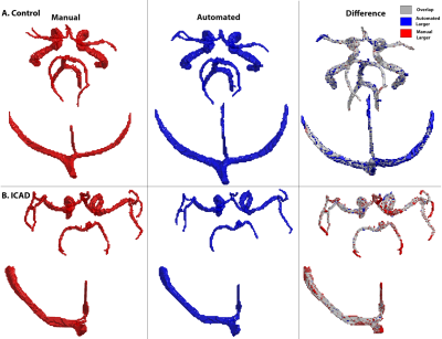 |
Fully Automated Intracranial Vessel Angiogram Segmentation from 4D flow MRI Data in Intracranial Stenosis Patients using Deep Learning
Haben Berhane1, Maria Aristova2, Yue Ma2, Michael Markl2, and Susanne Schnell2
1Lurie Children's Hospital of Chicago, Chicago, IL, United States, 2Northwestern University, Chicago, IL, United States
We developed and validated a convolutional neural network for the fully automated 3D segmentation of the cerebral vasculature from 4D flow MRI for rapid flow analysis. Using 53 4D flow MRI scans, including 16 patients with intracranial atherosclerotic disease, we trained and tested our CNN using 10-fold cross validation. We assessed net flow and peak velocities across all of the major arteries and veins of the intracranial vasulature between automated and manually performed analysis. Across all metrics and regions, we found the automated segmentation showed excellent agreement with the manual, while taking a fraction of the time to perform.
|
1666.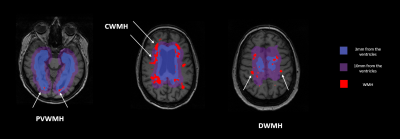 |
White matter hyperintensity features in Neuropsychiatric Systemic Lupus Erythematosus: a retrospective case-control study.
Francesca Inglese1, Myriam G. Jaarsma-Coes1, Gerda M. Steup-Beekman2, Tom Huizinga2, Mark A. van Buchem1, Itamar Ronen1, and Jeroen de Bresser1
1Department of Radiology, Leiden University Medical Center, Leiden, Netherlands, 2Department of Rheumatology, Leiden University Medical Center, Leiden, Netherlands
Systemic Lupus Erythematosus (SLE) often presents with neuropsychiatric (NP) symptoms. These symptoms can be directly associated with SLE (NP-SLE) or not (non-NP-SLE). We investigated whether white matter hyperintensity (WMH) features (such as type, volume and shape) may contribute to the diagnosis of NP-SLE. Patients with NP-SLE more often had deep WMH (DWMH) compared to patients with non-NP-SLE. There were no significant differences in WMH volume and shape features between NP-SLE patients compared to non-NP-SLE patients. In conclusion, NP-SLE patients more often had DWMH compared to non-NP-SLE patients and this may help in the diagnostic process of NP-SLE.
|
|
1667. |
Cerebral Neurovascular Coupling Altered in Patients with Narcolepsy
YuTing Li1
1Department of Radiology, Tangdu Hospital, Fourth Military Medical University, Xian,ShanXi province, China Our purpose is to find the cerebral neurovascular coupling altered in patients with narcolepsy.It was reported that Multifunctional MRI can explore the relationship between cerebral vascular perfusion and neural activity. And we found decreased NVC in right and left precentral gyrus, left occipital gyrus, left postcentral gyrus, left fusiform gyrus and right amygdala |
|
1668. |
MRI for studying yoga-induced cortical structural changes in stroke patients
Dushyant Kumar1, Rajesh Mishra1, Govind Maurya1, Chahat Kumar2, Priyanka Bhagat2, Padma Srivastava2, and Rama Jayasundar3
1Department of NMR, All India Institute of Medical Sciences, New Delhi, India, 2Department of Neurology, All India Institute of Medical Sciences, New Delhi, India, 3DEPARTMENT OF NMR, ALL INDIA INSTITUTE OF MEDICAL SCIENCES, NEW DELHI, India Poster Permission Withheld
This MRI study has evaluated for the first time, the structural changes effected by yoga in post-stroke (ischemic) recovery. Patients (n=6) with deficits practiced Hatha yoga for one hour daily for six months under the supervision of certified yoga trainers. Pre- and 6 months post-yoga intervention assessment involved assessment of NIHSS score and 3D-T1 MRI at 3T MR. Results show individual patients’ positive response to yoga reflected in the significantly reduced NIHSS scores, and in the increased gray matter volume in some areas in brain.
|
|
| 1669. | Non-uniform Brain Temperature in Healthy Subjects
Wen-Ching Liu1, Timothy Linghau Chen2, Kyler Shin2, and David Wang3
1Radiology, OSF Saint Francis Medical Center, Peoria, IL, United States, 2Medicine, University of Illinois College of Medicine at Peoria, Peoria, IL, United States, 3Neurology, Barrow's Neurological Institute, Phoenix, AZ, United States Poster Permission Withheld In order to establish the standard brain regional temperatures in normal and healthy people, we initiated a weekly MR spectroscopy quality assurance program, developed the single voxel MRS thermometry calibration curve with a MRS phantom and scanned 10 normal and healthy right handed men. Five region of interests, hypothalamus, cerebellum, thalamus, frontal lobe, and occipital lobe are selected. Interestingly we found a non-uniform temperature distribution in these ROIs. The hypothalamus and thalamus have the highest temperature while cerebellum has the lowest. |
|
1670. |
White Matter Alterations in Patients with End-Stage Renal Disease: A Diffusion Tensor Imaging Study
Yiwei Che1, Yanwei Miao1, Yuhan Jiang1, Peipei Chang1, Bingbing Gao1, Qingwei Song1, Ailian Liu1, Jiazheng Wang2, and Yanling Wu2
1Department of Radiology, The First Affiliated Hospital of Dalian Medical University, Dalian, China, 2Philips Healthcare, Beijing, China
End-stage renal disease (ESRD) has become risk factor for vascular disease, stroke and cognitive dysfunction, and the white matter lesions may be associated with these adverse outcomes. Silent white matter alterations may occur in the earlier stages, whereas conventional MR imaging may not be found, This study aim to detect subtle white matter alterations in patients with ESRD through diffusion tensor imaging (DTI). DTI can reflect early occult white matter damage in ESRD patients.
|
|
1671.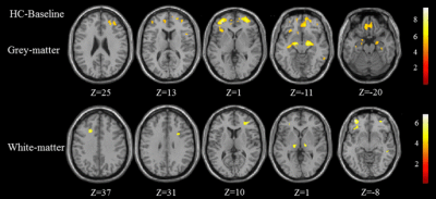 |
Brain atrophy was a risk factor for neurocognitive impairment in hemodialysis patients: a longitudinal voxel-based morphological study
Huiying Wang1,2, Chao Chai3, and Shuang Xia3
1Radiology, Tianjin First Central Hospital;Tianjin Medical Imaging Institute, Tianjin,, Tianjin, China, 2Qinghai University Affiliated Hospital, QingHai, China, 3Tianjin First Central Hospital;Tianjin Medical Imaging Institute, Tianjin,, Tianjin, China
This study aimed to demonstrate the longitudinal changes of brain volume in hemodialysis, and the relationship between brain volume and cognitive function. Thirty-three hemodialysis patients and thirty healthy controls (HCs) were recruited to this study. All patients underwent MR examinations to evaluate baseline and follow-up brain volume. Compared to the HCs, patients showed decreased gray matter (GM) volume and white matter (WM) volume. Compared to baseline patients, follow-up patients showed decreased GM volume and WM volume. The decreased left caudate volume was positively correlated with reduced neuropsychological scores in patients. Brain atrophy occurred in hemodialysis patients, leading to neurocognitive impairment.
|
|
1672.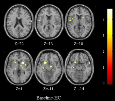 |
Longitudinal study on changes of regional cerebral blood flow in patients maintaining hemodialysis
Huiying Wang1,2, Chao Chai1, and Shuang Xia1
1Tianjin First Central Hospital;Tianjin Medical Imaging Institute, Tianjin,, Tianjin, China, 2Qinghai University Affiliated Hospital, QingHai, China
This was the first study focused on longitudinal CBF changes of ESRD patients with regular hemodialysis. This study used noninvasive pulsed arterial spin labeling technique to provide evidence for clinical evaluation of the changes in cerebral hemodynamics in hemodialysis patients.
|
|
1673.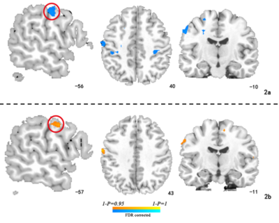 |
Abnormality of insular functional connectivity in uremic restless legs:a resting state functional magnetic resonance study
DUN DING1,2, Xueying Ma2,3, Peng Li4, Pan Zhang5, and Ming Zhang6
1The Second affiliated hospital of Xi'an JiaoTong Uuniversity, Xi'an, China, 2The second affiliated hospital of Xi 'an JiaoTong Uuniversity, Xi'an, China, 3The First affiliated hospital of Neimenggu medical Uuniversity, Hohhot, China, 4The first affiliated hospital of Xi'an JiaoTong Uuniversity, Xi'an, China, 5The second affiliated hospital of Xi'an JiaoTong Uuniversity, Xi'an, China, 6The First affiliated hospital of Xi'an JiaoTong Uuniversity, Xi'an, China
Uremic Restless Leg Syndrome (uRLS) is an uncomfortable complication of inferior limbs and involuntary movements in patients with end-stage renal disease during maintenance of insular, especially during sleep breaks. As an important information processing and processing area, the insular play an important role in sensory, motor metabolism, sensorimotor information processing,and mood regulation. Through this study,We found that the altered morphology of post insular was revealed in uRLS patients, abnormal sensorimotor activities in patients with RLS is closely associated with reduced functional connectivity of insular-S1.Low hemoglobin level was the most important risk factor related to brain functional connectivity feature.
|
|
1674.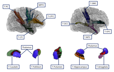 |
Thalamic structural connectivity abnormalities in minimal hepatic encephalopathy: a probabilistic tractography study
Hua-Jun Chen1, Tian-Xiu Zou1, and Nao-Xin Huang1
1Fujian Medical University Union Hospital, Fuzhou, China
Minimal hepatic encephalopathy (MHE) is characterized by a series of cognitive impairments. Thalamus is a critical pathological node of MHE. Thus, we applied probabilistic tractography to investigate the thalamic fiber pathway alterations in MHE, for the first time. The thalamic structural connection was disturbed in MHE patients (reflected by decreased CS/FA and increased MD/AD/RD). Abnormal thalamic connection primarily involved prefrontal cortex, sensorimotor cortex, parietal cortex, medial temporal cortex and hippocampus, and striatum. Thalamic connectivity abnormalities deteriorated with disease progression and correlated with patients’ impaired neuropsychological performance. The disturbed thalamic connectivity can serve as biomarker for diagnosis of MHE.
|
|
1675.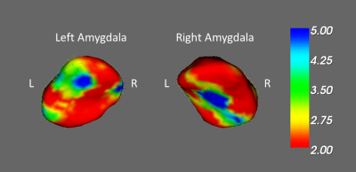 |
Investigation of chemotherapy-induced brain volume and shape change in breast cancer women
Yi-Fang Wu1, Vincent Chin-Hung Chen2,3, Yuan-Hsiung Tsai4, and Jun-Cheng Weng1,3,5
1Department of Medical Imaging and Radiological Sciences, Chang Gung University, Taoyuan, Taiwan, 2School of Medicine, Chang Gung University, Taoyuan, Taiwan, 3Department of Psychiatry, Chang Gung Memorial Hospital, Chiayi, Taiwan, 4Department of Diagnostic Radiology, Chang Gung Memorial Hospital, Chiayi, Taiwan, 5Medical Imaging Research Center, Institute for Radiological Research, Chang Gung University and Chang Gung Memorial Hospital at Linkou, Taoyuan, Taiwan
Adjuvant chemotherapy has been associated with cognitive impairment among a subgroup of breast cancer patients. A limited number of studies have observed the relationship between adjuvant chemotherapy, post-traumatic stress disorder (PTSD) and structural brain changes. Therefore, we employed voxel-based morphometry (VBM) and vertex-wise shape analyses to find the differences in brain volume and shape among pre- and post-chemotherapy breast cancer patients and healthy subjects. Our results showed gray and white matter volume alterations in frontal and temporal brain regions among each group. We also observed the change of shape in the limbic system.
|
1676. |
Relationship between GluCEST concentration and Perivascular Spaces (glia-lymphatic waste clearance pathways) at 7T in Acute Ischemic Stroke
Benjamin Sinclair1,2, John-Paul Nicolo1,2,3,4, David Wright1, Bradford Moffat5, Andrew Neale1,2,3,4, Nian Yu6,7, Elaine Lui8,9, Patricia Desmond8,9, Rebecca Glarin8,9, Kathryn Davis10, Ravi Prakash Nanga11, Ravinder Reddy11, Farshid Sepehrband12,
Bernard Yan3,4, Scott Kolbe1, Patrick Kwan1,2,3,4, Terence J O'Brien1,2,3,4, and Meng Law1,6
1Department of Neuroscience, Monash University, Melbourne, Australia, 2Department of Neurology, Alfred Hospital, Melbourne, Australia, 3Department of Neurology, Royal Melbourne Hospital, Melbourne, Australia, 4Department of Medicine, University of Melbourne, Melbourne, Australia, 5Melbourne Node of the National Imaging Facility, Department of Radiology, University of Melbourne, Melbourne, Australia, 6Department of Radiology, Alfred Hospital, Melbourne, Australia, 7Department of Radiology, The Nanjing Brain Hospital Affiliated to Nanjing Medical University, Nanjing, China, 8Department of Radiology, Royal Melbourne Hospital, Melbourne, Australia, 9Department of Radiology and Medicine, University of Melbourne, Melbourne, Australia, 10Penn Epilepsy Center, Department of Neurology, Hospital of the University of Pennsylvania, Philadelphia, PA, United States, 11Center for Magnetic Resonance & Optical Imaging, Department of Radiology, University of Pennsylvania, Philadelphia, PA, United States, 12USC Stevens Neuroimaging and Informatics Institute, Keck School of Medicine, University of Southern California, Los Angeles, CA, United States
In this pilot study we test the hypothesis that glutamate levels post-stroke are influenced by glia-lymphatic clearance pathways in the brain. Excess glutamate can be neurotoxic, and stroke is associated with focally elevated gluatamate concentrations. Perivascular spaces have recently been found to play a role in waste clearance in the brain, and often become enlarged when compromised. We used Chemical Exchange Saturation Transfer (CEST) imaging at 7T to characterise glutamate concentrations, and found that they were moderately positively correlated with scores of enlarged pervivascular spaces.
|
|
1677.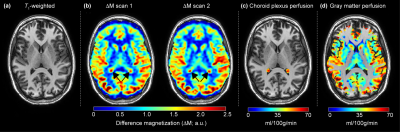 |
Choroid plexus and gray matter perfusion dissimilarity in patients with cerebrovascular disease: implications for CSF dynamics and glymphatics
Skylar E Johnson1, Colin McKnight1, Spencer Waddle1, Maria Garza1, Lori C Jordan1,2,3, Daniel O Claassen3, and Manus J Donahue1,3,4
1Radiology and Radiological Sciences, Vanderbilt University School of Medicine, Nashville, TN, United States, 2Pediatrics, Vanderbilt University School of Medicine, Nashville, TN, United States, 3Neurology, Vanderbilt University School of Medicine, Nashville, TN, United States, 4Psychiatry, Vanderbilt University School of Medicine, Nashville, TN, United States
The goal of this study is to investigate whether choroid plexus (CP) activity, quantified as perfusion measured from arterial spin labeling, is altered in patients with cerebrovascular disease. Cohorts comprised arterial intracranial stenosis (n=56), sickle cell disease (n=41), and healthy controls (n=31). In controls and stenosis patients, CP and gray matter perfusion were similar; in patients with sickle cell disease, CP-to-gray matter perfusion was reduced (p<0.001) relative to other cohorts. This finding is discussed in the context of how efficient CSF perivascular flow surrounding dilated arterial spaces may require reduced CSF production activity to subserve waste clearance.
|
|
1678.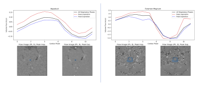 |
Continuous Golden-Angle Spiral Acquisition as a Flexible Tool for Studying CSF Flow Dynamics
Daniel D Borup1, Sandeep K Ganji2, Petrice Mostardi Cogswell1, and James G Pipe1
1Radiology, Mayo Clinic, Rochester, MN, United States, 2Philips Healthcare, Gainesville, FL, United States
The use of a 2D golden-angle spiral trajectory is demonstrated for the measurement of CSF flow at the cerebral aqueduct and foramen magnum. The results compare favorably with a standard cardiac-gated Cartesian scan when retrospectively binned using the cardiac waveform. The spiral data also show variation in the CSF flow waveforms across the respiratory cycle and can be reconstructed as a time-resolved image series for spectral analysis of the flow dynamics.
|
|
1679. |
Mesoscopic imaging of perivascular spaces at 7T
Farshid Sepehrband1, Samantha Ma2, Jeiran Choupan2, Sook-Lei Liew2, and Arthur W Toga2
1Stevens Neuroimaging and Informatics Institute, Keck School of Medicine, USC, Los Angeles, CA, United States, 2USC, Los Angeles, CA, United States
Imaging of the perivascular spaces (PVS) is of growing interest, especially due to its involvement in brain clearance and blood-brain barrier exchange [1]–[3]. PVS mapping is often limited to macroscopic PVS (>1mm), which limits the study of subtle morphological PVS alteration that is expected to occur in submillimeter resolution. Here we show the advantage of ultra-high field imaging for detailed quantification of PVS in vivoand noninvasively.
|
|
1680.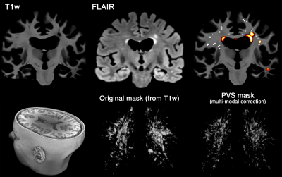 |
WMH and PVS mapping from clinical data using semi-supervised multi-modal convolutional neural network
Farshid Sepehrband1, Giuseppe Barisano2, Hyun-Joon Yang2, Jeiran Choupan2, and Arthur W Toga2
1Stevens Neuroimaging and Informatics Institute, Keck School of Medicine, USC, Los Angeles, CA, United States, 2USC, Los Angeles, CA, United States
Perivascular space (PVS), also known as Virchow-Robin space, is a pial-lined, fluid-filled structure that surrounds vessels in the cerebral cortex [1], [2], and occupies a large portion of the cerebral tissue. While PVS mapping becomes more clinically relevant, separation of PVS from white matter hyperintensities (WMH) make it challenging to map PVS on clinical data. Here we present a semi-supervised multi-modal approach to extract both PVS and WMH using T1w and FLAIR image modalities automatically. We also evaluated multi-site, multi-vendor reliability of the technique.
|
|
1681.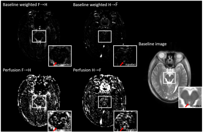 |
Cerebrospinal fluid flow measurement using alternate ascending/descending directional navigation (ALADDIN)
Jun-Hee Kim1, Seung Hong Choi2, and Sung-Hong Park1
1Department of Bio and Brain Engineering, Korea Advanced Institute of Science and Technology, Daejeon, Korea, Republic of, 2Department of Radiology, Seoul National University Hospital, Seoul National University College of Medicine, Seoul, Korea, Republic of
Imaging in-vivo movement of cerebrospinal fluid can be applied not only to diagnose the CSF disorders like hydrocephalus, but also to visualize and evaluate the CSF circulation for the clearance. In this study, we used single phase and multiphase alternate ascending/descending directional navigation (ALADDIN) to image the overall, dynamic CSF flow pattern. Single phase ALADDIN showed overall bulk flow of CSF, and multiphase ALADDIN showed dynamically‑tracked signals of labeled CSF. We expect that ALADDIN can be applied to study CSF circulation and clearance disorders.
|
|
| 1682. | Cerebral hemodynamics and CSF flow in patients with disorders of cerebrospinal fluid circulation
Andrey Tulupov1 and Olga Bogomyakova1
1Laboratory of neuroscience, The Institute International Tomography Center of the Russian Academy of Sciences, Novosibirsk, Russian Federation
In this work we study the most momentous disturbances of hemo- and cerebrospinal fluid dynamics in patients with idiopathic intracranial hypertension (IIH) and idiopathic non-obstructive hydrocephalus (INH). It is shown that ICC, AVD and PI are perspective diagnostic criteria for functional brain diseases, associated with disturbances in hemo- and cerebrospinal fluid dynamics.
|
|
1683.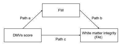 |
Venous disruption affects white matter integrity through increased interstitial fluid in cerebral small vessel disease
Ruiting Zhang1, Peiyu Huang1, and Minming Zhang1
1The Second Affiliated Hospital of Zhejiang University, School of Medicine, Hangzhou, China
We aimed to investigate whether and how venous disruption was related to white matter damage in cerebral small vessel disease(CSVD) patients. SWI was used to investigate venous disruption. Free water elimination DTI model was used to analyze interstitial fluid fraction(fraction of free water, fFW) and white matter integrity(tissue fractional anisotropy, FAt). In 104 CSVD patients, venous disruption was associated with FAt, and the effect was mediated by fFW. This relationship was independent of arterial cerebral blood flow. In conclusion, we discovered the venous disruption - increased interstitial fluid - white matter damage link in CSVD patients.
|
|
1684.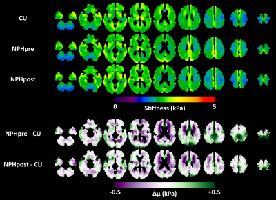 |
Mechanical response of the brain to shunt placement in normal pressure hydrocephalus
Matthew C Murphy1, Petrice M Cogswell1, Joshua D Trzasko1, Armando Manduca1, Matthew L Senjem1, Fredric B Meyer1, Clifford R Jack, Jr.1, Richard L Ehman1, and John Huston, III1
1Mayo Clinic, Rochester, MN, United States
Normal pressure hydrocephalus (NPH) is a neurological disorder characterized by abnormal gait, cognitive decline, and urinary incontinence. The hypothesized role of biomechanics in NPH pathogenesis supports a potential role for magnetic resonance elastography (MRE) in diagnosis and prediction of response to therapy. In this study, MRE was performed in 13 NPH patients before and after shunt placement to test the hypothesis that treatment would reverse NPH-driven changes to the brain’s mechanical properties. We observed that increased stiffness and decreased damping ratio at the vertex were largely reversed by shunting, while periventricular white matter softening was unaffected.
|
|
1685.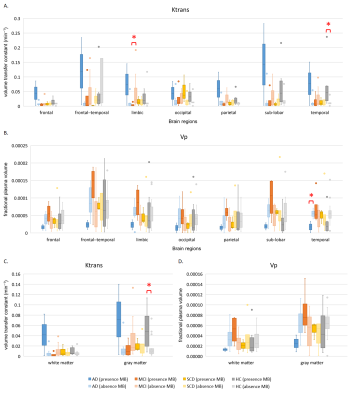 |
Association of cerebral microbleeds and blood-brain barrier function in patients with different severity levels of dementia
Szu-Hsuan Yu1, Wei-Chi Li1, Jiun Wei Chen2, Hong-Yuan Mark Liao2, Feng-Chi Chang3, Chih-Ping Chung4,5,6, Jong-Ling Fuh6,7,8, and Li-Fen Chen1,8,9,10
1Institute of Brain Science, School of Medicine, National Yang-Ming University, Taipei, Taiwan, 2Institute of Information Science, Academia Sinica, Taipei, Taiwan, 3Department of Radiology, Taipei Veterans General Hospital, Taipei, Taiwan, 4Department of Neurology, Taipei Veterans General Hospital, Taipei, Taiwan, 5Department of Neurology, National Yang Ming University, Taipei, Taiwan, 6Department of Neurology, Neurological Institute, Taipei Veterans General Hospital, Taipei, Taiwan, 7Faculty of Medicine, National Yang-Ming University School of Medicine, Taipei, Taiwan, 8Brain Research Center, National Yang-Ming University, Taipei, Taiwan, 9Integrated Brain Research Unit, Department of Medical Research, Taipei Veterans General Hospital, Taipei, Taiwan, 10Institute of Biomedical Informatics, National Yang-Ming University, Taipei, Taiwan
The contributions of blood vessels impairment to dementia are increasingly recognized, but the relationships between microbleeds and blood–brain barrier (BBB) in dementia patients are still unclear. We recruited subjects with Alzheimer’s disease (AD), mild cognitive impairment (MCI), subjective cognitive decline (SCD), and healthy control (HC). The susceptibility weighted images and dynamic contrast-enhanced images were acquired to detect cerebral microbleeds and to estimate BBB integrity. Our results showed high risk with BBB breakdown in AD patients with cerebral microbleeds, which suggests the association between neurovascular unit dysfunction and blood vessel impairment may be a biomarker for the progression of dementia.
|
|
1686.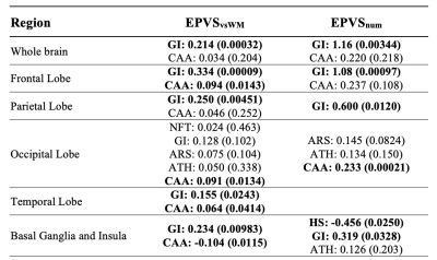 |
Enlarged Perivascular Spaces in Older Adults: Segmentation by Deep Learning; Correlations with Neuropathology and Cognitive Decline
Carles Javierre Petit1, Ashish A. Tamhane2, Arnold M. Evia1, Nazanin Makkinejad1, Gady Agam3, David A. Bennett2, Julie A. Schneider2, and Konstantinos Arfanakis1,2
1Biomedical Engineering, Illinois Institute of Technology, Chicago, IL, United States, 2Rush Alzheimer’s Disease Center, Rush University Medical Center, Chicago, IL, United States, 3Computer Science, Illinois Institute of Technology, Chicago, IL, United States
Enlarged perivascular spaces (EPVS) are common in aging and have been linked to increased risk of stroke, lower cognitive function, and vascular dementia. However, the neuropathologic correlates of EPVS, as well as the contributions of EPVS to cognition, are not well understood. In this work, we first developed a deep-learning algorithm for automatic segmentation and full quantification of whole brain and regional EPVS, and then studied the neuropathologic correlates of EPVS as well as the contributions of EPVS on cognition in a large community-based cohort of older adults.
|
|
1687.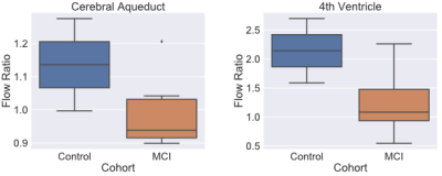 |
Investigating CSF Flow in Amnestic Mild Cognitive Impairment: A Preliminary Report
Arun Venkataraman1, F. Vankee Lin2, Rashid Deane3, and Jianhui Zhong4
1Physics, University of Rochester, Rochester, NY, United States, 2Neurology, University of Rochester, Rochester, NY, United States, 3Neuroscience, University of Rochester, Rochester, NY, United States, 4Imaging Sciences, University of Rochester, Rochester, NY, United States
Amnestic MCI (aMCI) is a prodromal stage to Alzheimer's Disease (AD), with around 10-15% of patients with aMCI converting to AD per year. It is unclear what mechanisms are involved in the initiation and progression through aMCI. One hypothesis involves the “glymphatic” system, a hypothesized pathway mediated by Cerebrospinal Fluid (CSF) flow that is essential to clearing toxins and cellular debris from the brain. In this study, we analyze how CSF flow dynamics are altered in subjects with aMCI. Overall, we found that lower flow in the 4th ventricle is linked with increased disease burden.
|
|
1688.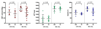 |
Diffusion and multi-echo ASL reveal lower blood-brain interface water permeability in mild cognitive impairment and early Alzheimer’s disease
Tabitha J Manson1,2, Xingfeng Shao3, Danny JJ Wang3, Matthias Günther4,5, Lynette Tippett2,6,7, Michael Dragunow2,7,8, David L Thomas9,10, Vinod Suresh1,11, and Catherine Morgan2,6,7
1Auckland Bioengineering Institute, University of Auckland, Auckland, New Zealand, 2Brain Research New Zealand, Auckland, New Zealand, 3Stevens Neuroimaging and Informatics Institute, Keck School of Medicine, University of Southern California, Los Angeles, CA, United States, 4Fraunhofer Institute for Digital Medicine MEVIS, Bremen, Germany, 5Faculty 01 (Physics/Electrical Engineering), University Bremen, Bremen, Germany, 6School of Psychology, University of Auckland, Auckland, New Zealand, 7Centre for Brain Research, University of Auckland, Auckland, New Zealand, 8Department of Pharmacology, University of Auckland, Auckland, New Zealand, 9Dementia Research Centre, UCL Queen Square Institute of Neurology, University College London, London, United Kingdom, 10Wellcome Centre for Human Neuroimaging, UCL Queen Square Institute of Neurology, University College London, UK, London, United Kingdom, 11Department of Engineering Science, University of Auckland, Auckland, New Zealand
Blood-brain barrier (BBB) dysfunction has been found in multiple neurodegenerative conditions, including mild cognitive impairment (MCI) and Alzheimer’s disease (AD). However recent concerns on the repeated use of Gadolinium based contrast agents (GBCAs), prompted us to investigate alternative, non-invasive methods for measuring BBB function. Both diffusion-weighted (DW) and multi-echo (ME) ASL was implemented at 3T to determine water transfer rates (kw) in the brain in MCI and early AD participants. We found kw to be lower in the cognitively impaired group, compared to controls, with both modalities, suggesting that these techniques may provide a marker of early AD pathology.
|
|
1689.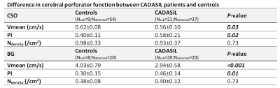 |
Velocity and pulsatility in small cerebral perforators of CADASIL patients compared to healthy controls
Tine Arts1, Hilde van den Brink2, Anna Kopczak3, Martin Dichgans3, Jeroen Hendrikse1, Jeroen Siero1,4, Jaco Zwanenburg1, and Geert Jan Biessels2
1Radiology, University Medical Center Utrecht, Utrecht, Netherlands, 2Neurology, University Medical Center Utrecht, Utrecht, Netherlands, 3Institute of Stroke and Dementia Research, Munich, Germany, 4Spinoza Centre for Neuroimaging, Amsterdam, Netherlands
CADASIL is a monogenic form of cerebral small vessel disease that leads to ischemic brain lesions at mid-adult age. We studied arterial perforator blood flow velocity and pulsatility in CADASIL patients and matched controls. We found significantly lower velocity and higher pulsatility in the basal ganglia and in normal appearing white matter (NAWM) of the centrum semi-ovale (CSO) in patients compared to controls. Within patients, no differences in velocity and pulsatility were found between CSO NAWM and white matter hyperintensities. This shows that small vessel disease in CADASIL patients is reflected in abnormal small vessel function also beyond lesioned areas.
|
|
1690. |
Dilated PVS affects surrounding white matter non-parenchymal water fraction differently in healthy young and elderly CSVD individuals
Yeerfan Jiaerken1,2 and Zhang Minming1
1Second Affiliated Hospital of Zhejiang University, School Of Medicine, hangzhou, China, 2UNC Chapel Hill, chapel hill, NC, United States
We investigated how dilated perivascular space (dPVS) affects water drainage in surrounding tissue and whether there is any distance-related pattern of water distribution around dPVS. We found that The isotropic diffusing water content was higher within dPVS, compared to contralateral normal white matter, and the CSVD subjects had higher within-dPVS water content than healthy subjects, and dPVS showed different distance-related pattern in healthy subjects and subjects with severe CSVD, suggesting dPVS played different roles in healthy subjects and CSVD.
|