
Digital Poster Session
Neuro: Neuro Acquisition and Processing
Neuro
1747 -1761 Neuro Acquisition and Processing - Neuroimaging: Acquisition 1
1762 -1776 Neuro Acquisition and Processing - Neuroimaging: Acquisition 2
1777 -1791 Neuro Acquisition and Processing - Neuroimaging: Image Processing
1792 -1806 Neuro Acquisition and Processing - Emerging Neuro Brain Imaging 1
1807 -1819 Neuro Acquisition and Processing - Emerging Neuro Brain Imaging 2
1820 -1834 Neuro Acquisition and Processing - Emerging Neuro Brain Imaging 3
1835 -1848 Neuro Acquisition and Processing - Emerging Neuro Brain Imaging 4
1849 -1861 Neuro Acquisition and Processing - All Things Neurospectroscopy
1747.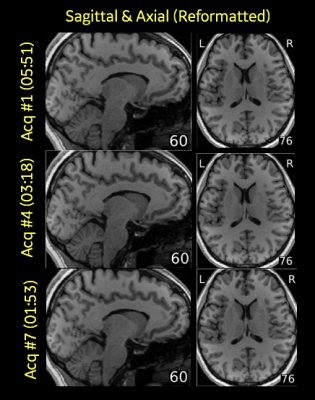 |
Compressed Sensed MPRAGE with Parallel Imaging: Image Quality Metrics and Morphometry Study at 3T
David D Shin1, Dan Rettmann2, Naoyuki Takei3, and Suchandrima Banerjee1
1GE Healthcare, Menlo Park, CA, United States, 2GE Healthcare, Rochester, MN, United States, 3GE Healthcare, Hino Tokyo, Japan
While there is a growing trend for using volumetric acquisitions for brain MRI, long acquisition times still limit their adoption. There is still a tremendous need to reduce scan time of these volumetric acquisitions to improve workflow productivity and to reduce the likelihood of motion during the scan. MPRAGE sequence is a key 3D brain acquisition because of its excellent gray-white contrast which is ideal for visualization of substructures, segmentation and morphometric measurements. This work takes a rigorous quantitative approach to investigating the effects of compressed sensing and parallel imaging factors on various tissue measurements derived from accelerated MPRAGE images.
|
|
1748. |
Detectability of the Dura Matter at 7T using Compressed Sensing
Olivier E Mougin1, Joshua McAteer1, Matthan Caan2, and Penny A Gowland1
1School of Physics and Astronomy, SPMIC, Nottingham, United Kingdom, 2Department of Biomedical Engineering & Physics, Amsterdam University Medical Center, Amsterdam, Netherlands
High resolution of the dura matter is useful to detect and follow meningeal pathology. We presented 0.5mm isotropic images obtained at 7T, using compressed sensing to reduce the acquisition time and improve the detectability of the dura matter. Acceleration up to a factor of 8 was possible with broadening of the PSF of the dura visible at acceleration factor higher than 6.
|
|
1749. |
The Feasibility of Accelerated Brain 3D T1WI MRI Using Compressed Sensing: Qualitative and VBM validation
Yunyun Duan1, Yaou Liu1, and Jiazheng Wang2
1Beijing Tiantan Hospital, Capital Medical University, Beijing, China, 2Philips (China) Investment Co., Ltd, Beijing, China
A major barrier of clinical application of 3D T1 sequence is the long acquisition time. Compressed SENSE (CS-SENSE) was introduced into the clinical routine and showed reduced scan time without reduced image quality. However, automatically quantitative assessment of brain structure has not been certificated up to date. This study evaluated the different acceleration factors of CS-SENSE or SENSE of a 3D brain T1 sequence with qualitative and quantitative measurements in health volunteers and patients, in order to assess the feasibility of 3D brain structural imaging acceleration.
|
|
1750. |
Evaluating brain MRE optimal conditions at low and high excitation frequencies
Fatiha Andoh1, Marion Tardieu2, Claire Barakat Pellot3, and Xavier Maître1
1Univ. Paris Saclay, IR4M Laboratory, Orsay, France, 2Institut du Cancer de Montpellier, ICM, Montpellier, France, 3INSERM, IMIV Laboratory, Orsay, France Mechanical parameters may be underestimated by MR-Elastography if data quality is not properly monitored to discarded inaccurate and unprecised biased data. Optimal condition defined by proper spatial data sampling upon λ/a≈[6;9] can alleviate the quality requirements by lowering Qthreshold, which is defined on each dataset at the drop of correlation between the quality factor and the targeted mechanical parameters. |
|
1751.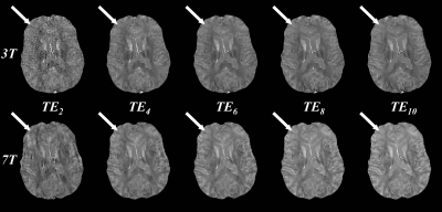 |
Quantitative susceptibility mapping: comparison of reproducibility between 3T and 7T
Pascal Spincemaille1, Julie Anderson2, Gaohong Wu2, Baolian Yang2, Maggie Fung2, Ke Li2, Shaojun Li1, Ilhami Kovanlikaya1, Ajay Gupta1, Doug Kelley, 2, Nissim Benhamo2, and Yi Wang1
1Weill Cornell Medicine, New York, NY, United States, 2General Electrical Healthcare, Waukesha, WI, United States
In this work, we compare the performance of QSM at 7T versus 3T in an intra-scanner test-retest experiment with varying echo numbers and in an image quality analysis. QSM was reproducible within fixed field strength when the same number of echoes were used. Across field strengths and with different numbers of echoes, QSM was reproducible between the 3T acquisition with 10 echoes and the 7T acquisition with 5 echoes. The preliminary data suggest that 7T can be used to shorten QSM acquisition time or enable higher resolution QSM in clinically acceptable scan times.
|
|
1752.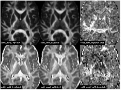 |
Comparison of advanced quantitative diffusion MRI parameter in a multi-site MR study using the traveling volunteer approach
Daniel Güllmar1, Renat Sibgatulin1, Stefan Ropele2, and Jürgen R Reichenbach1,3
1Medical Physics Group / IDIR, Jena University Hospital, Jena, Germany, 2Department of Neurology, Medical University of Graz, Graz, Austria, 3Michael-Stifel-Center for Data driven sciences, Friedrich-Schiller-University Jena, Jena, Germany
If quantitative diffusion measures are acquired at different sites but identical hardware and protocol settings, it remains unclear if a site bias would require a data homogenization in order to pool the data for analysis. A traveling volunteer (including four subjects) approach was applied and different complex diffusion measure as well as diffusion tensor metrics were computed based on the measurements at two different sites. Our results suggest that the inter-site differences are much smaller than the inter-subject differences in the ROI based analysis. The voxel-wise analysis was found to be more susceptible to incomplete artifact compensation and registration errors.
|
|
1753.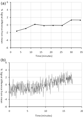 |
Assessing and comparing the suitability of 3D Gradient Echo and VIBE for quantifying subtle blood-brain barrier leakage
Nicholas G Dowell1, Nourah Alruwais2, Paul S Tofts1, and Jennifer Rusted2
1CISC, Brighton and Sussex Medical School, Brighton, United Kingdom, 2School of Psychology, University of Sussex, Brighton, United Kingdom
There is increasing interest in measuring leakage across the blood-brain barrier (BBB) to assess its response to pathology, inflammation or ageing. Quantifying permeability of the BBB is typically performed using dynamic contrast enhanced (DCE) MRI which involves repeated T1-weighted imaging following administration of a Gd contrast agent. In this work, we assess the suitability of two popular DCE acquisition approaches to BBB permeability: (1) a slow gradient echo approach and (2) fast T1-weighted imaging using the VIBE technique . We discuss the advantages and disadvantages of both techniques and recommend the optimum technique for quantification of subtle BBB permeability.
|
|
1754.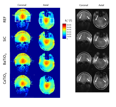 |
In vivo performance evaluation of silicon carbide dielectric pads for 7T MRI.
Zo Raolison1, Redha Abdeddaïm2, Marc Dubois2, Michel Luong3, Luisa Neves1, Franck Mauconduit3, Stefan Enoch2, Kaizad Rustomji1, Clarence Quinaux1, Nicolas Malléjac4, Pierre Sabouroux2, Fawzi Boumezbeur3, Patrick Berthault5,
Mikhail Zubkov6, Anne-Lise Adenot-Engelvin4, Lucie Hertz-Pannier3, and Alexandre Vignaud3
1Multiwave Imaging, Marseille, France, 2Aix Marseille Université, Marseille, France, 3CEA-DRF/Joliot/Neurospin, Gif-sur-Yvette, France, 4CEA-DAM Le Ripault, Monts, France, 5CEA-DRF/IRAMIS/NIMBE/LSDRM, Gif-sur-Yvette, France, 6Faculty of Physics and Engineering, ITMO University, Saint Petersburg, Russian Federation Dielectric pads have demonstrated to be a simple and yet efficient solution to mitigate locally RF transmission heterogeneities while using classic 7T MRI birdcage. Extensive research has not yet been carried on alternative candidate to water-based perovskites. The grail would be a long-lasting, comfortable, MR invisible, efficient, unalterable and high permittivity soft material. In this study, a novel material based on silicon carbide particles and addressing those requirements was successfully compared to perovskite pads from the literature in terms of B1+ homogeneity and image contrast through in vivo measurements. |
|
1755. |
Intraplatform Repeatability and Interplatform Agreement of Whole-Brain 3D MRE
Yuxiang Zhou1, Yuan Le2, Kevin J Glaser2, Jun Chen2, Roger C Grimm2, Arvin Arani2, Bradley D Bolster, Jr3, Stephan Kannengiesser4, John Huston III2, Joel P Felmlee2, Richard L Ehman2, and Joseph M Hoxworth1
1Radiology, Mayo Clinic, Phoenix, AZ, United States, 2Radiology, Mayo Clinic, Rochester, MN, United States, 3Siemens Medical Solutions USA, Inc., Salt Lake City, UT, United States, 4Siemens Healthcare GmbH, Erlangen, Germany Using MRE to evaluate the brain requires acquisition of the full 3D wave field and application of 3D inversion processing to generate valid stiffness maps. The goal of this study was to assess the precision and cross-platform reproducibility of whole brain stiffness measurements obtained with prototype 3D SE-EPI-based MRE techniques, as implemented on GE and Siemens MRI platforms, with the goal of standardization. Our data showed excellent intraplatform repeatability and good interplatform agreement. |
|
1756. |
The effect of distinct spatial resolution on quantitative maps using synthetic MRI
Tie-bao Meng1, Haoqiang He1, Huiming Liu1, Weijing Zhang1, Chenghui Huang1, Long Qian2, and Chuanmiao Xie1
1Department of Radiology, Sun Yat-Sen University Cancer Center, Guangzhou, China, 2MR Research, GE Healthcare, Beijing, China
In the field of neuroscience, an emerging technology named relaxation quantitative MRI (RQ-MRI) have demonstrated its great potential in both clinic and research. Among those RQ-MRI related technologies, the synthetic MRI has moved rapidly towards clinical application due to its acceptable acquisition time. However, how the spatial resolution of synthetic MRI impacts the quantitative maps is still largely unclear. To address this question, in current study, a total of 13 normal subjects with four distinct spatial resolutions were applied. Our results demonstrated that both the in-plane resolution and slice thickness have significantly influence on the measured quantitative values.
|
|
1757.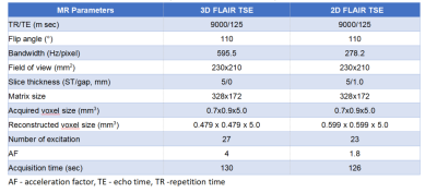 |
Evaluation of 3D FLAIR Combined Compressed Sense Technology in Cerebrospinal Fluid Flow Artifacts Reduction: Comparison with 2D FLAIR
Si Xu1, XiaoMing Liu1, and JiaZheng Wang2
1Departments of Radiology, Union Hospital, Tongji Medical College, Huazhong University of Science and Technology, WuHan, China, 2Philips Healthcare, BeiJing, China
3D FLAIR TSE imaging can effectively inhibit cerebrospinal fluid flow artifacts, but the scanning time is long, affecting clinical application. Compressed sensing (CS) is a technique to accelerate the acquisition of magnetic resonance imaging (MRI) by using sparsity constraints during readout measurements of k-space. This study aimed to compare the cerebrospinal fluid flow artifacts and overall image quality in CS 3D FLAIR TSE imaging combined and in 2D FLAIR TSE imaging. Results showed that CS 3D FLAIR TSE yielded better image quality, enhanced diagnosis performance, and reduced fluid flow artifacts when compared to the traditional 2D FLAIR TSE sequence.
|
|
1758.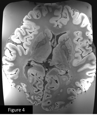 |
Try this at home: Ex vivo Human Brain at 200 Micron Resolution with a Simple Set Up.
Sanghoon Kim1, Ken Sakaie1, Mark J Lowe1, and Stephen E Jones1
1Cleveland Clinic Imaging Institute, CLEVELAND, OH, United States
Postmortem, whole-brain MRI provides an opportunity to investigate anatomy at spatial resolution approaching that of histology without the distortions and limited coverage inherent to histology. Previous work with ex vivo imaging has been done using a sophisticated process with additional steps, such as a custom-built vibration apparatus to remove air bubbles, proton-free oil to eliminate background signal and modifications to the image reconstruction hardware to deal with the large quantity of imaging data as well as a custom RF coil. The results shown here suggest that similar data can be readily acquired without taking extraordinary or costly measures.
|
|
1759. |
Postmortem Whole-Brain MP2RAGE Optimization at 3T: A New Imaging Window into Multiple Sclerosis Cortical Pathology
Matthias Weigel1,2,3, Riccardo Galbusera1,2, Reza Rahmanzadeh1,2, Muhamed Barakovic1,2, Po-Jui Lu1,2, Ludwig Kappos2, Wolfgang Brück4, Tobias Kober5,6,7, Peter Dechent8, and Cristina Granziera1,2
1Translational Imaging in Neurology (ThINk) Basel, Department of Medicine and Biomedical Engineering, University Hospital Basel and University of Basel, Basel, Switzerland, 2Neurologic Clinic and Policlinic, Departments of Medicine, Clinical Research and Biomedical Engineering, University Hospital Basel and University of Basel, Basel, Switzerland, 3Radiological Physics, Department of Radiology, University Hospital Basel, Basel, Switzerland, 4Institute of Neuropathology, University Medical Center Göttingen, Göttingen, Germany, 5Advanced Clinical Imaging Technology, Siemens Healthcare, Lausanne, Switzerland, 6Department of Radiology, Lausanne University Hospital and University of Lausanne, Lausanne, Switzerland, 7LTS5, École Polytechnique Fédérale de Lausanne (EPFL), Lausanne, Switzerland, 8Department of Cognitive Neurology, MR-Research in Neurology and Psychiatry, University Medical Center Göttingen, Göttingen, Germany
The MP2RAGE sequence provides fast volumetric T1 weighted MR imaging and offers the possibility to reconstruct quantitative T1 maps. Therefore, it is frequently applied for studying Multiple Sclerosis pathologies in recent years. The present work investigates and explains necessary protocol changes for applying MP2RAGE in fixated human brain acquisitions. Using the established protocol, it is shown that strong soft tissue contrast is reinstated and quantitative T1 values can be derived for normal appearing gray matter and lesions. Based on the feasibility of using long scan times, the isotropic resolution of the MP2RAGE could be even increased to 0.75mm.
|
|
1760.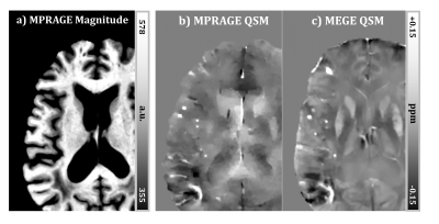 |
Quantifying Cerebral Microbleeds using MPRAGE-based Quantitative Susceptibility Mapping
Nashwan Naji1, Myrlene Gee1, Glen C. Jickling2, Richard Camicioli2, and Alan H. Wilman1
1Department of Biomedical Engineering, University of Alberta, Edmonton, AB, Canada, 2Division of Neurology, University of Alberta, Edmonton, AB, Canada
The MPRAGE sequence is commonly included in brain studies for structural imaging using magnitude images; however, its phase images can provide an opportunity to assess microbleed burden using Quantitative Susceptibility Mapping (QSM). In this study, the mean susceptibility and the cross-sectional area of cerebral microbleeds were assessed using a susceptibility map derived from the phase of the MPRAGE sequence, with comparison to QSM measurements based on standard multi-echo gradient-echo. Microbleeds were well visualized on MPRAGE-QSM with susceptibility generally higher on MPRAGE-QSM, mostly due to mismatch in spatial resolution and SNR.
|
|
1761. |
Improved presentation of brachial plexus at 3T using diffusion-weighted imaging with multiple interleaves in the phase-encode direction
Ke Jiang1, Jiazheng Wang1, and Yuefei Ma1
1Philips Healthcare Greater China, Beijing, China
Diffusion weighted imaging for brachial plexus can provide useful information for clinical applications. However, clinical DWI usually employs single-shot EPI for signal acquisition, which suffers from image distortion or signal loss especially in the imaging in the head-neck regions due to the spatial variation in B0 field. This work demonstrated the application of a multi-interleave DWI sequence that achieves quality brachial plexus imaging with a resolution of 1.9x1.9 mm2 at 3T system.
|
 View the Poster
 Watch the Video
1762.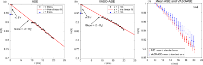 |
Vascular space occupancy asymmetric spin echo (VASO-ASE) for regional cerebral oxygen extraction fraction mapping
Spencer L. Waddle1, Maria E. Garza1, and Manus J. Donahue1,2,3
1Department of Radiology, Vanderbilt University Medical Center, Nashville, TN, United States, 2Department of Neurology, Vanderbilt University Medical Center, Nashville, TN, United States, 3Department of Psychiatry, Vanderbilt University Medical Center, Nashville, TN, United States
The asymmetric spin echo technique (ASE) can be used to collect maps of R2’ and calculate cerebral oxygen extraction fraction (OEF). However, these models require extravascular signal only, which is often not fully achieved in standard ASE approaches. Here, a vascular-space-occupancy (VASO) prepulse is used to null intravascular signal in a novel VASO-ASE method, and repeatability measurements of parenchymal versus extravascular R2’ are presented. VASO-ASE was found to provide physiological OEF (34.1±5.3%) and higher reproducibility in controls compared to standard ASE.
|
|
1763. |
Isotropic High-Resolution Brain T1rho Mapping with 3D FLAIR MAPSS at 3T
Qi Peng1, Can Wu2, Xiaojuan Li3, Michael L Lipton1, and Craig Branch1
1Gruss Magnetic Resonance Research Center, Department of Radiology, Albert Einstein College of Medicine and Montefiore Medical Center, Bronx, NY, United States, 2Philips Healthcare, Andover, MA, United States, 3Program of Advanced Musculoskeletal Imaging (PAMI), Cleveland Clinic, Cleveland, OH, United States
Quantitative T1rho (T1ρ) mapping MRI has recently gained wider clinical/research application in human brain imaging. However, high resolution 3D brain T1rho mapping on clinical scanners is still time consuming with compromised quantitative accuracy due to image blurring or artifacts. We propose a 3D FLAIR MAPSS T1rho imaging scheme, which combines CSF-suppression with 3D MAPSS sequence for improved quantitative accuracy in T1rho mapping. Phantom, volunteer, and patient studies validated its sensitivity and reliability for isotropic high resolution of voxel size (1.3mm)3 quantitative mapping using continuous-wave or adiabatic RF pulse T1rho preparation, acquired within clinically acceptable scan duration at 3T.
|
|
1764. |
Evaluation of 4D ultrashort TE MR Angiography using Variable Inversion Time.
Haruyuki Fukuchi1,2, Nao Takano3, Yutaka Ikenouchi2, Michimasa Suzuki2, Osamu Abe1, and Shigeki Aoki2
1Department of Radiology, Graduate School of Medicine, University of Tokyo, Tokyo, Japan, 2Department of Radiology, Juntendo University Graduate School of Medicine, Tokyo, Japan, 3Department of Radiology, Juntendo University Hospital, Tokyo, Japan
We developed a novel method, Variable TI, to improve the visibility of ASL based UTE 4D-MRA. In the phantom study and the volunteer study, the Variable TI UTE 4D-MRA offered a higher signal intensity and improved visualization of arteries in late phase compared to the conventional method. The Variable TI technique can improve clinical usability of 4D-MRA because this technique not only offered better visibility of arteries but also realized the data efficacy and reduced reconstruction time.
|
|
1765. |
Simultaneous Brain/Spinal Cord fMRI reveals Subject-Level Activation Differences under Noxious Thermal Stimulus
Christine Sze Wan Law1, Ken Arnold Weber1, Sean Mackey1, and Gary Glover1
1Stanford University, Stanford, CA, United States
Functional activation within brain has been studied extensively via BOLD fMRI. Limiting investigation to brain alone provides a truncated view of the central nervous system as it does not capture information exchange between brain and spinal cord. Simultaneously imaging brain, brainstem, and cervical spine provides insight into pain modulation pathways because dorsal horn is the first synapse connecting periaqueductal gray with cortical pain regions. Sprenger & Finsterbusch (2015) have shown group-level functional connection of brain to spinal cord under noxious thermal stimulus. Here, we investigate subject-level differences in neural activity in brain-spinal cord.
|
|
1766. |
Initial feasibility of a multi-band PSF-mapping based, reverse-gradient approach with geometric distortion correction for whole-brain fMRI
Myung-Ho In1, Daehun Kang1, Hang Joon Jo2, Uten Yarach1, Nolan K. Meyer1, Joshua D Trzasko1, Erin M Gray1, John III Huston1, Matt A Bernstein1, and Yunhong Shu1
1Department of Radiology, Mayo Clinic, Rochester, MN, United States, 2Department of Physiology, College of Medicine, Hanyang University, Seoul, Korea, Republic of
A point-spread-function mapping-based reverse-gradient approach was demonstrated as a viable method to correct severe susceptibility artifacts for deep-brain-stimulation fMRI in a pig model, but at the cost of reduced temporal resolution. Interleaved acquisition of the echo-planar-imaging was used with opposite phase-encoding polarities. In this work, feasibility was evaluated in in-vivo resting-state fMRI reliability in high-susceptibility regions. To compensate for the reduced temporal resolution, multi-band imaging was used, and the improved reliability in highly susceptible regions was evaluated on both standard whole-body and high-performance compact 3T scanners.
|
|
1767.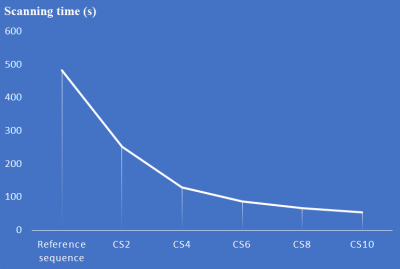 |
Accelerating magnetic resonance angiography using Compressed SENSE technology - a prospective multi-center study
Jinli Ding1, Yunyun Duan1, Zhizheng Zhuo1, Yawei Yuan2, Guiqing Zhang2, Qingwei Song3, Bingbing Gao3, Bing Zhang4, Maoxue Wang4, Linlin Yang5, Yang Hou5, Fenglian Zheng1, Xiaoya Chen6, Yishi Wang7,
and Yaou Liu1
1Department of Radiology, Beijing Tiantan Hospital, Capital Medical University, Beijing, China, 2Beijing Royal Integrative Medicine Hospital, Beijing, China, 3First Affiliated Hospital of Dalian Medical University, Dalian, China, 4Drum Tower Hospital, The Affiliated Hospital of Nanjing University Medical School, Jiangsu, China, 5Shengjing Hospital of China Medical University, Shenyang, China, 6the First Affiliated Hospital of Chongqing Medical University, Chongqing, China, 7Philips Healthcare, Beijing, China
Clinical feasibility of using Compressed SENSE (CS-SENSE) technology to shorten the scan time of Time-of-flight (TOF) magnetic resonance angiography (MRA) and optimal acceleration factor were investigated via a multi-center study. Ninety subjects underwent 8 customized TOF-MRA sequences including sequences with CS-SENSE technology, SENSE technology and without acceleration technology. Subjective assessments including evaluations of artery branches and artifacts, and objective measurements including signal-to-noise ratio, contrast-to-noise ratio and peak signal-to-noise ratio were performed. The results indicated that TOF-MRA using CS-SENSE with an optimal acceleration factor (4 to 6) provided comparable results compared with traditional TOF-MRA sequences, and significantly increased the scan efficiency.
|
|
1768.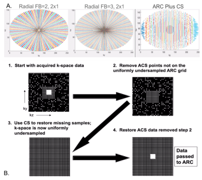 |
Evaluation of Compressed Sensing Approaches for Rapid Anatomical Imaging of Patients with Brain Tumors
Janine M Lupo1, Javier Villanueva-Meyer1, Maryam Vareth1,2, David Shin3, Emma Bahroos1, Angela Jakary1, Brian Burns3, and Suchandrima Banerjee3
1Radiology & Biomedical Imaging, UCSF, San Francisco, CA, United States, 2Berkeley Institute for Data Science, UC Berkeley, Berkeley, CA, United States, 3GE Healthcare, Menlo Park, CA, United States
Compressed Sensing with parallel imaging can allow for an accelerated anatomical imaging protocol for imaging patients with brain tumors, saving ~10 minutes of scan time over standard clinical protocols when incorporated into 3D T2-FLAIR, and 3D T1-weighted pre- and post- contrast imaging. We first determined the optimal undersampling pattern and acceleration for CS T1-weighted IR-SPGR sequences and then evaluated the quality of the images and lesion definition in patients with brain tumors. This shortened anatomical imaging protocol has the potential to reduce the frequency of motion artifacts and can allow time for more advanced, therapy specific imaging to be acquired.
|
|
1769.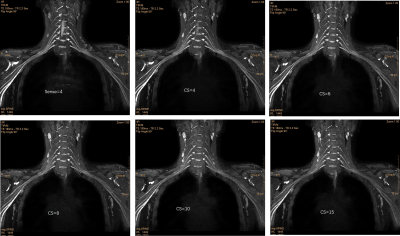 |
Combination of Compressed Sensing and Sensitivity Encoding (CS-SENSE) in MR brachial plexus imaging: a study of different acceleration factors
kong xiangchuang1, Tianjing Zhang2, Jiazheng Wang3, Qian Qi4, Zhenyang Zhou1, and Dingxi Liu1
1radiology, Union Hospital, Tongji Medical College, Huazhong University of Science and Technology, Wuhan, China, 2Clinical Science, Philips Healthcare, Shenzhen, China, 3Clinical Science, Philips Healthcare, Beijing, China, 4Philips Healthcare, Beijing, China The aim of this study was to reduce the scan time of 3D Nerve-view using Compressed Sensing-Sensitivity Encoding (CS), and evaluate the image quality and capability of diagnosis of accelerated 3D Nerve-view sequences. 3D Nerve-view sequences with 5 different CS (compressed sense technology) accelerating factors (4,6,8,10,15), and a traditional 3D Nerve-view with 4-fold parallel imaging (sense) as a clinical reference were obtained.The 3D-CS sequence offer comparable diagnostic quality to the clinical 3D scan with much less time, potentially increasing the productivity of MR scanners.CS-3D Nervview with factor 6 offer equilibrium between comparable clinical diagnostic quality with less scan time (235seconds). |
|
1770.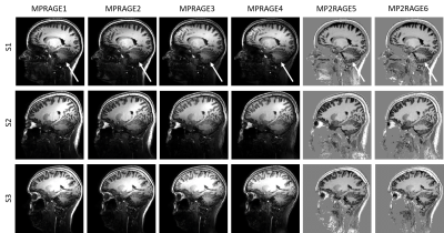 |
Can 7T MPRAGE match MP2RAGE for gray-white matter contrast?
Ícaro A F de Oliveira1,2, Thomas Roos1, Serge O Dumoulin1,2,3, and Wietske van der Zwaag1
1Spinoza Centre for Neuroimaging, Amsterdam, Netherlands, 2Experimental and Applied Psychology, VU University, Amsterdam, Netherlands, 3Experimental Psychology, Helmholtz Institute, Utrecht University, Utrecht, Netherlands
We compared MPRAGE and MP2RAGE at Ultra-High Field (UHF) in terms of signal separability in gray and white matter. Using a k-space shutter, kt-point universal pulses for signal excitation, and an efficient TR-FOCI for signal inversion, we obtained very good signal contrast throughout the brain, including the cerebellum, for the MPRAGE. Nevertheless, gray-white matter contrast was larger in the MP2RAGE data, leading to better segmentation results, especially in areas affected by low B1+. Hence, MP2RAGE appears more suitable for 7T T1-weighted anatomical data, despite the longer acquisition times.
|
|
1771.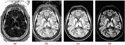 |
Improved combination of multi-inversion time images for fluid and white matter suppression
Dzung L Pham1, Neville Gai2, Yi-Yu Chou3, Abbey Goodyear2, Wen-Tung Wang3, and John A. Butman2
1Center for Neuroscience and Regenerative Medicine, Henry M. Jackson Foundation, Bethesda, MD, United States, 2National Institutes of Health, Bethesda, MD, United States, 3Henry M. Jackson Foundation, Bethesda, MD, United States
Fluid and White Matter Suppression (FLAWS) MRI uses two inversion times within a T1-weighted magnetization-prepared gradient echo sequence to enhance visualization of gray matter structures within the brain. FLAWS is based on the MP2RAGE sequence, but with different inversion times, followed by a voxel-wise minimum. In this work, we demonstrate that a variation of the regularized, ratio combination approach of MP2RAGE yields superior gray matter contrast-to-noise and intensity uniformity compared to the originally proposed FLAWS MRI combination based on minimum intensity.
|
|
1772.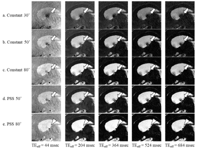 |
The effect of the refocusing flip angle on CSF dynamics imaging using multi-spin echo acquisition cine imaging (MUSACI)
Tatsuhiro Wada1, Chiaki Tokunaga1, Osamu Togao2, Yasuo Yamashita1, Kouji Kobayashi1, Masami Yoneyama3, and Toyoyuki Kato1
1Division of Radiology, Department of Medical Technology, Kyushu University Hospital, Fukuoka, Japan, 2Department of Clinical Radiology, Graduate School of Medical Sciences, Kyushu University, Fukuoka, Japan, 3Philips Japan, Fukuoka, Japan Poster Permission Withheld
Multi-spin echo acquisition cine imaging (MUSACI) is based on the multi-spin echo technique and used for detection of cerebrospinal fluid (CSF) movement. MUSACI detects CSF movement as a signal loss due to proton phase dispersion and flow void. To better detect the CSF loss caused by CSF movement and reduce the CSF signal loss caused by T2 decay, we modified the refocusing flip angle modulation using pseudo steady state sequence (PSS). The modulation of refocusing flip angle in PSS improved the CSF dynamic imaging using MUSACI.
|
|
1773. |
Imaging the Nigrosome 1 using Susceptibility Weighted Imaging and Quantitative Susceptibility Mapping: An Application to Parkinson’s Disease
Zenghui Cheng1, Naying He1, Pei Huang2, Sean K. Sethi3, Kiran Kumar Yerramsetty4, Vinay Kumar Palutla4, Weibo Chen5, Shengdi Chen2, Fuhua Yan1, and E.Mark Haacke1,3,6
1Radiology, Ruijin Hospital, school of medicine, Shanghai Jiaotong University, Shanghai, China, 2neurology, Ruijin Hospital, school of medicine, Shanghai Jiaotong University, Shanghai, China, 3Magnetic Resonance Innovations, Inc, Bingham Farms, MI, United States, 4MR Medical Imaging Innovations India Pvt. Ltd, Telangana, India, 5Philips Healthcare, Shanghai, China, 6Radiology, Wayne State University, Detroit, MI, United States Poster Permission Withheld
Imaging the nigrosome 1 has been reported to be promising in the diagnosis of Parkinson’s disease (PD), however, there is no uniform imaging protocol. This study was designed to create a rapid imaging protocol with high image quality to consistently visualize and characterize the N1 and to evaluate the loss of N1 in the diagnosis of PD. We found that the N1 sign could be consistently visualized using true SWI (tSWI) with a resolution of at least 0.67 x 0.67 x 1.34 mm3 and could be seen in 95% of HCs, while only 21.1% in PD and atypical parkinsonian syndromes.
|
|
1774.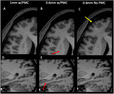 |
Difference in Morphometry Measures of 1mm and 0.6mm MPRAGE data with Prospective Motion Correction at 3T
Joelle E. Sarlls1, Francois Lalonde2, Joellyn Stolinski1, Maxim Zaitsev3, and Lalith Talagala1
1NMRF, National Institutes of Health, Bethesda, MD, United States, 2NIMH, National Institutes of Health, Bethesda, MD, United States, 3MR Development and Application Center, University Medical Center Freiburg, Freiburg, Germany
Higher resolution anatomical images can provide more accurate estimates of morphometric measures extracted from segmentation algorithms. Acquisition of submillimeter MPRAGE data at 3T requires long scan times making it prone to subject motion. Prospective motion correction (PMC) techniques can mitigate these effects by tracking brain motion and updating scan parameters accordingly. Here, we compare morphometric measures from 1.0mm and 0.6mm isotropic resolution MPRAGE data obtained at 3T while using PMC. Results show that good quality 0.6mm MPRAGE data can be acquired with PMC in approximately 30minutes. Increased cortical thickness in some brain regions is seen with higher resolution data.
|
|
1775. |
Feasibility of a quasi-volumetric synthetic MRI of the brain using 2D slice-interleaved MDME acquisition with deep learned reconstruction
Ho-Joon Lee1, Yeonah Kang1, Marc Lebel2, Min Soo Park3, and Joonsung Lee3
Video Permission Withheld
1Department of Radiology, Haeundae Paik Hospital, Busan, Republic of Korea, 2MR Collaboration and Development, GE Healthcare, Calagary, AB, Canada, 3GE Healthcare Korea, Seoul, Republic of Korea Sequences that allow volumetric parametric maps are being developed but when these techniques will be available clinically is uncertain. MDME sequence is a robust method, allowing rapid acquisition of T1, T2 relaxation times, PD from which one can generate synthetic multi-contrast images. However, acquisition at thin slices is challenging. With a deep learned reconstruction method, we demonstrate that interleaved thin slice acquisition of MDME, can produce quasi-volumetric synthetic MRI at an isotropic resolution. Limitations include motion misregistrations between acquisitions, parallel imaging/partial volume/pulsation related artifacts, which we believe can be overcome with technical development. |
|
1776. |
Voxel-Based Quantitative Susceptibility Mapping of Brain Using Compressed SENSE Acceleration
Aocai Yang1, Guangbin Wang1, Weibo Chen2, and Ye Li1
1Shandong Medical Imaging Research Institute,Shandong University, Jinan, China, 2Philips Healthcare, Shanghai, China
Quantitative susceptibility mapping (QSM) is an important technique for quantifying iron content in the brain. The conventional time of high spatial QSM data is too long. Compressed sensing acceleration (CS) technique can primarily reduce the acquisition time. We sought to evaluate the accuracy and stability of whole brain QSM on both the voxel-wise level and regional level by using several CS accelerations. We found significant differences in the magnetic susceptibility values on voxel-based QSM, but no statistically different in regions of interest. CS acceleration is feasible for QSM acquisition without influence the magnetic susceptibility values obviously.
|
1777. |
Comparison of EPI- and SSFP-PC with S-Transform for Separating Cardiac- and Respiratory-driven Intracranial CSF Motions under Free Breathing
Satoshi Yatsushiro1, Tomohiko Horie2, Mitsunori Matsumae3, and Kayagaki Kuroda1
1Department of Human and Information Science, School of Information Science and Technology, Tokai University, Hiratsuka, Kanagawa, Japan, 2Department of Radiological Technology, Tokai University Hospital, Isehara, Kanagawa, Japan, 3Department of Neurosurgery, School of Medicine, Tokai University, Isehara, Kanagawa, Japan
To compare the performance of separating cardiac- and respiratory-driven intracranial CSF motions under free breathing with Stockwell transform (ST), real time phase contrast imaging based on echo planar imaging (EPI-PC) and steady state free precession (SSFP-PC) were compared. In 3 healthy volunteers, 3 patients with idiopathic normal pressure hydrocephalus (iNPH), and 2 patients with Chiari malformation, both PC schemes yielded almost the same image quality, although EPI-PC had relatively higher frame rate than SSFP-PC. These preliminary results indicated that both techniques in conjunction with ST will be useful to seize the CSF motions under free breathing.
|
|
1778. |
Multi-planar, multi-contrast and multi-timepoint analysis tool (MOCHA) for intracranial vessel wall imaging review
Li Chen1, Duygu Baylam Geleri 1, Jie Sun 1, Hiroko Watase 1, Jiarui Cai1, Yin Guo1, Niranjan Balu 1, Dongxiang Xu 1, Thomas Hatsukami 1, Yongjun Wang 2, Jenq-Neng Hwang 1, and Chun Yuan 1
1University of Washington, Seattle, WA, United States, 2Beijing Tiantan Hospital, Capital Medical University, Beijing, China
A visualization technique (MOCHA) was developed to facilitate intracranial artery review using 3D Magnetic Resonance vessel wall imaging. Multiple intracranial MR scans, either from multiple contrasts or timepoints, are registered, then the artery of interest is traced and straightened using multiplanar reformation. Scans of 15 subjects with intracranial atherosclerosis were reviewed using MOCHA by a novice reader, with the traditional review method as comparison. The results showed higher sensitivity for plaque identification and higher accuracy for quantifying plaque features with MOCHA. MOCHA is promising for artery reviews using multiple scans, such as identifying plaque components and monitoring vessel wall thickening.
|
|
1779.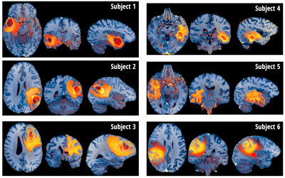 |
PCA-multiFuse: Visualising multi-dimensional data in a single colorised image
Daniel Gallichan1
1School of Engineering, Cardiff University, Cardiff, United Kingdom
A novel approach for image colorisation is presented, intended for situations where multiple contrasts are available covering the same anatomy. A colormap is created in a PCA-based hybrid parameter space, allowing signals from all contrasts to contribute to the final image colorization. Examples are shown from a dataset of high grade glioma patients, enabling rapid visual comparison between subjects. Within-subject there is clear contrast between healthy and pathological tissue, while contrast between healthy GM/WM/CSF is preserved.
|
|
1780.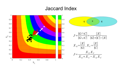 |
Towards Template-Invariant Voxel-Wise Analysis
Roman Fleysher1, Nelson Gil1, and Michael L Lipton 1
1Radiology, Albert Einstein College of Medicine, Bronx, NY, United States
Voxel-wise cluster analysis of any MR-derived metric necessitates non-linear registration to a common brain template. Because quality of inter-brain registrations is sensitive to the similarity between the brains, the results of such analyses are sensitive to the choice of the template. Despite judicious selection of the template may reduce registration errors, inherently the results should not depend on template choice. We show that a dramatic reduction in this sensitivity is achieved by filtering out poorly registered images. Consequently, voxel-wise cluster analyses of the remaining data become more robust and less sensitive to the choice of the template.
|
|
1781.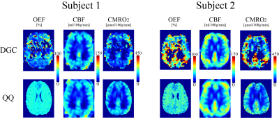 |
Cerebral oxygen extraction fraction mapping: comparison of dual-gas challenge calibrated BOLD and challenge-free gradient echo QSM+qBOLD
Junghun Cho1, Yuhan Ma2, Pascal Spincemaille3, Bruce Pike2,4, and Yi Wang1,3
1Biomedical Engineering, Cornell University, New York, NY, United States, 2McConnell Brain Imaging Centre, McGill University, Montreal, QC, Canada, 3Radiology, Weill Cornell Medical College, New York, NY, United States, 4Radiology, University of Calgary, Calgary, AB, Canada
In this study, we compare cerebral oxygen extraction fraction (OEF) and metabolic rate of oxygen consumption (CMRO2) maps obtained using dual-gas challenge calibrated-BOLD (DGC) and challenge-free gradient echo quantitative susceptibility mapping plus quantitative blood oxygen level-dependent modeling (QSM+qBOLD or QQ) to explore the important clinical advantage of challenging-free data acquisition. In n=11 healthy subjects, cortical gray matter average OEF was not significantly different (36.4±1.9% and 38.0±9.1%, P=0.63) as was CMRO2 (151.4±17.6 and 168.2±54.1 μmolO2/min/100g, P=0.26), for QQ and DGC, respectively. QQ can measure OEF and CMRO2 at both baseline and hypercapnia independently, showing a 14% CMRO2 decrease in hypercapnia (P=0.039).
|
|
1782.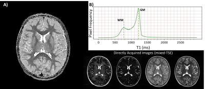 |
A Quantitative MRI-Based Method of Repolarization of T1 for the Mixed Turbo Spin Echo Pulse Sequence
Ryan McNaughton1, Mina Botros2, Ning Hua2, Xin Zhang1, and Hernan Jara2
1Mechanical Engineering, Boston University, Boston, MA, United States, 2Boston University Medical Center, Boston, MA, United States
Purpose: To develop quantitative MRI-based algorithms for correction of signal polarity for measurement of T1. Methods: Two polarity maps of T1 are calculated according to the mixed-TSE acquisition, and repolarized on a voxel-wise basis. The two polarity maps are compared to thresholds for PD and T2. The correct polarity is selected based on which T1 more accurately describes the selected voxel. Maps of the T1/T2 ratio repolarize the remaining unpolarized tissue. Results: Repolarized T1 maps exhibit an accurate bimodal distribution in vivo. Conclusion: A qMRI-based repolarization technique allows T1 measurement from mixed-TSE magnitude data, toward studying the extremely preterm brain.
|
|
1783.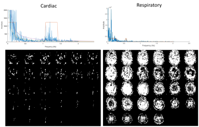 |
Spatially selective physiological noise suppression for high frequency resting state fMRI
Khaled Talaat1, Bruno Sa De La Rocque Guimaraes2, and Stefan Posse2
1Nuclear Engineering, University of New Mexico, Albuquerque, NM, United States, 2Neurology, University of New Mexico, Albuquerque, NM, United States
Assessing the extent of high frequency resting state connectivity (> 0.15 Hz) across different brain networks has been hampered by the presence of physiological noise. Much of the high frequency information is lost when global filters are applied to stop respiratory and cardiac frequency bands. A spatially selective automated filtering method is developed in order to preserve high frequency signal information in regions where physiological contamination is weak. Preliminary results show significant reduction in artifactual correlations compared to unfiltered data.
|
|
1784. |
Development of an unbiased population-specific brain atlas for adolescent collision-sport athletes
Yukai Zou1,2, Wenbin Zhu3, Ho-Ching (Shawn) Yang1, Nicole L Vike4, Diana O Svaldi1, Trey E Shenk5, Victoria N Poole1,4, Gregory G Tamer, Jr.1, Larry J Leverenz6, Ulrike Dydak7, Eric A Nauman1,4,8, Thomas M Talavage1,5, and Joseph V Rispoli1,5
1Weldon School of Biomedical Engineering, Purdue University, West Lafayette, IN, United States, 2College of Veterinary Medicine, Purdue University, West Lafayette, IN, United States, 3Department of Statistics, Purdue University, West Lafayette, IN, United States, 4Department of Basic Medical Sciences, Purdue University, West Lafayette, IN, United States, 5School of Electrical and Computer Engineering, Purdue University, West Lafayette, IN, United States, 6Department of Health and Kinesiology, Purdue University, West Lafayette, IN, United States, 7School of Health Sciences, Purdue University, West Lafayette, IN, United States, 8School of Mechanical Engineering, Purdue University, West Lafayette, IN, United States
Over years of practices and competitions, adolescent collision-sport (American football, soccer) athletes undergo repetitive subconcussive head impacts, and therefore may exhibit a neuroanatomical trajectory different from healthy adolescents in general. Targeting this vulnerable population, we constructed a specific brain atlas that includes templates (T1 and DTI) and semantic labels (cortical and white matter parcellations), and we demonstrated that the unbiased population-specific brain atlas can minimize bias introduced in spatial normalization, improve sensitivity of voxel-wise statistical analysis, and therefore better clarify the mechanisms that lead to traumatic brain injury in adolescent athletes.
|
|
| WITHDRAWN | ||
1786.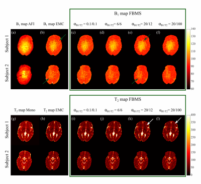 |
Optimizing Fusion Bootstrap Moves Solver (FBMS) regularization for improved B1+ mapping using Multi Spin-Echo brain sequences
Andreia C. Freitas1, Inês Sousa1, Andreia S. Gaspar1, Rui P.A.G. Teixeira2, Joseph V. Hajnal2, and Rita G. Nunes1,2
1ISR-Lisboa/LARSyS and Department of Bioengineering, Instituto Superior Técnico – Universidade de Lisboa, Lisbon, Portugal, 2Centre for the Developing Brain, King's College London, London, United Kingdom
T2 mapping provides valuable tissue-specific MR information. To enable shorter scan times, multi spin-echo (MSE) sequences are commonly used but the achieved T2 accuracy using conventional mono-exponential fitting is poor. Improvements are possible by matching the measured signal to a pre-computed dictionary. Although simultaneous B1+ estimation is feasible, previous work demonstrated a bimodal behaviour. We investigate further improvements in B1+ accuracy using an iterative pixel-neighborhood based method (the Fusion Bootstrap Moves Solver), comparing different levels of spatial regularization. Improved B1+ accuracy and recovery of spatially smooth maps was demonstrated both in simulated and in-vivo brain data.
|
|
1787.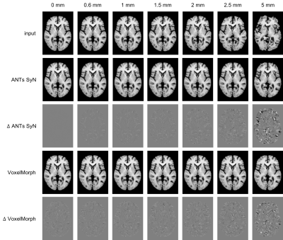 |
Evaluating VoxelMorph, a deep learning-based non-linear diffeomorphic registration algorithm, against native ANTs SyN
Victoria Madge1,2, Philip Novosad1,2, Daniel A. Di Giovanni1,3, and D. Louis Collins1,2,3
1McConnell Brain Imaging Centre, Montreal Neurological Institute and Hospital, Montreal, QC, Canada, 2Department of Biomedical Engineering, McGill University, Montreal, QC, Canada, 3Department of Neurology and Neurosurgery, McGill University, Montreal, QC, Canada
VoxelMorph is a deep-learning based non-linear diffeomorphic registration algorithm which claims to perform comparably to the state-of-the-art. However, the previous evaluation did not compare against manual gold-standard anatomical segmentations, used only the Dice metric for comparison, and compared against a modified version of a state-of-the-art algorithm, ANTs SyN. Here, VoxelMorph is evaluated against an unmodified version of ANTs SyN using multiple metrics based on manual labels. Results show VoxelMorph is less robust than ANTs SyN and underperforms in the presence of simulated deformations, and in registration of BrainWeb20 images to the VoxelMorph atlas.
|
|
1788.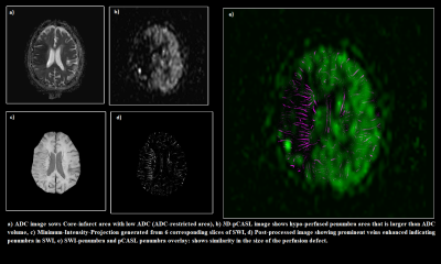 |
Penumbra Quantification from SWI visible Prominent Veins and its comparison with ASL-derived penumbra in patients with acute stroke
Rupsa Bhattacharjee1,2, Rakesh Kumar Gupta3, Vijay Kant Dixit4, Praveen Gupta5, and Anup Singh1,6
1Center for Biomedical Engineering, Indian Institute of Technology Delhi, New Delhi, India, 2Philips Health Systems, Philips India Limited, Gurugram, India, 3Department of Radiology, Fortis Memorial Research Institute, Gurugram, India, 4Department of Interventional Neuroradiology, Fortis Memorial research Institute, Gurugram, India, 5Department of Neurology, Fortis Memorial research Institute, Gurugram, India, 6Department of Biomedical Engineering, All India Institute of Medical Sciences, New Delhi, India
Objectives are to enhance the PHVS visibility in SWI, quantify stroke penumbra from it and calculate the mismatch ratio between PHVS(SWI) and DWI. For algorithm performance evaluation, 3D non-contrast pCASL as a gold standard was used to calculate true mismatch ratio with DWI. The proposed approach demonstrates high correlation between the two mismatch ratios, suggesting that PHVS and SWI based penumbra quantification has the potential to be used as an alternative for perfusion based methods. If mismatch ratio can accurately be produced from PHVS SWI, it could potentially reduce the scan time for acute stroke.
|
|
1789.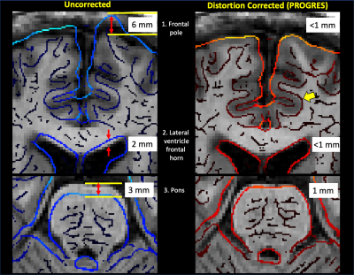 |
Distortion Correction in EPI Diffusion MRI within Clinical Workflow: A Quantitative Evaluation
Jaemin Shin1, Jungho Cha2, Jeffrey McGovern3, Patrick Quarterman1, Suchandrima Banerjee4, and Ki Sueng Choi2
1GE Healthcare, New York, NY, United States, 2Mount Sinai, New York, NY, United States, 3GE Healthcare, Waukesha, WI, United States, 4GE Healthcare, Menlo Park, CA, United States
This work does a quantitative evaluation of the effectiveness of a distortion correction method, clinically available at the scanner console, in diffusion MRI of the brain (N=10). After distortion correction, average cross-correlation coefficient between T1 and DWI was significantly increased. Distortion correction reduced average residual distance error to 1.1 mm or less (average 76% reduction) in all of three anatomical landmarks (frontal pole,lateral ventricle, pons). This on-console distortion correction method could help in the clinical applications that requires fast and accurate distortion correction such as deep brain stimulations surgery and cases with high susceptibility such as postoperative cohorts.
|
|
1790.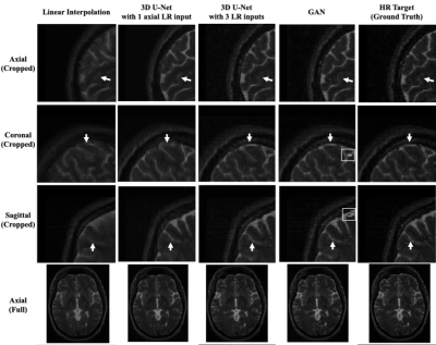 |
3D volume reconstruction from three orthogonal multi-slice 2D images using a super-resolution network
Xue Feng1, Huitong Pan2, Li Zhao3, and Craig H. Meyer1
1Biomedical Engineering, University of Virginia, Charlottesville, VA, United States, 2Springbok, Inc., Charlottesville, VA, United States, 3Children's National Health System, Washington DC, DC, United States
High-resolution 3D MRI can provide detailed anatomical information and is favorable for accurate quantitative analysis. However, due to the limited data acquisition time and other physical constraints such as breath-holding, multi-slice 2D images are often acquired. The 2D images usually have a larger slice thickness than the in-plane resolution. To reconstruct the high- resolution 3D MRI, we propose to use a super-resolution network with three orthogonal multi-slice 2D images as the input. We validated the proposed method on brain MRIs and achieved good results in terms of mean absolute difference, mean squared difference and image details with visual inspection.
|
|
1791.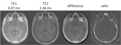 |
Importance of Off-resonance Effects in Ultrashort echo-time Imaging
Wen-Tung Wang1, Dzung Pham1, and John A Butman1,2
1Center for Neuroscience and Regenerative Medicine, NIH/USU, Bethesda, MD, United States, 2Radiology and Imaging Sciences, NIH, Bethesda, MD, United States
Ultrashort echo-time (UTE) imaging can detect short- and ultrashort-T2 tissue components. e.g. tendons, ligaments, and cortical bone. Multi-echo UTE is used to generate the tissue attenuation maps required for quantitative MRI PET, as short T2 skull is visible on the first echo but not on the second. For accurate classification, it is assumed that these are registered. Here we show that geometric scaling issues of fat and water may be different between the 1st and 2nd echoes – and hence lead to erroneous tissue classification.
|
1792.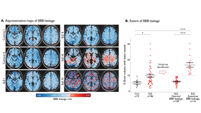 |
Blood-Brain Barrier Imaging as a Biomarker for Cognitive Decline in Systemic Lupus Erythematosus
Lyna Kamintsky1, Steven D Beyea2,3, John D Fisk4,5, Javeria A Hashmi6, Antonina Omisade7, Tim Bardouille8, Chris Bowen2,3, Maher Quraan2,3, Kara A Matheson9, Alon Friedman1,10, and John G Hanly11,12
1Department of Medical Neuroscience, Dalhousie University, Halifax, NS, Canada, 2Department of Diagnostic Radiology, Dalhousie University, Halifax, NS, Canada, 3Biomedical Translational Imaging Centre (BIOTIC), QEII Health Sciences Centre, Halifax, NS, Canada, 4Departments of Psychiatry, Psychology & Neuroscience and Medicine, Dalhousie University, Halifax, NS, Canada, 5Nova Scotia Health Authority, Halifax, NS, Canada, 6Department of Anesthesia, Pain Management and Perioperative Medicine, Dalhousie University, Halifax, NS, Canada, 7Acquired Brain Injury (Epilepsy Program), Nova Scotia Health Authority, Halifax, NS, Canada, 8Department of Physics, Dalhousie University, Halifax, NS, Canada, 9Research Methods Unit, Nova Scotia Health Authority, Halifax, NS, Canada, 10Department of Physiology and Cell Biology, Medicine, Ben-Gurion University of the Negev, Beer Sheva, Israel, 11QEII Health Sciences Center, Halifax, NS, Canada, 12Department of Medicine and Division of Rheumatology, Dalhousie University, Halifax, NS, Canada
This study addresses the need for mechanism-based understanding of cognitive impairment in systemic lupus erythematosus (SLE). Using dynamic contrast-enhanced MRI we identified extensive blood-brain barrier (BBB) leakage in 16 of 46 SLE patients. Extensive BBB leakage was associated with worse overall cognitive performance, affecting primarily information processing speed and executive abilities. Our study provides the first compelling evidence for BBB damage in SLE, and links BBB leakage to cognitive dysfunction. These findings highlight the diagnostic potential of BBB imaging and call for research into BBB-targeting therapies.
|
|
1793. |
White matter neuroplasticity: Motor learning modifies hemodynamic responses in the internal capsule
Lukas A. Grajauskas1,2,3, Tory Frizzell2,4, Sujoy Hajra2,4, Caressa Liu2,4, Xiaowei Song2,3, and Ryan C.N. D'Arcy4,5
1Cumming School of Medicine, University of Calgary, Calgary, AB, Canada, 2Surrey Memorial Hospital ImageTech Laboratory, Fraser Health, Surrey, BC, Canada, 3Biomedical Physiology and Kinesiology, Simon Fraser University, Burnaby, BC, Canada, 4Faculty of Applied Sciences, Simon Fraser University, Burnaby, BC, Canada, 5Djavad Mowafaghian Centre for Brain Health, University of British Columbia, Vancouver, BC, Canada
Though white matter has a noted role in motor learning, there have been no MRI studies of functional neuroplasticity in this tissue. Therefore, in this work, twelve healthy participants underwent a motor training program designed to drive behavioral changes in the non-dominant hand. Using BOLD fMRI, we noted an associated change in the temporal dispersion of the white matter hemodynamic response over the training period. This is in line with previous DTI studies that show increases in white matter myelination with training, and BOLD investigations that show hemodynamic responses differ between grey and white matter, and between white matter tracts.
|
|
1794.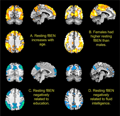 |
Resting brain entropy in the default mode network and the executive network may serve as a functional brain reserve
Ze Wang1
1Radiology, University of Maryland School of Medicine, Baltimore, MD, United States
Human brain relies on long-range coherent activity to execute complex function. The long-range coherence can be measured by brain entropy mapping, which has gained increasing research interest in recent years. The purpose of this study is to examine the relationship between brain entropy and brain functions using large data.
|
|
1795.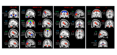 |
Patterns of grey matter atrophy in patients with MS: a multivariate analysis using source-based morphometry
Paola Valsasina1, Maria A. Rocca1,2, Alessandro Meani1, Claudio Gobbi3, Chiara Zecca3, Alex Rovira4, Xavier Montalban5, Hugh Kearney6, Olga Ciccarelli6, Lucy Matthews7, Jacqueline Palace7, Antonio Gallo8, Alvino Bisecco8,
Carsten Lukas9, Barbara Bellenberg9, Frederik Barkhof10,11, Hugo Vrenken10, Paolo Preziosa1,2, and Massimo Filippi1,2,12
Video Permission Withheld
1Neuroimaging Research Unit, Institute of Experimental Neurology, Division of Neuroscience, IRCCS San Raffaele Scientific Institute, Milan, Italy, 2Neurology Unit, IRCCS San Raffaele Scientific Institute, Milan, Italy, 3Department of Neurology, Neurocenter of Southern Switzerland, Civic Hospital, Lugano, Switzerland, 4Section of Neuroradiology and MRI Unit, Department of Radiology, Hospital Universitari Vall d'Hebron, Barcelona, Spain, 5Department of Neurology/Neuroimmunology, Multiple Sclerosis Centre of Catalonia, Hospital Universitari Vall d'Hebron, Barcelona, Spain, 6NMR Research Unit, Queen Square MS Centre, Department of Neuroinflammation, UCL Institute of Neurology, London, United Kingdom, 7Nuffield Department of Clinical Neurosciences, University of Oxford, Oxford, United Kingdom, 8Department of Advanced Medical and Surgical Sciences, and 3T MRI Center, University of Campania “Luigi Vanvitelli”, Naples, Italy, 9Department of Radiology and Nuclear Medicine, and Institute of Neuroradiology, St. Josef Hospital, Ruhr-University Bochum, Bochum, Germany, 10Department of Radiology and Nuclear Medicine, MS Center Amsterdam, Amsterdam Neuroscience, Amsterdam UMC, location VUmc, Amsterdam, Netherlands, 11Institutes of Neurology and Healthcare Engineering, University College London, London, United Kingdom, 12Vita-Salute San Raffaele University, Milan, Italy
In this study, we used source-based morphometry to identify patterns of grey matter tissue loss in a large, multicenter cohort of patients with multiple sclerosis (MS) acquired at 8 European sites. We detected a differential involvement of grey matter networks across the different stages of the disease. Cortical and subcortical grey matter atrophy progressed significantly in MS patients over 1-year of follow-up. Grey matter atrophy, especially in the sensorimotor network, was able to explain patients’ clinical disability, while cerebellar atrophy was able to predict clinical disability worsening over 1-year follow-up.
|
|
1796. |
Higher b-value diffusion improves correlation with cortical myelin content
Sandy Mournet1,2, Gosuke Okubo1,2,3, Ismail Koubiyr1,2, Valentin H Prevost4, Clémence Bal2, Bei Zhang5, Hiroshi Kusahara 4, Nobuyasu Ichinose4, Bruno Triaire4, Bassem Hiba6, Vincent Dousset1,2,7, and Thomas Tourdias1,2,7
1Neurocentre Magendie, INSERM U1215, Bordeaux, France, 2Université de Bordeaux, Bordeaux, France, 3Department of Radiology, Tenri Hospital, Nara, Japan, 4Canon Medical Systems Corporation, Otawara, Japan, 5Canon Medical systems Europe, Paris, France, 6Centre de neuroscience cognitive, CNRS UMR 5229, Université Claude Bernard Lyon, Lyon, France, 7Neuroimagerie diagnostique et thérapeutique, CHU de Bordeaux, Bordeaux, France
In diffusion MRI, the use of very high b-value remains very challenging. In this study, we scanned 9 volunteers with a protocol of dMRI sequences using b values from 1000 to 5000 s/mm². We compared cortical surface map of myelin (T1wi/T2wi) to maps of mean diffusivity (MD) computed from each b value. As opposed to b1000 s/mm², MD maps from b3000 and b5000 inversely mirrored the myelin maps. With increasing b-values, multiple regression models confirmed an increasing negative association between myelin and MD. The MD obtained with high b-value is sensitive to subtle cellular variations such as the cortical myeloarchitecture.
|
|
1797. |
The impact of cerebrocortical-cerebellar loops on brain dynamics in simulations using The Virtual Brain
Fulvia Palesi1, Roberta Lorenzi1, Claudia Casellato1, Petra Ritter2, Viktor Jirsa3, Claudia AM Gandini Wheeler-Kingshott1,4,5, and Egidio D'Angelo1,5
1Department of Brain and Behavioural Sciences, University of Pavia, Pavia, Italy, 2Department of Neurology with Experimental Neurology, Charité – Universitätsmedizin Berlin, Berlin, Germany, 3Institut de Neurosciences des Systèmes - Inserm UMR1106, Aix-Marseille Université, Marseille, France, 4NMR Research Unit, Queen Square MS Centre, Department of Neuroinflammation, UCL Institute of Neurology, London, United Kingdom, 5IRCCS Mondino Foundation, Pavia, Italy
The Virtual Brain(TVB) has been developed to simulate brain dynamics starting from individual structural and functional connectivity(FC) MRI data. Nowadays, only cerebrocortical circuits have been considered. Here, we provided the first TVB simulations including cerebellar nodes on single-subject datasets. The brain dynamics simulated by either including or excluding cerebrocortical-cerebellar connectivity were compared, revealing that the predictive power of empirical FC is not significantly modified by inclusion of cerebro-cerebellar loops. To improve the present results and apply this pipeline to predict disease states involving cerebrocortical-cerebellar loops, specific neural mass models accounting for cerebellar microcircuit physiology need to be integrated in TVB.
|
|
1798. |
180 vertebrate brains for an open data set of comparative neuroanatomy
Katja Heuer1, Mélanie Didier2, Stéphanie Anastacio2, Antoine Burgos2, Romain Valabregue2, Marc Herbin3, Roberto Toro1,4, and Mathieu David Santin2
1Groupe de Neuroanatomie appliquée et théorique, Unité de Génétique humaine et fonctions cognitives, Département de neuroscience, Institut Pasteur, Paris, France, 2Centre de Neuroimagerie de Recherche, Institut Cerveau Moelle – ICM, CENIR, UPMC-Inserm U1127, CNRS 7225, Paris, France, 3Département Adaptations du Vivant, UMR MECADEV 7179 Equipe FUNEVOL Sorbonne Universités-MNHNUPMC- CNRS-IRD, Muséum National d'Histoire naturelle, PAris, France, 4Center for Research and Interdisciplinarity (CRI), Université Paris Descartes, Paris, France This work presents the MR acquisition of 180 post mortem brains from different species coming from a Museum collection. 3D-High resolution images were obtained at 3 and 11.7T, depending on brain size. The images where then preprocessed in order to be available online for dowload on The Brain Catalogue (https://braincatalogue.org) web portal designed for comparative neuroanatomy studies. |
|
1799. |
Bedside Stroke Imaging at 64mT
Rafael OHalloran1, Laura Sacolick1, Hadrien Dyvorne1, Carole Lazarus1, Samantha By1, Brian Welch1, Bradley A Cahn2, Jill T Shah2, Mercy H Mazurek2, Matthew M Yuen2, Gordon Sze2, W. Taylor Kimberly3, Matthew S Rosen4,5, and
Kevin N Sheth2
1Hyperfine Research, Inc., Guilford, CT, United States, 2Yale New Haven Hospital, New Haven, CT, United States, 3Division of Neurocritical Care, Department of Neurology, Massachusetts General Hospital, Boston, MA, United States, 4Athinoula A. Martinos Center for Biomedical Imaging, Department of Radiology, Massachusetts General Hospital and Harvard Medical School, Charlestown, MA, United States, 5Department of Physics, Harvard University, Cambridge, MA, United States
Initial evidence of the feasibility of clinical stroke imaging using a portable 64mT MRI system is presented. Twelve patients with a diagnosis of ischemic stroke diagnosis admitted to a neuroscience intensive care unit were imaged with a portable, point-of-care, MRI system.
|
|
1800.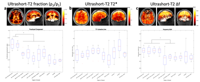 |
Ultrashort-T2* component differences in white matter regions of the brain
Nikhil Deveshwar1 and Peder E. Z. Larson1
1Department of Radiology and Biomedical Imaging, University of California, San Francisco, San Francisco, CA, United States
This work presents a study detailing how ultrashort-T2* measurements can provide new and complimentary characterizations and differentiations of white matter anatomy in comparison to classic DTI measurements. Specifically the magnitude and frequency shift components of the measured signal are significantly different between white matter with thick versus thin myelin sheaths and provides new methods of assessing myelin content in white matter when compared to FA, MD, and V1 values. These results show that this method could be used to better characterize and study demyelinating diseases such as multiple sclerosis.
|
|
1801.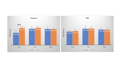 |
Glial changes induced by lipopolysaccharide inflammatory challenge in humans detected with diffusion-weighted MRS
Itamar Ronen1, Francesca Branzoli2, Alessandro Colasanti3, Iris Asllani3, Riccardo De Marco3, Neil Harrison4, and Mara Cercignani3
1Department of Radiology, Leiden University Medical Center, Leiden, Netherlands, 2Centre for NeuroImaging Research - CENIR, Brain and Spine Institute - ICM, Paris, France, 3Department of Neuroscience, Brighton and Sussex Medical School, University of Sussex, Brighton, United Kingdom, 4Cardiff University Brain Research Imaging Centre (CUBRIC), School of Medicine, Cardiff University, Cardiff, United Kingdom
Neuroinflammation is a pathomechanism implicated in several neruological, neurodegenerative and psychiatric disorders and is expressed in activation of microglia, the resident brain macrophages. Currently the only neuroimaging method for detecting microglial activation is PET with translocator protein (TSPO) ligands. A well-known model of experimentally inducing systemic inflammation is the administration of lipopolysaccharides (LPS). Here we report a significant increase in the apparent diffusion coefficient of the glial metabolite choline (tCho) in the human thalamus, following injection of LPS. This finding suggests that the ADC(tCho) is a putative marker for glial activation and may be useful in measuring neuroinflammation in disease.
|
|
1802.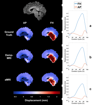 |
Comparing aMRI to DENSE for the assessment of brain tissue motion
Ayodeji L Adams1, Itamar Terem2, Allen A Champagne3, Samantha J Holdsworth4, and Jaco Zwanenburg5
1Radiology, University Medical Center Utrecht, Utrecht, Netherlands, 2Radiology, Stanford University, Stanford, CA, United States, 3School of Medicine, Queen’s University, Kingston, ON, Canada, 4Anatomy and Medical Imaging, University of Auckland, Auckland, New Zealand, 5University Medical Center Utrecht, Utrecht, Netherlands
aMRI holds great potential for assessing brain motion/strain using images acquired from readily-available sequences. However, registration is needed to extract displacements from the motion-amplified images, which may limit its accuracy. In this study we separately assessed the errors induced by registration limitations and by imperfections in the aMRI amplification. Displacements were extracted from aMRI and DENSE-amplified images using a common registration algorithm, which were then compared to a ground truth. Although significant differences were found between DENSE-amplified images and aMRI, the aMRI-derived displacements were comparable to the ground truth, strengthening the potential of aMRI for investigating brain motion in disease.
|
|
1803.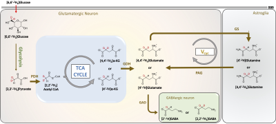 |
Assessing gray and white matter glutamatergic turnover in human brain non-invasively using 1H MRS and deuterated glucose
Puneet Bagga1, Laurie J Rich1, Abigail Cember1, Ravi Prakash Reddy Nanga1, Deepa Thakuri1, Mark Elliott1, Mohammad Haris2,3, John A Detre4, and Ravinder Reddy1
1Department of Radiology, University of Pennsylvania, Philadelphia, PA, United States, 2Sidra Medicine, Doha, Qatar, 3LARC, Qatar University, Doha, Qatar, 4Department of Neurology, University of Pennsylvania, Philadelphia, PA, United States
Cerebral metabolism is reported to be monitored by various techniques including 13C/2H MR spectroscopy and 18F-FDG Positron emission tomography. It is well known that 1H MRS allows the non-invasive detection and quantification of neurochemicals. In this study, we performed 1H MRS in conjuction with the oral administration of [6,6′-2H2]glucose in a healthy volunteer at 3T and 7T to measure turnover of glutamate in gray and white matter. As 2H is invisible on 1H MRS, the 2H enrichment of glutamate leads to a corresponding drop in its 1H
MR resonance measured via LCModel.
|
|
1804. |
Automatic Classification of MR Image Contrast
Julia Cluceru1, Yannet Interian2, Janine M. Lupo3, Riley Bove4, Atul Butte5, and Jason Crane3
1Radiology and Biomedical Imaging, UCSF, San Francisco, CA, United States, 2Program in Data Science, USF, San Francisco, CA, United States, 3Radiology and Biomedical Imagin, UCSF, San Francisco, CA, United States, 4Department of Neurology, UCSF, San Francisco, CA, United States, 5Bakar Computational Health Sciences Institute, UCSF, San Francisco, CA, United States
To perform large-scale analyses of disease progression, it is necessary to automate the retrieval and alignment of MR images of similar contrast. The goal of this study is to create an algorithm that can reliably classify brain exams by MR image contrast. We use two modeling strategies (SVM and CNN) and two training/testing cohorts to compare within-disease and between-disease transferability of the algorithms. For both cohorts, deep ResNets for extract imaging features combined in a random forest with DICOM metadata perform the best, resulting in 95.6% accuracy on the within-disease comparison, and 99.6% overall accuracy on between-disease comparison.
|
|
1805.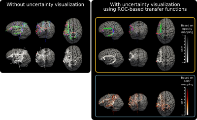 |
Visualization of uncertainty in tractograms using ROC-based transfer functions for real-time TMS applications
Dogu Baran Aydogan1
1Department of Neuroscience and Biomedical Engineering, Aalto University, Helsinki, Finland
Transcranial magnetic stimulation (TMS) is a non-invasive brain stimulation technique that is widely used in both research and clinical settings. However, state-of-the-art TMS guidance systems do not leverage the valuable structural connectivity in real-time applications. This is mainly due to the limitations of tractography which has been increasingly pointed out in validation studies. In order to address this problem, in this work we propose a novel visualization approach that is capable of providing information about the uncertainty of obtained streamlines. Our technique offers an easy and intuitive way for the TMS operator to interpret the displayed streamlines in real-time.
|
|
1806. |
Amide Proton Transfer Brain Imaging with Compressed Sense: Comparison of Different Acceleration Factors and Conventional Parallel Imaging
Nan Zhang1, Qingwei Song1, Renwang Pu1, Haonan Zhang1, Jiazheng Wang2, Zhongping Zhang2, and Yu Song1
1The First Affiliated Hospital of Dalian Medical University, Dalian, China, 2Philips Healthcare, Beijing, China
Amide proton transfer (APT) imaging is a novel and promising MRI contrast method, but it can be time-consuming. Common parallel imaging methods, like SENSE, can lead to reduced quality of APT. Here, compressed sensing technique is evaluated for the acceleration of APT in brain. The purpose of the present study is to explore feasibility of different acceleration factors in brain APT. The results show that it is feasible to apply accelerator factor 5 to APT display of brain tissue and brain tumor, which could reduce the scan time with in 1 min.
|
1807. |
Optimization of k-space Filtering for Compensating T2 Blurring in 3D ASL-MRI: Application to GBM
Yiming Wang1, Limin Zhou1, Joshua S. Greer1,2, Edward Pan3,4,5, Marco C. Pinho1,6, Joseph A. Maldjian1,6, and Ananth J. Madhuranthakam1,6
1Radiology, UT Southwestern Medical Center, Dallas, TX, United States, 2Pediatrics, UT Southwestern Medical Center, Dallas, TX, United States, 3Neurology and Neurotherapeutics, UT Southwestern Medical Center, Dallas, TX, United States, 4Neurological Surgery, UT Southwestern Medical Center, Dallas, TX, United States, 5Harold C. Simmons Cancer Center, UT Southwestern Medical Center, Dallas, TX, United States, 6Advanced Imaging Research Center, UT Southwestern Medical Center, Dallas, TX, United States
3D arterial spin labeled (ASL) MRI using turbo spin echo (TSE) acquisition suffers from image blurring, due to T2 decay along the echo train. In this study, a k-space filtering method proposed earlier was optimized to compensate T2 blurring, incorporating ASL T2 measurements in healthy volunteers for T2 decay estimation. This method was then applied to 3D ASL images acquired in healthy volunteers and patients with glioblastoma (GBM) from an ongoing clinical trial. Results showed reduced image blurring in both volunteers and patients.
|
|
1808.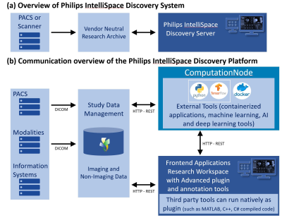 |
Implementation of the academic image processing pipeline ExploreASL in an outpatient center using IntelliSpace Discovery
Sandeep Ganji1, Nandor Pinter2, Jan Petr33, Bela Ajtai2, Joseph V Fritz2, Laszlo Mechtler2, Shahrukh Husain2, Alexander Fischer4, Frederik Barkhof5,6, and Henri Mutsaerts5,7
1Philips, Gainesville, FL, United States, 2Dent Neurologic Institute, Amherst, NY, United States, 3Helmholtz-Zentrum Dresden-Rossendorf, Dresden, Germany, 4Philips Research, Aachen, Germany, 5Dept of Radiology and Nuclear Medicine, Amsterdam University Medical Center, Amsterdam, Netherlands, 6Institute of Biomedical Engineering and Neurology, University College London, London, United Kingdom, 7Ghent Institute for Functional and Metabolic Imaging, Ghent, Belgium
The use of standardized image processing pipelines is continuously increasing in radiological research with developments in computing power, image processing, and machine learning techniques. Early integration of academic processing methods into clinical research workflow would accelerate the translation of promising novel MRI techniques into the clinic. However, the integration of such tools is both resource and time consuming. While most of neurological imaging takes place in outpatient centers, resource and workflow limitations of such clinics do not allow for the application of advanced image analysis. Here, we present the integration the “ExploreASL” into the PACS-connected research platform IntelliSpace Discovery.
|
|
1809. |
Multiparametric brain MR Imaging for the assessment of Multiple Sclerosis disease progression over 6 months
Xiaomeng Zhang1, Xiaoqing Yang2, Karthikeyan Subramanian3, Bradley A Hooker1, Mark Haacke3, Ivonne Suridjan1, Qi Guo1, Robert Comley1, Adam Ziemann4, and Yanping Luo1
1Translational imaging, iSAT, Abbvie Inc., North Chicago, IL, United States, 2Data & Statistical Sciences, Abbvie Inc., North Chicago, IL, United States, 3Wayne state university, Detroit, MI, United States, 4Neuroscience Development, Abbvie Inc., North Chicago, IL, United States
Quantitative brain MRI was used to evaluate 18 RRMS patients and 10 age matched controls at baseline and 6 months. The study aimed to assess test-retest reproducibility in healthy controls, disease variability in the RRMS population, and sensitivity to disease progression of each MRI-derived parameter. The results will inform the selection of MRI measures to be used as biomarkers in future clinical trials. In addition, longitudinal multiparametric assessment of MS lesions may improve our understanding of different pathological components of such lesions (e.g. inflammation, demyelination, and iron accumulation) during disease progression.
|
|
1810. |
Improved Cerebral Microbleed vs Calcification Discrimination using QSM compared to SWI Phase Maps
Salil Soman1, Kyuwon Lee2, Magdy Selim3, Aristotelis Filippidis4, Ajit Thomas4, Pascal Spincemaille5, and Yi Wang5
1Beth Israel Deaconess Medical Center / Harvard Medical School, Boston, MA, United States, 2Radiology, Beth Israel Deaconess Medical Center / Harvard Medical School, Boston, MA, United States, 3Neurology, Beth Israel Deaconess Medical Center / Harvard Medical School, Boston, MA, United States, 4Neurosurgery, Beth Israel Deaconess Medical Center / Harvard Medical School, Boston, MA, United States, 5Radiology, Weill Cornell Medicine, New York, NY, United States Cerebral microbleeds are bleeds < 1 cm seen on MRI, not visible on CT, which play a role in diagnosing disease and identifying risks of developing multiple diseases. Many susceptibility based techniques, such as SWI require a phase map to distinguish CMB from calcification. However, studies have demonstrated that for non-CMB bleeds on GRE and SWI, the phase dominant sign may differ across slices, preventing clinical interpretation. We compared the dominant sign for candidate CMBs on SWI phase maps and MEDI QSM images, and found that QSM images showed much less change in dominant sign than SWI phase maps.
|
|
1811. |
Optimized Structural Imaging at 7T using ME-MP2RAGE
Eberhard Daniel Pracht1, Philipp Ehses1, Hankyeol Lee2, Kamil Uludag2,3, and Tony Stoecker1,4
1German Center for Neurodegenerative Diseases, Bonn, Germany, 2Center for Neuroscience Imaging Research, Institute for Basic Science & Department of Biomedical Engineering, Sungkyunkwan University, Suwon, Korea, Republic of, 3Techna Institute & Koerner Scientist in MR Imaging, University Health Network, Toronto, ON, Canada, 4Department of Physics and Astronomy, University of Bonn, Bonn, Germany
In this work, we present an optimized, scan-time efficient multi-echo MP2RAGE sequence (ME-MP2RAGE) for improved brain segmentation at ultra-high field. Apart from removing B1-, M0, and T2* dependencies (MP2RAGE features), the proposed method removes susceptibility induced image distortions along the readout direction. Additionally, an optimized reordering scheme was implemented for scan-time optimization.
|
|
1812.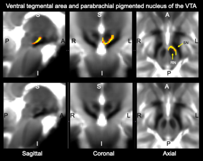 |
Probabilistic structural atlas of human ventral tegmental area, mesencephalic and isthmic reticular formation using in-vivo 7 Tesla MRI
KAVITA SINGH1, María Guadalupe García-Gomar1, and Marta Bianciardi1
1Brainstem Imaging Laboratory, Department of Radiology, Athinoula A. Martinos Center for Biomedical Imaging, Massachusetts General Hospital and Harvard Medical School, Boston, MA, United States
Human ventral tegmental area and its parabrachial pigmented nucleus are involved in attention, memory, reward, drug abuse, and motivation. Mesencephalic and isthmic reticular formation nuclei are implicated in the control of head/eye movement and position. Currently, their assessment in radiological investigations is difficult due to the limited-resolution and contrast of clinical MRI. We precisely delineated these nuclei on 7 Tesla MRI of healthy humans and generated a validated in-vivo stereotaxic probabilistic atlas of these structures. Upon coregistration to clinical MRI, this atlas might improve the evaluation of lesions and assessment of connectivity pathways underlying clinical conditions relating to these nuclei.
|
|
1813.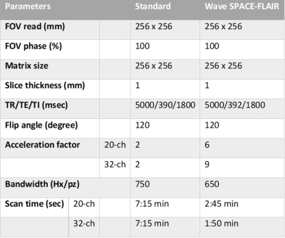 |
Evaluation of Ultrafast Wave-CAIPI 3D FLAIR Versus Standard 3D FLAIR for Quantitative Analysis of White Matter Lesions
Chanon Ngamsombat1,2, Maria Gabriela Figueiro Longo1,3, Augusto Lio M. Gonçalves Filho1,3, Stephen F. Cauley1,4, Kawin Setsompop1,4,5, Qiyuan Tian1, Qiuyun Fan1, Daniel Polak6,7, Wei Liu8, Wei-Ching Lo7, Ramon Gilberto González3,4, Pamela W. Schaefer3,4, John E. Kirsch1,3,
Otto Rapalino3,4, John Conklin1,3, and Susie Y. Huang1,3,5
1Athinoula A. Martinos Center for Biomedical Imaging, Department of Radiology, Massachusetts General Hospital, Boston, MA, United States, 2Faculty of Medicine, Siriraj Hospital Mahidol Univerity, Bangkok, Thailand, 3Department of Radiology, Massachusetts General Hospital, Boston, Massachusetts, Boston, MA, United States, 4Harvard Medical School, Boston, MA, United States, 5Harvard-MIT Division of Health Sciences and Technology, Massachusetts Institute of Technology, Cambridge, MA, United States, 6Department of Physics and Astronomy, Heidelberg University, Heidelberg, Heidelberg, Germany, 7Siemens Healthineers AG, Erlangen, Erlangen, Germany, 8Siemens Shenzhen Magnetic Resonance Ltd., Shenzhen, Shenzhen, China
Quantification of cerebral white matter lesion volume has become increasingly feasible for routine clinical evaluation and research due to the availability of automated segmentation tools and 3D FLAIR sequences. However, these sequences suffer from long acquisition times, limiting their widespread use. We demonstrate that quantitative white matter lesion volumes estimated using ultrafast Wave-CAIPI SPACE-FLAIR obtained in <3 minutes show excellent agreement with standard SPACE-FLAIR requiring >7 minutes of scanning in patients undergoing clinical evaluation for suspected MS and epilepsy. Wave-CAIPI SPACE-FLAIR may facilitate the adoption of 3D FLAIR sequences for lesion evaluation in patients with MS and other white matter diseases.
|
|
1814.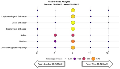 |
Highly Accelerated Wave-CAIPI Post-Contrast 3D-T1 Compared to Standard Post-Contrast 3D-T1 SPACE for Detection of Abnormal Enhancing Lesions.
Augusto Lio Mota Goncalves Filho1,2, Maria Gabriela Figueiro Longo1,2, John Conklin1,2, Stephen Cauley1,2, Daniel Polak3, Wei Liu4, John Kirsch1,2, Kawin Setsompop1,2, Ramon Gilberto Gonzalez1, Pamela Schaefer1, Susie Yi Huang1,2,5, and Otto Rapalino1
1Radiology, Massachusetts General Hospital, Boston, MA, United States, 2Athinoula A. Martinos Center for Biomedical Imaging, Charlestown, MA, United States, 3Siemens Healthineers, Erlangen, Germany, 4Siemens Shenzhen Magnetic Resonance Ltd., Shenzhen, China, 5Harvard Medical School, Boston, MA, United States
We present the first large-scale evaluation of the diagnostic performance and image quality of highly accelerated Wave-CAIPI post-contrast 3D-T1 SPACE (Wave-T1 SPACE) compared to standard post-contrast 3D-T1 SPACE for the detection of intracranial enhancing lesions in patients undergoing 3T MRI scanning. Two neuroradiologists assessed the images in a head-to-head comparison, and found no significant difference between the two sequences for detection of abnormal intracranial enhancement and overall diagnostic quality, despite a nearly 3-fold decrease in acquisition time of post-contrast Wave-T1 SPACE. The application of highly-accelerated 3D imaging may improve use of MRI resources while reducing motion artifacts and patient anxiety.
|
|
1815.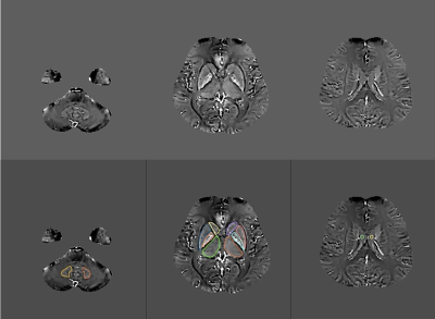 |
Quantitative susceptibility mapping reveals increased iron content of subcortical nuclei in hypertensive patients
Xin Li1, Yue Qin1, Shaoyu Wang2, Xiang Feng3, Yifan Qian1, Juan Tian1, Liyao Liu1, Yinhu Zhu1, Boyuan Jiang1, and Yanqiang Qiao1
1XI’AN DAXING HOSPITAL, ShaanXi, Xi’an, China, 2Siemens Healthcare Ltd., ShaanXi, Xi’an, China, 3Siemens Healthcare Ltd., Beijing, China
Hypertension is known to be a major risk factor for damage to target organs, including the brain. Hypertension may be associated with increased heme content and non-heme iron contents. QSM is a potent imaging method that can provide a noninvasive and accurate measurement of iron content in the brain for diagnosing diseases and monitoring progression and treatment. This study evaluated the feasibility of QSM in the measurement of brain iron deposition in patients with hypertension. We found increased iron accumulation primarily in the deep gray matter nucleus in hypertensive patients.
|
|
1816.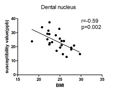 |
Correlation analysis between brain iron deposition and physiological parameters in hypertensive patients
Xin Li1, Yue Qin1, Shaoyu Wang2, Xiang Feng3, Juan Tian1, Yifan Qian1, Yinhu Zhu1, Liyao Liu1, Yanqiang Qiao1, and Boyuan Jiang1
1XI’AN DAXING HOSPITAL, ShaanXi, Xi’an, China, 2Siemens Healthcare Ltd, ShaanXi, Xi’an, China, 3Siemens Healthcare Ltd., Beijing, China
Hypertension is becoming a common health problem worldwide with increasing life expectancy and increasing prevalence of risk factors. Hypertension was positively associated with body mass index. Previous studies have detected that increased iron accumulation mainly in deep gray matter nucleus and CSF in hypertensive patients. There has no study about the correlates of physiological parameters and magnetic susceptibility of brain in hypertension. The present study used Pearson's correlation test to compare the relationship between the magnetic susceptibility of brain tissue and BMI, maximum systolic blood pressure and maximum diastolic blood pressure in hypertensive patients.
|
|
1817.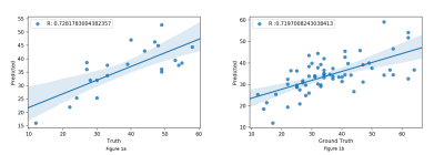 |
Evaluating normative radiomics in T2-FLAIR MRI of the brain
Umang Pandey1, Jitender Saini2, Rakesh Gupta3, and Madhura Ingalhalikar1
1Symbiosis Centre For Medical Image Analysis, Symbiosis International University, Pune, India, 2Department of Radiology, National Institute of Mental Health and Neurosciences, Bengaluru, India, 3Department of Radiology, Fortis Hospital, Gurgaon, India
Radiomics has gained momentum as a non-invasive prediction tool to assess MRI based tumor data in neuro-oncology. However, there’s little understating about the robustness and reproducibility of these features in normative population which is an important first step before translating to pathology. This work investigates and demonstrates the reproducibility of radiomic features on T2-FLAIR images and variation within hemispheres and lobes and its gender and age-related effect. Overall, our findings suggest that care must be taken while interpreting these features for pathological inference.
|
|
1818.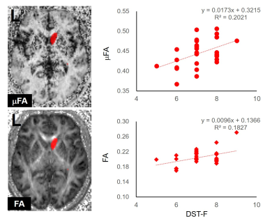 |
Diffusion Imaging Correlates of Neurocognition in Healthy Adults: The Potentials of Sequences with Multiple Diffusion Encoding Schemes
Khin Khin Tha1,2, Daisuke Sawamura3, Xinnan Li3, Yuta Urushibata4, Keita Ogawa5, Kohsuke Kudo1,2, and Hiroki Shirato2
1Department of Diagnostic and Interventional Radiology, Hokkaido University Hospital, Sapporo, Japan, 2Global Station for Quantum Medical Science and Engineering, Hokkaido University, Sapporo, Japan, 3Department of Biomarker Imaging Science, Hokkaido University Graduate School of Biomedical Science and Engineering, Sapporo, Japan, 4Siemens Healthcare, Tokyo, Japan, 5Department of Rehabilitation, Hokkaido University Hospital, Sapporo, Japan
This prospective study was aimed to identify the diffusion imaging correlates of neurocognition. Newly developed diffusion-imaging sequences, i.e., oscillating gradient spin-echo (OGSE), pulsed gradient spin-echo (PGSE), and double diffusion encoding (DDE) sequences, were acquired in 30 healthy adults. The diffusion imaging indices were tested for correlation with the neuropsychological test scores. The microscopic fractional anisotropy (υFA) and fractional anisotropy (FA) of the right caudate nucleus, the υFA of the right temporal lobe, and the OGSE sequence-derived apparent diffusion coefficient (ADC) of the right sublobar white matter showed significant correlations with forward digit span test or trail-making test part-B.
|
|
1819.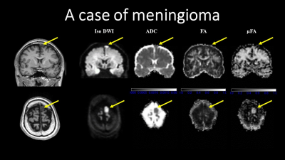 |
Microscopic fractional anisotropy measurements from double diffusion encoding for human brain tumor diagnosis
Masaaki Hori1,2, Masanori Ozaki3, Hiroshi Kusahara4, Masahiro Abe4, Shuji Sato5, Seiko Shimizu6, Kouhei Kamiya1,7, Toshiaki Akashi2, Koji Kamagata2, and Shigeki Aoki2
1Toho University Omori Medical Center, Tokyo, Japan, 2Radiology, Juntendo University School of Medicine, Tokyo, Japan, 3Research and Development Center, Canon Medical Systems Corporation, Kanagawa, Japan, 4Advanced MRI development PJ Team, Canon Medical Systems Corporation, Kanagawa, Japan, 5Juntendo University Hospital, Tokyo, Japan, 6Canon Medical Systems Corporation, Tokyo, Japan, 7Radiology, The University of Tokyo Graduate School of Medicine, Tokyo, Japan
We investigated the values and image contrast of μFA using double diffusion encoding in patients with brain tumors in vivo, as a preliminary study. In general, μFA showed higher values than FA did in the brain tumors. There is no correlation among μFA and FA and ADC, in the meningiomas and solid part of glioblastoma and malignant lymphoma. However, more investigation will be needed to establish the usefulness of μFA in clinical use.
|
 |
1820.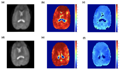 |
Quantitative Measurement of Cerebral Oxygen Extraction in Patients with Sickle Cell Disease after Diamox Infusion Using Asymmetric Spin Echo
Jian Shen1, Chau Vu1, Soyoung Choi2, Aart Nederveen3, and John Wood1,4
1Biomedical Engineering, University of Southern California, Los Angeles, CA, United States, 2Neuroscience Graduate Program, University of Southern California, Los Angeles, CA, United States, 3Academic Medical Center, Amsterdam, Netherlands, 4Division of Cardiology, Children's Hospital Los Angeles, Los Angeles, CA, United States
This study demonstrated tissue oxygen extraction change in patients with sickle cell disease after Acetazolamide injection using Asymmetric Spin Echo technique, and compared with global oxygen extraction change revealed by TRUST. The results suggested that global oxygen extraction decreased as blood flow increased, while tissue oxygen extraction remained stable. And this mismatch might be a proof to the existence of "physiological shunting".
|
1821.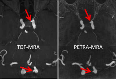 |
Pointwise Encoding Time Reduction with Radial Acquisition in Subtraction-Based MRA: Assessing Intracranial Saccular Aneurysms
Qing Fu1, Xiao-yong Zhang2, Ding-xi Liu1, and Xian-bo Deng1
1Department of Radiology, Union Hospital, Tongji Medical College, Huazhong University of Science and Technology, Wuhan, China, 2MR Collaborations, Siemens Healthcare Ltd., Shenzhen, China
This study compared the clinical utility of the pointwise encoding time reduction with radial acquisition (PETRA) sequence in subtraction-based magnetic resonance angiography (MRA) with conventional time-of-flight MRA (TOF-MRA) for evaluating intracranial saccular aneurysms. The results showed no substantial differences between PETRA-MRA and TOF-MRA in detecting, localizing, and determining the size of aneurysms. Furthermore, PETRA-MRA produced better image quality and provided a higher correlation with computed tomography angiography (CTA) in assessing morphologic parameters of aneurysms than TOF-MRA.
|
|
1822.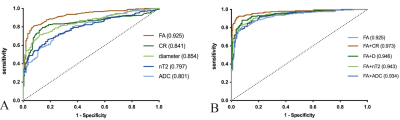 |
Multimodal quantitative magnetic resonance neurography in chronic inflammatory demyelinating polyneuropathy
Su Xiaoyun1, Zheng Chuansheng1, Kong Xiangquan1, Lu Zuneng2, Osamah Alwalid1, and Zhang Huiting3
1Radiology, union hospital, tongji medical college, Huazhong University of science and technology, Wuhan, China, 2Neurology, Renming Hospital of Wuhan University,, Wuhan, China, 3MR Scientific Marketing, Siemens Healthcare, Shanghai, China
Conventional imaging is insufficient for diagnosis and management of patients with chronic inflammatory demyelinating polyneuropathy (CIDP). Brachial and lumbosacral plexus of 37 CIDP patients and 37 age and gender-matched controls were examined by using multi-sequences. Our study showed that fractional anisotropy (FA) had the most sensitivity, while contrast-enhanced ratio (CR) had the most specificity in single-parameter. The combination of FA and CR value had the best diagnostic efficiency. FA had a negative correlation with course duration, and CR positive correlation with CSF protein. SPACE combined with DTI and contrast enhancement sequences serve as a recommended composite protocol.
|
|
1823.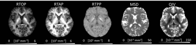 |
Detection of microstructural abnormalities of gray matter in migraineurs without aura using Mean Apparent Propagator (MAP) MRI model
Su Xiaoyun1, Wang Jing1, Chi Bin1, Zhu Qing2, Zhang Huiting3, Zhang Xiaoyong4, and Zheng Chuansheng1
1Radiology, union hospital, tongji medical college, Huazhong University of science and technology, Wuhan, China, 2Neurology, union hospital, tongji medical college, Huazhong University of science and technology, Wuhan, China, 3MR Scientific Marketing, Siemens Healthcare, Shanghai, China, 4Shanghai Key Laboratory of Magnetic Resonance,East China Normal University, Shanghai, China
The mean apparent propagator (MAP) MRI is an advanced diffusion model proposed recently for brain microstructure imaging. In this study, the MAP-MRI model was applied in diagnosis of migraineurs patient without aura. The results found that the parameter of MAP-MRI showed significant differences in in superior frontal gyrus, temporal lobe, cingulate gyrus, parahippocampal gyrus, hippocampus and left amygdata in MWoAs between patients and healthy controls. These findings further suggest involvement of the gray matter in pathology of migraine without aura.
|
|
1824.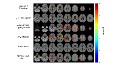 |
Viscerotoxic Brain Infarcts: A Voxel-Based Neuroanatomic Localization Study
Ken Chang1, James Brown2, Praveer Singh1, Jay Patel1, Katharina Hoebel1, Andrew Beers3, Bruce Rosen1, Jayashree Kalpathy-Cramer1, and Hakan Ay1
1Martinos Center for Biomedical Imaging, Charlestown, MA, United States, 2University of Lincoln, Lincoln, United Kingdom, 3University of Washington, Seattle, WA, United States
It is currently not fully known what parts of the human brain predispose to neurogenic organ injury when injured. In this study, we aimed to identify the neuroanatomic correlates of a broad range of cardiac and systemic alterations occurring after ischemic stroke. Using a mapping technique that is free from the bias of a-priori hypothesis as to any specific location, we show that both cardiac and systemic abnormalities occurring after stroke map to specific infarct locations on diffusion-weighted MR. We show that these maps are predictive of the abnormalities as well as patient outcomes.
|
|
 |
1825.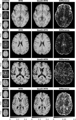 |
QuaSI-MTR (qualitative scans for imaging MTR): deep-learned MTR from routine scans using U-nets.
Antonio Ricciardi1,2, Francesco Grussu1,3, Ferran Prados1,2,4, Baris Kanber2, Rebecca S Samson1, Daniel C Alexander3, Declan T Chard1,5, and Claudia A M Gandini Wheeler-Kingshott1,6,7
1NMR Research Unit, Queen Square MS Centre, Department of Neuroinflammation, UCL Queen Square Institute of Neurology, Faculty of Brain Sciences, University College London, London, United Kingdom, 2Centre for Medical Image Computing, Department of Medical Physics and Biomedical Engineering, University College London, London, United Kingdom, 3Centre for Medical Image Computing, Department of Computer Science, University College London, London, United Kingdom, 4Universitat Oberta de Catalunya, Barcelona, Spain, 5National Institute for Health Research (NIHR) University College London Hospitals (UCLH) Biomedical Research Centre, London, United Kingdom, 6Department of Brain and Behavioral Sciences, University of Pavia, Pavia, Italy, 7Brain MRI 3T Research Center, IRCCS Mondino Foundation, Pavia, Italy
Magnetisation transfer ratio (MTR) is a popular MR-modality for the identification of brain anomalies in multiple sclerosis due to its sensitivity to myelin changes. It however requires dedicated sequences with long acquisition times, which make its applicability in clinics less feasible. In this work, deep learning U-net architectures have been used to extract MTR information directly from routine qualitative images, bypassing the need for specialised acquisitions. Results show strong correlation with MTR and agreement between regional distributions in normal appearing tissues, both in healthy controls and multiple sclerosis patients.
|
1826.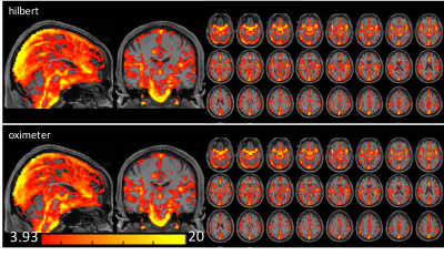 |
Measurement of cardiac pulsatile map in the brain without monitoring an external cardiac signal.
Tae Kim1 and James T Becker1
1University of Pittsburgh, Pittsburgh, PA, United States
In order to obtain cardiac waveforms in the brain for studies with incomplete measurement by pulse-oximeter, we developed a technique to map cardiac pulsation directly from fMRI data itself. We compared the cardiac-phase from our method with that from pulse-oximeter and found they are highly correlated (r > 0.8).
|
|
|
1827.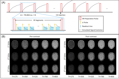 |
MR Multitasking-based Dynamic Imaging for Cerebrovascular Evaluation (MT-DICE): Development and Feasibility Study
Zhehao Hu1,2, Anthony G. Christodoulou1, Nan Wang1,2, Yibin Xie1, Bin Sun3, Xiaoming Bi4, Fei Han4, Shlee S. Song5, Marcel M. Maya6, Debiao Li1,2,7, and Zhaoyang Fan1,2,7
1Biomedical Imaging Research Institute, Cedars-Sinai Medical Center, Los Angeles, CA, United States, 2Bioengineering Department, University of California, Los Angeles, Los Angeles, CA, United States, 3Department of Radiology, Fujian Medical University Union Hospital, Fuzhou, China, 4Siemens Healthineers, Los Angeles, CA, United States, 5Department of Neurology, Cedars-Sinai Medical Center, Los Angeles, CA, United States, 6Department of Imaging, Cedars-Sinai Medical Center, Los Angeles, CA, United States, 7Department of Medicine, University of California, Los Angeles, Los Angeles, CA, United States
Perfusion MR imaging (i.e. DCE, DSC) has evolved as an increasingly common modality for evaluating a variety of cerebrovascular diseases, such as stroke and brain tumors, and susceptibility weighted imaging (SWI) is suitable for detection of intracranial hemorrhage. In this work, we developed an MR MultiTasking based Dynamic Imaging for Cerebrovascular Evaluation (MT-DICE) technique that can provide DCE, DSC and SWI information simultaneously with one 8-minute scan and a single contrast agent injection. Feasibility of MT-DICE was demonstrated in healthy volunteers and patients with hemorrhagic stroke or brain cancer.
|
|
1828.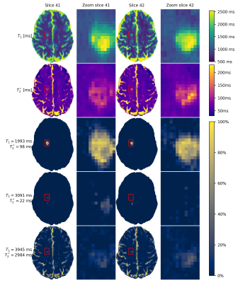 |
Detection of small cerebral lesions using multi-component MR Fingerprinting with local joint sparsity
Martijn Nagtegaal1, Ingo Hermann1,2, Sebastian Weingärtner1, Jeroen de Bresser3, and Frans Vos1
1Department of Imaging Physics, Delft University of Technology, Delft, Netherlands, 2Medical Faculty Mannheim, Heidelberg University, Mannheim, Germany, 3Department of Radiology, Leiden University Medical Centre, Leiden, Netherlands
We propose a novel multi-component analysis for MR fingerprinting that enables detection of small lesions, while taking partial volume effects into account. The algorithm uses a joint sparsity constraint limiting the number of components in local regions. It is evaluated in simulations and on MRF-EPI data from a patient with multiple sclerosis (MS). MS-lesions are separated from other tissues based on having increased T2* relaxation times. The improved sensitivity to multiple components makes it possible to detect components with long relaxation times within the lesion, possibly increasing our insight into these small pathologies.
|
1829.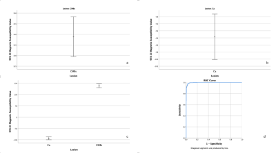 |
Quantitative Evaluate the Differential Between Cerebral Microbleeds and Calcification in Susceptibility Weight Imaging Mapping
Ke Gao1, Yu Luo1, and Yong Zhou1
1Department of Radiology, Shanghai Fourth People's Hospital Affiliated to Tongji University School of Medicine, Shanghai, China
To quantitative assess the differential between cerebral microbleeds and calcification in susceptibility weight imaging mapping. We quantified the magnetic susceptibility value of calcifications and cerebral microbleeds(CMBs) in susceptibility weight imaging mapping. Significant difference of magnetic susceptibility value was found between them. The 95% confidence interval for the magnetic susceptibility value of the calcification and CMBs was different. The optimal threshold to detecting CMBs was selected by receiver operating characteristic. The conclusion was that magnetic susceptibility value can used to differential diagnose cerebral microbleeds and calcification in the susceptibility weighted imaging mapping.
|
|
1830.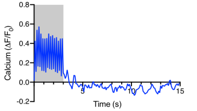 |
Development of simultaneous calcium photometry and fMRI of the mouse brain
Shabnam Khorasani Gerdekoohi1, Hsu-Lei Lee1, Helena Hung-Yin Huang1, Pankaj Sah1, and Kai-Hsiang Chuang1,2
1Queensland Brain Institute, University of Queensland, Brisbane, Australia, 2Centre for Advanced Imaging, University of Queensland, Brisbane, Australia Neural calcium activity of genetic reporter GCaMP6f was successfully recorded in 9.4T MRI. Event-related BOLD activation can be measured using short stimuli. Susceptibility artefacts from the optic fiber and dental cement affect fMRI quality. |
|
1831.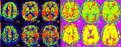 |
Quantitative measurement of cerebral blood flow in healthy volunteers and hypertension patients by multi-TI arterial spin labeling at 3 T MRI
Fan Qiuju1, Tan Hui1, Yang Zhen1, Yu Nan1, Yu Yong1, Wang Shaoyu2, Xue Yu3, and Li Yue3
1Affiliated Hospital of Shaanxi University of Chinese Medicine, Shaanxi, China, 2MR senior scientific marketing specialist, Shaanxi, China, 3Shaanxi University of Chinese Medicine, Shaanxi, China
This study aimed to quantitative measurement of cerebral blood flow in different brain regions of different levels of blood pressure by mTI ASL. In the first part of this study, we evaluated the reproducibility of mTI-ASL in healthy subjects, and then mTI-ASL was used to evaluate 55 primary hypertension patients. There was no major variation between two consecutive mTI-ASL measurements in healthy volunteers. Furthermore, the rCBF and ATT values provided valuable information for the classification of hypertension, which can be used as a non-invasive quantitative monitoring MRI tool for hypertension progression.
|
|
1832.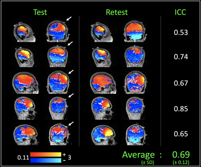 |
In-vivo imaging of current induced magnetic fields during High Definition transcranial direct current stimulation (HD-tDCS)
Mayank A Jog1, Kay Jann1, Lirong Yan1, Chenyang Zhao1, Bhargavi Kannan1, and Danny JJ Wang1
1University of Southern California, Los Angeles, CA, United States
High-definition transcranial direct current stimulation (HD-tDCS) is an emerging neuromodulation technique that uses a multi-electrode montage to focally modulate regional brain activities. However, the effects of stimulation have been shown to be variable across subjects, and efforts to investigate this have been hampered by an inability to visualize the electric current distribution in-vivo. To address this, we developed a MR technique that maps the tDCS current induced magnetic fields in-vivo. The measured fields were robust (avg. ICC = 0.69) and show different hotspot-locations between subjects, potentially demonstrating the sensitivity of this technique to track individual differences in responses to HD-tDCS.
|
|
1833.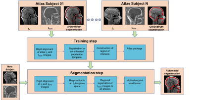 |
Automated Segmentation of Human Skull to plan Craniofacial Surgery using dual-Radiofrequency dual-Echo, 3D Ultrashort Echo Time MRI sequence
Pulkit Khandelwal1, Carrie E. Zimmerman2, Long Xie3, Hyunyeol Lee3, Cheng-Chieh Cheng3, Scott P. Bartlett2, Paul Yushkevich3, and Felix W. Wehrli3
1Department of Bioengineering, University of Pennsylvania, Philadelphia, PA, United States, 2Division of Plastic Surgery, Children’s Hospital of Philadelphia, Philadelphia, PA, United States, 3Department of Radiology, University of Pennsylvania, Philadelphia, PA, United States
Bone-selective Magnetic Resonance Imaging (MRI) produced by a Dual Radiofrequency (RF), dual‐echo, 3D Ultrashort Echo Time (UTE) pulse sequence and bone‐selective image reconstruction process provides a radiation-free imaging modality with high concordance to computed tomography (CT). This imaging technique is of specific interest to craniofacial surgeons. Here, we pilot the use of an automated segmentation pipeline on the bone-selective MR images with the goal of reducing time required for the 3D segmentation step. This automated segmentation pipeline, used with bone-selective MRI could eliminate the need for radiative CT thereby reducing the risk of malignancy in pediatric craniofacial patients.
|
|
1834.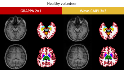 |
Comparison between MPRAGE GRAPPA 2×1 with MPRAGE Wave-CAIPI 3×3
Azusa Sakurama1, Yasutaka Fushimi1, Satoshi Nakajima1, Yusuke Yokota1, Sonoko Oshima1, Sayo Otani1, Krishna Pandu Wicaksono1, Tomohisa Okada2, Wei Liu3, and Kaori Togashi1
1Department of Diagnostic Imaging and Nuclear Medicine, Kyoto University Graduate School of Medicine, Kyoto, Japan, 2Human Brain Research Center, Kyoto University Graduate School of Medicine, Kyoto, Japan, 3Siemens Healthineers AG, Shenzhen, China Poster Permission Withheld
This comparison study between MPRAGE with GRAPPA and MPRAGE with Wave-CAIPI has been performed at the clinical MR scanner. MPRAGE with Wave-CAIPI 3×3 shows relatively good contrast despite its short scan time of 1 m 42 s. No segmentation error was found in MPRAGE with Wave-CAIPI 3×3 in our cohort. The correlation of cerebral cortex and cerebral white matter was very high between 2 MPRAGEs. In deep gray matters except pallidum and hippocampus, the correlation of VOIs were also high. These results suggest that volumes derived from automated segmentation of MPRAGE with Wave-CAIPI are reliable measures.
|
1835.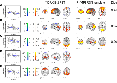 |
Identifying RSN-like spatially covarying sources in synaptic density PET (11C-UCB-J) with independent component analysis
Xiaotian T. Fang1, Takuya Toyonaga1, Ansel T. Hillmer1,2, Patrick D. Worhunsky2, and Richard E. Carson1
1Radiology and Biomedical Imaging, Yale University, New Haven, CT, United States, 2Psychiatry, Yale School of Medicine, New Haven, CT, United States
Resting-state networks, functionally connected brain regions, are canonically investigated with resting-state functional-MRI. 11C-UCB-J is a recently developed PET tracer that binds and visualizes synaptic vesicle protein 2A. The aim is to identify networks of coherent 11C-UCB-J covariation patterns using independent component analysis. We find modest similarity between five 11C-UCB-J PET-based synaptic density sources and rs-fMRI template RSNs. In several sources, there are varying rates of age-related change in subject loading weight, consistent with functional changes in RSNs and age-related gray matter decline. Our findings support the independency of such networks and potential physiological links between synaptic density and brain function.
|
|
1836. |
Prediction-Identification Landscape for Brain Structure and Connectivity
Sina Mansour L.1, Ye Tian2, Vanessa Cropley2, and Andrew Zalesky1,2
1Department of Biomedical Engineering, The University of Melbourne, Melbourne, Australia, 2Melbourne Neuropsychiatry Centre, Department of Psychiatry, The University of Melbourne, Melbourne, Australia
Neuroimaging-derived brain phenotypes can be used to identify individuals and predict behavior. We investigated the prediction and identification performance of brain structure and connectivity phenotypes derived from structural, functional and diffusion MRI. Five behavioral domains were predicted: cognition, illicit substance use, tobacco use, personality-emotion traits, and mental health. We arranged the phenotypes on a two-dimensional prediction-identification landscape. Functional connectivity performed better at prediction than identification, whereas the converse was found for curvature and other structural phenotypes. Structural connectivity performed well for both tasks. High-resolution measures outperformed atlas-based counterparts. Our work can aid brain phenotype selection in future neuroimaging studies.
|
|
1837.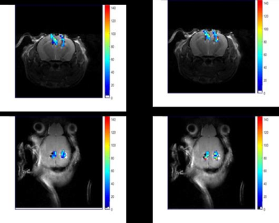 |
Quantitative mapping of brain tissue oxygenation around neural interfaces
Babak Moghadas1, Arati Sridharan1, Livia De Mesquita1, Yuka Sugamura1, Jitendran Muthuswamy1, and Vikram D Kodibagkar1
1Biomedical engineering, Arizona State University, Tempe, AZ, United States
There is a significant need to spatially map the pO2 levels around chronically implanted neural devices to assess the impact on surrounding tissue. Here we apply a quantitative MRI based oximetry approach to map pO2 around neural interfaces. Electrodes were coated with polydimethylsiloxane matrix and soaked in tetradecamethylhexasiloxane, which was characterized and used as a 1H MR pO2 reporter along with the previously developed PISTOL technique. Tissue pO2 in response to air and oxygen breathing was successfully measured from around implanted electrodes. Future studies will combine single-unit electrophysiology with quantitative, high resolution spatial maps of pO2 levels in vivo.
|
|
1838.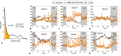 |
CMRO2 and slow-wave activity are inversely related during sleep: findings from concurrent EEG and OxFlow MRI.
Alessandra Caporale1, Hyunyeol Lee1, Hui Lei2, Hengyi Rao3, Michael C Langham1, Alessandra A Caporale2, and Felix W Wehrli1
1Radiology, University of Pennsylvania Perelman School of Medicine, Philadelphia, PA, United States, 2Neurology, University of Pennsylvania Perelman School of Medicine, Philadelphia, PA, United States, 3Psychiatry, Division of Sleep and Chronobiology, University of Pennsylvania Perelman School of Medicine, Philadelphia, PA, United States
Radial OxFlow, a technique able of measuring cerebral metabolic rate of oxygen (CMRO2) dynamically, has been combined with simultaneous electroencephalography (EEG) recording, during the natural onset of sleep and maintenance during a 50-min period of scanning. During sleep, CMRO2 (measured continuously with an effective temporal resolution of 3.4 seconds) decreased up to 24% with respect to pre-sleep wakefulness. CMRO2 changes were positively correlated with heart rate (r2=0.25-0.48, P<0.0005), and negatively with changes in EEG slow-wave activity (r2=0.41-0.67, P<0.005), when sleep onset and maintenance occurred.
|
|
1839.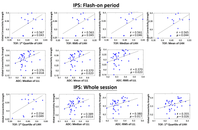 |
MR radiomic features Correlate with EEG Connectivity during Intermittent Photic Stimulation in Patients with Cavernous Malformation
Nai-Chi Chen1, Cheng-Chia Lee2,3,4, Yo-Tsen Liu3,5,6, Chien-Chen Chou2, Chung-Jung Lin7, Wan-Yuo Guo3,7, Wen-Yuh Chung2,3, and Chia-Feng Lu1
1Department of Biomedical Imaging and Radiological Sciences, National Yang-Ming University, Taipei City, Taiwan, 2Department of Neurosurgery, Neurological Institute, Taipei Veteran General Hospital, Taipei, Taiwan, 3School of Medicine, National Yang-Ming University, Taipei, Taiwan, 4Brain Research Center, National Yang-Ming University, Taipei, Taiwan, 5Department of Neurology, Neurological Institute, Taipei Veterans General Hospital, Taipei, Taiwan, 6Institute of Brain Science, National Yang-Ming University, Taipei, Taiwan, 7Department of Radiology, Taipei Veteran General Hospital, Taipei, Taiwan
Cavernous malformation (CM) is one of the common cause for seizure attacks. Till now, the relationships between the structural characteristics of CM and the resultant abnormality of neural activity are still less explored. We employed MR radiomics analysis and EEG functional connectivity analysis to investigate whether quantitative and non-invasive features derived from these approaches can be used to differentiate CM patients with and without seizure. Furthermore, the association between structural and functional characteristics of CM were unraveled.
|
|
1840.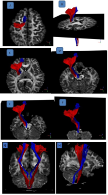 |
Multimodal Quantitative Magnetic Resonance Imaging in Adolescent Idiopathic Scoliosis
Bertwin Zhicheng Chen1, Reuben Chee Cheong Soh1,2, Septian Hartono2,3, Chu Ning Ann3, Ming-Ching Wen3, Weiling Lee1, Soo Lee Lim4, Julian Gan5, Yew Long Lo2,3, and Ling Ling Chan1,2
1Singapore General Hospital, Singapore, Singapore, 2Duke-NUS Medical School, Singapore, Singapore, 3National Neuroscience Institute, Singapore, Singapore, 4National Heart Centre Singapore, Singapore, Singapore, 5Siemens Healthineers, Singapore, Singapore
The aetiopathogenesis of adolescent idiopathic scoliosis (AIS) remains elusive, with studies suggesting a neurological basis. Using a case control, multimodal approach incorporating volumetry and diffusion tensor imaging, we characterised brain MRI changes in AIS. The pontine volume was significantly larger while left insula was smaller in patients than controls. Whole brain Tract-Based Spatial Statistics (TBSS) showed no difference between subject cohorts. Tractography of the corticoreticular pathway (CRP), which innervates the axial muscles, showed significant differences in mean and axial diffusivity between the left and right CRP only in the patient cohort, but no such asymmetry in the control cohort.
|
|
1841.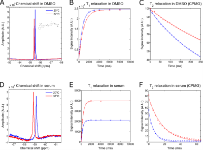 |
Fluorine-19 MR Characterisation of Siponimod as MR Theranostic Approach in the Treatment of Multiple Sclerosis
Christian Prinz1, Fatima Sherazi1, Ludger Starke1, Paula Ramos Delgado1, Andre Kuehne2, Thoralf Niendorf1,2,3, and Sonia Waiczies1
1Berlin Ultrahigh Field Facility (B.U.F.F.), Max Delbrück Center for Molecular Medicine in the Helmholtz Association, Berlin, Germany, 2MRI TOOLS GmbH, Berlin, Germany, 3Experimental and Clinical Research Center, a joint cooperation between the Charité Medical Faculty and the Max Delbrück Center for Molecular Medicine in the Helmholtz Association, Berlin, Germany
Siponimod is a trifluorinated anti-inflammatory drug indicated for Multiple Sclerosis that could be potentially tracked in pharmacokinetic studies by fluorine-19 (19F) MR techniques in order to support therapeutic monitoring. In this study, we investigated the 19F MR properties of siponimod and its dependency on environmental factors. We found a temperature dependence of the relaxation times T1 and T2 and identified a T2-drop in the presence of human serum. We identified UTE as SNR-efficient MR pulse sequence to study siponimod physiologically and show the potential of tailoring MR methods in the future to individual patient therapeutic needs.
|
|
1842.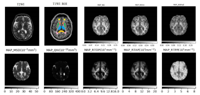 |
The performance of mean-apparent-propagator diffusion model in assessing brain parenchymal microstructure changes of hypertensive patients
shenghui Xie1, shaoyu Wang2, xu Yan2, huapeng Zhang2, guang Yang3, and yang Gao1
1Department of radiology, affiliated hospital of Inner Mongolia medical university, Hohhot, China, 2MR Scientific Marketing, Shanghai, China, 3Shanghai Key Laboratory of Magnetic Resonance, Shanghai, China
The purpose of this study was to evaluate the value of diffusion spectrum imaging (DSI) MAP diffusion model in the changes of brain tissue microstructure in patients with primary essential hypertension, as well as the differences of various parameters including RTOP, RTAP, RTPP, QIV and MSD between the hypertensive group and the normal control group. The results showed that there were significant differences between RTOP and QIV. All the MAP-MRI parameters showed obvious differences in detecting the changes of brain structure microstructure in the early stage of hypertension, especially the RTOP and QIV parameters.
|
|
1843.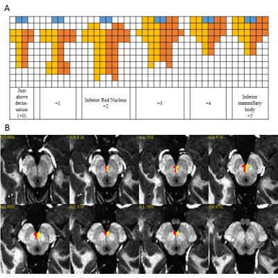 |
Definition of Brainstem Nuclei: Ventral Tegmental Area (VTA) and Dorsal Raphe Nucleus (DRN)
Anna Crawford1, Stephen Jones1, and Mark Lowe1
1Imaging Institute, Cleveland Clinic Foundation, Cleveland, OH, United States
Many advanced imaging application require the identification of seed and target ROIs. Due to size, and signal characteristics some brain structures including brainstem nuclei are difficult to identify with MR imaging, while other types of imaging such as PET have other drawbacks such as poor spatial resolution. We have come up with a “recipe” for defining the ventral tegmental area (VTA) as well as the dorsal raphe nucleus (DRN) using three anatomic atlases. The definition of these ROIs were confirmed with PET imaging. Our method allows for robust, reproducible ROIs using only a high resolution T1 weighted image.
|
|
1844.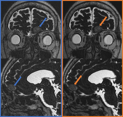 |
Clinical Application of Six-fold Accelerated Submillimeter Whole Brain 3D T2-weighted Imaging with Deep Learning Reconstruction
Sayo Otani1, Yasutaka Fushimi1, Satoshi Nakajima1, Yusuke Yokota1, Sonoko Oshima1, Azusa Sakurama1, Krishna Pandu Wicaksono1, Yuichiro Sano2, Ryo Matusda2, Masahito Nambu2, Koji Fujimoto3, Hitomi Numamoto4, Kanae Kawai Miyake4,
Tsuneo Saga4, and Kaori Togashi1
1Department of Diagnostic Imaging and Nuclear Medicine, Kyoto University Graduate School of Medicine, Kyoto, Japan, 2MRI Systems Division, Canon Medical Systems Corporation, Kyoto, Japan, 3Human Brain Research Center, Kyoto University Graduate School of Medicine, Kyoto, Japan, 4Department of Advanced Medical Imaging Research, Kyoto University Graduate School of Medicine, Kyoto, Japan We have demonstrated six-fold accelerated submillimeter whole brain 3D T2-weighted imaging with deep learning reconstruction (DLR) showed better coefficient of variation and signal ratio compared with that without DLR. Scan time of around 2.5 min is clinically feasible and the delineation of fine structure is preserved after DLR processing. 3D T2-weighted image with better image quality derived from DLR will help clinicians and radiologists evaluate CSF space abnormalities, and its short scan time will be feasible for routine clinical examinations. |
|
1845.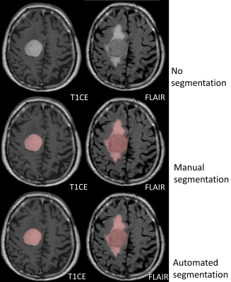 |
Clinical evaluation of automated deep-learning based meningioma segmentation in multiparametric MRI
Kai Laukamp1, Lenhard Pennig1, Frank Thiele1, Robert Reimer1, Lukas Goertz1, David Zopfs1, Georgy Shakirin1, Marco Timmer1, Michael Perkuhn1, and Jan Borggrefe1
1UKK, Cologne, Germany
We trained an established deep-learning-model architecture (3D-Deep-Convolutional-Neural-Network, DeepMedic) on manual segmentations from 70 meningiomas independently segmented by two radiologists. The trained deep-learning model was then validated in a group of 55 meningiomas. Ground truth segmentations were established by two further radiologists in a consensus reading. In the validation-group the comparison of the automated deep-learning-model and manual segmentations revealed average dice-coefficients of 0.91±0.08 for contrast-enhancing-tumor volume and 0.82±0.12 for total-lesion-volume. In the training-group, interreader-variabilities of the two manual readers were 0.92±0.07 for contrast-enhancing-tumor and 0.88±0.05 for total-lesion-volume. Deep-learning based automated segmentation yielded high segmentation accuracy, comparable to manual interreader-variability.
|
|
1846.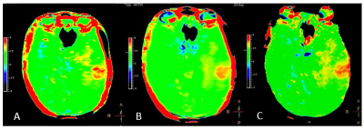 |
Fast APTw imaging with Compress Sense technique in Glioma
Cong Xie1, Yaou Liu1, Rongkai Ju2, Zhiwei Shen2, Zhizheng Zhuo1, Yunyun Duan1, Xiaobo Wang1, Qiong Qiu1, and Fenglian Zheng1
1Department of Radiology, Beijing Tiantan Hospital, Beijing, China, 2Philips,healthcare,China, Beijing, China
explorer the performance of in APTw with Compress Sense (CS) in the evaluation of glioma with high grade and low grade.
|
|
1847.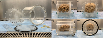 |
A Container and Detailed Preparation Protocol for Ex Vivo Whole Human Brain MRI
Alan C Seifert1,2,3, Joseph A Borrello1,2,3, Jessie Laffey4, Etty Cortes4, Tamjeed Sikder4, Enna Selmanovic5, Bradley N Delman2, John Crary4, Kristen Dams-O'Connor5, and Junqian Xu1,2,3,6
1Biomedical Engineering and Imaging Institute, Icahn School of Medicine at Mount Sinai, New York, NY, United States, 2Department of Radiology, Icahn School of Medicine at Mount Sinai, New York, NY, United States, 3Graduate School of Biomedical Sciences, Icahn School of Medicine at Mount Sinai, New York, NY, United States, 4Department of Pathology, Icahn School of Medicine at Mount Sinai, New York, NY, United States, 5Department of Rehabilitation Medicine, Icahn School of Medicine at Mount Sinai, New York, NY, United States, 6Department of Neuroscience, Icahn School of Medicine at Mount Sinai, New York, NY, United States
Whole-brain ex vivo MRI has proven extremely valuable in neuroscience research, but detailed descriptions of the specific preparation and packaging steps to minimize motion, susceptibility artifacts, and background signal are often missing from reports of experimental findings. We present the design of a reproducible container and a detailed protocol that yield whole-brain images uncorrupted by susceptibility artifacts. The most important steps to eliminate susceptibility artifacts due to residual air bubbles are removal of the leptomeninges, application of vacuum to dislodge bubbles, immersion in Fluorinert, and rolling of the sealed container, although removal of leptomeninges may be undesirable in some cases.
|
|
1848.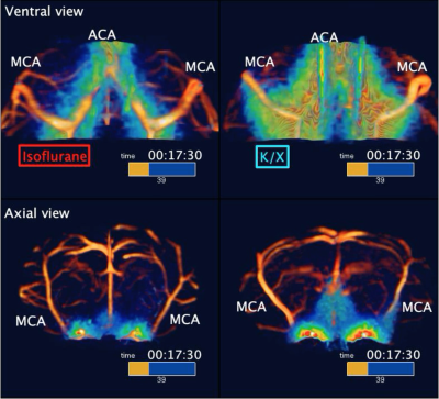 |
High temporal resolution glymphatic influx/efflux analysis with 3D-FISP sequence
Evan Hunter Stanton1, Niklas Daniel Åke Persson1, Björn Sigurdsson1, Tuomas Lilius1, Humberto Mestre2, Maiken Nedergaard1, and Yuki Mori1,3
1Division of Glial Disease and Therapeutics, University of Copenhagen, Center for Translational Neuromedicine, Copenhagen, Denmark, 2Division of Glial Disease and Therapeutics, University of Rochester Medical Center, Center for Translational Neuromedicine, Rochester, NY, United States, 3Faculty of Health and Medical Sciences, University of Copenhagen, Copenhagen, Denmark, Panum NMR Core Facility, Copenhagen, Denmark
The glymphatic system is a brain waste clearance pathway based on cerebrospinal fluid (CSF) and interstitial fluid exchange. As such, this pathway plays important roles in both acute and neurodegenerative conditions. With 3D-FISP-based fast imaging, we investigated how anesthetic regiments, i.e., ketamine-xylazine (K/X) versus isoflurane, influences the movement of tracer through the brain. This was achieved by conducting acute cisterna magna cannulations, tracer scanning using 3D-FISP, and calculating dynamic tracer distribution maps. Our analysis demonstrates that anesthesia alters the spatial and temporal distribution of CSF tracer, with K/X showing higher influx, and isoflurane shunting CSF out via deep cervical nodes.
|
1849.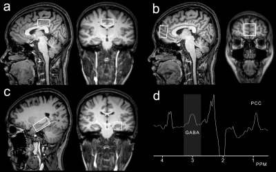 |
Brain GABA levels distinguish multiple sclerosis and neuromyelitis optica using edited magnetic resonance spectroscopy
fei gao1, fuxin ren1, weibo chen2, and guangbin wang1
1Shandong Medical Imaging Research Institute, Shandong University, Jinan, China, 2Philips Healthcare, shanghai, China
Distinguishing relapsing-remitting multiple sclerosis (RRMS) and neuromyelitis optica (NMO) is clinically important because they differ in prognosis and treatment. Edited magnetic resonance spectroscopy (MRS), using the MEGA-PRESS sequence, is the most widely used technique for detecting GABA in the human brain. However, none of the existing studies are performed to compare the brain GABA levels between patients with MS and NMO. In this study, we found GABA levels differences in the LHC and PCC between the RRMS and NMO patients, which may be helpful in distinguishing RRMS from NMO after further validation.
|
|
1850.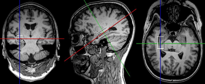 |
Cross sectional evaluation of neurometabolic alterations in RRMS with fingolimod and injectable treatments using advanced MRS
Oun Al-iedani1,2, Karen Ribbons2, Saadallah Ramadan1,2, Rodney Lea2, and Jeannette Lechner-Scott2,3,4
1School of Health Sciences, University of Newcastle, Newcastle, Australia, 2Hunter Medical Research Institute (HMRI), Newcastle, Australia, 3School of Medicine and Public Health, University of Newcastle, Newcastle, Australia, 4Department of Neurology, John Hunter Hospital, Newcastle, Australia
This novel study evaluated the amount of axonal injury with neurometabolite in RRMS patients on fingolimod(N=52) and injectable(N=46) treatments compared with 51 HCs in hippocampus using advanced MRS. PRESS-MRS of the hippocampal voxel(30x15x15mm3) and clinical assessments for RRMS and HCs were acquired. Hippocampal MR-spectra were analysed by LCModel. Hippocampal-NAA/tCr decreased (fingolimod:-17%,p=0.001;injectables:-15%,p=0.01) and in Glx/tCr increased (fingolimod:+16%,p=0.001;injectables:+15%,p=0.02) in RRMS compared to HCs. Total-ARCS (r=0.402) and memory in particular (r=0.428) displayed associations with hippocampal NAA/tCr. This study is the first cross-sectional in-vivo investigation comparing the impact of fingolimod and injectable treatments on the hippocampal metabolism in RRMS patients.
|
|
1851.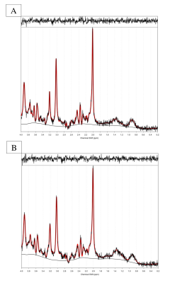 |
Human cerebral blood flow-metabolic uncoupling during acute hypoxia: A 1H Magnetic Resonance Spectroscopy & Arterial Spin Labelling study
Matthew Rogan1,2, Alexander Friend2, Gabriella Rossetti2, Jamie Macdonald2, Sam Oliver2, and Paul Mullins1
1The Bangor Imaging Centre, School of Psychology, Bangor University, Bangor, United Kingdom, 2Extremes Research Group, School of Sport Health and Exercise Science, Bangor University, Bangor, United Kingdom
Acute exposure to a moderate hypoxic environment leads to regional specific reduction in cerebral blood flow (rCBF) to the posterior cingulate cortex. Despite this, there is no concomitant reduction in the main excitatory neurotransmitter glutamate, as would be expected if the reductions in rCBF were coupled to neural activity. Our findings obtained using a combined 1H Magnetic Resonance Spectroscopy and Arterial Spin Labelling paradigm indicate that hypoxia disrupts neurovascular coupling, as it is presently understood, in a regionally specific manner.
|
|
1852.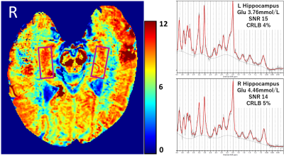 |
Hippocampal concentration of glutamate: A comparative study of CEST and 1HMRS measurements at 7T.
Tomohisa Okada1, Ravi Prakash Reddy Nanga2, Koji Fujimoto1, Dinh Ha Duy Thuy1, Ravinder Reddy2, Hideto Kuribayashi3, Yuta Urushibata3, Ravi Teja Seethamraju4, Sinyeob Ahn5, and Tadashi Isa1
1Kyoto University, Kyoto, Japan, 2University of Pennsylvania, Philadelphia, PA, United States, 3Siemens Healthcare K.K., Tokyo, Japan, 4Siemens Healthineers USA, Burlington, MA, United States, 5Siemens Healthineers USA, Berkeley, CA, United States
Chemical exchange saturation transfer of the glutamate (GluCEST) contrast is considered from glutamate to be about 70 - 75 %. Its values at the bilateral hippocampus were compared to those of 1HMRS in 10 healthy subjects. Correlation between GluCEST contrast and glutamate from 1HMRS measurements of all subjects was marginally significant (r = 0.44, p = 0.054), but it was statistically significant (r = 0.56, p = 0.030) when analysis was limited to 1HMRS measurements with SNR ≥ 10. The GluCEST map gives regional difference in glutamate concentration in high resolution and is expected to facilitate investigation of the hippocampus.
|
|
1853. |
Brain and immune system crosstalk: Examining the link between proinflammatory markers and glial markers measured with 1H-MRS at 7T
Carl-Johan Boraxbekk1,2,3, Anna Lind1, Esben Thade Petersen1,4, Hartwig Roman Siebner1,5,6, and Anouk Marsman1
1Danish Research Centre for Magnetic Resonance, Centre for Functional and Diagnostic Imaging and Research, Copenhagen University Hospital, Hvidovre, Denmark, 2Department of Radiation Sciences, Umeå University, Umeå, Sweden, 3Institute of Sports Medicine Copenhagen (ISMC), Copenhagen University Hospital Bispebjerg, Copenhagen, Denmark, 4Center for Magnetic Resonance, Department of Health Technology, Technical University of Denmark, Lyngby, Denmark, 5Department of Clinical Medicine, Faculty of Health and Medical Sciences, University of Copenhagen, Copenhagen, Denmark, 6Department of Neurology, Copenhagen University Hospital Bispebjerg, Copenhagen, Denmark
Inflammation is proposed as one of the central pillars driving ageing. This study was the first to link peripheral inflammation to brain metabolite differences during normal ageing and to associate this to visuo-spatial working memory performance. Our data shows how peripheral inflammatory markers were associated with performance on visuo-spatial working memory and glial metabolic markers in ACC, Hippocampus, and Thalamus. The findings highlight the role of glial cells as part of the neurobiological link between peripheral inflammation and cognitive performance during ageing.
|
|
1854. |
Neurochemistry and cognition across age: Examining the link between working memory and neuronal and glial markers measured with 1H-MRS at 7T
Anouk Marsman1, Anna Lind1, Esben Thade Petersen1,2, Hartwig Roman Siebner1,3,4, and Carl-Johan Boraxbekk1,5,6
1Danish Research Centre for Magnetic Resonance, Centre for Functional and Diagnostic Imaging and Research, Copenhagen University Hospital Hvidovre, Hvidovre, Denmark, 2Center for Magnetic Resonance, Department of Electrical Engineering, Technical University of Denmark, Lyngby, Denmark, 3Department of Clinical Medicine, Faculty of Health and Medical Sciences, University of Copenhagen, Copenhagen, Denmark, 4Department of Neurology, Copenhagen University Hospital Bispebjerg, Copenhagen, Denmark, 5Department of Radiation Sciences, Umeå University, Umeå, Sweden, 6Institute of Sports Medicine Copenhagen, Copenhagen University Hospital Bispebjerg, Copenhagen, Denmark
Ageing has been associated with widespread neurochemical changes and cognitive decline. The increased sensitivity and resolution of 1H-MRS at 7T could shed new light on the potential relationship between neurochemistry and cognition during ageing. Our data show that primarily levels of glial metabolic markers, and not neuronal markers, differed between younger, middle-aged and older normal individuals and were correlated with visuo-spatial working memory performance across these age groups. This suggests that glial markers may be more affected by ageing than neuronal markers and that there is a potential association between glial markers and visuo-spatial working memory performance during ageing.
|
|
1855. |
Diffusion of NAA as a marker for tau pathology in Alzheimer's disease - a combined diffusion weighted MRS and positron emission tomography study.
Nicola Spotorno1, Markus Nilsson2, Boel Hansson2, Felix Andersson3, Antonie Leuzy3, Danielle van Westen2, Oskar Hansson3, and Itamar Ronen4
1Department of Medicine, University of Pennsylvania, Philadelphia, PA, United States, 2Department of Diagnostic Radiology, Lund University, Lund, Sweden, 3Department of clinical sciences Malmö, Lund University, Lund, Sweden, 4Department of Radiology, Leiden University Medical Center, Leiden, Netherlands
Intraneuronal tau accumulation is a hallmark of Alzheimer's disease. Currently, positron emission tomography with tau specific ligands (Tau-PET) is the only neuroimaging method for measuring tau in AD. In this study that combines Tau-PET with diffusion weighted MRS we show that the apparent diffusion coefficient of NAA, a neuronal metabolite, is significantly lower in AD patients compared to healthy controls in a brain region that shows elevated levels of uptake of the tau-PET ligand. Standard MRS data shows elevated level of glial metabolites, suggesting concomitant gliosis of neuroinflamation. We propose ADC(tNAA) as a putative marker for tau aggregation in AD.
|
|
|
1856.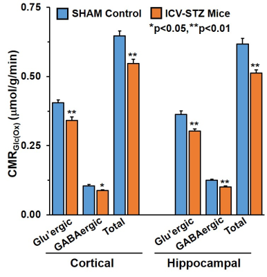 |
Neuronal and Astroglial Metabolism in icv-STZ Mouse Model of Alzheimer’s Disease: A 1H-[13C]-NMR Study
Narayan Datt Soni1, Dipak Roy2, Akila Ramesh2, and Anant Bahadur Patel2
1NMR spectroscopy and Microimaging, CSIR-CCMB, Hyderabad, India, 2NMR Spectroscopy and Microimaging, CSIR-CCMB, Hyderabad, India
Alzheimer’s Disease (AD) has been categorized into familial (~5%) and sporadic AD (~95%). Familial cases are genetic whereas sporadic AD (sAD) is acquired during individual’s lifespan. Intracerebroventricular(icv) streptozotocin(stz) administered animals has been shown to resemble AD phenotype through many molecular studies. In this study we have investigated the impact of icv-stz treatment on memory and neurometabolic activity by infusion of [1,6-13C2]Glucose and [2-13C]Acetate in conjunction with NMR spectroscopy. Our findings have shown the compromised memory, perturbed neurometabolic homeostasis along with decreased synaptic neurotransmission across glutamatergic and GABAergic synapse in cortical and hippocampal brain regions of icv-stz treated mouse.
|
1857.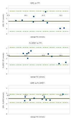 |
Comparison of three tissue sodium concentration quantification methods at 3T and 7T MRI.
Lazar Fleysher1, Mounir El Mendili2, Guilaume Madelin3, Fernando Boada 3, and Matilde Inglese2
Video Permission Withheld
1Mount Sinai Medical Center, New York, NY, United States, 2Icahn School of Medicine at Mount Sinai, New York, NY, United States, 3NYU School of Medicine, New York, NY, United States
We report results of a comparison of three methods of tissue sodium concentration (TSC) quantification which use GRE, FLORET and TPI acquisition techniques at different field strengths. Results demonstrate a good inter-method agreement (better than 10%) in TCS quantification.
|
|
1858.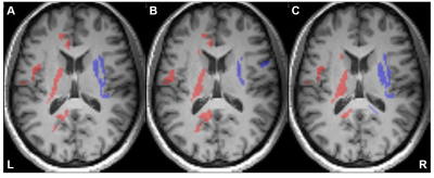 |
Microstructural Attributes of Hemispheric Lateralization: A Combined Evaluation of Diffusion Spectrum Imaging and MR Spectroscopic Imaging
Xinnan Li1, Kagari Abiko2, Yuta Urushibata3, Hiroyuki Hamaguchi1, Sinyeob Ahn4, and Khin Khin Tha5,6
1Department of Biomarker Imaging Science, Hokkaido University Graduate School of Biomedical Science and Engineering, Sapporo, Japan, 2Hokkaido University Hospital, Sapporo, Japan, 3Siemens Healthineers, Tokyo, Japan, 4Siemens Healthineers, San Francisco, CA, United States, 5Department of Diagnostic and Interventional Radiology, Hokkaido University Hospital, Sapporo, Japan, 6Global Station for Quantum Medical Science and Engineering, Hokkaido University, Sapporo, Japan
We evaluated the extent of laterality of the diffusion spectrum imaging (DSI) indices, and tested the association of their laterality to laterality of neurometabolite concentrations derived from whole-brain MR spectroscopic imaging (MRSI). Leftward asymmetry was observed mainly in the motor structures and rightward asymmetry in the structures associated with attention and emotion. A similar pattern of GFA, NQA and FA laterality in NAA/Cr and Cho/Cr and a reversed pattern in ADC were observed. The combination of DSI and MRSI can be helpful for a better understanding of ongoing neural processes. Hemispheric lateralization needs to be paid attention when interpreting pathologies.
|
|
1859.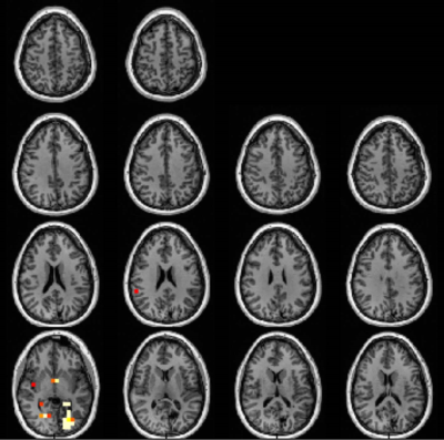 |
Rosette spectroscopic imaging of epilepsy at 3T
Jullie W Pan1, Arun Antony2, Victor W Yushmanov1, Claud W Schirda2, and Hoby P Hetherington1
1Radiology, University of Pittsburgh, Pittsburgh, PA, United States, 2UPMC, Pittsburgh, PA, United States
Our group has implemented the rosette trajectory at 3T with 3dimensional phase and Hadamard encoding to achieve multi- and whole-slice spectroscopic images covering a total longitudinal 60mm. These acquisitions are applied in n=25 surgical epilepsy patients (15 medial temporal lobe; 10 neocortical epilepsy), evaluating metabolic dysfunction in terms of overlap with surgical target and outcome. In 19/25 patients the MRSI identified gyral regions of overlap between the abnormal MRSI and surgical therapy. Based on a 4% cut segregation of fractional volume of MRSI abnormalities, the outcome ILAE score was significantly lower (better) in patients with <=4% abnormality compared to >4%.
|
|
1860.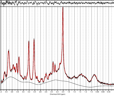 |
CDP-choline may influence metabolite levels in the anterior cingulate of people in remission of depression
Reggie Taylor1, Emma Lynn1,2, Pierre Blier1,2, Gerd Melkus3,4, Verner Knott1,2, and Natalia Jaworska1,2
1Institute of Mental Health Research, Ottawa, ON, Canada, 2Cellular and Molecular Medicine, University of Ottawa, Ottawa, ON, Canada, 3The Ottawa Hospital, Ottawa, ON, Canada, 4Radiology, University of Ottawa, Ottawa, ON, Canada
Cognitive deficits often persist beyond remission in major depressive disorder (MDD). CDP-choline has shown improvements in cognition in other populations characterized by cognitive deficits. These improvements may be due to interactions with the glutamatergic and cholinergic systems. The aim of this ongoing study was to assess the neurochemical response through magnetic resonance spectroscopy to the acute administration of CDP-choline compared with placebo in the anterior cingulate in a group of remitted MDD participants. Early results indicate possible relationships between glutamate and myo-inositol concentrations, and CDP-choline.
|
|
1861. |
N-Acetyl Aspartate and Myo-Inositol as biomarkers of HIV infection in the brain
Teddy Salan1, Sameer Vyas2, Paramjeet Singh2, Sulaiman Sheriff3, Mahendra Kumar4, and Varan Govind1
1Radiology, University of Miami, Miami, FL, United States, 2Postgraduate Institute for Medical Education & Research, Chandigarh, India, 3University of Miami, Miami, FL, United States, 4Psychiatry and Behavioral Sciences, University of Miami, Miami, FL, United States
The purpose of this study is to determine whether magnetic resonance spectroscopic imaging (MRSI) can identify metabolite biomarkers of HIV infection in the brain. Concentrations of N-acetyl aspartate (NAA), choline (CHO), creatine (CR), myo-inositol (MI), and glutamate and glutamine (GLX) were measured at the whole-brain level and compared between HIV+ and healthy control subjects. Significant reductions in NAA and increases in MI are observed throughout the brain, while the concentrations of other metabolites show no significant change.
|