
Digital Poster Session
Neuro: Preclinical Studies
Neuro
1916 -1930 Preclinical studies - Preclinical Studies: CNS Disease 1
1931 -1944 Preclinical studies - Preclinical Studies: CNS Disease 2
1945 -1960 Preclinical studies - Preclinical Studies: Technical Developments & Normal Brain
1916.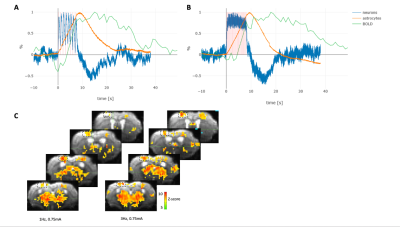 |
Investigating the Biological Basis of the BOLD fMRI Signal in Mice
Zhiva Skachokova1, Felix Schlegel1, Horea-Ioan Ioanas1, Aileen Schroeter1, and Markus Rudin1,2
1ETH Zurich, Zurich, Switzerland, 2University of Zurich, Zurich, Switzerland
How is the BOLD fMRI signal generated still remains unclear. By using a combination of simultaneous calcium imaging and fMRI, we studied the involvement of both astrocytes and neurons in blood flow regulation during electrical hindpaw stimulation in mice. The observed response duration varies based on the anesthesia used, however a strong correlation between astrocytic calcium and BOLD signal was present. Our method allows the study of neurovascular coupling mechanisms in the intact brain and points to the importance of astrocytes in this process, and can be further used to study BOLD signal alterations e.g. in models of neurodegeneration.
|
|
1917.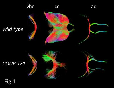 |
Quantitative white matter fibers in a mouse model of a neurodevelopmental disease: Insights from high spatial resolution 3D-DTI
Maxime Leclercq1, Jean Christophe Deloulme2, Michèle Bertacchi3, Michèle Studer3, and Hana Lahrech1
1BrainTech Lab Inserm U1205, Grenoble, France, 2GIN Inserm U836, Grenoble, France, 3iBV Inserm U1091, Nice, France
Microscopic 3D-DTI was applied to detect brain connection defects in COUP-TFI-mutant mice. Several tractography abnormalities were identified supporting a major role of COUP-TFI gene acting in the formation and guidance of forebrain commissures. DTI results are in agreement with those using fluorescent dyes, but identifies deficiencies of other cortical tracts not previously described. As COUP-TFI (NR2F1 in humans) mutations were also linked to a complex neurodevelopmental disease in humans, this work underlines the interest of 3D-DTI to study the whole brain in patients as those affected with Bosch-Boonstra-Schaaf optic-atrophy syndrome due to NR2F1 gene mutations/deletions, an emerging rare neurodevelopmental disease.
|
|
1918.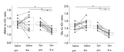 |
Detecting Synaptic Dysfunction caused by Pathological α-Synuclein Accumulation in Mouse Brain using MRS
Yuhei Takado1, Maiko Ono2, Keiichiro Minatohara2, Masafumi Shimojo2, Nobuhiro Nitta2, Sayaka Shibata2, Naruhiko Sahara2, Ichio Aoki2, Masato Hasegawa3, and Makoto Higuchi2
1Department of Functional Brain Imaging Research, National Institutes for Quantum and Radiological Science and Technology, Chiba, Japan, 2National Institutes for Quantum and Radiological Science and Technology, Chiba, Japan, 3Dementia Research Project, Tokyo Metropolitan Institute of Medical Science, Tokyo, Japan Poster Permission Withheld
To develop therapeutic strategies, in vivo detection of the early pathological changes in Parkinson’s disease is critically important. In this work, we aimed to detect early pathological changes caused by the propagation of α-synuclein in mouse brain using MRS. Recombinant α-synuclein and fibrils were prepared and injected into C57BL6 mice. 8 weeks after the injection, glutamate levels were decreased significantly compared to saline-injected control mice, which was in accordance with decreased synapsin staining in the cortex. We demonstrated that MRS can detect synaptic dysfunction caused by α-synuclein propagation in vivo.
|
|
 |
1919.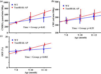 |
Age-related hyperactivity of brain oxygen metabolism in a novel Tau-APP mouse model of Alzheimer’s disease
Zhiliang Wei1,2, Jiadi Xu1,2, Kerstin E. Braunstein3, Lin Chen1,2, Tong Li3, Philip C. Wong3, and Hanzhang Lu1,2,4
1Russell H. Morgan Department of Radiology and Radiological Science, Johns Hopkins University School of Medicine, Baltimore, MD, United States, 2F.M. Kirby Research Center for Functional Brain Imaging, Kennedy Krieger Research Institute, Baltimore, MD, United States, 3Department of Pathology, Johns Hopkins University School of Medicine, Baltimore, MD, United States, 4Department of Biomedical Engineering, Johns Hopkins University School of Medicine, Baltimore, MD, United States
Alzheimer’s disease (AD) has been the leading cause of cognitive impairment and decline in elder individuals. Cross-sectional human studies have reported declined cerebral oxygen metabolism in AD patients tentatively attributed to reduced neuron cells. However, longitudinal change in oxygen metabolism remains unclear. We, therefore, performed a multi-modality (MRI, behavior test, and histology) study on a novel AD mouse model, dubbed as Tau4RΔK-AP, which largely mimics the pathological processes of tau-tangles and amyloid plaques as in human AD. Enhanced oxygen metabolism has been found in Tau4RΔK-AP mice, possibly indicating a compensatory response or an inefficiency of the brain energy consumption.
|
1920.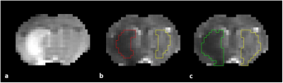 |
Human Mesenchymal Stem Cell Donor Effects on Apparent Diffusion Coefficient in a Rodent Stroke Model at 21.1 T
F. Andrew Bagdasarian1,2, Shannon Helsper1,2, Xuegang Yuan1,2, Jens T. Rosenberg1, and Samuel Colles Grant1,2
1National High Magnetic Field Laboratory, Florida State University, Tallahassee, FL, United States, 2Chemical & Biomedical Engineering, FAMU-FSU College of Engineering, Tallahassee, FL, United States
This study extends diffusion weighted imaging at 21.1-T to identify the pattern of potential recovery of apparent diffusion coefficient (ADC) in rodent ischemic stroke with novel stem cell therapy using dissociated aggregate human mesenchymal stem cells (d-hMSC) from different donors. All scanning was performed at 21.1 T with the goal of quantitatively assessing treatment efficacy longitudinally, spanning a 3 weeks post ischemia. Results show donor resultant ADC discrepancies at various time points and regions.
|
|
1921.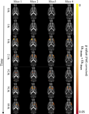 |
Long-term Structural Brain Changes in Adult Rats After Focal Ischemic Stroke
Warda Syeda1,2,3, Charlotte Ermine1, David Wright4, Vanessa Brait1, Lachlan Thompson1, Jess Nithianantharajah1, Scott Kolbe4, Leigh Johnston1,2, and Amy Brodtmann1
1The Florey Institute of Neuroscience and Mental Health, Melbourne, Australia, 2The Melbourne Brain Centre Imaging Unit, The University of Melbourne, Melborne, Australia, 3Melbourne Neuropsychiatry Centre, The University of Melbourne, Melborne, Australia, 4Department of Neuroscience, Monash University, Melbourne, Australia
Accumulating clinical evidence suggests remote neurodegeneration occurs in non-ischemic brain regions distant to the site of infarction in stroke, driven by both axonal degeneration and global brain inflammation. Prior preclinical studies of remote degeneration have mainly focused on brain changes over a few days or weeks post-stroke. We investigated long-term structural brain changes in an endothelin-1 model of mild focal ischemic stroke in rats, using a clinically relevant period of 48-weeks. Serial structural and diffusion-weighted MRI data were used to assess dynamic volume and white matter trajectories. We found significant cortical atrophy and white matter alterations, suggesting widespread stroke-related degenerations.
|
|
1922.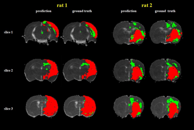 |
Machine-Learning-Based Segmentation of Ischemic Penumbra By Using Diffusion Tensor Metrics in a Rat Model
Cheng-Yu Chen1,2,3,4, Po-Chih Kuo5, Yung-Chieh Chen1, Yu-Chieh Jill Kao2, Ching-Yen Lee6, Hsiao-Wen Chung7, and Duen-Pang Kuo1
1Department of Medical Imaging, Taipei Medical University Hospital, Taipei, Taiwan, 2Translational Imaging Research Center, Taipei, Taiwan, 3Department of Radiology, School of Medicine, College of Medicine, Taipei Medical University, Taipei, Taiwan, 4Radiogenomic Research Center, Taipei Medical University Hospital, Taipei, Taiwan, 5Institute of Statistical Science, Academia Sinica, Taipei, Taiwan, 6TMU Research Center for Artifical Intelligence in Medicine, Taipei, Taiwan, 7Graduate Institute of Biomedical Electrics and Bioinformatics, National Taiwan University, Taipei, Taiwan
In the present study, we developed a 2-level classification model with an overall accuracy of 88.1 ± 6.7% for discriminating the stroke hemisphere into the infarct core (IC), ischemic penumbra (IP), and normal tissue regions on a voxel-wise basis in a permanent left middle cerebral artery occlusion model. According to the analysis results, we suggest that a single diffusion tensor imaging (DTI) sequence combined with machine learning (ML) algorithms can dichotomize ischemic tissue into the IC and IP, which are comparable to the conventional perfusion–diffusion mismatch.
|
|
1923.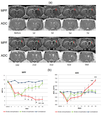 |
Macromolecular proton fraction as a marker of myelin recovery in ischemic stroke
Marina Khodanovich1, Ilya Gubskiy 2, Darya Namestnikova 2, Marina Kudabaeva1, Valentina Glazacheva1, Tatyana Anan'ina1, and Vasily Yarnykh1,3
1Tomsk State University, Tomsk, Russian Federation, 2Pirogov Russian National Research Medical University, Moscow, Russian Federation, 3University of Washington, Seattle, WA, United States
The study, performed on a rat model of ischemic stroke, aimed to evaluate a recently proposed myelin biomarker, macromolecular proton fraction (MPF) as a non-invasive tool for monitoring recovery after stroke. Longitudinal observations showed the different time courses of MPF evolution in the infarct zones undergoing subsequent demyelination (DZ) or remyelination (RZ). After a sharp decrease at days 1-5 after MCAO, MPF showed a further decline in the DZ and a restoration in the RZ to day 56. These findings were confirmed by histology which showed similar tissue evolution zones and enhancement of neurogenesis and oligodendrogenesis in the ischemic core.
|
|
1924.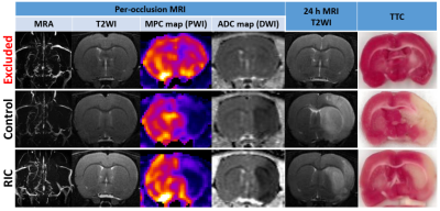 |
Remote ischemic conditioning in a rat model of acute ischemic stroke: a two-centre study with translational longitudinal MRI
Marlene Wiart1, Maryna Basalay2, Fabien Chauveau3, Chloe Dumot1, Christelle Leon1, Camille Amaz4, Radu Bolbos5, Diana Cash6, Eugene Kim6, Tae-Hee Cho7, Norbert Nighoghossian7, Sean Davidson2, Michel Ovize1, and Derek Yellon2
1Université Lyon, CarMeN laboratory, Inserm U1060, Lyon, France, 2The Hatter Cardiovascular Institute, London, United Kingdom, 3Université Lyon, Lyon Neuroscience Research Center, CNRS UMR5292, Inserm U1028, Lyon, France, 4Clinical Investigation Center, HCL, Lyon, France, 5CERMEP-Imagerie du Vivant, Bron, France, 6Neuroimaging, Institute of Psychiatry, Psychology and Neuroscience, King’s College London, London, United Kingdom, 7Stroke Medicine, Université Lyon, CREATIS CNRS UMR 5220-INSERM U1206, Lyon, France
The main objective of this study was to test the neuroprotective effects of remote ischemic conditioning (RIC: 4 cycles of 5-min hind limb ischemia interleaved with 5-min reperfusion) in a rat model of transient ischemic stroke (90 minutes) in a two-center study using translational MR imaging endpoints. Neuroscores and edema-corrected infarct size measured at 24h on T2-weighted MRI and expressed as percentage of the area at risk on per-occlusion MRI were significantly reduced in the RIC-treated group compared to the control group. The use of longitudinal MRI increases results robustness, which is greatly needed for successful RIC clinical translation.
|
|
1925.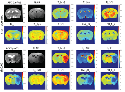 |
Changes in Quantitative Magnetization Transfer MR in the Mouse Brain After Transient Cerebral Ischemia
Grzegorz Kwiatkowski1, Georgios Louloudis1, Jan Klohs1, and Sebastian Kozerke2
1Institute for Biomedical Engineering, ETH Zurich, Zurich, Switzerland, 2ETH Zurich, Zurich, Switzerland
Quantitative analysis of magnetization transfer (qMT) based on a two-pool model was employed to characterized changes in the mouse brain following a transient middle cerebral artery occlusion (tMCAO) model of cerebral ischemia.The changes in qMT were compared to the standard MR metrics of an ischemic lesion (T1, T2, ADC, FLAIR) to examine the possible overlap of mechanisms affecting these magnetic resonance imaging contrasts. Notable changes of all MR metrics were found in the brain of the tMCAO group while only the qMT analysis revealed significant alterations in the sham-operated animals.
|
|
1926.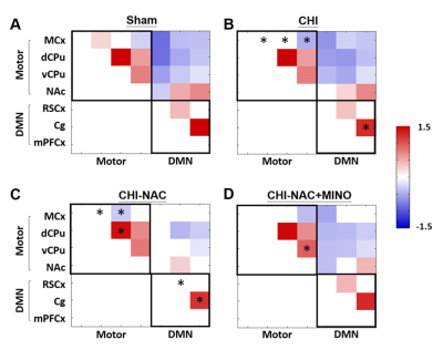 |
Functional Reorganization and Behavioral Recovery in Rats with Repetitive Closed-head Injury after Drug Treatment
Yu-Chieh Jill Kao1,2,3, Chia-Feng Lu4, Bao-Yu Hsieh5, and Cheng-Yu Chen1,2,3,6
1Neuroscience Research Center, Taipei Medical University, Taipei, Taiwan, 2Department of Radiology, School of Medicine, College of Medicine, Taipei Medical University, Taipei, Taiwan, 3Translational Imaging Research Center, Taipei Medical University Hospital, Taipei, Taiwan, 4Department of Biomedical Imaging and Radiological Sciences, National Yang-Ming University, Taipei, Taiwan, 5Department of Biomedical Imaging and Radiological Science, China Medical University, Taichung, Taiwan, 6Department of Medical Imaging, Taipei Medical University Hospital, Taipei, Taiwan
Significant improvement of locomotor activity and anxiety-like behavior along with reorganization of motor and default mode network was observed after NAC or NAC plus MINO treatment after repetitive closed-head injury, suggesting tentative treatment using drugs in patients with repetitive mild traumatic brain injury.
|
|
1927.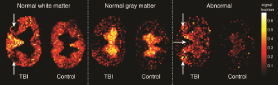 |
Highly specific and direct assessment of microscopic anisotropy following traumatic brain injury using diffusion correlation imaging (DCI)
Dan Benjamini1,2, Michal Komlosh1,2, Elizabeth Hutchinson3, and Peter Basser1
1National Institutes of Health, Bethesda, MD, United States, 2Uniformed Service University of the Health Sciences, Bethesda, MD, United States, 3The University of Arizona, Tucson, AZ, United States Diffusion MRI techniques that extend beyond diffusion tensor imaging (DTI) – including double diffusion encoding (DDE) – could provide more specific tools to probe abnormalities in disease or injury states. Here, DDE is applied to reveal the diffusion correlation spectrum on a voxelwise basis, which allows to directly assess the microscopic anisotropy in healthy and injured ferret spinal cords. |
|
1928.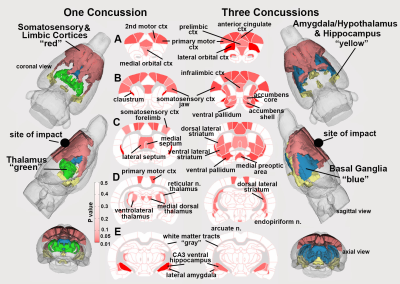 |
Imaging Changes in Blood Brain Barrier Permeability Following Repetitive Mild Traumatic Brain Injury.
Praveen Kulkarni1, Ju Qiao2, and Craig Ferris1
1Psychology, Northeastern University, Boston, MA, United States, 2A.A. Martinos Center for Biomedical Imaging, Massachusetts General Hospital, Charlestown, MA, United States
Failure in the blood brain barrier (BBB) lies at the foundation of small vessel disease that is precursor to neurodegenerative diseases. There are multiple risk factors leading to increased permeability in the BBB; one prevalent risk factor is repetitive mild TBI. Imaging the subtle changes in BBB permeability is not possible with standard imaging protocols. With quantitative ultra-short time-to-echo, contrast-enhanced (QUTE-CE) MRI, using the superparamagnetic iron oxide nanoparticle ferumoxytol, we can image changes in BBB permeability following rmTBI. With this novel methodology we can measure the immediate effects of rmTBI on changes in BBB permeability across the entire rat brain.
|
|
1929.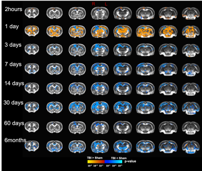 |
Diffusion tensor imaging reveals persistent microstructural alterations up to six month in an open-head traumatic brain injury
Abdalla Z Mohamed1 and Fatima Abdalla Nasrallah1
1Queensland Brain institute, The University of Queensland, Brisbane, Australia
Traumatic brain injury (TBI) is a severe problem worldwide. The non-invasive investigation of the microstructural alterations is of significant benefit for early diagnosis and interventions. In this study, we applied diffusion tensor imaging (DTI) to monitor the longitudinal microstructural changes in a controlled cortical impact rodent model of TBI from 2 hours and up to 6 months post-injury. Using DTI, we observe ongoing white matter changes following TBI, that initiate in corpus callosum and injury location at early timepoints and persists to exist up to 6 months, suggesting the temporal sensitivity of DTI to detect ongoing microstructural changes following TBI.
|
|
 |
1930.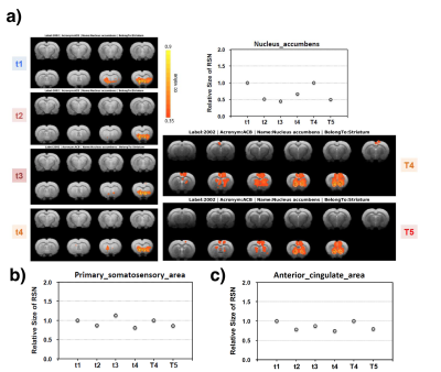 |
Longitudinal Study of Resting State Connectivity in a Rat Model of Alcohol Use Disorder at Ultrahigh Fields
Hannes M. Wiesner1, Wei Zhu1, Yi Zhang1, Manuel Esguerra2, Colleen Hutchison2, Mark J. Thomas2, Xiao-Hong Zhu1, and Wei Chen1
1Department of Radiology, CMRR, University of Minnesota Medical School, Minneapolis, MN, United States, 2Departments of Neuroscience and Psychology, University of Minnesota, Minneapolis, MN, United States Poster Permission Withheld
Resting state fMRI (rs-fMRI) has been used to investigate alcohol use disorder (AUD), mostly in humans; and a wide array of commonly induced neurological changes due to acute and chronic use have been reported. In this work, we established a rat model of AUD to longitudinally study the effects of chronic alcohol abuse on the functional brain connectivity using rs-fMRI at ultrahigh fields. Preliminary results revealed potential changes in reward-processing circuit connectivity and global brain activity. Further research is advised to quantify early effects observed in and between different neural structures (NAc/IBST/PreL/IL and ACC) during withdrawal stress and relapse.
|
 View the Poster
 Watch the Video
1931.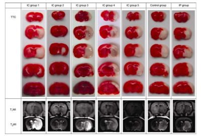 |
Simple diffusion delivery via brain interstitial route for the treatment of cerebral ischemia
Yu Song1,2, Hongbin Han2,3,4, Qingwei Song1, Yajuan Gao2,3,4, Rui Wang2,3,4, Xianjie Cai2,3,4, Yumeng Cheng2,3,4, and Zeqing Tang2,3,4
1Department of Radiology, the First Affiliated Hospital of Dalian Medical University, Dalian, China, 2Key Laboratory of Magnetic Resonance Imaging Equipment and Technique, Beijing, China, 3Department of Radiology, Peking University Third Hospital, Beijing, China, 4Institute of Medical Technology(IMT)of Peking University Health Science Center(PKUHSC), Beijing, China
Delivering pharmacologic agents directly into the brain has been proposed as a means of bypassing the blood brain barrier. Within this study we propose a novel system for delivering drugs into the brain named the simple diffusion (SDD) system. To validate this technique, rats were subjected to a sin- gle intracranial (at the caudate nucleus), or intraperitoneal injection, of the compound citicoline, followed two hours later by a permanent middle cerebral artery occlusion (pMCAO). These results suggest that given the appropriate injection point, through SDD a pharmacologically effective concentration of citicoline can be administered.
|
|
1932.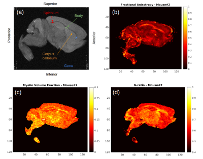 |
White Matter Microstructural Changes in an Absence Epilepsy Mouse Model
Gustavo Chau Loo Kung1, Juliet Knowles2, Erpeng Dai3, John Huguenard2, Michelle Monje2, and Jennifer McNab3
1Bioengineering Department, Stanford University, Stanford, CA, United States, 2Neurology Department, Stanford University, Stanford, CA, United States, 3Radiology Department, Stanford University, Stanford, CA, United States
A previously unexplored possibility is that maladaptive myelination may contribute to both the predisposition to seizures and cognitive impairment in diseases such as absence epilepsy. This work presents MRI white matter microstructural measurements in ex vivo mouse brains from the Scn8amed+/- model of absence epilepsy. We obtained estimates of fractional anisotropy, myelin volume fraction and g-ratio. Our results are suggestive of thicker myelin sheaths, which is consistent with g-ratio data obtained by electron microscopy. We look to expand the current analysis to perform longitudinal measurements of the evolution of this particular type of epilepsy and its interplay with aberrant myelination.
|
|
1933.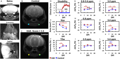 |
Chemical Exchange Saturation Transfer MRI of Disease-Associated Lymph Nodes in a Mouse Model of Multiple Sclerosis
Aline M. Thomas1,2, Peter A. Calabresi3,4, Michael T. McMahon1,5, Peter C.M. van Zijl1,5, and Jeff W.M. Bulte1,2
1Russell H. Morgan Department of Radiology and Radiological Science, Johns Hopkins University School of Medicine, Baltimore, MD, United States, 2Institute for Cell Engineering, Imaging Section and Vascular Biology Program, Johns Hopkins University School of Medicine, Baltimore, MD, United States, 3Department of Neurology, Johns Hopkins University School of Medicine, Baltimore, MD, United States, 4Solomon H. Snyder Department of Neuroscience, Johns Hopkins University School of Medicine, Baltimore, MD, United States, 5Department of Radiology, Kennedy Krieger Institute, Baltimore, MD, United States
In multiple sclerosis (MS), immune cells damage the brain and spinal cord, often causing irreversible disability. Current imaging strategies visualize the resulting damage, but the heterogeneity of the damage observed complicates image interpretation. Evidence has emerged that the immunological attacks in MS are initiated in central nervous system-draining lymph nodes. Here, we demonstrate the potential of chemical exchange saturation transfer (CEST) MRI to monitor changes in these lymph nodes as disability progressed in a mouse model of experimental autoimmune encephalomyelitis.
|
|
1934.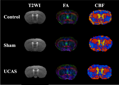 |
Association of white matter deficits with cerebral blood flow in cerebral hypoperfusion mouse model as revealed by in-vivo multi-modal MRI
Fangrong Zong1, Yan Zhuo2,3, Chengya Dong4, Baoshan Qiu4, Shunyin Zhao4, Baogui Zhang5, Yilong Wang4, and Xiangrong Liu4
1Centre for Advanced Magnetic Resonance Research, Institute of Biophysics, Chinese Academy of Sciences, Beijing, China, 2State Key Laboratory of Brain and Cognitive Science, Institute of Biophysics, Chinese Academy of Sciences, Beijing, China, 3The Innovation Center of Excellence on Brain Science, Chinese Academy of Sciences, Beijing, China, 4China National Clinical Research Center for Neurological Diseases, Beijing, China, Beijing, China, 5Brainnetome Center, Institute of Automation, Chinese Academy of Sciences, Beijing, China
The underlying mechanism of vascular dementia remains unclear which leads to difficulties in developing specific disease treatments. This contribution firstly developed a multi-modal MRI approach on a mouse model with unilateral carotid artery stenosis as non-invasive measures. A positive correlation was built between fractional anisotropy and cerebral blood flow under hypoperfusion conditions, which provides insights into understanding the pathological mechanism of vascular dementia.
|
|
1935.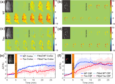 |
Functioning of the glucose transporter and glymphatic systems in the tauopathy AD mouse brain studied by onVDMP MRI and D-glucose infusion
Lin Chen1,2, Zhiliang Wei1,2, Kannie W.Y. Chan1,2,3, Jianpan Huang4, Xiang Xu1,2, Philip C. Wong5,6, Hanzhang Lu1,2, Peter C.M. van Zijl1,2, Tong Li5,6, and Jiadi Xu1,2
1Department of Radiology and Radiological Science, The Johns Hopkins University School of Medicine, Baltimore, MD, United States, 2F.M. Kirby Research Center for Functional Brain Imaging, Kennedy Krieger Research Institute, Baltimore, MD, United States, 3Department of Biomedical Engineering, City University of Hong Kong, Hong Kong, China, 4City University of Hong Kong, Hong Kong, China, 5Department of Pathology, The Johns Hopkins University School of Medicine, Baltimore, MD, United States, 6Department of Neuroscience, Johns Hopkins University School of Medicine, Baltimore, MD, United States
In this study, we used onVDMP MRI to detect glucose uptake in tauopathy Alzheimer's disease (AD) mouse brain. Compared to wild-type mice, significantly reduced glucose uptake was observed in both cerebrospinal fluid (CSF) and parenchyma of AD mouse brain. Clearance of glucose through CSF was found in wild-type mice, but not in AD mice, which implicates dysfunction of the glymphatic system in AD mouse brain. The results in this study suggest that onVDMP MRI could be a cost-effective and widely available method for evaluating the functions of glucose transporter and glymphatic system, and hence diagnosing AD.
|
|
|
1936.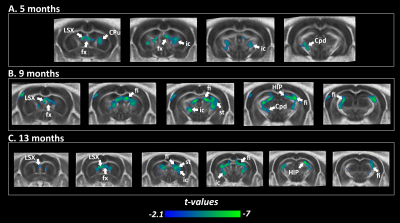 |
Longitudinal brain connectome reorganization in a tauopathy mouse model of Alzheimer's disease
Laetitia Degiorgis1, Marion Sourty1,2, Julien Lamy1, Vincent Noblet1, Meltem Karatas1,3,4, Thomas Bienert4, Marco Reisert4, Anne-Laurence Boutillier5, Jean-Paul Armspach1, Frédéric Blanc1,6, and Laura Harsan1,7
1University of Strasbourg and CNRS, ICube Laboratory UMR 7357, Strasbourg, France, 2The University of Sydney, Faculty of Engineering, School of Aerospace, Mechanical and Mechatronic Engineering, Sydney, Australia, 3CNRS, University of Strasbourg, INCI, UMR 7168, Strasbourg, France, 4Department of Radiology, Medical Physics, University Medical Center Freiburg, Freiburg, Germany, 5Laboratoire de Neuroscience Cognitives et Adaptatives, Strasbourg, France, 6University Hospital of Strasbourg, CM2R (Memory Resource and Research Centre), Day Hospital, Geriatrics Department, Strasbourg, France, 7Department of Biophysics and Nuclear Medicine, University Hospital of Strasbourg, Strasbourg, France Poster Permission Withheld
MRI is a unique tool to understand the complexity of the brain functional and structural communication evolution over time. Among the main mechanisms of AD, tauopathy remains poorly studied in preclinical imaging. We used graph theory approaches and DTI analysis in a longitudinal study of Thy-Tau22 mice, associated with behavioral evaluation. Alterations of the cholinergic septal circuitry, supporting memory and emotional processes, were found as the main hallmark of the progression of the pathology, associated with default mode network dysfunction, both starting before the first memory deficits.
|
1937. |
Optogenetic modulation and rsfMRI mapping of depression related functional brain networks in the mouse
Laura-Adela Harsan1, Laetitia Degiorgis1, Julien Todeschi2, Lea Becker3, Maxence Thomas de la Pintière1, Victor Mathis3, Chrystelle Po1, and Ipek Yalcin3
1ICube: Engineering science, computer science and imaging laboratory, University of Strasbourg, Strasbourg, France, 2ICube: Engineering science, computer science and imaging laboratory, Neurosurgery Department, University Hospital Strasbourg, University of Strasbourg, Strasbourg, France, 3Institute de Neurosciences Cellulaires et Intégratives, University of Strasbourg-CNRS, Strasbourg, France
The main objective of this study is to modulate and map the anterior cyngulate cortex (ACC) functional connectivity (FC) pathways underlying depression development in a mouse model. We use the optogenetic approaches to create the depression phenotype in mice, by activating the pyramidal ACC neurons expressing Channel rhodopsin 2 (ChR). We further use resting state functional MRI (rsfMRI) as non-invasive read-out of the effects at the level of functional brain connectivity. Four consecutive sessions of optogenetic ACC stimulation induced strong depression phenotype and major modifications of the functional mouse brain connectivity including perturbed mesocorticolimbic pathways and default mode network patterns
|
|
1938.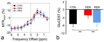 |
Temporal alterations of GluCEST contrast in a rat model of cuprizone-induced demyelination and remyelination
Do-Wan Lee1, Chul-Woong Woo2, Hwon Heo3, Jae-Im Kwon2, Yeon Ji Chae3, Su Jung Ham4, Jeong Kon Kim1, Kyung Won Kim1,4, Dong-Cheol Woo2,3, and Dong-Hoon Lee5
1Department of Radiology, Asan Medical Center, University of Ulsan College of Medicine, Seoul, Republic of Korea, 2Convergence Medicine Research Center, Asan Institute for Life Sciences, Asan Medical Center, Seoul, Republic of Korea, 3Department of Convergence Medicine, Asan Medical Center, University of Ulsan College of Medicine, Seoul, Republic of Korea, 4Asan Image Research, Asan Institute for Life Sciences, Asan Medical Center, Seoul, Republic of Korea, 5Faculty of Health Sciences and Brain & Mind Centre, The University of Sydney, Sydney, Australia
Research on changes to glutamate signaling in the white matter of demyelinating diseases may provide important biophysical information for diagnostic and prognostic assessment. We attempted to evaluate glutamate signals in a cuprizone-induced rat model of demyelination by GluCEST imaging. GluCEST imaging provides in vivo image contrast of changing glutamate concentrations during demyelination and subsequent remyelination. We also performed histological validation to analyze the state of myelinated axons. GluCEST imaging could be a useful tool to evaluate brain metabolism in demyelination and remyelination models and provide quantitative results that are highly representative of changes to glutamate levels in vivo.
|
|
1939.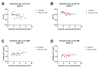 |
MR elastography and DTI for monitoring microstructural changes in the developing brain of Poly (I:C)-induced maternal immune activated rats.
Lucy Liu1,2, Andre Bongers2, Lynne Bilston1,2, and Lauriane Jugé1,2
1Neuroscience Research Australia, Sydney, Australia, 2University of New South Wales, Sydney, Australia
Maternal infection during pregnancy can cause enlarged ventricles and compromise brain microstructure, which conventional imaging techniques cannot identify. We investigated the potential of magnetic resonance elastography (MRE) and diffusion tensor imaging (DTI) in detecting brain microstructural changes in offspring of Poly (I:C)-induced maternal immune activated rats. Results showed that DTI and MRE were sensitive to neurodevelopmental microstructural changes, but not to subtle longitudinal microstructural changes related to maternal infection. DTI showed that white matter injury differs between perinatal and adolescent Poly(I:C) rats, while MRE was not sensitive enough to detect subtle compromise of gray matter microstructure in Poly (I:C) rats.
|
|
1940.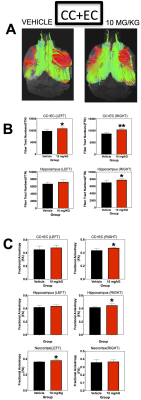 |
High Resolution Volumetric and Connectomic Analyses in a Mouse Model of CNS Lupus
Talaignair N Venkatraman1, Haichen Wang2, Chris Petty1, Allen W Song1, and Christopher D Lascola1
1Radiology, Duke University Medical Center, Durham, NC, United States, 2Neurology, Duke University Medical Center, Durham, NC, United States
Ex-Vivo high resolution (3D) volumetric, tractographic and connectomic analysis carried out on a mouse model of CNS Lupus at 7T. The results show strong therapeutic effect of a novel QR II inhibitor.
|
|
1941.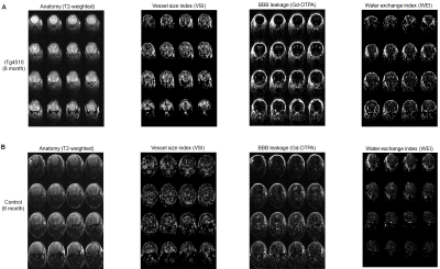 |
MRI assessment of vascular disruption in tau pathology animal model (rTg4510 mouse) of Alzheimer disease
Kwangyeol Baek1, Rachel Bennett2, Bradley Hyman2, Woo Hyun Shim3, and Young Ro Kim4,5
1School of Biomedical Convergence Engineering, Pusan National University, Busan, Republic of Korea, 2Department of Neurology, Massachusetts General Hospital and Harvard Medical School,, Boston, MA, United States, 3Department of Radiology, Asan Medical Center, Seoul, Korea, Republic of, 4Athinoula A. Martinos Center for Biomedical Imaging, Massachusetts General Hospital, Charlestown, MA, United States, 5Department of Radiology, Harvard Medical School, Boston, MA, United States
We assessed vascular disruption in rTg4510 mouse (age of 6 and 9 months), a tau expressing transgenic animal model of Alzheimer’s disease, using MRI metrics such as vessel size index (VSI), Gd-DTPA leakge through BBB and water exchange index (WEI). The rTg4510 mice showed age-dependent expansion of the hippocampal lesion and profound vascular alteration around the hippocampal lesion: Increase in VSI, mild Gd-DTPA leakage and increased WEI. Our findings suggest the role of vascular components in development of Alzheimer’s disease with tau pathology. Vascular MRI assessments might have a potential application in early diagnosis and monitoring of Alzheimer’s disease.
|
|
1942. |
Neurite orientation dispersion and density imaging of rat brain microstructural changes due to middle cerebral artery occlusion at a 3T MRI
Zhenxiong Wang1,2, Wenzhen Zhu1, Shun Zhang1, Guiling Zhang1, Mehran Shaghaghi2, and Kejia Cai2
1Tongji Hospital, Tongji Medical College, Huazhong University of Science and Technology, Wuhan, China, 2Departments of Radiology, Department of Bioengineering, and the Center for MR Research, Chicago, IL, United States
In this study we demonstrated the feasibility of longitudinal neurite orientation dispersion and density imaging (NODDI) in characterizing the microstructural alterations in rat brain tissues due to middle cerebral artery occlusion at a 3T MRI and validated the NODDI parameters with histology.
|
|
1943. |
Cerebral metabolic rate at three hours post injury predicts 24-hour neurological outcome in a rat model of cardiac arrest
Zhiliang Wei1,2, Qihong Wang3, Sung-Min Cho4, Romergryko Geocadin4, Hiren R. Modi3, Nitish V. Thakor3, and Hanzhang Lu1,2,3
1Russell H. Morgan Department of Radiology and Radiological Science, Johns Hopkins University School of Medicine, Baltimore, MD, United States, 2F.M. Kirby Research Center for Functional Brain Imaging, Kennedy Krieger Research Institute, Baltimore, MD, United States, 3Department of Biomedical Engineering, Johns Hopkins University School of Medicine, Baltimore, MD, United States, 4Department of Neurology, Johns Hopkins University School of Medicine, Baltimore, MD, United States
Cardiac arrest (CA) is associated with low survival rate and unfavorable outcomes despite maximal medical care. For determining the timing of acute brain injury and delivering aggressive intervention at early stage to improve neurologic outcomes, an early-stage biomarker is compulsory. Here, we utilized MRI techniques to reveal the temporal trajectories of brain’s blood supply, oxygenation, and energy consumption in the first few hours following return-of-spontaneous-circulation, and found early physiologic measure is associated with 24-h neurologic deficit score. This finding may potentially facilitate the future research on CA management by providing a sensitive physiologic biomarker to determine appropriate medical intervention.
|
|
1944. |
Early postoperative cerebral perfusion changes after direct revascularization surgery in moyamoya disease based on perfusion territory study
Jing Yuan1, Zhizheng Zhuo1, Bing Wu2, and Yaou Liu1
1Beijing Tiantan Hospital, Capital Medical University, Beijing, China, 2GE Healthcare, China, Beijing, China
Early postoperative cerebral perfusion changes after direct revascularization surgery in moyamoya disease were studied. The preoperative and postoperative perfusion parameters from ASL and CT perfusion studied were compared based on perfusion territory showed by ssASL technique. Two types of perfusion changes were found, group I represented perfusion territory redistribution and group II represented perfusion improvement.
|
1945.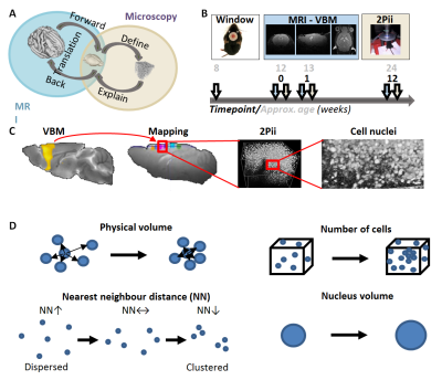 |
Cellular correlates of gray matter volume changes identified with two-photon microscopy and magnetic resonance morphometry in the mouse brain
Claudia Falfán-Melgoza1, Livia Asan2, Johannes Knabbe2, Carlo Beretta2,3, Thomas Kuner2, and Wolfgang Weber-Fahr1
1RG Translational Imaging, Central Institute of Mental Heath, Mannheim, Germany, 2Department of Functional Neuroanatomy, Institute for Anatomy and Cell Biology, Heidelberg University, Heidelberg, Germany, 3CellNetworks Math-Clinic, Bioquant BQ001, Heidelberg University, Heidelberg, Germany
Insights gained from Voxel Based morphometry (VBM) tremendously advance the understanding of neurologic and psychiatric diseases. However, the cellular basis of VBM changes remains largely unclear. We used longitudinal two-photon fluorescence and magnetic resonance imaging in mice to explore the cellular basis of VBM. Our data shows that MRI volume changes are only limited reflected by physical volume changes, yet dominated by cellular composition and cytoarchitectural characteristics. This has great implications for findings in neuroimaging in general, and the novel approach introduced by this study can be applied to various disease models to potentially unravel key mechanisms of brain pathophysiology.
|
|
1946.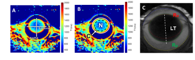 |
In-vivo measurements of physiological optics of mouse crystallin lens using MRI
Xingzheng Pan1, Eric R. Muir2, Paul J. Donaldson1,3, Ehsan Vaghefi1, Zhao Jiang2, Caterina Sellitto4, and Thomas W. White4
1School of Optometry and Vision Science, University of Auckland, Auckland, New Zealand, 2Department of Radiology, School of Medicine, Stony Brook University, Stony Brook, NY, United States, 3Department of Physiology, School of Medical Sciences, University of Auckland, Auckland, New Zealand, 4Department of Physiology & Biophysics, School of Medicine, Stony Brook University, Stony Brook, NY, United States
The physiological optics of the crystallin lens depend on its water content, water-bound protein ratios and surface geometry1,2. To maintain these properties, the lens generate a circulating flux of ions that actively removes water from the lens center via an intracellular pathway mediated by gap junction channels3. In this study, we established and optimised T1&T2 mapping and structural scan to study the physiological optics the mouse lens at 7T. These protocols were then applied to a transgenic mouse model in which we have genetically modified the number of gap junction channels to alter the removal of water from the lens.
|
|
1947.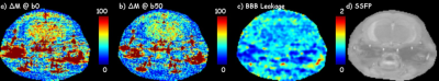 |
Implementation and optimization of steady state free precession MRI for mapping BBB leakage in mouse brain at 9.4T
Frederick C. Damen1, Steve Zaldua2, Jin Gao3, Weiguo Li3, Leon Tai2, and Kejia Cai1
1Radiology, University of Illinois at Chicago, CHICAGO, IL, United States, 2Anatomy and Cell Biology, University of Illinois at Chicago, CHICAGO, IL, United States, 3Research Resources Center, University of Illinois at Chicago, CHICAGO, IL, United States
In neurodegenerative diseases, e.g., Alzheimer’s disease, it is important to study cerebral blood flow (CBF) and blood brain barrier (BBB) permeability. Arterial Spin Labeling (ASL), used to measure CBF, is hampered by physiological variability, requiring many averages, and thus, fast imaging. Echo Planner Imaging, the most used fast imaging method, suffers from massive distortions and blurring for mice brain imaging at high field MRI. Alternatively, in this study we successfully implemented steady state free precession fast imaging for the measurement of the BBB leakage in mouse brain at 9.4T.
|
|
1948.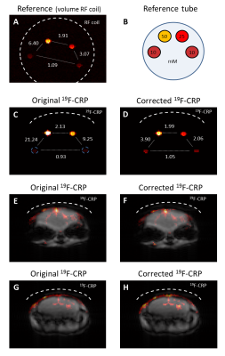 |
Quantitative in vivo 19F MRI of mouse brain inflammation using a cryogenic RF surface probe and RARE
Paula Ramos Delgado1, Christian Prinz1, Jason M. Millward1, Helmar Waiczies2, Ludger Starke1, Joao Periquito1, Laura Boehmert1, Thoralf Niendorf1,3, Andreas Pohlmann1, and Sonia Waiczies1
1Berlin Ultrahigh Field Facility (B.U.F.F), Max Delbrück Center for Molecular Medicine in the Helmholtz Association, Berlin, Germany, 2MRI.tools GmbH, Berlin, Germany, 3Experimental and Clinical Research Center, a joint cooperation between the Charité Medical Faculty and the Max Delbrück Center for Molecular Medicine in the Helmholtz Association, Berlin, Germany
The low SNR inherent to fluorine (19F) MRI necessitates sensitivity-enhancing methods. SNR-efficient imaging techniques such as RARE and SNR-enhancing cryogenically-cooled RF coils (CRP) create new challenges. The strong spatially-varying B1 fields of transceive surface RF coils hamper quantification and no analytical signal intensity equation for B1+ correction exists for RARE. We developed a B1 correction method that makes use of experimental data to model the signal intensity from RARE and we established a workflow to correct and quantify 19F MR signals originating from inflammatory regions of the mouse brain that were acquired using a 19F-CRP.
|
|
1949.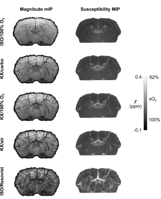 |
Effects of anesthetic conditions on the mouse cerebral oxygen saturation measured by QSM
Jérémie P. Fouquet1, Luc Tremblay1, Andreas Deistung2, Renat Sibgatulin3, Martin Krämer3, Karl-Heinz Herrmann3, Réjean Lebel1, Jürgen R. Reichenbach3, and Martin Lepage1
1Sherbrooke Molecular Imaging Center (CIMS), Department of Nuclear Medicine and Radiobiology, Université de Sherbrooke, Sherbrooke, QC, Canada, 2Faculty of Medicine, Universitätsklinikum Halle, Halle, Germany, 3Medical Physics Group, Institute of Diagnostic and Interventional Radiology, Jena University Hospital - Friedrich Schiller University Jena, Jena, Germany
Anesthetic conditions may have important effects on the blood oxygen saturation in rodents. However, the size of those effects has not been directly quantified in the mouse brain. We use Quantitative Susceptibility Mapping to provide a direct estimate of the oxygen saturation in the mouse brain under anesthesia. Three commonly used anesthetics, namely isoflurane, dexmedetomidine, and ketamine-xylazine, are evaluated. The effect of breathing gas is also evaluated.
|
|
1950.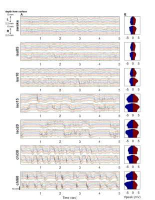 |
Effect of anesthetics on spontaneous neural activity and functional connectivity in the bilateral somatosensory cortices in rats
Kwangyeol Baek1, Chae Ri Park2, Siwan Jang3, Woo Hyun Shim2, and Young Ro Kim1
1Massachusetts General Hospital, Boston, MA, United States, 2Asan Medical Center, Seoul, Korea, Republic of, 3Portsmouth Abbey School, Portsmouth, RI, United States
Resting state fMRI study in animal model often involved use of anesthetics, but the effect of anesthetics on the spontaneous neural activity is not well known. We investigated how different types and doses of anesthetics (isoflurane and α-chloralose) influence the LFP activity and functional connectivity in comparison with the awake state. We observed distinct effects of these anesthetics on spontaneous neural activity (e.g. burst-suppression pattern and nonspecific correlation for high dose of isoflurane) and evoked response (e.g. potentified sensory-evoked response in bilateral cortices with α-chloralose). The effect of anesthetics should be taken into account in animal resting state fMRI studies.
|
|
1951.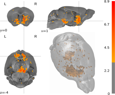 |
A Whole-Brain Map and Assay Parameter Analysisof Mouse VTA Dopaminergic Activation
Horea-Ioan Ioanas1, Bechara John Saab2, and Markus Rudin1
1Institute for Biomedical Engineering, ETH and University of Zurich, Zurich, Switzerland, 2Preclinical Laboratory for Translational Research into Affective Disorder, University of Zurich Psychiatric Hospital, Zurich, Switzerland
The definition of neurophenotypes for the function of key neurotransmitter systems is vital to the improvement of drug development in psychopharmacology. In this study we present a whole-brain map of stimulus-evoked VTA dopaminergic function in mice, alongside a parameter analysis for this novel assay. This study shows overall coherence between functional and known structural connectivity, but provides additional evidence for functional connectivity towards the dorsal striatum (first indicated in the rat model). We propose that this thoroughly explored new assay can provide an invaluable read-out for dopaminergic dysfunction and dopaminergic intervention modelling.
|
|
1952.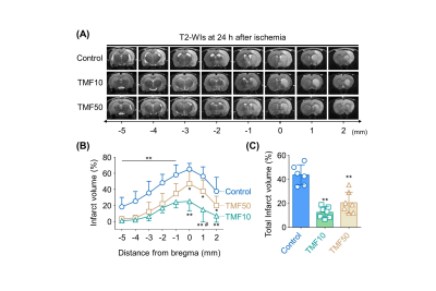 |
Aryl hydrocarbon receptor antagonism before reperfusion attenuates cerebral ischemia/reperfusion injury in rats
Jae-Im Kwon1, Hwon Heo2, Sang Tae Kim1, Su Jeong Ham1, Yeon Ji Chae1, Young Jin Kim1, Do-Wan Lee2, Kyung Won Kim3, Dong Cheol Woo1,2, and Chul-Woong Woo1
1Asan Institute for Life Sciences, Asan Medical Center, Seoul, Korea, Republic of, 2Convergence Medicine, University of Ulsan College of Medicine, Seoul, Korea, Republic of, 3Radiology, Asan Medical Center, Seoul, Korea, Republic of Poster Permission Withheld
Emerging evidence suggests that aryl hydrocarbon receptor (AhR) antagonism could attenuate neuronal damage caused by transient ischemic stroke. However, the optimal timing of AhR antagonist administration for maximum neuroprotective efficacy has yet to be established. The present study explored this issue via MRIs in rats with transient ischemic stroke and demonstrated that infarct volume and apoptosis were alleviated in rats that received AhR antagonists 10 or 50 minutes after ischemia. In addition, the timing of AhR antagonist administration was shown to affect edema formation caused by transient ischemic stroke.
|
|
1953.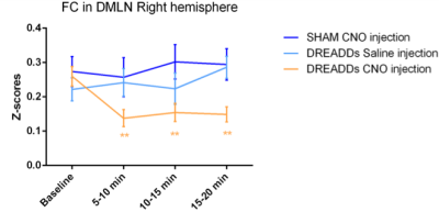 |
Stimulation of basal forebrain cholinergic neurons induces decreased functional connectivity in the default mode-like network in rats
Lore M. Peeters1, Monica Van den Berg1, Rukun Hinz1, and Georgios A. Keliris1
Video Permission Withheld
1Bio-Imaging Lab, University of Antwerp, Antwerp, Belgium Understanding the role of the basal forebrain (BFB) in controlling the dynamics of brain processes is of great interest. We aimed to investigate the influence of selective unilateral stimulation of BFB cholinergic neurons on functional connectivity (FC) in the rodent default-mode like network (DMLN). By combining resting-state functional MRI (rsfMRI) with chemogenetics, we demonstrate that stimulation of the cholinergic neurons in the right BFB significantly decreased right intra-hemispheric and inter-hemispheric FC in the DMLN in rats. These findings provide new critical insights into the interplay between attentional networks and DMLN in rodents. |
|
1954.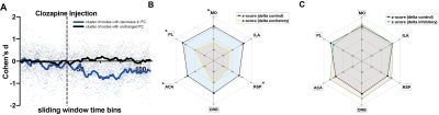 |
Chemogenetic neuromodulation of striato-cortical direct pathway projections cause alterations in brain-wide network connectivity
Marija Markicevic1, Oliver Sturman 2, Johannes Bohacek2, Markus Rudin3,4, Nicole Wenderoth1, and Valerio Zerbi1
1Neural Control of Movement lab, DHEST, ETH Zurich, Zurich, Switzerland, 2Laboratory of Molecular and Behavioural Neuroscience, DHEST, ETH Zurich, Zurich, Switzerland, 3Institute for Biomedical Engineering, University and ETH Zurich, Zurich, Switzerland, 4Institute of Pharmacology and Toxicology, University of Zurich, Zurich, Switzerland
RsfMRI is used to investigate healthy and diseased brains via temporal correlations of distinct brain regions i.e. functional connectivity (FC). However, this gives little insight into the contribution of specific cell-types to the recorded signals. To tackle this, we measured rsfMRI in mice while chemogenetically exciting or inhibiting D1 expressing cells in dorsomedial striatum (dmCP) via DREADDs. Initial results illustrate opposing effects of excitation and inhibition on FC, observed only within anatomically connected nodes of corticostriatal circuitry. These results are the first, necessary step for further disentangling the role of dmCP on motor control and behavior.
|
|
1955.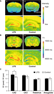 |
In vivo detection of neuroinflammation in rats using Manganese-Enhanced MRI (MEMRI)
Satoshi Fujiwara1, Sosuke Yoshinaga1, Shigeto Iwamoto1, Sayaka Shibata2, Aiko Sekita2, Nobuhiro Nitta2, Tsuneo Saga2, Ichio Aoki2, and Hiroaki Terasawa1
1Faculty of Life Sciences, Kumamoto University, Kumamoto, Japan, 2Institute for Quantum Life Science, National Institutes for Quantum and Radiological Science and Technology (QST), Chiba, Japan Poster Permission Withheld
Neuroinflammation is initiated by many types of neural disorders as a defensive response of the innate immune system in the central nervous system (CNS). Neuroinflammation is typically accompanied by the disruption of Ca2+ homeostasis. Manganese chloride (MnCl2) is a useful positive MRI contrast agent that enters activated cells through Ca2+ channels, and is utilized in Manganese-Enhanced MRI (MEMRI) for functional neuroimaging. We sought to determine whether MEMRI could be used to assess the cellular/molecular alterations caused by acute neuroinflammation in vivo, by focusing on Mn2+ accumulation in the rodent brain.
|
|
1956. |
Regional Mapping of Glutamate Distributions in a Healthy Rat Brain Using Glutamate Chemical Exchange Saturation Transfer (GluCEST) MRI
Do-Wan Lee1, Hwon Heo2, Chul-Woong Woo3, Jae-Im Kwon3, Su Jung Ham4, Yeon Ji Chae2, Kyung Won Kim1,4, Jeong Kon Kim1, Dong-Cheol Woo2,3, and Dong-Hoon Lee5
1Department of Radiology, Asan Medical Center, University of Ulsan College of Medicine, Seoul, Republic of Korea, 2Department of Convergence Medicine, Asan Medical Center, University of Ulsan College of Medicine, Seoul, Republic of Korea, 3Convergence Medicine Research Center, Asan Institute for Life Sciences, Asan Medical Center, Seoul, Republic of Korea, 4Asan Image Research, Asan Institute for Life Sciences, Asan Medical Center, Seoul, Republic of Korea, 5Faculty of Health Sciences and Brain & Mind Centre, The University of Sydney, Sydney, Australia
Detecting glutamate signals in vivo within the brain may have a diagnostic potential. Recently, with many translational research efforts leading to clinical trials, presenting the normal in vivo glutamate distribution in pre-clinical data to establish a database for pre-clinical studies can be valuable. In this abstract, we investigated glutamate signal distributions in multiple brain regions of a healthy rat brain using GluCEST imaging. Quantified GluCEST signals showed significant differences between white and gray matter regions. Our findings and investigations yield a valuable database and insights for comparing glutamate signal changes in pre-clinical brain diseases.
|
|
1957.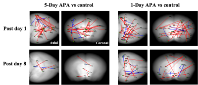 |
Training intensity induced differential functional networks in rodent
Zengmin Li1, Hsu-Lei Lee1,2, Elizabeth Coulson1,3, Pankaj Sah1, Patricio Opazo1, and Kai-Hsiang Chuang1,2
1Queensland Brain Institute, The University of Queensland, St Lucia, Australia, 2Centre for Advance Imaging, The University of Queensland, St Lucia, Australia, 3School of Biomedical Sciences, The University of Queensland, St Lucia, Australia · After spatial learning, large-scale plasticity in functional networks were detected in mice using resting-state fMRI. · Different training intensity recruited different functional networks. · Functional connectivity reorganized to be less hippocampus dependent after 1 week of consolidation. |
|
1958.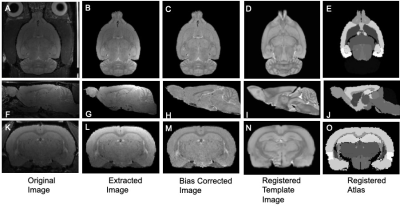 |
Quantitative MRI Volumetric Analysis of Rodent Brains as a Function of Age
Loi Do1, Adam Scott Bernstein1, Pradyumna Bharadwaj2, Chidi Ugonna1, Marc A Zempare2, Nan-kuei Chen1, Gene E Alexander2, Carol A Barnes2, and Theodore Trouard1
1Biomedical Engineering, University of Arizona, Tucson, AZ, United States, 2Psychology, University of Arizona, Tucson, AZ, United States
High resolution 3D-RARE magnetic resonance imaging (MRI) was carried out in the brain of rats (n=114) of varying ages and cognitive performance. Volumetric comparison of total intracranial volume (TIV) and total ventricular system (TVS) was performed using an atlas based analysis. The TIV increases from young adult to middle age but plateaus through old age. Average body weight increases from young adult to middle age but decreases from middle age to old age. No significant difference was found in TVS volume between ages or cognitive ability.
|
|
1959.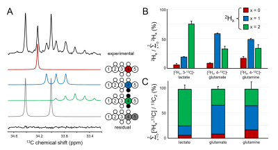 |
Characterization of kinetic isotope effects and label loss in deuterium-based isotopic labeling studies.
Robin A. de Graaf1, Monique A. Thomas1, Kevin L. Behar2, and Henk M. De Feyter1
1Dept. of Radiology and Biomedical Imaging, Yale University, New Haven, CT, United States, 2Dept. of Psychiatry, Yale University, New Haven, CT, United States
Deuterium metabolic imaging (DMI) is a novel, non-invasive method to map metabolism from deuterated substrates in 3D. The replacement of protons with deuterons could potentially lead to kinetic isotope effects (KIEs), and loss of deuterons. Knowledge of the KIE levels, and label losses is required for DMI-based measurements of absolute metabolic rates. Here the deuterium KIE and label loss is investigated for glucose and acetate in rat brain in vivo using a double substrate/double labeling strategy. Significant, but predictable and reproducible label losses were observed in the metabolic products lactate, glutamate and glutamine. The measured KIE was relatively small (4-6%).
|
|
1960.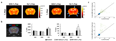 |
Improving T2 Sensitivity of Magnetic Resonance Fingerprinting for Simultaneous Quantification of Gadolinium and 17O-Water Concentration
Yuning Gu1, Yong Chen2, Jesse I. Hamilton3, Kihwan Kim1, Ciro Ramos-Estebanez4, Chris A. Flask2, Nicole Seiberlich2,3, Charlie Androjna5, and Xin Yu1,2,6
1Department of Biomedical Engineering, Case Western Reserve University, Cleveland, OH, United States, 2Department of Radiology, Case Western Reserve University, Cleveland, OH, United States, 3Department of Radiology, University of Michigan, Ann Arbor, MI, United States, 4Department of Neurology, Case Western Reserve University, Cleveland, OH, United States, 5Cleveland Clinic Pre-Clinical Magnetic Resonance Imaging Center, Cleveland Clinic Foundation, Cleveland, OH, United States, 6Department of Physiology and Biophysics, Case Western Reserve University, Cleveland, OH, United States
Simultaneous quantification of intravenously injected Gadolinium (Gd) and 17O-water through T1 and T2 shortening effect enables evaluation of BBB permeability to large and small molecules under pathological conditions. Magnetic resonance fingerprinting allows fast and simultaneous T1 and T2 mapping, though improvement in T2 sensitivity is required for accurate quantification of 17O-water in brain. This study demonstrates that the combination of T2-preparation module and small flip angle improves the accuracy in T2 estimation in mouse brain. Further, the feasibility of using MRF method to quantify Gd and 17O-water
concentration was explored in a phantom study.
|