
Digital Poster Session
Neuro: Psychoradiology
Neuro
1961 -1972 Psychoradiology - Neuropsychiatric Disorders
1973 -1984 Psychoradiology - Pairs of Psychoradiology Topics
1985 -1998 Psychoradiology - Obsessive-Compulsive, Major Depression & Bipolar Disorders
Session Topic: Psychoradiology
Session Sub-Topic: Neuropsychiatric Disorders
Digital Poster
Neuro
1961.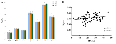 |
Disrupted integration of single-subject gray matter networks in suicidality of MDD
Huiru Li1, Jing Yang1, Huawei Zhang1, Li Yin2, Qiyong Gong1,3, and Zhiyun Jia1,4
1Huaxi MR Research Center (HMRRC), Department of Radiology, West China Hospital of Sichuan University, Chengdu, China, 2Department of Psychiatry, West China Hospital of Sichuan University, Chengdu, China, 3Psychoradiology Research Unit of Chinese Academy of Medical Sciences (2018RU011), Chengdu, China, 4Department of Nuclear Medicine, West China Hospital of Sichuan University, Chengdu, China, Chengdu, China
In this study, we explore suicidal brain of depression from the level of network connection to capture the complex connectivity alterations of gray matter networks that support higher cognitive and affective processes. We constructed single-subject morphological brain gray matter networks and small-world parameters (Cp, Lp, γ, λ and σ), network efficiency parameters (Eloc and Eglob) and nodal properties (nodal degree, efficiency and centrality). We found decreased σ/Eglob and increased Lp/λ in SU compared to PC and HC. In summary, suicidality involves complex neocortical network organization and showed a weaker integration compared PC and HC.
|
|
1962.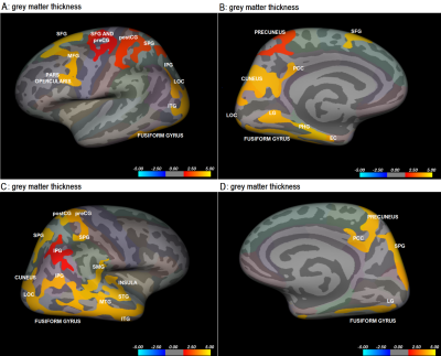 |
A 3T magnetic resonance study of structural brain alterations in neuroticism
Barbara Hrast1, Gašper Zupan1,2, Andrej Vovk1, and Dušan Šuput1
1Faculty of Medicine, University of Ljubljana, Ljubljana, Slovenia, 2Institute of Radiology, University Medical Centre Ljubljana, Ljubljana, Slovenia
This study investigated the association between alterations in cerebral morphology and neuroticism. Grey matter thickness, cortical area, and volume were calculated in 110 healthy right-handed subjects and correlations with neuroticism scores performed. Negative correlations between neuroticism and cortical area/volume and positive correlations between neuroticism and grey matter thickness were found in occipitotemporal regions. The importance of the ventral visual pathway in emotion processing has been highlighted by structural changes in the occipital and temporal lobe. Alterations in the prefrontal cortex also show the importance emotional control plays in neuroticism. This study provided an additional explanation of structural alterations in neuroticism.
|
|
1963.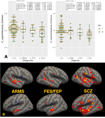 |
Structural Brain Deterioration in the Course of Schizophrenia
Akiko Uematsu1,2, Hidenori Yamasue3, Kiyoto Kasai4, and Shinsuke Koike4,5
1Graduate School of Arts and Sciences, University of Tokyo, Tokyo, Japan, 2RIKEN CBS, Saitama, Japan, 3Departmentof Psychiatry, Hamamatsu University School of Medicine, Hamamatsu, Japan, 4The University of Tokyo Hospital, Tokyo, Japan, 5Center for Evolutionary Cognitive Science, University of Tokyo, Tokyo, Japan
In this study, we examined structural brain deterioration in the course of schizophrenia with multi aspects by utilizing a variety of information derived from multi-contrast MRI data. Our findings suggested superior temporal gyrus is the key to understand the onset mechanism of Schizophrenia.
|
|
1964.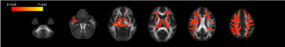 |
Evaluation of white matter neuroinflammation in children with autism spectrum disorder using free-water imaging
Wataru Uchida1,2, Koji Kamagata1, Eiji Kirino3,4, Christina Andica1, Yuya Saito1,2, Akifumi Hagiwara1, Toshiaki Akashi1, Akihiko Wada1, Syo Murata1, Masaaki Hori5, and Shigeki Aoki1
1Radiology, Juntendo University Graduate School of Medicine, Tokyo, Japan, 2Radiological Sciences, Tokyo Metropolitan University Graduate School of Human Health Sciences, Tokyo, Japan, 3Psychiatry, Juntendo University Graduate School of Medicine, Tokyo, Japan, 4Radiology, Juntendo University Shizuoka Hospital, Shizuoka, Japan, 5Radiology, Toho University Omori Medical Center, Tokyo, Japan
We investigated the differences in white matter pathology between children with autism spectrum disorder (ASD) and typically developing children using single-tensor diffusion tensor imaging (DTI) and bi-tensor free-water (FW) imaging. Tract-based spatial statistics analysis demonstrated significantly increased FW volume fraction in children with ASD compared with that in typically developing children. Increased FW volume fraction may reflect neuroinflammation in children with ASD as a result of inflammatory cytokine accumulation in the white matter. Furthermore, we showed that FW imaging is more sensitive and specific than single-tensor DTI in the evaluation of white matter in children with ASD.
|
|
1965.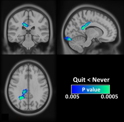 |
Brain alternations after smoking cessation: An arterial spin labeling study
Guan-Jie Wang1, Chun-Ming Chen2, and Shin-Lei Peng1
1Department of Biomedical Imaging and Radiological Science, China Medical University, Taichung, Taiwan, 2Department of Radiology, China Medical University Hospital, Taichung, Taiwan
Although the unfavourable effects of cigarette smoking on the brain have been demonstrated in current smokers, it is unclear whether the neurotoxic effects of smoking on the brain are permanent or reversible after smoking cessation. Our results showed that ex-smokers had a decreased CBF when compared to never-smokers, especially in the posterior cingulate cortex (PCC). The present findings may explain in part the frequently reported cognitive dysfunctions in ex-smokers. However, the affected brain region was less extensive than the previous studies which compared current smokers and never smokers, suggesting the potential to partially recover from smoking-related CBF deficit.
|
|
1966.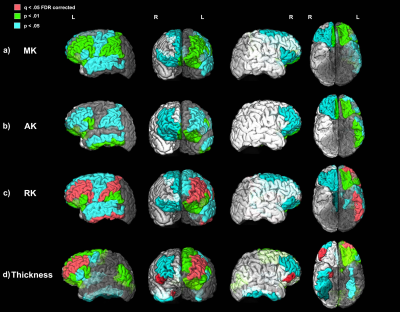 |
Progressive Microstructural Cortical Changes in Psychotic Spectrum Disorders
Faye McKenna1, Yu Veronica Sui1, Hillary Bertisch2, Donald Goff3, and Mariana Lazar1
1Radiology, New York University School of Medicine, New York, NY, United States, 2Department of Rehabilitation Medicine, New York University School of Medicine, New York, NY, United States, 3Psychiatry, New York University School of Medicine, New York, NY, United States
Widespread progressive cortical thinning is a long-standing finding in Psychotic Spectrum Disorders (PSD). While previous histological studies have documented an array of microstructural deficits in the thinning cortex in PSD few techniques have been available to examine these microstructural changes in vivo. We employed the diffusion kurtosis imaging (DKI) technique alongside the classical T1-weighted measure of cortical thickness to describe microstructural changes in the PSD cortex over the first 10 years of the illness. We found, for the first time, that DKI metrics of tissue complexity were significantly positively related to PSD illness progression in the cortex.
|
|
1967.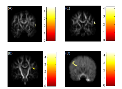 |
Association between brain structural alteration and emotional change in depressive patients with suicidal ideation with GQI
Man Teng Cheok1, Vincent Chin-Hung Chen2,3, Yuan-Hsiung Tsai4, and Jun-Cheng Weng1,3,5
1Department of Medical Imaging and Radiological Sciences, Chang Gung University, Taoyuan, Taiwan, 2School of Medicine, Chang Gung University, Taoyuan, Taiwan, 3Department of Psychiatry, Chang Gung Memorial Hospital, Chiayi, Taiwan, 4Department of Diagnostic Radiology, Chang Gung Memorial Hospital, Chiayi, Taiwan, 5Medical Imaging Research Center, Institute for Radiological Research, Chang Gung University and Chang Gung Memorial Hospital at Linkou, Taoyuan, Taiwan
Over 300 million people are suffering from various degrees of depression worldwide. The level of depression severity is a significant risk factor for suicidal behavior. In this study, we used generalized q-sampling imaging (GQI) integrating with clinical diagnostic scale in the suicidal ideation and non-suicidal ideation depressive patients. The multiple regression was performed to identify the association between the GQI indices and the clinical scales. Our results indicated that the demyelination of precuneus may relate to the suicidal ideation in depressive patients.
|
|
| 1968. | Structural alterations in functional movement disorders: a diffusion weighted imaging study
Silvina G Horovitz1, Jacob Parker1, Patrick Bedard1, Carine Maurer1,2, and Mark Hallett1
1MNB - HMCS, NINDS - NIH, Bethesda, MD, United States, 2School of Medicine, Stony Brook University, Stony Brook, NY, United States Functional movement disorders (FMD) patients have an overactive limbic system and an abnormal sense of self-agency. Using diffusion data on a population of 44 (FMD) patients and 44 matched controls, we identified structural anomalies in motor and limbic related areas. ‘Stronger’ (higher FA) limbic white matter reinforces the idea of enhanced limbic effects. Less integrity (higher trace) in areas of the motor system might contribute to the lack of agency. |
|
1969.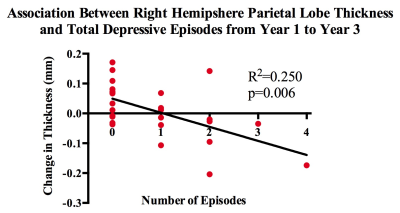 |
Quantification of cortical thickness changes in bipolar disorder patients following first episode mania: A prospective study
Ariana Cahn1, Wayne Su1, Trisha Chakrabarty1, Kamyar Keramatian1, and Lakshmi Yatham1
1Psychiatry, University of British Columbia, Vancouver, BC, Canada
A long disease course of Bipolar I Disorder (BD-I) brings not only with it mood episodes and cognitive impairments, but a progressive change in grey matter morphology. To ascertain how mood episode variables such as type, amount and duration might have an effect on cortical thickness, we conducted a preliminary study in 32 patients from a larger longitudinal study. We found a negative correlation between right parietal lobe thickness and amount of depressive episodes between years 1 and 3 of the disease. These data suggest that early disease course morphological changes may be affected by presence of multiple depressive episodes.
|
|
1970.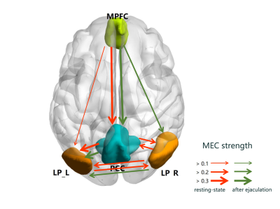 |
An Integrated PET/MR Study on Functional Connection and Glucose Metabolism of Default Mode Network Nodes for Premature Ejaculation Patients
Lei Wang1, Long Jin1, Yarong Wang2, and Menghui Yuan1
1Nuclear Medicine, Tangdu Hospital of Air Force Medical University, Xi'an, China, 2Radiology, The First Affiliated Hospital of Xi'an Jiaotong University, Xi'an, China
Integrated PET/MR platform was used to observe the functional and metabolic effective connectivity (FC, MEC) patterns between 4 default mode network nodes for premature ejaculation patients, at resting-state and after-ejaculation-state. Significant increased FC between medial prefrontal cortex and posterior cingulate cortex (PCC) and decreased metabolic in PCC were found, after ejaculation. MEC pathways were not the same in different state, but the strength differences of MEC were not significant. There was significant positive correlation between intravaginal ejaculation latency time and the resting-state metabolism in PCC, controlling age factor. These findings gave new thoughts of PE diagnosis and treatment.
|
|
1971.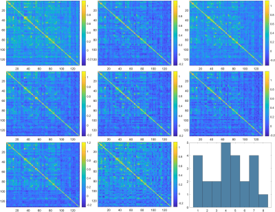 |
Altered transient connectivity states at rest in Tourette’s syndrome
Shukti Ramkiran1,2,3, Ravichandran Rajkumar1,2,3, N. Jon Shah1,3,4,5, and Irene Neuner1,2,3
1Institute of Neuroscience and Medicine - 4 (INM-4), Forschungszentrum Juelich, Juelich, Germany, 2Department of Psychiatry, Psychotherapy and Psychosomatics, RWTH Aachen University, Aachen, Germany, 3JARA – BRAIN – Translational Medicine, Juelich, Germany, 4Department of Neurology, RWTH Aachen University, Aachen, Germany, 5Institute of Neuroscience and Medicine - 11 (INM-11), JARA, Forschungszentrum Juelich, Juelich, Germany
Analysis of dynamic functional connectivity enables identification of spatio-temporal variations in functional connectivity. This is useful in identifying transient states of abnormalities in various brain disorders. Tourette syndrome (TS) is a neurodevelopmental disorder characterized by multiple motor and at least one vocal or phonic tic. We identified eight distinct states in TS patients and healthy controls (HC) using spectral clustering. Our results show preferential state dominance and increased inter-state variability in TS patients as compared to healthy controls.
|
|
1972.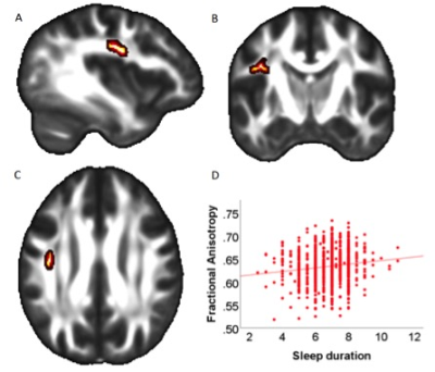 |
Sleep Duration is Associated with Cognitive Performance and White Matter Microstructure in Healthy, Young Adults
Harald Kugel1, Pascal Grumbach2, Nils Opel2, Susanne Meinert2, Elisabeth J Leehr2, Ronny Redlich2, Verena Enneking2, Janik Goltermann2, Bernhard T Baune2,3, Udo Dannlowski2, and Jonathan Repple2
1Institute of Clinical Radiology, University of Muenster, Muenster, Germany, 2Department of Psychiatry, University of Muenster, Muenster, Germany, 3Department of Psychiatry, University of Melbourne, Melbourne, Australia
This cross-sectional study showed that real-world differences in sleep duration but not subjective sleep quality are related to cognitive performance measures and - as indicated by fractional anisotropy measures in the SLF - white matter integrity in healthy, young adults, suggesting a detrimental effect of shorter sleep duration on brain structure and function.
|
 View the Poster
 Watch the Video
Session Topic: Psychoradiology
Session Sub-Topic: Pairs of Psychoradiology Topics
Digital Poster
Neuro
1973.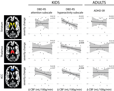 |
The association between stimulant medication, pharmacological MRI, and ADHD symptom severity in the mature and developing brain.
Antonia Kaiser1, Anouk Schrantee1, Ramon Lindauer2, Marco Bottelier3, Paul J. Lucassen4, and Liesbeth Reneman1
1Department of Radiology and Nuclear Medicine, Amsterdam University Medical Center, University of Amsterdam, Amsterdam, Netherlands, 2Department of Child and Adolescent Psychiatry, Amsterdam University Medical Center, University of Amsterdam, Amsterdam, Netherlands, 3Department of Child- and Adolescent Psychiatry, GGZ Noord-Holland Noord, Triversum, Alkmaar, Netherlands, 4Brain Plasticity group, Swammerdam Institute for Life Sciences, University of Amsterdam, Amsterdam, Netherlands
We previously used pharmacological MRI (phMRI) to demonstrate that four months of methylphenidate treatment alters dopamine function after washout in medication-naïve children, but not adults, with ADHD. Here, we show in the same children at a 4-5 year follow-up, that the baseline phMRI response in the thalamus and anterior cingulate cortex predicts ADHD severity (hyperactivity scale). However, this association was not moderated by cumulative medication dose. Moreover, no association between phMRI response and symptom severity was found in adults.
|
|
 |
1974.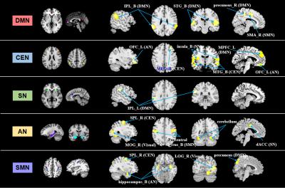 |
Hyperconnectivity among larger-scale intrinsic functional networks in drug-naïve children with attention-deficit/hyperactivity disorder
Yingxue Gao1, Xuan Bu1, Kaili Liang1, Hailong Li1, Lianqing Zhang1, Lu Lu1, Shi Tang1, Yanlin Wang1, Xinyu Hu1, and Xiaoqi Huang1
1Huaxi MR Research Center (HMRRC), Functional and Molecular Imaging Key Laboratory of Sichuan Province, Department of Radiology, West China Hospital, Sichuan University, Chengdu 610041, China, Chengdu, China
We used seed-based analysis and independent component analysis to investigate functional connectivity among five resting-sate networks including the default mode network (DMN), central executive network (CEN), salience network (SN) sensorimotor network (SMN) and affective network (AN) in children with attention-deficit/hyperactivity disorder (ADHD). Hyperconnectivity was found both within and between these networks. Our findings highlight the “hub” role of the DMN and the CEN among all these functional networks in the mechanism of network dysfunction in ADHD children.
|
1975.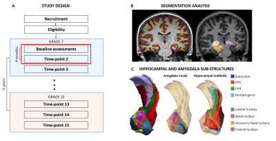 |
The longitudinal adolescent brain study: subcortical volume correlates of psychological distress in early adolescence
Kathryn Broadhouse1, Amanda Boyes1, Larisa McLoughlin1, Marcella Parker1, Denise Beaudequin1, Gabrielle Simcock1, Jim Lagopoulos1, and Daniel Hermens1
1University of the Sunshine Coast, Sunshine Coast, Australia
Mapping structural trajectories across adolescence provides valuable insights into the “typical” pathway as well as the developmental emergence of mental illness during this dynamic period. Here we present preliminary findings investigating the relationship between the subcortical structures, hippocampus and amygdala (sub-structures known to play important roles in fear pathways and higher order executive functions) and psychological distress measures from the first two time-points in the Longitudinal Adolescent Brain Study. By determining the neuronal changes that present with the psychological symptoms of mental illness, potential efficacious, targeted interventions become a possibility.
|
|
1976.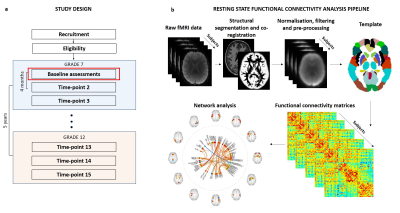 |
Resting-state functional neurobiological signatures associated with psychological distress in early adolescence
Kathryn Broadhouse1, Amanda Boyes1, Larisa McLoughlin1, Marcella Parker1, Denise Beaudequin1, Gabrielle Simcock1, Jim Lagopoulos1, and Daniel Hermens1
1University of the Sunshine Coast, Sunshine Coast, Australia
By determining the neuro-functional changes that present with the psychological symptoms of mental illness, potential efficacious, targeted interventions become a possibility. Here we have shown that network dysfunction signatures present in several mental health disorders that underpin emotional regulation and social domain deficits are evident in 12-year-olds with increased psychological distress. Follow-up data from LABS mapping the functional connectome through adolescence and corresponding cognitive and psychological scores will provide valuable insight into the specific neuronal signatures and subsequent divergence pathways that underpin mental health and mental illness.
|
|
1977. |
Increased Connectivity Correlated to Severity of Autism in a Cohort of High Functioning Autism and Major Depressive Disorder
Mario Serrano-Sosa1, Chuan Huang1,2,3, Christine DeLorenzo1,2, and Kenneth Gadow2
1Biomedical Engineering, Stony Brook University, Stony Brook, NY, United States, 2Psychiatry, Stony Brook University, Stony Brook, NY, United States, 3Radiology, Stony Brook University, Stony Brook, NY, United States
This is the first study to investigate connectivity in a cohort of High Functioning Autism (HFA) and Major Depressive Disorder (MDD) subjects with comorbid autism. Using DSI studio for connectometry analysis, we investigated the relationship between Social Responsiveness Scale (SRS) scores and Spin Distribution Function (SDF) values derived from diffusion MRI images. Tractography based analysis showed corpus callosum, longitudinal fasciculus and corticothalamic pathways to have increased connectivity relating to SRS scores (FDR=0.011). Post hoc analysis showed trend-level partial correlation between SRS and SDF while controlling for IQ scores (p=0.056, r=0.480).
|
|
1978. |
Dynamic Sliding-Window MRS Method for Measuring Changes in Glutamate and GABA in Patients with Major Depressive Disorder
John Port1, Balwinder Singh2, and Mark A Frye2
1Mayo Clinic, Rochester, MN, United States, 2Department of Psychiatry, Mayo Clinic, Rochester, MN, United States
We have developed a method of optimally frequency/phase adjusting individual FIDS of an MRS acquisition such that patient motion and scanner artifacts can be eliminated from long MRS acquisitions. This method uses MRS peak symmetry in order to optimally phase the Cho and Cr peaks acquired using a frequency-locked MEGA-PRESS sequence. We tested our method subjects receiving an IV ketamine infusion for MDD and noticed dramatic changes in GABA and Glx levels in one subject. Our method allows for dynamic MRS studies that can measure neurotransmitter changes over time.
|
|
1979.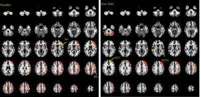 |
Relative modulations of left DLPFC connectivity with four patho-physologically relevant targets after repetitive TMS in major depression
Pallab K Bhattacharyya1,2, Murat Altinay3, Xuemei Huang1, Mark Lowe1, and Amit Anand3
1Imaging Institute, Cleveland Clinic, Cleveland, OH, United States, 2Radiology, Cleveland Clinic Lerner College of Medicine, CLEVELAND, OH, United States, 3Neurogolical Institute, Cleveland Clinic, Cleveland, OH, United States
Abnormalities of resting state functional connectivity of several networks have been implicated in the pathology of major depressive disorder. Modulations of functional connectivity of left dorsolateral prefrontal cortex (DLPFC), the site of repetitive transcranial magnetic stimulation (rTMS) for patients inadequately responsive to medication, with four patho-physiologically relevant nodes were studied following rTMS therapy. While the group (N=6) showed improvement in depression following the therapy, only connectivity of left DLPFC with bilateral inferior parietal lobule changed its course – no such change was apparent in connectivity with bilateral anterior cingulate, anterior insula and middle temporal gyrus.
|
|
1980.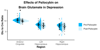 |
A 7T MRS study of Psilocybin administration in patients with major depressive disorder
Michal Povazan1,2, Manoj Doss3,4, Alan K Davis3,4,5, Matthew W Johnson3,4, Roland R Griffits3,4,6, Peter B Barker1,2, and Frederick S Barrett3,4
1Russell H. Morgan Department of Radiology and Radiological Science, The Johns Hopkins University, School of Medicine, Baltimore, MD, United States, 2F. M. Kirby Research Center for Functional Brain Imaging, Kennedy Krieger Institute, Baltimore, MD, United States, 3Center for Psychedelic and Consciousness Research, The Johns Hopkins University School of Medicine, Baltimore, MD, United States, 4Department of Psychiatry and Behavioral Sciences, The Johns Hopkins University School of Medicine, Baltimore, MD, United States, 5College of Social Work, The Ohio State University, Columbus, OH, United States, 6Department of Neuroscience, The Johns Hopkins University School of Medicine, Baltimore, MD, United States
Recent studies have shown that the administration of psilocybin may reduce depression severity. A role of glutamate was hypothesized in the antidepressant efficacy, however the exact neurobiological mechanisms remain unknown. Proton MR spectroscopy enables a valuable insight into glutamatergic metabolism and provides information about other important neuronal markers such as N-acetylaspartate. Here, we have utilized STEAM MR spectroscopy at 7T to observe the changes of cortical metabolites after psilocybin administration in patients with major depressive disorder. Two high dose sessions of psilocybin decreased glutamate and NAA levels of the anterior cingulate in the cohort.
|
|
1981.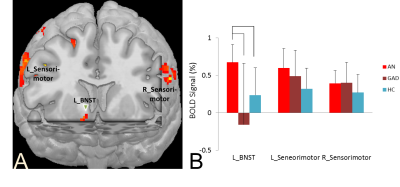 |
Evaluating the Role of Bed Nucleus of Stria Terminalis in Anorexia Nervosa Restricting Type
Jianli Wang1, Lauren Spreen2, Xiaoyu Wang1, Christopher Freet3, Jeffrey Vesek2, Fauzia Mahr3, Lydia Petrovic-Dovat3, and Scott Bunce3
1Radiology, Penn State College of Medicine, Hershey, PA, United States, 2Molecular Biology, Penn State College of Medicine, Hershey, PA, United States, 3Psychiatry, Penn State College of Medicine, Hershey, PA, United States
There is growing evidence for the contribution of neurocircuitry regulating emotion, as well as appetite and body weight, in the etiology of anorexia nervosa (AN). The high prevalence of anxiety disorders in AN implicates a critical node in the neurocircuitry of anxiety. The bed nucleus of the stria terminalis (BNST) modulates not only responses to anxiogenic (versus fear) situations, but mediates anxious temperament and feeding as well, through efferent connections to the lateral hypothalamus. In this study, we sought to identify if there is significant functional and structural changes of BNST in AN related to food.
|
|
1982.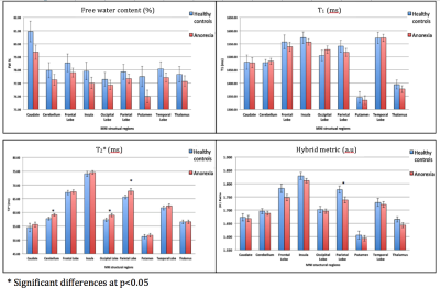 |
Quantitative volumetric and parametric alterations in patients suffering from anorexia nervosa
Zaheer Abbas1, Syed Hammad Akhter1, Dominik Ridder1, and N. Jon Shah1,2,3
1Medical Imaging Physics, Institute of Neuroscience and Medicine 4, Jülich, Germany, 2Institute of Neuroscience and Medicine 11, INM 11, JARA, Jülich, Germany, 3Department of Neurology, Faculty of Medicine, Aachen, Germany
Anorexia nervosa (AN) is an eating disorder characterized by severe weight loss due to a fear of gaining weight and obsessions relating to body shape. AN is a psychiatric illness, and although psychiatric co-morbidity in AN has already been extensively studied, the extent to which it can affect mortality is unclear. In this work, a quantitative and volumetric study to monitor the disease-related integrity of the brain tissue in a cohort of AN patients and healthy controls was performed. The preliminary results suggest that psychological treatment may be useful in terms of brain tissue integrity, even after only three months.
|
|
1983. |
Subanesthetic ketamine infusion increases oxidative metabolism in human prefrontal cortex
Naomi R. Driesen1,2, Peter Herman3, Margaret Rowland1,2, Maolin Qiu3, George He4, Peter T. Morgan1, Andrea Diaz-Stansky1, Sarah Fineberg1, Daniel Barron1, Lars Helgeson5, Robert Chow5, Ralitza Gueorguieva6, Teo-Carlo Straun7,
John H. Krystal1,2, and Fahmeed Hyder3,8
1Psychiatry, Connecticut Mental Health Center, Yale University, New Haven, CT, United States, 2VA Connecticut Health Care System, West Haven, CT, United States, 3Radiology and Biomedical Imaging, Yale University, New Haven, CT, United States, 4Psychology, Yale University, New Haven, CT, United States, 5Anesthesiology, Yale University, New Haven, CT, United States, 6Yale School of Public Health, Yale University, New Haven, CT, United States, 7Straun Health and Wellness, New Haven, CT, United States, 8Biomedical Engineering, Yale University, New Haven, CT, United States
We compared the effect of subanesthetic infusion of the NMDA receptor antagonist ketamine on metabolic activity to a similar volume of saline infusion in healthy volunteers. Since BOLD fMRI depends on neurovascular-neurometabolic couplings which can be confounded by pharmacological agents, we measured transverse relaxation rates (R2, R2*) and blood flow (CBF) to calculate oxidative metabolism (CMRO2) with calibrated fMRI. We found CBF and CMRO2 increased with ketamine infusion in nearly all Brodmann areas of the cortex. The CMRO2 increase was significant in prefrontal (0.16±0.06, p=0.026) and visual cortex (0.22±0.07, p=0.01), but not in sensorimotor cortex (0.17±0.14, p=0.258).
|
|
1984.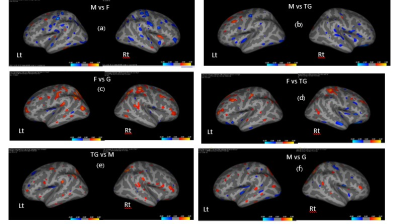 |
MRI Brain Morphometry in Heterosexual Men and Women, Gay Men, and Transgender Women: A Preliminary Study
Suwit Saekho1, Artit Rodkong2, Diana E Peragine3, Malvina N Skorska3, Pongpun Saokhieo4, Taweewat Supintham4, Oranicha Kaewthip4, Kittichai Wantanajittikul1, Suwat Chariyalertsak4, and Doug P VanderLaan3
1Radiologic Technology, Chiang Mai University, Chiang Mai, Thailand, 2Radiology, Chiang Mai University, Chiang Mai, Thailand, 3Psychology, University of Toronto Mississauga, Mississauga, ON, Canada, 4Research Institute for Health Sciences, Chiang Mai University, Chiang Mai, Thailand
This study investigated brain bases of sexual and gender diversity. Heterosexual men and women, gay men, and transgender women underwent structural MRI. Regional volumes and cortical thickness were examined. Heterosexual sex differences were replicated. Men had greater gray and white matter volumes and women had thicker cortices. Overall, transgender women had a brain pattern that more closely reflected that of heterosexual women, whereas gay men were more similar to heterosexual men. Yet, transgender women also showed some volume and cortical thickness similarities to heterosexual men. These findings help identify brain regions related to gender identity.
|
Session Topic: Psychoradiology
Session Sub-Topic: Obsessive-Compulsive, Major Depression & Bipolar Disorders
Digital Poster
Neuro
1985.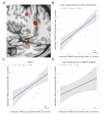 |
Identifying the brain network underlying symptom improvement following neuromodulation in Obsessive Compulsive Disorder.
Jurgen Germann1, Gavin JB Elias1, Clemens Neudorfer1, Alexandre Boutet1, Clement T Chow1, Peter Giacobbe2, Se Jo Kim3, Hyun Ho Jung3, Walter Kucharczyk1, Jin Woo Chang4, and Andres M Lozano1
1University Health Network, Toronto, ON, Canada, 2Sunnybrook Health Sciences Centre, Toronto, ON, Canada, 3Department of Psychiatry, Yonsei University College of Medicine, Seoul, Republic of Korea, 4Department of Neurosurgery, Yonsei University College of Medicine, Seoul, Republic of Korea
Neuromodulatory interventions have shown promise in the treatment of Obsessive-Compulsive Disorder (OCD). However, the nature of the aberrants circuits that cause the disease as well as the network changes that are the basis of improvement following treatment are not well understood. Here we analyzed OCD patients treated with either MRI-guided focused ultrasound capsulotomy or deep brain stimulation targeting the inferior thalamic peduncle. Using neuroimaging analysis and modeling techniques we found that a fronto-limbic network (DLPFC, dorsal ACC, and amygdala) predicts individual improvement across groups. These results provide new insight into OCD and how to possibly refine neuromodulatory therapies.
|
|
|
1986.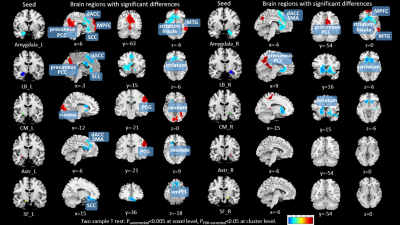 |
Disrupted Resting-State Functional Connectivity of Amygdala Subregions in Obsessive-Compulsive Disorder
Lingxiao Cao1, Hailong Li1, Xinyu Hu1, Jing Liu1, Yingxue Gao1, Xuan Bu1, Lianqing Zhang1, Lu Lu1, Shi Tang1, Yanlin Wang1, Xinyue Hu1, Qiyong Gong1, and Xiaoqi Huang1
1Huaxi MR Research Center (HMRRC), Functional and molecular imaging Key Laboratory of Sichuan Province, Department of Radiology, West China Hospital, Sichuan University, Chengdu, China
Using seed based FC analysis, we investigated alterations in functional connectivity of amygdala subregions in OCD. Our findings (ⅰ) indicated OCD patients, relative to HC, have anomalous amygdala resting state network connections to brain regions that play a pivotal role in emotional processing; (ⅱ) identified CM/Astr-PCG hyperconnectivity and CM-cuneus hyperconnectivity, which were not detected when using the whole amygdala as a seed; (ⅲ) demonstrated FC measures of significant regions were correlated with illness duration and symptom severity. Besides, we found that functional aberrations were accompanied with anatomical changes in OCD. These findings may contribute to revealing the pathophysiology underlying OCD.
|
1987.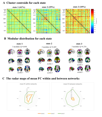 |
Disorder-specific alternations of intrinsic brain functional network dynamics in obsessive-compulsive disorder and schizophrenia
Fei Li1,2, Lekai Luo1,2, Qian Li1,2, Wanfang You1,2, and Qiyong Gong1,2
1Huaxi MR Research Center (HMRRC), Functional and Molecular Imaging Key Laboratory of Sichuan Province, Department of Radiology, West China Hospital of Sichuan University, Chengdu, China, 2Psychoradiology Research Unit of Chinese Academy of Medical Sciences (2018RU011), West China Hospital of Sichuan University, Chengdu, China
Dynamic functional connectivity (dFC) analyses were used to compare the intrinsic brain functional network dynamics in patients with obsessive-compulsive disorder (OCD) and schizophrenia (SZ), by using resting-state functional MRI (rfMRI). We found that patients with OCD and SZ showed distinct disorder-specific alternations of brain dynamics and the higher fractional time in state 2 may indicate the more anxiety ratings in OCD patients.
|
|
1988.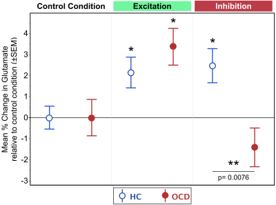 |
dACC Dysfunction During Inhibitory But Not Excitatory Motor Control in OCD: Glutamate Dysmodulation Estimated using ¹H fMRS
Jeffrey A. Stanley1, Asadur Chowdury2, Dalal Khatib2, Phillip Easter2, David R. Rosenberg2, and Vaibhav A. Diwadkar2
1Psychiatry and Behavioral Neurosciences, Wayne State University School of Medicine, Detroit, MI, United States, 2Wayne State University School of Medicine, Detroit, MI, United States
Loss of the E/I balance of the dACC in OCD was assessed using a combination of a) motor control tasks with distinct excitatory and inhibitory modes of responding, and b) ¹H fMRS to quantify evoked glutamate changes. Basal (task-independent) glutamate levels were comparable to healthy controls, as were levels during excitatory modes of responding. However, the inhibitory mode of responding induced a specific deficit in the degree of dACC glutamate modulation in OCD patients. In surmounting shortcomings of fMRI, ¹H fMRS successfully identified loss of excitation in the E/I balance of the dACC during specific phases of motor control.
|
|
1989.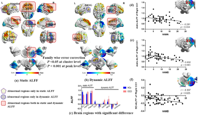 |
Temporal variability of local brain activity in medication-free patients with Obsessive Compulsive Disorder
Jing Liu1, Yingxue Gao1, Hailong Li1, Xuan Bu1, Lingxiao Cao1, Lianqing Zhang1, Suming Zhang1, Xinyu Hu1, and Xiaoqi Huang1
1Huaxi MR Research Center (HMRRC), Functional and molecular imaging Key Laboratory of Sichuan Province, Department of Radiology, West China Hospital, Sichuan University, Chengdu 610041, China, Chengdu, China
In current study, we use static and dynamic amplitude of low-frequency fluctuation / fractional low-frequency fluctuation (ALFF/fALFF) to assess the local brain activity on patients with obsessive-compulsive disorder (OCD). We found that both static and dynamic ALFF/fALFF can detect certain cerebral region with abnormal activity, however, dynamic ALFF/fALFF can demonstrate additional brain regions that are ignored by static ALFF/fALFF. In addition, we found that there is significant correlation between cerebellum activity and clinical scale, which suggested the crucial role of cerebellum in the pathophysiology of OCD.
|
|
1990.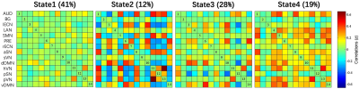 |
Dynamic characteristics of functional connectivity in the first-episode and treatment-naive patients with obsessive-compulsive disorder
Junhong Liu1, Xiaoming Li1, Kaiyu Wang2, and Jingliang Cheng1
1The First Affiliated Hospital of Zhengzhou University, Zhengzhou, China, 2GE Healthcare, MR Research China, Beijing, China
Compared with task-activation studies, dynamics are potentially even more prominent during resting-state, when mental activity is unconstrained. To assess whole-brain dynamic functional connectivity of first-episode and treatment-naive patients with obsessive-compulsive disorder (OCD), we used a series of methods including independent component analysis, sliding windows and k-means clustering. Our results indicated that OCD groups displayed more transitions between different states than healthy controls. This change was positively correlated with clinical scale scores, potentially contributing to better understanding of the dynamic neural mechanism of OCD.
|
|
1991.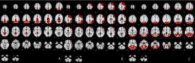 |
Analysis of resting-state networks alterations of first-episode obsessive–compulsive disorder patients by independent component analysis
Junhong Liu1 and Jingliang Cheng1
1The First Affiliated Hospital of Zhengzhou University, Zhengzhou, China
So far, there is no consensus on the cause of obsessive-compulsive disorder(OCD). We used a data-driven method, independent component analysis (ICA), to study the change of whole brain resting-state functional connectivity of first-episode and treatment-naïve patients with OCD. Three abnormal resting-state networks, posterior default-mode network, right frontoparietal network and lateral visual network, have been found in patients. In addition, patients showed increased functional connection in bilateral cuneus, right inferior parietal lobule and right middle occipital lobule compared with controls. It’s considered that changes of abnormal resting-state networks might reveal the possible neural mechanism of OCD.
|
|
1992.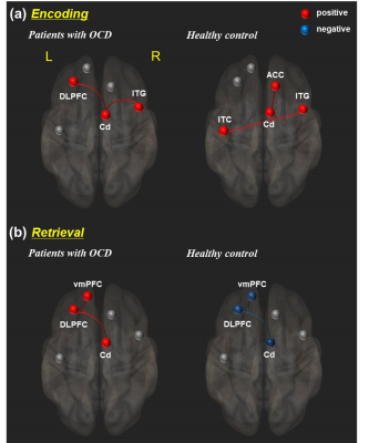 |
The OCD symptoms affect to abnormal functional activity and connectivity at caudate nucleus during memory retrieval, and lead to GM volume changes
Shin-Eui Park1, Gwang-Woo Jeong2, and Chulhyun Lee1
1Center for research equipment, Korea Basic Science Institue, Cheongju, Republic of Korea, 2Department of Radiology, Medical school, Chonnam National University, Gwangju, Republic of Korea
For verifying how the OCD symptom affects to abnormal functional activity and connectivity at Cd during memory processing, and whether leading to brain volume changes in direct or indirect, we performed multi study combined with fMRI and VBM study. Twenty OCD patients and 20 controls took part in VBM and fMRI studies with explicit memory task. The functional and morphological profiles of Cd could be emphasized to abnormal processing of explicit memory by OCD symptoms. It puts forward a possibility that focal functional connectivity and brain volume changes may be affected by lasting OCD symptoms, and leading to memory dysfunction.
|
|
1993.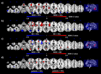 |
Common and distinct patterns of intrinsic brain activity alterations in major depression and bipolar disorder: voxel-based meta-analysis
JiaYing Gong1,2, Junjing Wang3, Shaojuan Qiu1, Long Qian4, Li Huang1, and Ying Wang1
1Medical Imaging Center, First Affiliated Hospital of Jinan University, Guangzhou, China, 2Department of Radiology, Six Affiliated Hospital of Sun Yat-sen University, Guangzhou, China, 3Department of Applied Psychology, Guangdong University of Foreign Studies, Guangzhou, China, 4MR Research, GE Healthcare, Beijing, China
This is the largest voxel-based meta-analysis conducted to date of amplitude of low-frequency fluctuation (ALFF) studies in major depressive disorder (MDD) and bipolar disorder (BD). MDD and BD show a common pattern of aberrant regional intrinsic brain activity which predominantly includes the insula, medial prefrontal cortex (mPFC), and cerebellum. These results expand on a growing literature exploring resting-state activity in MDD and BD, which provide useful insights for understanding the underlying pathophysiology of brain dysfunction in affective disorders, and developing more targeted and efficacious treatment and intervention strategies.
|
|
|
1994.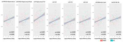 |
Smaller Hippocampal Subfields Associate with Poorer Logical Memory Performance in a Large Sample of MDD Patients
Lianqing Zhang1, Xinyu Hu1, Lu Lu1, Xuan Bu1, Yingxue Gao1, Shi Tang1, Kaili Liang1, Lingxiao Cao1, Yanlin Wang1, Xinyue Hu1, Jing Liu1, Qiyong Gong1, and Xiaoqi Huang1
1Radiology, Huaxi MR Research Center (HMRRC), Sichuan University, Chengdu, China
Smaller hippocampus is a consistent finding in studies of major depressive disorder (MDD), and subfield-level anatomic analyses could provide new insights to the role of hippocampal deficit in MDD. In the current study, we recruited a relatively large sample of MDD patients and demonstrated that (1) “core structures” including CA and dentate gyrus are more anatomically involved in MDD than other structures in hippocampus; (2) hippocampal tail might be important in MDD, but the relationship between hippocampal tail and MDD could be complex; (3) smaller hippocampus could contribute to memory deficit in MDD patients by failure in long-term memory formation.
|
1995.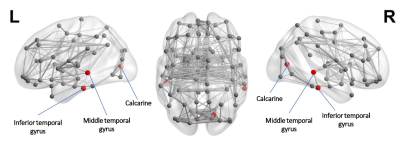 |
Altered network efficiency is associated with mood disturbance in adults with major depressive disorder
Xiaopei Xu1 and Lanying Liu2
1Department of Radiology, Second Affiliated Hospital of Zhejiang University School of Medicine, Hangzhou, China, 2Department of Psychiatry, Tongde Hospital of Zhejiang Province, Hangzhou, China
To better understand the underlying mechanisms for emotional disturbances in patients with major depressive disorder (MDD), we used structural brain connectivity analysis to investigate the differences in global and local network organization of MDD and healthy controls. Our results demonstrated that both global and local efficiency of patients with MDD were significantly decreased due to depressive symptoms, and that higher depression severity, anxiety somatization and cognitive disturbance were significantly associated with decreased network efficiency. These results indicated that brain network analysis is a useful tool to link psychological disorders with their underlying anatomical substrate.
|
|
1996. |
Glutamatergic involvement of left DLPFC and left anterior insula in treatment resistant major depressive disorder
Pallab K Bhattacharyya1,2, Murat Altinay3, Xuemei Huang1, Jian Lin1, Mark Lowe1, and Amit Anand3
1Imaging Institute, Cleveland Clinic, Cleveland, OH, United States, 2Radiology, Cleveland Clinic Lerner College of Medicine, CLEVELAND, OH, United States, 3Neurogolical Institute, Cleveland Clinic, Cleveland, OH, United States
Six subjects with treatment resistant major depressive disorder (MDD) were scanned with resting state functional connectivity (rsfMRI)and GABA/Glx editing protocol. rsFMRI between left dorsolateral prefrontal cortex (lDLPFC) and left anterior insula (lAI), two nodes of physiological importance in MDD, inversely correlated with Glx at DLPFC. No such association was observed between rsfMRI and GABA. lDLPFC-lAI rsfMRI, lDLPFC GABA or Glx levels did not correlate with disease severity. The results suggest a collective role of lDLPFC and lAI in MDD via a glutamatergic mechanism.
|
|
|
1997.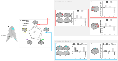 |
Ketamine modulation of Cerebro-Cerebellar Circuitry During Response Inhibition in Major Depression
Joana R. A. Loureiro1, Ashish K. Sahib1, Megha Vasavada1, Antoni Kubicki1, Shantanu Joshi1, Benjamin Wade1, Amber Leaver2, Roger Woods1, Randall Espinoza3, and Katherine Narr1
1Neurology, Ahamason-Lovelace Brain Mapping Center, UCLA, Los Angeles, CA, United States, 2Radiology, Center for Translational Imaging, Northwestern University, Chicago, IL, United States, 3Psychiatry and Biobehavioral Sciences, UCLA, Los Angeles, CA, United States
Treatments for major depressive disorder (MDD) target communication between large-scale cortical networks and the cerebellum to improve cognitive control [2]. Here, we examined how repeated-ketamine perturbs cerebro-cerebellar circuitry during response-inhibition using a NoGo/Go task (43 MDD-patients receiving ketamine, 31 controls). We implemented a psychophysiological-interaction analysis to investigate ketamine-related changes in connectivity between cerebellum-regions and cortical-networks and respective nodes. Results showed significant decreases in connectivity between a cerebellum-dorsal-attention region and the default-network. A time-by-response interaction for the somatomotor-network was observed in treatment-responders only. Results support ketamine modulates cerebello-cerebro circuitry, which may be of impact for identifying new biomarkers of MDD response.
|
1998. |
Single and repeated ketamine treatment induces perfusion changes in sensory and limbic networks in major depressive disorder
Ashish Kaul Sahib1, Joana Loureiro1, Megha Vasavada1, Antoni Kubicki1, Shantanu H Joshi1, Kai Wang2, Roger P Woods1, Eliza Congdon1, Danny J.J. Wang2, Michael Boucher1, Randall Espinoza1, and Katherine L Narr1
1UCLA, Los Angeles, CA, United States, 2University of Southern California, Los Angeles, CA, United States
Ketamine infusion therapy is now well-replicated to produce fast-acting antidepressant effects in patients with major depressive disorder (MDD). However, brain systems-level hemodynamic changes in response to single and repeated ketamine treatment remain uncharacterized. Using advanced MB pCASL MRI we examined CBF changes occurring with single and repeated ketamine treatment. Initial changes in blood flow were observed in the posterior cingulate and precuneus and primary and higher order visual areas. However, repeated exposure to ketamine engaged deeper limbic structures and the insula. Findings demonstrate that ketamine treatment perturbs distinct functional networks including sensory and limbic regions
|