
Digital Poster Session
Cardiovascular: Myocardial Tissue Characterization and Perfusion
Cardiovascular
2045 -2057 Myocardial Tissue Characterization and Perfusion - Myocardial Tissue Characterization 1
2058 -2073 Myocardial Tissue Characterization and Perfusion - Myocardial Tissue Characterization 2
2074 -2089 Myocardial Tissue Characterization and Perfusion - Myocardial Tissue Characterization 3
2090 -2104 Myocardial Tissue Characterization and Perfusion - CMR Perfusion Imaging
2045.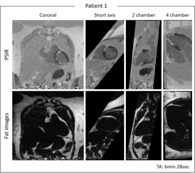 |
3D Whole-heart Motion Compensated Grey-blood Late Gadolinium Enhancement Imaging
Giorgia Milotta1, Camila Munoz1, Karl Kunze1, Radhouene Neji1, Stefano Figliozzi1, PierGiorgio Masci1, Claudia Prieto1, and Rene Botnar1
1Biomedical Engineering Department, School of Biomedical Engineering and Imaging Sciences, King's College London, London, United Kingdom
Grey-blood phase-sensitive inversion-recovery (PSIR) late gadolinium enhancement (LGE) imaging has shown promising and robust results for the assessment of myocardium viability. Conventionally 2D grey-blood LGE images are acquired under several breath-holds in different image orientations to depict scar extension. However, these approaches achieve limited spatial resolution and coverage and can be challenging in un-collaborative patients. In this work, we have proposed a free-breathing 3D whole-heart LGE sequence with water/fat Dixon encoding and blood nulling which provides grey-blood PSIR images with isotropic resolution for scar visualization and complementary 3D fat images for pericardial and myocardial adipose tissue detection in ~6min.
|
|
2046.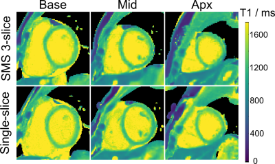 |
Model-based reconstruction for simultaneous multi-slice myocardial T1 mapping using single-shot inversion-recovery radial FLASH
Xiaoqing Wang1,2, Sebastian Rosenzweig1,2, and Martin Uecker1,2
1Department of Interventional and Diagnostic Radiology of the University Medical Center Göttingen, Göttingen, Germany, 2DZHK (German Centre for Cardiovascular Research), partner site Göttingen, Göttingen, Germany
Fast multi-slice myocardial T1 mapping is of great interest in clinical cardiovascular magnetic resonance (CMR) imaging. This work extends a simultaneous-multi-slice (SMS) model-based reconstruction method for 3-slice myocardial T1 mapping using single-shot Inversion-recovery radial FLASH. Initial results on experimental phantom and one healthy volunteer have demonstrated simultaneous 3-slice myocardial T1 mapping (1.6 x 1.6 x 8 mm3 ) might be feasible within a single inversion recovery of 4 s. More clinical validations of the proposed method will be explored in future studies.
|
|
2047.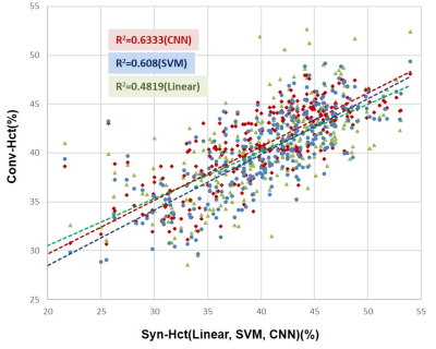 |
AI-driven Multi-Feature based Blood Hematocrit Prediction for Myocardial Extracellular Volume Quantification
Young-Jung Yang1, Pan Ki Kim1, Jinho Park1, Yoo Jin Hong1, Chul Hwan Park2, and Byoung Wook Choi1
1Department of Radiology and Research Institute of Radiological Science, Severance Hospital, Yonsei University College of Medicine, Seoul, Korea, Republic of, 2Department of Radiology and Research Institute of Radiological Science, Gangnam Severance Hospital, Yonsei University College of Medicine, Seoul, Korea, Republic of Poster Permission Withheld
Blood hematocrit is needed for myocardial ECV. To determine the hematocrit, blood sampling is the standard way, but it is invasive and time-consuming. To avoid the inconvenience of blood sampling, synthetic derivation of hematocrit was suggested in recent studies. In here, we derived the Hct using three prediction methods with multi-features of patient. Investigated methods include the linear regression and AI apporaches. We hypothesized that AI driven multi-feature based synthetic Hct would be more precise than the linear regression. The results of synthetic methods were compared with the laboratory Hct (Lab-Hct) and conventional ECV (Conv-ECV) as the reference.
|
|
2048.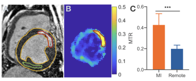 |
Magnetization Transfer Contribution to Balanced SSFP Image Contrast in Chronic Myocardial Infarction
Xinheng Zhang1,2, Hsin-Jung Yang2, Richard Tang2, and Rohan Dharmakumar2,3
1Bioengineering Department, University of California, Los Angeles, CA, United States, 2Biomedical Imaging Research Institute, Cedars-Sinai Medical Center, Los Angeles, CA, United States, 3Department of Medicine, University of California, Los Angeles, CA, United States
Magnetization Transfer (MT) effect in balanced steady-state free precession (bSSFP) has been demonstrated in acute myocardial infarction (MI). However, whether MT effect influences image contrast in bSSFP acquisitions of chronic MI has not been investigated. We studied this using a collagen phantom and large animal models with chronic MI. We found the bSSFP MT ratio to be more than 2-fold higher in the chronic MI zone compared to uninfarcted myocardium. Our findings support the notion that a strong MT contrast in bSSFP acquisitions, likely from collagen deposition within chronic MI, is available for discriminating chronic MI territories without contrast agents.
|
|
2049.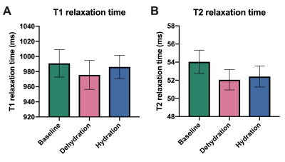 |
Influence of hydration status on myocardial T1 and T2 relaxation time measurements: an intraindividual study in healthy volunteers
Julian A. Luetkens1, Marilia Voigt1, Anton Faron1, Alexander Isaak1, Darius Dabir1, Alois M. Sprinkart1, Daniel Kuetting1, Ulrike Attenberger1, and Daniel Thomas1
1Radiology, Universityhospital Bonn, Bonn, Germany
Myocardial T1 and T2 mapping allow for non-invasive quantification of myocardial tissue alterations. Several confounders can influence correct relaxation time assessment. However, the effect of physiological changes in myocardial water content has not been investigated yet. In an intraindividual cardiac magnetic resonance imaging (MRI) study, we investigated 12 healthy volunteers at baseline, after 10-12 hours of fasting and after rehydration. Especially after dehydration, T1 and T2 relaxation times were reduced compared to baseline indicating a detectable effect of the hydration status on relaxation time assessment. Hydration status should therefore be recognized as a possible confounder of T1 and T2 mapping.
|
|
2050.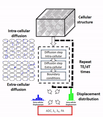 |
Sensitivity of Cardiac Diffusion Encoding Waveforms to Myocardial Extracellular Volume: A simulation study
Kevin Moulin1, Magalie Viallon2, Nyasha Maforo3,4, Valentina Mazzoli1, Pierre Croisille2, and Daniel Ennis1,5,6
1Department of Radiological Sciences, Stanford University, Stanford, CA, United States, 2Université de Lyon, UJM-Saint-Etienne, INSA, CNRS UMR 5520, INSERM U1206, CREATIS, F-42023, Saint Etienne, France, 3Department of Radiological Sciences, University of California Los Angeles, Los Angeles, CA, United States, 4Physics and Biology in Medicine Interdepartmental Program, University of California Los Angeles, Los Angeles, CA, United States, 5Division of Radiology, Veteran Affairs Health Care System, Palo Alto, CA, United States, 6Cardiovascular Institute, Stanford University, Palo Alto, CA, United States
In vivo cardiac DWI is sensitive to cardiac bulk motion and requires second-order motion compensated gradient designs. Even for a fixed b-value, motion and non-motion compensated diffusion encoding gradient waveforms have different lobe durations, number of encoding lobes, and diffusion encoding times (Δ). It remains unclear if these gradient waveforms have the same sensitivity to intra- and extra-cellular diffusion and to ECV changes. The objective of this work was to analyze the sensitivity of various gradient waveforms (fixed b-value) to a change in ECV by simulating molecular displacements in a two-compartment model.
|
|
2051. |
Right ventriclar myocardial strain in chronic total occlusion patients by cardiac magnetic resonance tissue tracking: a polit study
Xin Li 1, Zhiyong Li1, Qingwei Song1, and Ailian Liu1
1First Affiliated Hospital of Dalian Medical University, Dalian, China
We try to valuate right ventricular myocardial strain in CTO patients using cardiac magnetic resonance tissue tracking. In this study we found that in CTO patients, even though the structural and functional parameters of right ventricle were normal, the strain condition had already compensated. Besides,compared with LGE area, strain is more correlated with LVEF.
|
|
2052.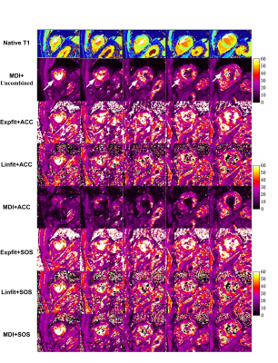 |
Optimizing Myocardium T2* Mapping using a Novel Multi-Dimension Integration (MDI) Method
Wenbo Sun1, Lan Lan1, Junpu Hu2, Yanqun Teng2, Yongquan Ye3, Jingyuan Lyu3, Jian Xu3, Haibo Xu1, and XiaoChun Zhang1
1Department of Radiology, Zhongnan Hospital of Wuhan University, Wuhan University, Wuhan, China, 2United Imaging Healthcare, Shanghai, China, 3UIH America Inc., Houston, TX, United States
Traditional myocardium T2* mapping based on curve fitting methods generally suffers from artifacts, suboptimal SNR and poor reproducibility. This study aimed to evaluate the benefits of a novel multi-dimension integration (MDI) method on myocardium T2* mapping. Five patients with known myocardial infarction were recruited. The MDI method was compared with curve fitting methods based on exponential and linear models. Results showed our MDI method had higher image quality and diagnose sensitivity, as well as much faster computation speed than curve fitting methods. MDI has the potential to provide accurate and reliable measurements for myocardial iron content.
|
|
2053.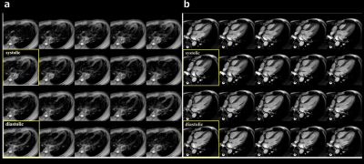 |
Motion-Sensitive (MoSe) CINE imaging: utility for improving robustness of myocardial quantitative mapping
Isao Shiina1, Michinobu Nagao2, Masami Yoneyama3, Yasuhiro Goto1, Kazuo Kodaira1, Yutaka Hamatani1, Mamoru Takeyama1, Isao Tanaka1, and Shuji Sakai2
1Department of Radiological Services, Women's Medical University Hospital, tokyo, Japan, 2Department of Diagnostic imaging & Nuclear Medicine, Women's Medical University Hospital, tokyo, Japan, 3Philips Japan, tokyo, Japan
Motion-Sensitive (MoSe) CINE imaging, based on T2FFE (also known as PSIF) sequence, could clearly visualize the motion-insensitive cardiac timing due to its motion sensitivity and it point out when is the best timing to trigger for both systole and diastole to prevent signal loss and/or motion artifacts effectively. Confirming the MoSe CINE imaging in addition to conventional bSSFP CINE imaging could be useful to determine accurate trigger delay (TD) setting which leads to increase the robustness of image quality in quantitative myocardial mapping and other imaging such as T2-weighted imaging and diffusion-weighted imaging.
|
|
2054.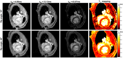 |
Fast T1rho mapping in mice using an optimized Bloch simulation based radial sampling pattern
Maximilian Gram1,2, Daniel Gensler1,3, Patrick Winter1,2, Michael Seethaler2,3, Peter Nordbeck1,3, and Peter Jakob2
1Department of Internal Medicine I, University Hospital Würzburg, Würzburg, Germany, 2Experimental Physics 5, University of Würzburg, Würzburg, Germany, 3Comprehensive Heart Failure Center (CHFC), University Hospital Würzburg, Würzburg, Germany
Myocardial T1ρ-mapping in small animal studies is still a challenging procedure. Commonly, T1ρ-mapping requires long measurement times or only provides insufficient image quality due to a low signal-to-noise-ratio. Using a novel approach based on an optimized radial sampling pattern and high flip angles, we were able to overcome both restrictions. By a special sorting of golden angles based on Bloch simulations the image quality of the method could be significantly increased, and a high quantification accuracy could be realized. Thus, the new approach is a reliable method for fast T1ρ-mapping in future studies on small animals.
|
|
2055. |
Reproducibility of diffusion tensor imaging (DTI) on 12 clinical scanners: Towards validation of cardiac DTI sequences.
Irvin Teh1, William Romero2, Erica Dall'Armellina1, Daniel Ennis3, Pedro F. Ferreira4, Prateek Kalra5, Arunark Kolipaka5, Sebastian Kozerke6, David Lohr7, Kevin Moulin8, Christopher Nguyen9, Sonia Nielles-Vallespin4, Beau Pontre10,
Laura M. Schreiber7, Andrew Scott4, David Sosnovik9, Christian T. Stoeck6, Cyril Tous11, Elizabeth Tunnicliffe12, Vicky Wang13, Andreas M. Weng14, Alistair Young15, Pierre Croisille2, Magalie Viallon2, and Jürgen E. Schneider1
1Leeds Institute of Cardiovascular and Metabolic Medicine, University of Leeds, Leeds, United Kingdom, 2Université de Lyon, UJM-Saint-Etienne, INSA, CNRS UMR 5520, INSERM U1206, CREATIS, F-42023, Saint Etienne, France, 3Department of Radiology, VA Palo Alto Health Care System, Palo Alto, CA, United States, 4Royal Brompton Hospital and Imperial College, London, United Kingdom, 5Department of Radiology, The Ohio State University Wexner Medical Center, Columbus, OH, United States, 6Institute for Biomedical Engineering, University and ETH Zurich, Zurich, Switzerland, 7Department of Cardiovascular Imaging, Comprehensive Heart Failure Center, Würzburg, Germany, 8Stanford University, Stanford, CA, United States, 9Massachusetts General Hospital and Harvard Medical School, Boston, MA, United States, 10Department of Anatomy and Medical Imaging, The University of Auckland, Auckland, New Zealand, 11Department of Radiology, Radiation-Oncology and Nuclear Medicine and Institute of Biomedical Engineering, Université de Montréal, Montréal, QC, Canada, 12University of Oxford, Oxford, United Kingdom, 13San Francisco Veteran Affairs Medical Center, San Francisco, CA, United States, 14Department of Diagnostic and Interventional Radiology, University Hospital Würzburg, Würzburg, Germany, 15Department of Biomedical Engineering, King's College London, London, United Kingdom Poster Permission Withheld
Cardiac diffusion tensor imaging (DTI) is increasingly used for non-invasive in vivo characterisation of cardiac microstructure. To help reconcile the variation in DTI metrics in the literature, we investigated the inter- and intra-site variation of DTI across twelve clinical scanners. Data were acquired at two time points, with a standardised isotropic phantom, acquisition, and post-processing pipeline. In water at 0°C, the coefficient of variation of mean diffusivity (MD) across sites was 1.9 ± 1.4% (mean ± SD), while the mean difference across two scans was (-0.010 ± 0.098) × 10-3 mm2/s (mean ± 1.96SD), indicating low bias and good reproducibility.
|
|
2056. |
Fast Myocardial T1 Mapping in Mice at 11.7 T using DESPOT and continuous tiny golden Angle Acquisitions
Tobias Speidel1, Hao Li1, Micha Bischoff1, Patrick Metze2, and Volker Rasche2
1Core-Facility Small Animal Imaging, Ulm University, Ulm, Germany, 2Experimental Cardiovascular MRI, Ulm University Medical Center, Ulm, Germany
In magnetic resonance imaging, T1 mapping is a valuable tool for investigating structural alterations of the heart due to cardiomyopathy. This work presents a fast T1 mapping method based on DESPOT in combination with continuous radial tiny golden angle acquisitions. The suggested method was tested at 11.7 T and compared to DESPOT based on respiratory and cardiac gated FLASH data. Average values of both measurements are in good accordance to one another as well as to recent literature findings. The presented method is capable of acquiring qualitative and quantitative 2D T1 maps of the myocardium in under 25 s.
|
|
2057. |
Impact of calibration-based coil combination on myocardial fibrosis assessment with a 3D Delayed Myocardial Enhancement sequence prototype
Gaspar Delso1, Paz Garre2, Francisco Alarcón2, Daniel Lorenzatti2, Julián Vega2, Teresa M. Caralt2, Adelina Doltra2, José T. Ortiz-Pérez2, Rosario J. Perea2, Susanna Prat2, Lluís Mont2, Marta Sitges2, and Martin Janich3
1ASL MR, GE Healthcare, Barcelona, Spain, 2Hospital Clínic de Barcelona, Barcelona, Spain, 3GE Healthcare, Munich, Germany
We present the results of a comparative study of the impact of clinically available MR coil combination methods on the diagnostic value of 3D Delayed Myocardial Enhancement data, with a focus on arrhythmogenic tissue analysis.
|
 Watch the Video
 View the Poster
2058.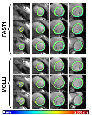 |
Fast single-breathhold 2D multi-slice myocardial T1 mapping (FAST1) at 3T for time-efficient full left ventricular coverage
Li Huang1, Radhouene Neji1,2, Muhummad Sohaib Nazir1, Amedeo Chiribiri1, Reza Razavi1, and Sébastien Roujol1
1School of Biomedical Engineering and Imaging Sciences, Faculty of Life Sciences and Medicine, King's College London, London, United Kingdom, 2MR Research Collaborations, Siemens Healthcare Limited, Frimley, United Kingdom
Modified Look-Locker inversion recovery (MOLLI) as a commonly used myocardial T1 mapping approach shows high precision and reproducibility. Its limited capability in multi-slice acquisition per breathhold prolongs examination time in cases of desired full left ventricular coverage. The previously proposed fast single-breathhold 2D multi-slice myocardial T1 mapping (FAST1) technique can achieve time-efficient full left ventricular coverage at 1.5T. In this work, this capability of FAST1 at 3T is optimized and characterized. Compared to MOLLI, FAST1 can yield 4-fold increase of spatial coverage, limited penalty of T1 spatial variability, no significant difference of T1 repeatability and linearly correlated T1 values.
|
|
2059.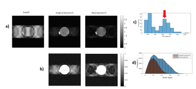 |
Improved Image Quality for 3D LGE Scar Imaging in Patients with Fast and Variable Heart Rate: a Simulation Study.
Jack Allen1,2, Peter Gatehouse1,2, Rick Wage1, David Firmin1,2, and Jennifer Keegan1,2
1Cardiovascular Magnetic Resonance Unit, Royal Brompton and Harefield NHS Trust, London, United Kingdom, 2National Heart and Lung Institute, Imperial College London, London, United Kingdom
3D LGE is used to assess scar in patients with atrial fibrillation. However, the fast and variable heart rate in these patients results in poor image quality. An existing dynamic-TI method varies inversion time on a beat-by-beat basis (according to the previous cardiac cycle length) to improve myocardial nulling, but blood signal variations are incompletely corrected and cause ghosting. We have developed an improved technique which bases the beat-by-beat TI on the history of RR intervals (rather than the previous one) and reduces blood signal variations while maintaining myocardial nulling. Simulations with patient RR interval distributions show significantly improved results.
|
|
2060.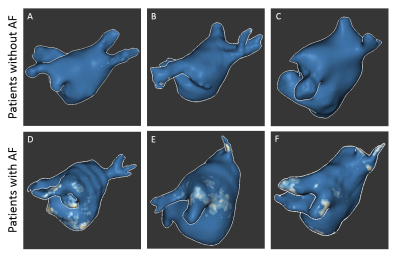 |
3D left atrial LGE MRI at 1.5 Tesla: calibration of fibrosis cut point and initial evaluation in patients with and without atrial fibrillation
Suvai Gunasekaran1, Mohammed S.M. Elbaz1, Daniel Lee1, Michael Markl1, Rod Passman1, and Daniel Kim1
1Northwestern University, Chicago, IL, United States
Given the thin nature of the left atrial (LA) wall and the need to perform respiratory gating, the clinical translation of LA late gadolinium-enhanced (LGE) MRI has proven challenging, particularly at 1.5 Tesla. To address this, a recent study described development of a self-navigated, free-breathing 3D LA LGE pulse sequence using stack-of-stars k-space sampling and XD-GRASP reconstruction. Here, we aim to derive the cut point for LA fibrosis for this 3D LA LGE technique, by using image intensity ratio (LA wall/blood signal) of healthy volunteers, and evaluate LA fibrosis in patients with and without atrial fibrillation.
|
|
2061.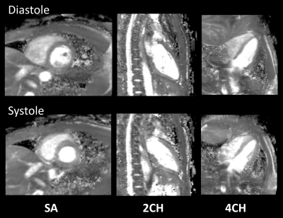 |
Rapid free-breathing 3D isotropic whole-heart diastolic and systolic myocardial T2mapping
Masami Yoneyama1, Michinobu Nagao2, Yasuhiro Goto3, Shuo Zhang4, Isao Shiina3, Kazuo Kodaira3, Yutaka Hamatani3, and Marc Van Cauteren5
1Philips Japan, Tokyo, Japan, 2Department of Diagnostic Imaging and Nuclear Medicine, Tokyo Women’s Medical University, Tokyo, Japan, 3Department of Radiological Services,, Tokyo Women’s Medical University, Tokyo, Japan, 4Philips Healthcare, Hamburg, Germany, 5Philips Healthcare, Best, Netherlands
Current myocardial T2-mapping based on either segmented or single-shot readout obtains one 2D slice per breath hold. To overcome the limitations in spatial coverage and breath hold dependence, we report free-breathing 3D isotropic whole-heart T2 mapping using a T2-prepared segmented gradient echo sequence with interleaved scan, which enables quantitative mapping at multiple cardiac phases such as late diastole and systole. We demonstrate the feasibility of this approach with comparison to the conventional techniques in healthy volunteer examination.
|
|
2062.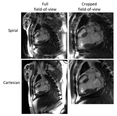 |
Late gadolinium enhancement imaging at low field using through-time spiral GRAPPA
Dominique Franson1, Rajiv Ramasawmy2, Nicole Seiberlich1,3, and Adrienne Campbell-Washburn2
1Case Western Reserve University, Cleveland, OH, United States, 2National Heart, Lung, and Blood Institute, National Institutes of Health, Bethesda, MD, United States, 3University of Michigan, Ann Arbor, MI, United States
A free-breathing late gadolinium enhancement scan may be beneficial for patients who have difficulty with breath-holds. However, the fast data acquisition needed may be difficult to achieve at low field strengths. Here, an undersampled spiral (R=3) acquisition with a through-time spiral GRAPPA reconstruction is used to achieve high spatial (1.25 x 1.25 mm2) and temporal (139 ms acquisition window) resolution for LGE imaging at 0.55T. The resulting images have qualitatively good contrast-to-noise between lesions and healthy myocardium, with acceptable SNR and without motion blurring.
|
|
2063.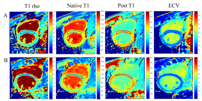 |
Quantitative assessment of the effect of alcohol on myocardial tissues with T1 rho and T1 mapping
Xiaomeng Wu1, Shuai Liu2, Zhangxuan Hu2, Tan Gong1, Xue Lin3, Hua Guo2, Weitian Chen4, Fei Shang1, and Xihai Zhao2
1Department of Biomedical Engineering, Beijing Institute of Technology, Beijing, China, 2Center for Biomedical Imaging Research, Department of Biomedical Engineering, School of Medicine, Tsinghua University, Beijing, China, 3Department of Cardiology, Peking Union Medical College Hospital, Beijing, China, 4Department of Imaging and Interventional Radiology, The Chinese University of Hong Kong, Hong Kong, China
Quantitative Cardiovascular Magnetic Resonance (CMR) imaging has been widely used in detection of myocardial diseases. In this study, T1 rho and T1 mapping MR imaging were performed in subjects with and without alcohol consumption to assess the impact of drinking on the heart. The results suggested that compared with healthy controls, heavy alcohol consumption may lead to detectable changes in myocardial tissues, while moderate alcohol consumption showed no significant difference. In addition, T1 mapping was more sensitive to detect myocardial changes caused by alcohol than T1 rho.
|
|
2064.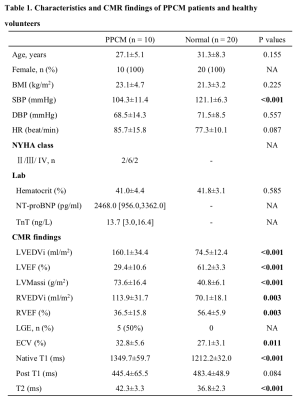 |
Dynamic changes of tissue characteristics by T1 and T2 mapping in patients with peripartum cardiomyopathy
Yaodan Liang1, Yuanwei Xu2, Weihao Li2, Jing An3, and Yucheng Chen2
1Department of Cardiology, Beijing Hospital, Beijing, China, 2Department of Cardiology, West China Hospital, Chengdu, China, 3Siemens Shenzhen Magnetic Resonance Ltd, Shenzhen, China
This study aimed to demonstrate the dynamic changes of tissue characteristics in patients with peripartum cardiomyopathy (PPCM). Cardiovascular magnetic resonance (CMR) examination was performed using a 3.0T MR scanner, consisted of a MOLLI sequence for T1 mapping, and a T2 prep with bSSFP for T2 mapping. Ten PPCM patients with repeated CMR and 20 healthy age-matched females were analyzed. After a median 15.3-month follow-up, five patients had a recovered left ventricular ejection fraction. PPCM patients showed significantly higher native T1, extracellular volume, and T2 than normal controls. Furthermore, native T1 decreases significantly in the follow-up CMR of PPCM patients.
|
|
2065.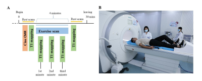 |
The repeatability in the assessment of hyperemic myocardium native T1 using a MRI-compatible ergometer pedal exerciser.
Bo He1, Jie Zheng2, and Fabao Gao1
1Radiology, Medical Imaging,West China School of Medicine, Sichuan University, Chengdu, Sichuan, China, 2Mallinckrodt Institute of Radiology, Washington, WA, United States
10 healthy volunteers underwent rest and stress Free-breathing myocardial native T1 mapping CMR scanning twice in a 3.0T MR scanner in midventricular short-axis position using a custom-made MRI-compatible ergometer pedal exerciser. It was demonstrated that T1 reactivity (ΔT1 values) had a moderate reliability and an excellent repeatability. The assessment of native myocardial T1 value using the pedal exerciser CMR stress/rest T1 mapping holds promise for myocardium ischemia detection without injecting gadolinium contrast agents and pharmacologic agents, also shows an excellent repeatability to demonstrates for the first time that normal myocardium has distinctive ranges of T1 reactivity to exercise stress.
|
|
2066.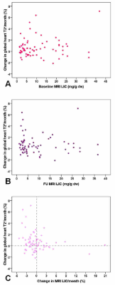 |
Correlation between changes in cardiac iron and hepatic iron in pediatric patients with thalassemia major
Antonella Meloni1, Maddalena Casale2,3, Aldo Filosa2, Laura Pistoia1, Vincenzo Positano1, Emanuele Grassedonio4, Elda Chiara Resta5, Gennaro Restaino6, Riccardo Righi7, Lorella Pitrolo8, Antonella Quarta9, and Alessia Pepe1
1MRI Unit, Fondazione G. Monasterio CNR-Regione Toscana, Pisa, Italy, 2Azienda Ospedaliera di Rilievo Nazionale "A. Cardarelli", Napoli, Italy, 3Università degli Studi della Campania Luigi Vanvitelli, Napoli, Italy, 4Policlinico "Paolo Giaccone", Palermo, Italy, 5Ospedale “SS. Annunziata”, Taranto, Italy, 6Fondazione di Ricerca e Cura "Giovanni Paolo II", Campobasso, Italy, 7Ospedale del Delta, Lagosanto (FE), Italy, 8Ospedale "V. Cervello", Palermo, Italy, 9Ospedale "A. Perrino", Brindisi, Italy
In pediatric thalassemia major patients who performed a baseline and a follow-up MRI study at 18±3 months the percentage changes in global heart T2* values per month were not influenced by initial hepatic iron levels and were not correlated to final hepatic iron. Moreover, the correlation between % changes in global heart T2* and MRI liver iron concentration values did not reach the statistical significance So, our data seem not supporting the hypothesis for which it is necessary to clean the liver before removing iron from the heart.
|
|
2067.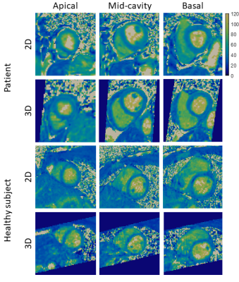 |
Accelerated and non-rigid motion-corrected 3D T2 mapping in a 3T PET-MR system
Alina Psenicny1, Camila Munoz1, Aurélien Bustin1, Karl P Kunze2, Radhouene Neji1,2, Pier-Giorgio Masci1, René M Botnar1, and Claudia Prieto1
1Biomedical Engineering Department, School of Biomedical Engineering and Imaging Sciences, King's College London, London, United Kingdom, 2MR Research Collaborations, Siemens Healthcare Limited, Frimley, United Kingdom
Simultaneous 18F-FDG PET-MR imaging has shown promise for improved diagnostic accuracy of inflammatory cardiac diseases, such as cardiac sarcoidosis. However, respiratory motion and mis-registration between free-breathing 3D PET and conventional 2D breath-held MR images remain a challenge that has hindered clinical adoption of this technique. Here we introduce a 3x-accelerated free-breathing motion-corrected 3D whole-heart T2-mapping prototype sequence, which provides characterisation of myocardial inflammation while additionally providing non-rigid respiratory deformation fields to correct simultaneously acquired PET data. Results from phantom, healthy subjects and patients show that the method produces good-quality high-resolution 3D T2 maps from an efficient scan of ~10 minutes.
|
|
2068.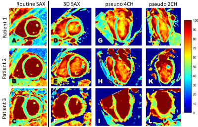 |
Respiratory motion-registered isotropic 3D whole-heart T2 mapping in patients with suspected inflammatory myocardial injury
Karolina Dorniak1, Lorenzo Di Sopra2, Agniezka Sabisz3, Anna Glinska3, Christopher W Roy2, Kamil Gorczewski4, Davide Piccini2,5, Jérôme Yerly2,6, Jadwiga Fijałkowska3, Edyta Szurowska3, Matthias Stuber2,6, and Ruud B van Heeswijk2
1Department of Noninvasive Cardiac Diagnostics, Medical University of Gdansk, Gdansk, Poland, 2Radiology, Lausanne University Hospital (CHUV) and University of Lausanne (UNIL), Lausanne, Switzerland, 32nd Department of Radiology, Medical University of Gdansk, Gdansk, Poland, 4Siemens Healthineers, Erlangen, Germany, 5Advanced Clinical Imaging Technology, Siemens Healthcare AG, Lausanne, Switzerland, 6Center for Biomedical Imaging (CIBM), Lausanne, Switzerland
T2 mapping can be used to effectively detect myocardial edema and inflammation. However, the focal nature of myocardial inflammation may render 2D approaches suboptimal and make whole-heart isotropic 3D mapping desirable. Unfortunately, at 1.5T, self-navigated 3D radial bSSFP results in too noisy images for adequate T2 mapping. In this study, we therefore used a respiratory motion-resolved reconstruction together with image registration to improve the 3D T2 mapping precision and accuracy at 1.5T in patients with inflammatory myocardial injury. The resulting myocardial T2 values matched those of the routine 2D T2 maps, with no discernible bias and slightly lower precision.
|
|
2069.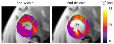 |
Multiphase CINE T2*-mapping of the mouse heart at 9.4 tesla
Sandra Lehmann1,2, Min-Chi Ku1,3, Joao dos Santos Periquito1, Han Haopeng1, Andreas Pohlmann1, and Thoralf Niendorf1,3,4
1Berlin Ultrahigh Field Facility (B.U.F.F.), Max Delbrück Center for Molecular Medicine in the Helmholtz Association, Berlin, Germany, 2Technische Universität Berlin, Berlin, Germany, 3DZHK (German Centre for Cardiovascular Research), partner site Berlin, Germany, 4Experimental and Clinical Research Center, Charite Medical Faculty and the Max Delbrueck Center for Molecular Medicine in the Helmholtz Association, Berlin, Germany
Hypertrophic cardiomyopathy (HCM) is the most frequent inherited monogenic heart disease and could lead to heart failure or even sudden cardiac death. Effective relaxation time T2* is related to different physiological parameters. Dynamic CINE mapping of T2* covering the entire cardiac cycle facilitates distinction of healthy and pathologic myocardial tissue. Here we propose, implement, evaluate and apply an effective approach of retrospective cardiac gating that affords for the first time CINE T2*-mapping in mice at 9.4T and provides the technological basis for translational research into a deeper understanding of the mechanisms underlying hypertrophic cardiomyopathy.
|
|
2070.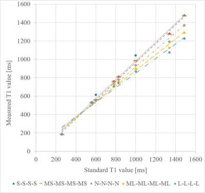 |
Evaluation of factors contributing to the correction of the effect of heartrate variation on MOLLI using BlochSolver
Yuta Endo1, Rei Ikegawa1, Kuninori Kobayashi1, Makoto Amanuma1, and Shigehide Kuhara1
1Kyorin University Faculty of Heaith Science, Mitaka, Japan
Fast cardiac T1 mapping including MOLLI with ECG-gating leads to poorer T1 measurement accuracy, since the recovery time of the longitudinal magnetization changes with heart rate variation. Ikekawa et al. proposed correction methods for the changes in the inversion recovery time at each sampling point and in that of longitudinal magnetization on the heartrate variation; however, the specific factor that predominantly improves T1 measurement remains unclear. Here, we investigated the dominant effect of the proposed correction method in the effect of heartrate variation on T1 measurement using BlochSolver.
|
|
2071.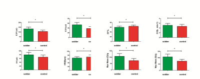 |
Cardiac Remodeling After Military Endurance Training: A Preliminary Study of Cardiac MRI Cine
Hongqin Liang1, Liqiang Zhu2, Bing Ji1, Qing Li3, Xiaoyue Zhou4, and Jian Wang1
1Southwest Hospital, Army Medical University, Chongqing, China, 2The Central Theater Command Air Force Hospital of PLA, Datong, China, 3Siemens Healthineers Ltd., Shanghai, China, 4Siemens Healthcare Ltd, Shanghai, China
Long-term military training leads to the remodeling of the heart and may cause exercise-induced myocardial injury (EIMI). The mechanism of heart remodeling is studied by using cardiac cine MRI to estimate left ventricular cardiac function and global stress. This study found that prolonged and intensive training lasting 4 years leads to cardiac morphological adaptations. Contrary to conventional thinking, left ventricular function changes happens earlier than the mechanical direction.
|
|
2072.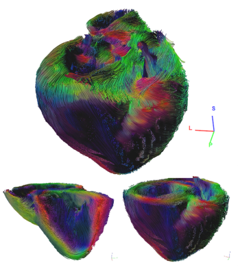 |
High Resolution Imaging of the Ex-Vivo Porcine Heart at 7T Using a Dedicated Custom Built 8TX/16RX Array: Quality Assessment
David Lohr1, Maxim Terekhov1, Ibrahim A. Elabyad1, Franziska Veit2, and Laura Maria Schreiber1
Video Permission Withheld
1Chair of Cellular and Molecular Imaging, Comprehensive Heart Failure Center (CHFC), University Hospital Würzburg, Würzburg, Germany, 2Chair Tissue Engineering and Regenerative Medicine (TERM), University Hospital Würzburg, Würzburg, Germany
MRI measurements of ex-vivo hearts at ultrahigh field strengths (≥7T) can provide high resolution, high fidelity ground truth data that complement clinical cardiac MRI. In this study we demonstrate ex-vivo sample preparation steps and capabilities of a custom built, multiple element transceiver array dedicated to high resolution imaging of ex-vivo hearts. Measurements included whole heart data for T2* maps, a high resolution 3D FLASH, as well as high resolution DTI. We found that receive sensitivity of the dedicated coil was superior to a commercial head coil and that SNR was similar, even without the application of B1-shim.
|
|
2073.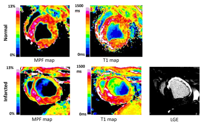 |
Macromolecular Proton Fraction Mapping Enables Endogenous Contrast Between Normal and Infarcted Myocardium.
Anna V Naumova1, Lauren E Neidig2,3,4, Hiroshi Tsuchida2,3, Kenta Nakamura5, Charles E Murry2,3, and Vasily L Yarnykh1
1Radiology, University of Washington, Seattle, WA, United States, 2Pathology, University of Washington, Seattle, WA, United States, 3Institute for Stem Cells and Regenerative Medicine, University of Washington, Seattle, WA, United States, 4Comparative Medicine, University of Washington, Seattle, WA, United States, 5Cardiology, University of Washington, Seattle, WA, United States
This pilot study shows the feasibility of fast MPF mapping in cardiac applications in the clinical magnetic field using a large-animal model. The results demonstrate that MPF maps present the effective source of the quantitative soft tissue endogenous contrast in sub-acute myocardial infarction and can be obtained with high spatial and temporal resolution.
|
2074.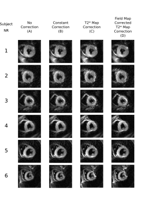 |
Correcting for T2* related blurring in spiral MR: A novel method compensating for spatial variations in T2*
Malte Roehl1,2, Peter Gatehouse1,2, Pedro Ferreira1,2, Sonia Nielles-Vallespin1,2, Sonya Babu-Narayan1,2, Margarita Gorodezky1,2, Hui Xue3, Peter Kellman3, Dudley Pennell1,2, David Firmin1,2, and Andrew Scott1,2
1Imperial College London, London, United Kingdom, 2Royal Brompton Hospital, London, United Kingdom, 3NIH, Bethesda, MD, United States
In regions of short T2*decay during long spiral k-space covering readouts can cause significant image blurring. In this work we introduce a novel correction for spatially varying T2* including a stimulated echo-based spiral T2* mapping sequence and compare it to an existing correction for constant T2*. Using both computational simulations and in-vivo spiral cardiac diffusion tensor data we show that the spatially varying correction improves the sharpness compared to the constant correction.
|
|
2075.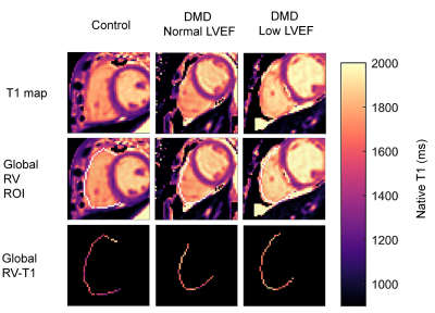 |
Right ventricular T1-mapping in boys with Duchenne muscular dystrophy and healthy controls at 3T
Seraina A. Dual1,2, Nyasha G. Maforo3,4, Patrick Magrath1,5, Doff B. McElHinney6, Ashley Prosper5, Holden H. Wu4,5, Nancy Halnon7, Shiraz Maskatia6,8, Pierangelo Renella3,5, and Daniel B. Ennis1,8,9
1Department of Radiology, Stanford University, Palo Alto, CA, United States, 2Department of Cardiothoracic Surgery, Stanford University, Palo Alto, CA, United States, 3Physics and Biology in Medicine Interdepartmental Program, University of California, Los Angeles, CA, United States, 4Department of Radiological Sciences, University of California, Los Angeles, CA, United States, 5Department of Bioengineering, University of California, Los Angeles, CA, United States, 6Department of Pediatrics, Stanford University, Palo Alto, CA, United States, 7Department of Pediatrics, University of California, Los Angeles, CA, United States, 8Maternal & Child Health Research Insitute, Palo Alto, CA, United States, 9Cardiovascular Insitute, Stanford University, Palo Alto, CA, United States
Early diagnosis of cardiac involvement in boys with Duchenne muscular dystrophy (DMD) allows for timely therapy. The aim of this study was to assess the association between native T1 in the right ventricle (RV) and cardiac involvement as marked by decreased left ventricular (LV) or RV ejection fraction (EF). Healthy boys (N=10) and boys with DMD (N=16) underwent 3T cardiac MR using motion-corrected gradient-MOLLI T1-mapping, a clinically routine protocol. RV-T1 did not correlate with LVEF or RVEF. Longer native LV-T1 was associated with lower LVEF. Based on this data, native RV-T1 does not provide insight to cardiac involvement in DMD.
|
|
2076.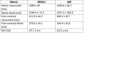 |
Arrhythmia Insensitive Rapid versus Modified Look Locker Inversion Recovery T1 mapping in mitral valve prolapse patients
Ernest Cheung1, Hui-Chen Han1, Emma Hornsey1, Leonid Churilov2,3, Kyung Pyo Hong4, Han Lim1,2, Julie Smith1, Daniel Kim4, and Ruth Lim1,2
1Austin Health, Melbourne, Australia, 2The University of Melbourne, Melbourne, Australia, 3The Florey Institute of Neuroscience and Mental Health, Melbourne, Australia, 4Northwestern University, Evanston, IL, United States
We compare two cardiac T1 mapping techniques, arrhythmia insensitive rapid (AIR) which is a saturation recovery technique acquired in a short breath-hold and robust to arrhythmia, and Modified Look-Locker inversion recovery (MOLLI), in 55 patients with mitral valve prolapse. There was excellent inter-reader agreement in T1 values and extracellular volume (ECV) between techniques. However, higher T1 values were observed in AIR compared to MOLLI and vice versa for ECV, consistent with previous studies reporting significantly different T1 and ECV values between inversion recovery and saturation recovery techniques. These differences are important to consider when applying T1 mapping to clinical practice.
|
|
2077.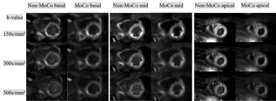 |
Improvement of distortion-free cardiac diffusion weighted imaging (DWI) using motion-compensated single-shot turbo spin-echo (MoCo-TSE) DWI
Yasuhiro Goto1, Michinobu Nagao2, Masami Yoneyama3, Isao Shiina1, Kazuo Kodaira1, Yutaka Hamatani1, Mamoru Takeyama1, Isao Tanaka1, and Shuji Sakai2
1Department of Radiological Services, Tokyo Women's Medical University Hospital, Tokyo, Japan, 2Department of Diagnostic imaging & Nuclear Medicine, Tokyo Women's Medical University Hospital, Tokyo, Japan, 3Philips Japan, Tokyo, Japan
We compared the image quality of the motion-compensated (MoCo) and conventional (non-MoCo) cardiac TSE-DWI by visual evaluation and homogeneity evaluation. MC-TSE-DWI achieved superior image quality compared with non-MoCo-TSE-DWI in all b-values and all segments. MoCo-TSE-DWI improves the image quality and may increase the diagnostic potential.
|
|
2078.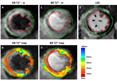 |
Dark-blood Prepared T2* Cardiac MRI Based Assessment of Intramyocardial Hemorrhage in Patients with Reperfusion Acute MI at 1.5T
Xingmin Guan1,2, Yinyin Chen1,3,4, Hsin-Jung Yang1, and Rohan Dharmakumar1
1Cedars-Sinai Medical Center, Los Angeles, CA, United States, 2University of California, Los Angeles, Los Angeles, CA, United States, 3Radiology, Zhongshan Hospital, Fudan University, Shanghai, China, 4Medical Imaging, Shanghai Medical School, Shanghai Institute of Medical Imaging, Shanghai, China
Double inversion recovery preparation is often used in T2* cardiac MRI to acquire dark-blood images. We investigated the impact of dark-blood preparation on imaging of intramyocardial hemorrhage in patients with acute MI at 1.5T. SNR and CNR were compared between dark-blood and bright-blood T2* weighted images. Hemorrhage size, T2* were evaluated and compared between methods. Inter-observer reliability were reported as intraclass correlation coefficients. Our findings here support the notion that when choosing between bright-blood and dark-blood T2* cardiac MRI for the determination of intramyocardial hemorrhage in patients at 1.5T, bright-blood T2* cardiac MRI is likely the preferred approach.
|
|
2079.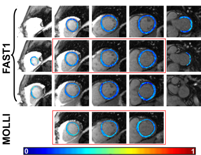 |
Extra-cellular volume (ECV) mapping using fast single-breathhold 2D multi-slice myocardial T1 mapping (FAST1) at 1.5T
Li Huang1, Radhouene Neji1,2, Filippo Bosio1, Amedeo Chiribiri1, Reza Razavi1, and Sébastien Roujol1
1School of Biomedical Engineering and Imaging Sciences, Faculty of Life Sciences and Medicine, King's College London, London, United Kingdom, 2MR Research Collaborations, Siemens Healthcare Limited, Frimley, United Kingdom
Extra-cellular volume (ECV) mapping using combined native and post-contrast myocardial T1 maps shows promise in assessing cardiomyopathies. However, its feasibility for full left ventricular (LV) coverage is limited using conventional myocardial T1 mapping techniques such as modified Look-Locker inversion recovery (MOLLI) with single-slice acquisition per breathhold. The previously proposed fast single-breathhold 2D multi-slice myocardial T1 mapping (FAST1) technique can provide time-efficient full LV coverage. In this work, its capability for ECV mapping with full LV coverage at 1.5T is evaluated. Compared to MOLLI, FAST1 yields 4-fold increased spatial coverage, limited penalty of ECV spatial variability and highly correlated ECV values.
|
|
2080.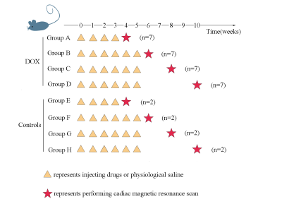 |
Identifying early stages of doxorubicin-induced cardiotoxicity in rat model using 7.0 tesla cardiac magnetic resonanc
Shiyu Wang1 and Fabao Gao1
1Department of Radiology, West China Hospital / West China School of Medicine, Chengdu, China
the aim of this study is to identify early stages of doxorubicin (DOX)-induced cardiotoxicity in rat model using 7.0 tesla cardiac magnetic resonance (CMR) combining creatine kinase isoenzymes (CKMB).Comparison of Ejection fraction (EF), Global radial (GRS), circumferential (GCS) and longitudinal strain (GLS) parameters of LV and CKMB between experimental groups and controls at 4,6, 8, 10 weeks.CKMB can detect DOX-induced cardiotoxicity at the earliest time. Decreased GLS during treatment identifies impaired LV mechanic as an early marker of DOX-induced cardiotoxicity, in the absence of EF. CKMB and CMR-TT are an effective method to evaluate DOX-induced cardiotoxicity at early stage.
|
|
2081.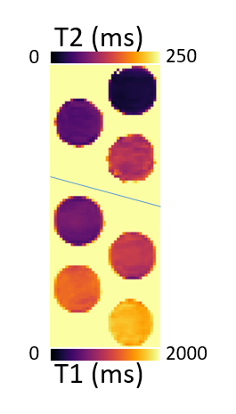 |
3D cardiac T1 and T2 mapping in a single breath-hold via compressed SENSE at 3T
Tiago Ferreira da Silva1,2, Paula Montesinos1, Carlos Galan-Arriola2, Robert Austin Benn2, Borja Ibañez2,3, and Javier Sánchez-González1
1Philips Healthcare Iberia, Madrid, Spain, 2Centro Nacional de Investigaciones Cardiovasculares Carlos III, Madrid, Spain, 3Department of Cardiology, IIS-Fundación Jiménez Díaz Hospital, Madrid, Spain We propose a new 3D simultaneous T1 and T2 mapping technique to acquire accurate co-registered 3D T1 and T2 maps of the entire left ventricle in a single breath-hold. The technique combines T2-prep and saturation pulses with compressed SENSE. The proposed sequence successfully acquired co-registered 3D T1 and T2 maps in a single breath-hold of 17 s (HR=60 bpm). Phantom estimated T1 and T2 values were in agreement with gold-standard values obtained from IR-SE and GraSE, respectively. Accuracy and precision results were similar to previously published values for saturation recovery at 3T. |
|
2082.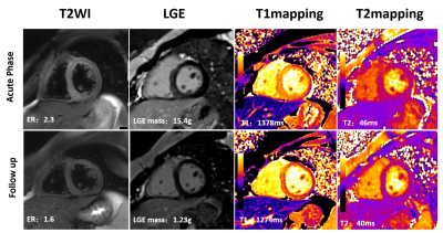 |
Tissue Characterization by Mapping and Strain Cardiac MR for Detection and Monitoring of Myocardial Injury in Fulminant Myocarditis
Haojie Li1, Hui Zhu1, Zhaoxia Yang1, and Liming Xia1
1Radiology, Tongji Hospital, Tongji Medical College, Huazhong University of Science and Technology, Wuhan, China
Fulminant myocarditis is a rare form of acute severe myocarditis and accurate detection and monitoring of the inflammatory myocardial edema is essential for clinical decision-making. We therefore evaluated the ability of multiparametric CMR to detect and monitor the inflammation myocardial alterations and to differentiate acute and convalescent stage of fulminant myocarditis.
|
|
2083.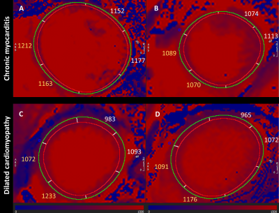 |
T1 mapping in chronic myocarditis, dilated cardiomyopathy and hypertensive heart disease using two Modified Look-Locker schemes
Maria Teodora Antuaneta Wetscherek1, Christian Lücke2, Philipp Lurz3, and Matthias Gutberlet2
1Radiology Department, Cambridge University Hospitals NHS Foundation Trust, Cambridge, United Kingdom, 2Department of Diagnostic and Interventional Radiology, University Leipzig – Heart Center Leipzig, Leipzig, Germany, 3Department of Cardiology, University Leipzig – Heart Center Leipzig, Leipzig, Germany
.The purpose of our study was to evaluate T1-mapping for the comparison of chronic myocarditis, nonischemic dilated cardiomyopathy (DCM) and hypertensive heart disease using two MOLLI schemes in a clinical setting. We prospectively enrolled 81 patients investigated for suspected myocarditis that underwent a cardiac magnetic resonance protocol including consecutively acquired 3(3)3(3)5MOLLI and 5(3)3 MOLLI T1-mapping. We performed an overall and segmental analysis on a single mid ventricular short-axis slice. The results from the two MOLLI schemes are very similar. A segmental evaluation of 5(3)3MOLLI data could enable refining diagnosis in clinical practice, particularly when DCM is within the differential diagnoses.
|
|
2084.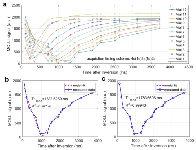 |
A novel generalized modeling of modified Look-Locker inversion recovery (MOLLI) signal to allow a shorter breath-hold and accurate T1 mapping
Seonghwan Yee1 and Seonghwan Yee2
1CU Anschutz medical, Aurora, CO, United States, 2Radiology, University of Colorado Anschutz Medical Campus, Aurora, CO, United States
A novel generalized modeling method is presented here for the MOLLI signal. When compared with the conventional 3-parameter exponential modeling, this new modeling demonstrates the better accuracy, particularly with the larger T1 values in the case of using enhanced acquisition timing scheme (e.g. 4s(1s)3s(1s)2s). The fact that this novel modeling method does not require full recovery of longitudinal magnetization in successive inversion pulses strongly suggests the possibility of further reducing the total MOLLI scan time to the level a few seconds less than 10 s, which would be much beneficial for those patients who have difficulty in holding the breath.
|
|
2085.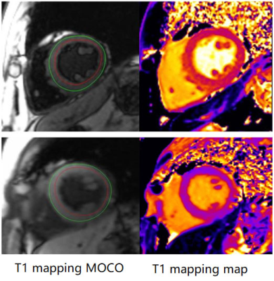 |
Detection of myocardial change in primary aldosteronism and essential hypertension with T1 mapping
Tao Wu1, Yan Ren2, Wei Cheng1, Wei Wang2, Fangli Zhou2, Xiaoyue Zhou3, Yucheng Chen4, and Jiayu Sun1
1Department of Radiology, West China Hospital, Chengdu, China, 2Department of Endocrinology and Metabolism, Adrenal Center, West China Hospital, Chengdu, China, 3MR Collaboration, Siemens Healthineers Ltd, Shanghai, China, 4Department of Cardiology, West China Hospital, Chengdu, China
Primary aldosteronism (PA) increases cardiovascular morbidity. In order to elucidate the early features of heart damage of PA, we used the cardiac MRI and T1 mapping technique to evaluate and compare cardiac function and myocardium changes in PA and essential hypertension (EH) patients. Our results found that the PA patients had higher native T1 than the EH group, while other functional parameters had no significant differences between two groups. The change of myocardium was earlier in PA patients than in EH patients, suggesting that it is mainly due to myocardial fibrosis induced by inappropriate secretion of aldosterone.
|
|
2086.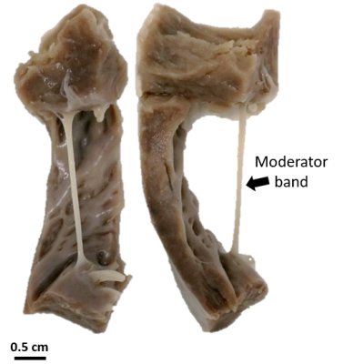 |
Evaluation of Inhomogeneous Magnetization Transfer ihMT at 9.4T for the visualization of the Purkinje Network in the heart
Kylian Haliot1,2,3, Andreea Hertanu4, Olivier Girard4, Lucas Soustelle4, Guillaume Duhamel4, Julie Magat1,2,3, and Bruno Quesson1,2,3
1IHU Liryc, Fondation Bordeaux Université, Pessac, France, 2U1045 CRCTB, Université de Bordeaux, Bordeaux, France, 3INSERM, CRCTB, U1045, Bordeaux, France, 4Aix-Marseille Univ, CNRS, CRMBM, Marseille, France
The Purkinje network (PN) of the heart plays a major role in cardiac electrical diseases such as in sudden cardiac death. Imaging of this fine network is of central interest to better characterize the links between electrical disorders and structural modifications of the heart. PN is composed of fibers containing cardiomyocytes surrounded by a collagen sheath (PF). In this study, we implemented inhomogeneous magnetization transfer (ihMT) sequence at 9.4T and evaluate this technique on ex vivo samples of pig heart fixed into formalin. We show that ihMT is a suitable candidate to increase the contrast between PF and cardiac muscle.
|
|
2087.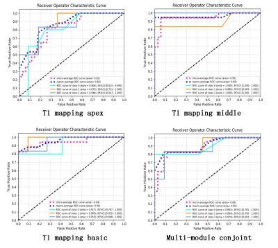 |
Radiomics Analysis for the Differential Diagnosis of Cardiac Hypertrophy with MR T1-mapping
Chenao Zhan1, Tao Ai1, Yayuan Geng2, and Liming Xia1
1Department of Radiology, Tongji Hospital, Tongji Medical College, Huazhong University of Science and Technology, Wuhan, China, 2Huiying Medical Technology Co., Ltd, Beijing, China
In clinical setting, there are three distinguishable entities presented mainly with left ventricular cardiac hypertrophy, including HCM, HHP and CA. Imaging differential diagnosis relies on LGE-CMR, and recently proposed native T1 mapping, which has high diagnostic accuracy and is potentially more sensitive for detecting disease than LGE imaging. However, both modalities are insufficient robustness for the differential diagnosis of three diseases. In this study, radiomics analysis was adopted into the differential diagnosis of cardiac hypertrophy with MR T1-mapping. And diagnostic accuracy of 0.95/0.96 can be achieved with test dataset, which is significantly improved as compared with conventional native T1 mapping.
|
|
2088.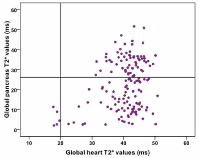 |
Pancreatic iron and cardiac iron and function in patients with thalassemia intermedia
Antonella Meloni1, Laura Pistoia1, Vincenzo Positano1, Giuseppe Peritore2, Paolo Preziosi3, Ada Riva4, Massimiliano Missere5, Antonino Vallone6, Valentina Carrai7, F. Mehtap Pasin3, Mauro Murgia8, and Alessia Pepe1
1MRI Unit, Fondazione G. Monasterio CNR-Regione Toscana, Pisa, Italy, 2"ARNAS" Civico, Di Cristina Benfratelli, Palermo, Italy, 3Ospedale "Sandro Pertini", Roma, Italy, 4Ospedale “SS. Annunziata” ASL Taranto, Taranto, Italy, 5Fondazione di Ricerca e Cura "Giovanni Paolo II", Campobasso, Italy, 6Azienda Ospedaliera "Garibaldi" Presidio Ospedaliero Nesima, Catania, Italy, 7Universitaria Careggi, Firenze, Italy, 8Ospedale San Martino di Oristano, Oristano, Italy
In patients with thalassemia intermedia (TI) pancreatic iron overload is frequent (58.1%), especially if regular transfusions are performed. Pancreatic T2* values were correlated with cardiac T2* values and a normal pancreas T2* showed negative predictive value of 100% for cardiac iron. Global pancreas T2* values were not correlated to biventricular volumes and ejection fraction or to myocardial fibrosis.
|
|
2089.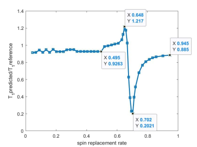 |
Flow Effects on MOLLI T1 Mapping
Zhengyang Ming1,2, Hengjie Liu1,2, Caroline Colbert1,2, Kim-Lien Nguyen1,2,3, and Peng Hu1,2
1Department of Radiological Sciences, David Geffen School of Medicine, University of California, Los Angeles, Los Angeles, CA, United States, 2Physics and Biology in Medicine Inter-Departmental Graduate Program, University of California, Los Angeles, Los Angeles, CA, United States, 3Division of Cardiology, Department of Medicine, David Geffen School of Medicine, University of California, Los Angeles, Los Angeles, CA, United States
T1 relaxation time for myocardial tissue represents a biomarker for a variety of pathologies. High resolution and pixel-wise T1 mapping can be acquired with Modified Look-Locker Imaging1 (MOLLI). Besides, T1 measurements of blood can be used as a reference value to calculate function parameters like extracellular volume (ECV) fraction2,3 and myocardial blood volume4.5 (MBV). Blood flow may affect blood T1 measurements and cause further deviation in calculating function parameters 6. In our work, we proposed a MOLLI-based flow model to simulate flow effect. Substantial T1 deviation may happen when the spin replacement rate is between 0.65 and 0.70.
|
2090.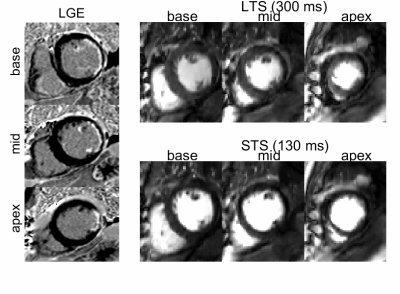 |
Dual-contrast and dual-phase first-pass myocardial perfusion using simultaneous multi-slice imaging
Giulio Ferrazzi1, Sarah McElroy1, Radhouene Neji1,2, Karl Kunze1,2, Muhummad Sohaib Nazir1, Peter Speier3, Daniel Stäb4, Christoph Forman3, Reza Razavi1, Amedeo Chiribiri1, and Sébastien Roujol1
1School of Biomedical Engineering and Imaging Sciences, King's College London, London, United Kingdom, 2MR Research Collaborations, Siemens Healthcare Limited, Frimley, United Kingdom, 3Cardiovascular MR predevelopment, Siemens Healthcare GmbH, Erlangen, Germany, 4MR Research Collaborations, Siemens Healthcare Limited, Melbourne, Australia The clinical need of covering multiple slices in myocardial perfusion constrains the saturation delay time to be short. However, this may be suboptimal in terms of myocardial/defect tissue contrast and blood/myocardium signal ratios. Moreover, it is not possible to acquire the same slice twice, and all slices are at different cardiac phases. In this study, we develop a perfusion sequence which provides dual phase and dual contrast data using simultaneous multi-slice at two different saturation delay times. Simulations and in-vivo data acquired in patients demonstrated a 150% increase of myocardial/defect contrast, and decreased blood/myocardium signal ratio by 60 to 80%. |
|
2091.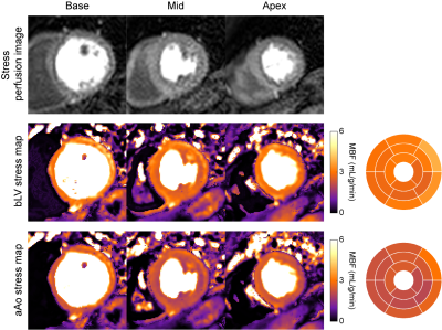 |
Myocardial perfusion quantification by cardiovascular magnetic resonance is significantly affected by the arterial input sampling location
Xenios Milidonis1, Russell Franks1, Kajol Verma1, Torben Schneider1,2, Javier Sánchez-González3, Sven Plein1, and Amedeo Chiribiri1
1King's College London, London, United Kingdom, 2Philips Healthcare, Guilford, United Kingdom, 3Philips Healthcare, Madrid, Spain
The arterial input function (AIF) describes the contrast agent input to the myocardium and is required for blood flow quantification. However, the impact of the AIF sampling location on quantification by cardiovascular magnetic resonance and the diagnosis of coronary artery disease (CAD) has yet to be determined. In this study, perfusion imaging was performed in patients with suspected CAD and blood flow was quantified for seven different locations. It was found that the sampling location has a significant impact on blood flow measurements, while the ascending aorta led to the most accurate prediction of inducible perfusion abnormalities.
|
|
2092.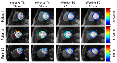 |
The importance of myocardial SNR in myocardial blood flow quantification derived from first-pass cardiac perfusion CMR
Sungtak Hong1, Kyungpyo Hong2, Li-Yueh Hsu3, Daniel C Lee4, and Daniel Kim1
1Radiology, Northwestern University, Chicago, IL, United States, 2Translational and Molecular Imaging Institute and Department of Radiology, Icahn School of Medicine at Mount Sinai, New York, NY, United States, 3National Heart, Lung, and Blood Institute, National Institutes of Health, Bethesda, MD, United States, 4Division of Cardiology, Internal Medicine, Northwestern University, Chicago, IL, United States
We have recently developed a perfusion CMR pulse sequence using golden angle radial acquisitions and CS reconstruction. The radial k-space sampling allows retrospective manipulation of saturation-recovery time (TS) using k-space weighted image contrast (KWIC) filtering. This study was conducted to determine the importance of SNR in myocardial blood flow quantification derived from cardiac perfusion CMR. Our analysis shows that mean resting MBF values decreases with TS, where the result with the highest TS = 95 ms agrees best with literature value reported by cardiac PET studies.
|
|
2093.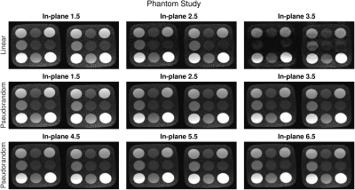 |
SMS-bSSFP perfusion imaging with high spatial resolution and coverage using pseudorandom undersampling and compressed sensing reconstruction
Sarah McElroy1, Giulio Ferrazzi1, Sohaib Nazir1, Karl Kunze2, Radhouene Neji2, Peter Speier3, Daniel Staeb4, Christoph Forman3, Reza Razavi1, Amedeo Chiribiri1, and Sébastien Roujol1
1School of Biomedical Engineering and Imaging Sciences, King's College London, London, United Kingdom, 2MR Research Collaborations, Siemens Healthcare Limited, Frimley, United Kingdom, 3Cardiovascular MR Predevelopment, Siemens Healthcare GmbH, Erlangen, Germany, 4MR Research Collaborations, Siemens Healthcare Pty Ltd, Melbourne, Australia
CMR perfusion imaging requires high temporal resolution, which limits the achievable spatial coverage and spatial resolution using conventional acquisition techniques. Simultaneous multi-slice (SMS) bSSFP perfusion imaging has been previously demonstrated at 1.5 T with matched spatial resolution and doubled spatial coverage compared to conventional protocols. In this work, we have implemented a pseudorandom undersampling scheme for SMS-bSSFP perfusion with compressed sensing reconstruction to increase in-plane acceleration of SMS-bSSFP imaging, enabling high spatial coverage and spatial resolution perfusion imaging at 1.5 T.
|
|
2094.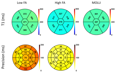 |
Improved precision of T1 estimation for 1.5T quantitative myocardial perfusion imaging using a high flip angle SSFP reference image
Sarah McElroy1, Sohaib Nazir1, Karl Kunze2, Radhouene Neji2, Amedeo Chiribiri1, and Sébastien Roujol1
1School of Biomedical Engineering and Imaging Sciences, King's College London, London, United Kingdom, 2MR Research Collaborations, Siemens Healthcare Limited, Frimley, United Kingdom
Accurate quantification of myocardial blood flow using cardiac MR perfusion imaging requires a linear relationship between the arterial input function and myocardial signal intensity curves. Typically this condition is not fulfilled with standard contrast agent doses, but can be achieved using signal calibration techniques for direct contrast concentration quantification. These methods use a low flip angle (LFA) reference image for normalisation. However, normalisation based on a low SNR reference image results in poor precision of T1 estimates. In this study the use of a high flip angle reference image shows improved precision compared to a LFA technique.
|
|
2095. |
Deep Learning for Radial Myocardial Perfusion Reconstruction using 3D residual booster U-Nets
Johnathan Le1,2,3, Ye Tian2,3,4, Jason Mendes2,3, Mark Ibrahim5, Brent Wilson5, Edward DiBella1,2,3, and Ganesh Adluru1,2,3
1Department of Biomedical Engineering, University of Utah, Salt Lake City, UT, United States, 2Department of Radiology and Imaging Sciences, University of Utah, Salt Lake City, UT, United States, 3Utah Center for Advanced Imaging Research (UCAIR), University of Utah, Salt Lake City, UT, United States, 4Department of Physics, University of Utah, Salt Lake City, UT, United States, 5Department of Cardiology, University of Utah, Salt Lake City, UT, United States
Although dynamic contrast enhanced (DCE) MRI has been successfully applied for characterizing coronary artery diseases, an acquisition scheme limited to 2-4 short axis slices restricts coverage of the left ventricle. Radial simultaneous multi-slice (SMS) has been shown to improve DCE cardiac perfusion by providing complete coverage of the left ventricle but also requires an increase in reconstruction time. Here we propose using a 3D residual booster U-Net to improve reconstruction time of spatio-temporal constrained reconstruction methods for radial SMS datasets. Results demonstrate promising improvements with a speed up in reconstruction by a factor of ~200.
|
|
2096. |
Flow Effects in AIF Measurements for Quantitative Myocardial Perfusion
Ye Tian1, Qi Huang1, Jason Mendes1, Ganesh Adluru1, and Edward DiBella1
1Radiology and Imaging Sciences, University of Utah, Salt Lake City, UT, United States
We study the flow effects in AIF estimation for myocardial perfusion MRI. Simulations were performed and radially acquired datasets were retrospectively analyzed. We reconstruct images using
|
|
2097.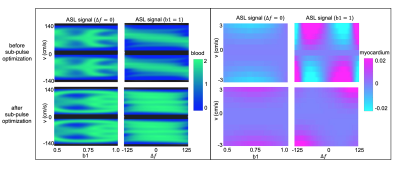 |
Fourier Transform based Velocity-Selective Labeling Pulses for Myocardial ASL
Vanessa Landes1, Ahsan Javed2, Terrence Jao3, Qin Qin4,5, and Krishna Nayak2
1Biomedical Engineering, University of Southern California, Los Angeles, CA, United States, 2Electrical Engineering, University of Southern California, Los Angeles, CA, United States, 3Keck School of Medicine, University of Southern California, Los Angeles, CA, United States, 4Radiology, Johns Hopkins University, Baltimore, MD, United States, 5F.M. Kirby Research Center for Functional Brain Imaging, Kennedy Krieger Institute, Baltimore, MD, United States
We present an improved velocity-selective (VS) labeling pulse for myocardial arterial spin labeling (ASL) that addresses limitations of prior pulses. The proposed pulse is designed using a Fourier Transform based Velocity-Selective (FT-VS) pulse train and optimized to label coronary blood while not labeling myocardium. Myocardial VSASL experiments were performed in healthy adult volunteers. The proposed pulse provided comparable measurements to FAIR ASL and a 2.5-fold reduction in physiological noise compared to a prior VS-ASL pulse.
|
|
2098.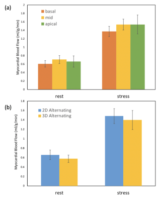 |
Quantitative myocardial perfusion with an alternating radial 2D simultaneous multi-slice and 3D stack-of-stars sequence
Qi Huang1,2, Ye Tian2, Jason Mendes2, Ganesh Adluru1,2, and Edward DiBella1,2
1Biomedical Engineering, University of Utah, Salt Lake City, UT, United States, 2Utah Center for Advanced Imaging Research (UCAIR), University of Utah, salt lake city, UT, United States
Here we propose a unique perfusion acquisition that applies a 2D/3D alternating acquisition method to obtain 2D simultaneous multi-slice (SMS) and 3D stack-of-stars (SoS) data every other heartbeat. Within each heartbeat, 2D SMS and 3D acquisitions are performed following a saturation pulse. Potential advantages include different spatio-temporal resolution and artifacts, accurate AIF acquisitions and the ability to compare the sequences directly with a single injection. Preliminary quantitative results of 7 dog and 3 human studies show the promise of this approach.
|
|
2099.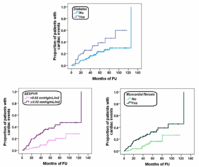 |
The additive prognostic value of end-systolic pressure-volume relation by stress CMR in patients with known or suspected coronary artery disease
Antonella Meloni1, Antonio De Luca2, Cinzia Nugara3, Chiara Cappelletto2, Camilla Cavallaro4, Chrysanthos Grigoratos1, Giovanni Donato Aquaro1, Giancarlo Todiere1, Andrea Barison1, Gianfranco Sinagra2, Giuseppina Novo3, Germano Di Sciascio4, and Alessia Pepe1
1MRI Unit, Fondazione G. Monasterio CNR-Regione Toscana, Pisa, Italy, 2University of Trieste, Trieste, Italy, 3University of Palermo, Palermo, Italy, 4Università Campus Bio-Medico, Roma, Italy
We assessed for the first time the prognostic value of the ΔESPVR (difference between peak and rest end-systolic pressure-volume relation) index evaluated during dipyridamole stress-CMR in 196 patients with known or suspected coronary artery disease. During a mean follow-up time of 53.17±28.21 months 50 cardiac events were recorded. In the multivariate analysis the independent predictive factors for cardiac events were diabetes, a ΔESPVR index≤0.02 mmHg/mL/m2, and myocardial fibrosis.
|
|
2100.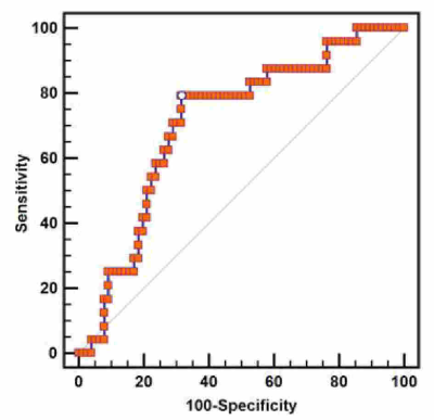 |
Pressure-volume Relationship by Stress Cardiovascular Magnetic Resonance: feasibility and clinical implications
Antonella Meloni1, Antonio De Luca2, Cinzia Nugara3, Chiara Cappelletto2, Camilla Cavallaro4, Giovanni Donato Aquaro1, Chrysanthos Grigoratos1, Giancarlo Todiere1, Andrea Barison1, Gianfranco Sinagra2, Giuseppina Novo3, Germano Di Sciascio4, and Alessia Pepe1
1MRI Unit, Fondazione G. Monasterio CNR-Regione Toscana, Pisa, Italy, 2University of Trieste, Trieste, Italy, 3University of Palermo, Palermo, Italy, 4Università Campus Bio-Medico, Roma, Italy
We showed for the first time that the ΔESPVR index, an index of myocardial contractile function easily obtained during routine stress echocardiography, can be noninvasively calculated during a dipyridamole stress-CMR exam. The ΔESPVR index was independent from baseline LV dimensions and function while it was lower in patients with myocardial fibrosis and in patients with abnormal stress CMR. At receiver-operating characteristic curve analysis, a ΔESPVR<0.02 predicted the presence of future cardiac events, being useful for additional prognostic stratification.
|
|
2101.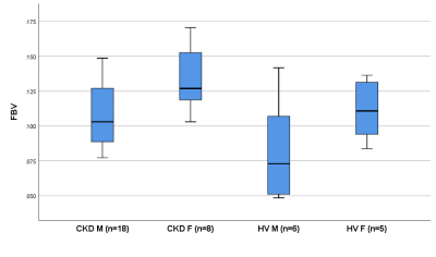 |
Assessment of myocardial fractional blood volume using Fe-MRI and 3-compartment model of capillary level water exchange in patients with CKD
Douglas Black1, Sokratis Stoumpos1, Michael Jerosch-Herold2, Neil Chatterjee3, Peter Gatehouse4, Geeshath Jayasekera1, Timothy Carroll5, David Kingsmore1, Colin Berry1, Patrick Mark1, Giles Roditi1, and Aleksandra Radjenovic1
1University of Glasgow, Glasgow, United Kingdom, 2Harvard Medical School, Boston, MA, United States, 3University of Pennsylvania, Philadelphia, PA, United States, 4Imperial College, London, United Kingdom, 5University of Chicago, Chicago, IL, United States
Estimates of myocardial fractional blood volume (FBV), capillary level vascular-interstitial water exchange rate and permeability surface area product were obtained in a cohort of chronic kidney disease (CKD) patients and a control set of healthy volunteers using iron-enhanced MRI (Fe-MRI). FBV was elevated in females, in both CKD patients and control subjects. Fe-MRI also demonstrated reduced permeability surface area product in CKD patients compared to healthy controls, and may provide unique insights into microvascular pathophysiology, particularly in patients with contraindications to gadolinium-based contrast agents.
|
|
2102.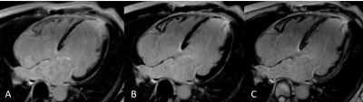 |
Myocardial Late Gadolinium Enhancement: Accuracy of Auto TI Inversion-Recovery Imaging vs Magnitude and Phase-Sensitivity IR images
Rui Wang1, Zixu Yan2, Tianjing Zhang3, Xinyan Tao2, Zhen Zhou4, Hongwei Wang5, Qian Qi6, and Lei Xu2
1Radiology, Beijing Anzhen Hospital, Captial Medical University, Beijing, China, 2Beijing Anzhen Hospital, Capital Medical University, Beijing, China, 3Philips Healthcare, Guangzhou, China, 4Beijing Anzhen Hospital,Capital Medical University, Beijing, China, 5Beijing Anzhen Hospital, Beijing, China, 6Philips Healthcare, Beijing, China
Recently, we proposed a novel LGE approach that could help radiologists/technicians automatically specify TI values .We propose a PSIR-specific TI optimization that could help automatically nulls TI while maintaining the scar-to-myocardium contrast. As a TFE preparation pulse is user defined on scanner, clinical application is readily available on current MR systems without the need for extensive optimizations, software modifications, and/or additional training.
|
|
2103. |
Feasibility Study of Whole Heart Myocardial Perfusion Imaging with tSMS and CS Reconstruction
Lixian Zou1,2, Yu Ding3, Junpu Hu4, Lele Zhao4, Jian Xu3, Hairong Zheng1, Xin Liu1, and Yuan Zheng3
1Shenzhen Institutes of Advanced Technology,Chinese Academy of Sciences, Shenzhen, China, 2Shenzhen College of Advanced Technology, University of Chinese Academy of Sciences, Shenzhen, China, 3UIH America Inc., Houston, TX, United States, 4Shanghai United Imaging Healthcare Co., Ltd, Shanghai, China
Auto-calibrated multiband CAIPIRINHA with through-time encoding (tSMS) has been proposed to acquire multiple slices simultaneously without extra reference scans. Reference images are estimated from the consecutive cardiac phases at a lower temporal resolution for subsequent slice separation. We implemented the tSMS method in a myocardial perfusion sequence and explored the feasibility of whole heart perfusion imaging with tSMS and CS reconstruction retrospectively. The preferable in-vivo results demonstrated the whole heart perfusion imaging is feasible using the tSMS+CS method.
|
|
2104. |
Impact on Cerebral Perfusion by Black Blood Vessel Wall Imaging Combining with 3D-ASL: Plaque Vulnerability vs. Intracranial Stenosis
Shaoyang Lei1, Chunhui Shan1, Jie Wu1, Jing Wu1, Yunfeng Bao1, Yingmin Chen1, and Shuqian Zhang1
1Hebei General Hospital, Shijiazhuang, China
Black Blood Vessel Wall Imaging, a valuable tool to detect abnormalities of intracranial vessel walls, was combined with 3D-ASL, a noninvasive approach for detecting cerebral perfusion, to explore the effects of plaque vulnerability and intracranial stenosis on cerebral perfusion. Absolute and relative values of CBF were determined in regions drawn manually based on the territory of blood supply in ASL images. Multivariate regression analysis indicated plaque vulnerability had no significant impact on perfusion while different degrees of luminal narrowing had statistically differences, signifying intracranial stenosis might be a more critical factor for decreased perfusion.
|