
Digital Poster Session
Cardiovascular: Cardiovascular Applications
Cardiovascular
2105 -2119 Cardiovascular Applications - Clinical Applications of Cardiac Flow
2120 -2135 Cardiovascular Applications - CMR: Atherosclerosis Imaging
2136 -2148 Cardiovascular Applications - Coronary & Thoracic MR Angiography
2149 -2161 Cardiovascular Applications - MR Angiography: Body
2162 -2175 Cardiovascular Applications - Cardiac Function 2
2176 -2191 Cardiovascular Applications - Cardiac Function 1
2105.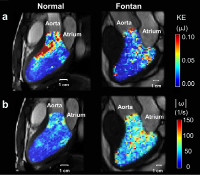 |
Impact of Intraventricular Vortex on Kinetic Energy in Patients with Fontan Circulation
Xue-Zhe Lu1, Ming-Ting Wu2, Ken-Pen Weng3,4, and Hsu-Hsia Peng1
1Department of Biomedical Engineering and Environmental Sciences, National Tsing Hua University, Hsinchu, Taiwan, 2Department of Radiology, Kaohsiung Veterans General Hospital, Kaohsiung, Taiwan, 3Department of Pediatrics, Kaohsiung Veterans General Hospital, Kaohsiung, Taiwan, 4Department of Pediatrics, National Yang-Ming University, Taipei, Taiwan
For patients with Fontan circulation, the intraventricular hemodynamics such as kinetic energy and vorticity might could be helpful with evaluating the cardiac function and the risks of negative long-term outcome. To investigate the impact of intraventricular vortex on kinetic energy in the of Fontan patients, we employed 4D flow magnetic resonance and evaluate intraventricular kinetic energy and vorticity in Fontan patients and found prolonged peaks in systolic kinetic energy and increased mean systolic vorticity. The strong correlation between kinetic energy and vorticity in systole showed a potential impact of vortex on kinetic energy during systole.
|
|
2106.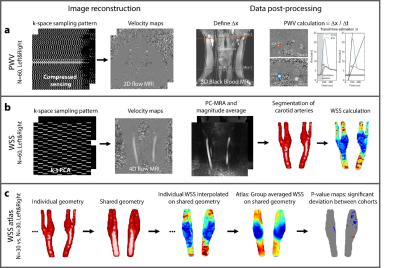 |
Pulse wave velocity and wall shear stress in the carotid arteries of familial hypercholesterolemia patients
Eva S Peper1, Ilse K Luirink2, Pim van Ooij1, Bram F Coolen3, Gustav J Strijkers3, Albert Wiegman2, Barbara A Hutten4, and Aart J Nederveen1
1Department of Radiology and Nuclear Medicine, Amsterdam UMC, Amsterdam, Netherlands, 2Department of Paediatrics, Amsterdam UMC, Amsterdam, Netherlands, 3Department of Physics and Biomedical Engineering, Amsterdam UMC, Amsterdam, Netherlands, 4Department of Clinical Epidemiology, Biostatistics and Bioinformatics, Amsterdam UMC, Amsterdam, Netherlands
Familial hypercholesterolemia (FH) is characterized by elevated low-density lipoprotein (LDL) cholesterol levels form birth onwards and premature cardiovascular disease. Statins are currently the preferred pharmacological therapy. Results from a recent study suggest that initiation of lipid-lowering treatment during childhood reduces the risk for cardiovascular disease in adulthood. With advanced acquisition strategies of 2D and 4D flow MRI we investigated PWV and WSS in the carotid arteries of 43 FH patients and 18 healthy controls of a similar age (33 years), as PWV and WSS have both been associated with atherosclerosis.
|
|
2107. |
The impact of different Middle Cerebral Artery Stenosis Severity on Blood Flow Lateralization
Wenwen Chen1, Xiaowei Song2, Jian Wu2, and Rui Li1
1Tsinghua University, Beijing, China, 2Beijing Tsinghua Changgung Hospital, Beijing, China
Middle cerebral artery stenosis induces flow lateralization. However, the impact of stenosis degree on flow lateralization is not fully understood. This study investigated the impact of MCA stenosis severity on MCA blood flow lateralization using 4D flow MRI. Twenty-seven patients with symptomatic MCA stenosis were included. Absolute flow rates of MCA on stenosis and contralateral sides were obtained. Paired samples t-test was used to compare the flows on both sides. We found a significant difference between the flows in the severe stenosis group, while there was no significant difference in non-severe group.
|
|
2108.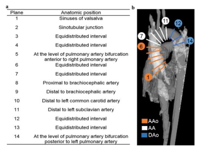 |
Aortic Wall Shear Stress and Oscillatory Shear Index in Patients with Repaired Tetralogy of Fallot
Qing-Xiao Zhang1, Meng-Chu Chang1, Ming-Ting Wu2, Ken-Pen Weng3,4, and Hsu-Hsia Peng1
1Department of Biomedical Engineering and Environmental Sciences, National Tsing Hua University, Hsinchu, Taiwan, 2Department of Radiology, Kaohsiung Veterans General Hospital, Kaohsiung, Taiwan, 3Department of Pediatrics, Kaohsiung Veterans General Hospital, Kaohsiung, Taiwan, 4Department of Pediatrics, National Yang-Ming University, Taipei, Taiwan
We aimed to explore the difference of the wall shear stress (WSS) and oscillatory shear index (OSI) between control and rTOF patients. The rTOF patients were divided into rTOF1and rTOF2 groups according to their indexed right ventricular end-systolic volume (RVESVi). The rTOF patients presented decreased WSSaxial and OSIcirc in the ascending aorta. In rTOF2 group the WSSaxial and OSIcirc correlated with resistance index and flow velocity. In conclusion, higher resistance index and slower flow velocity correlated with decreasd WSSaxial and OSIcirc in rTOF2. The deceasing OSIcirc may slightly improve the stress condition of vascular wall.
|
|
2109.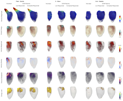 |
Left Ventricular Reverse Remodeling in Dilated Cardiomyopathy: Does Ejection Fraction Reflect Subtle Ventricular Dysfunction?
Pamela Alejandra Franco1,2,3, Julio Sotelo1,2,3, Bram Ruijsink4,5, David Nordsletten4,5, Eric Kerfoot4,5, Joaquín Mura6, and Sergio Uribe1,3,7
1Biomedical Imaging Center, Pontificia Universidad Católica de Chile, Santiago, Chile, 2Electrical Engineering, Pontificia Universidad Católica de Chile, Santiago, Chile, 3Millennium Nucleus for Cardiovascular Magnetic Resonance, Santiago, Chile, 4Guy's and St Thomas' NHS Foundation Trust, London, United Kingdom, 5School of Imaging Sciences and Biomedical Engineering, King's College London, London, United Kingdom, 6Department of Mechanical Engineering, Universidad Técnica Federico Santa María, Santiago, Chile, 7Radiology Department, School of Medicine, Pontificia Universidad Católica de Chile, Santiago, Chile
An important number of patients with dilated cardiomyopathy have improved their left ventricular function with an optimal treatment. However, it is not well understood whether remodeling represents a recovery in left ventricular (LV) hemodinamics1. In this abstract, we discuss the capacity of the ejection fraction to represent disease remission, by analyzing LV blood flow.
|
|
2110.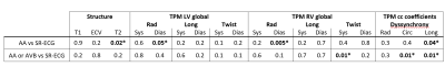 |
Myocardial Structural and Velocity Analysis in Patients with Arrhythmias Post-Heart Transplantation
Roberto Sarnari1, Allison Blake1, Muhannad Aboud Abbasi1, Ashitha Pathrose1, Julie Blaisdell1, Alyssa Singer1, Kambiz Ghafourian2, Jane Wilcox2, Sadiya Khan2, Esther Vorovich2, Jonathan Rich2, Allen Anderson2, Clyde Yancy2,
James Carr1, and Michael Markl1,3
1Radiology, Northwestern University, Chicago, IL, United States, 2Cardiology, Northwestern University, Chicago, IL, United States, 3Biomedical Engineering, Northwestern University, Evanston, IL, United States
Cardiac magnetic resonance including tissue phase mapping was performed in a cohort of heart transplanted (HTx) patients developing atrial arrhythmias or atrio-ventricular blocks: T1-T2-ECV values and 3-directional myocardial velocities were calculated and compared to the same parameters acquired in HTx patients with normal sinus rhythm during the follow up. Alterations in myocardial structural properties (increased T2) and cardiac dynamics were evidenced in cardiac grafts showing hyperkinetic arrhythmias or atrio-ventricular blocks.
|
|
2111.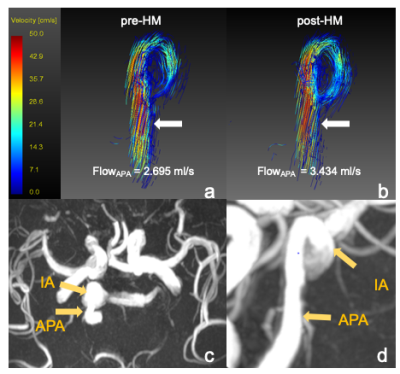 |
Hemodynamic assessments of intracranial aneurysm (IA) following hypotensor medication: A pilot study
Miaoqi Zhang1, Fei Peng2, Yunduo Li1, Aihua Liu2, and Rui Li1
1Center for Biomedical Imaging Research, Department of Biomedical Engineering, Tsinghua University, Beijing, China, 2Department of Interventional Neuroradiology, Beijing Neurosurgical Institute and Beijing Tiantan Hospital, Capital Medical University, Beijing, China
In this study, we utilize 4D-flow MRI to investigate the relationship between hemodynamics and blood pressure change following hypotensor medication. Statistics indicated that FlowAPA significant increased when blood pressure drops. No differences were found in FlowIA, VAPA and VIA. This is probably because hypotensor medication has a vasodilatory effect, the widening of blood vessel will result in an increased flow. The results of this pilot study suggested that an acute blood pressure drop affects hemodynamic conditions of APA rather than IA.
|
|
2112.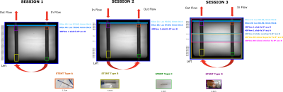 |
Stent effects in 2D and 4DFlow investigation: should we stop using stainless-steel stents?
Ana Beatriz Solana1, Margarita Gorodezky2, Ann Shimakawa3, Peng Lai3, and Christian Meierhofer4,5
1GE Healthcare, Munich, Germany, 2GE Healthcare, London, United Kingdom, 3GE Healthcare, Sunnyvale, CA, United States, 4German Heart Center Munich, Munich, Germany, 5TUM, Munich, Germany
Flow measurements using Phase Contrast MRI in the present of stents is of clinical relevance, especially in vessels with no alternative to measure elsewhere. Here, we evaluate 4 different stent types in a pulsatile flow phantom using both 2D and 4DFlow including different acquisition schemes for 4DFlow, e.g. 1 slab whole-volume, 1slab with reduced number of slices and multislab whole-volume. The stent made of platinum-iridium was the only stent under evaluation that allowed in-stent accurate net flow measurements for both 2D and 4DFlow (1 slab with reduced number of slices and multislab whole-volume).
|
|
2113.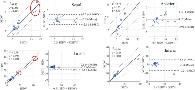 |
Measuring left ventricular diastolic function with dual-echo dual velocity encoding PC-MRI: Clinical validation
Afis Ajala1,2, Jiming Zhang3, Erick Buko4, Janie Swaab3, Melissa Dotson3, Benjamin Cheong3, Pei-Herng Hor4, and Raja Muthupillai3
1Physics, University of Houston [Main Campus], Houston, TX, United States, 2Department of Diagnostic and Interventional Radiology, Baylor St. Luke's Medical Center, Houston, TX, United States, 3Baylor St. Luke's Medical Center, Houston, TX, United States, 4University of Houston [Main Campus], Houston, TX, United States
A common clinical index of left ventricular (LV) diastolic function is the ratio of peak trans-mitral blood velocity (E) to mitral annular velocities (Em) during early diastole. Here, we provide clinical validation of a single TR, dual-echo approach wherein the first and second echoes are velocity sensitized to encode E and Em respectively.
|
|
2114.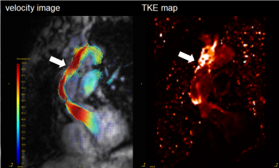 |
4D Flow MRI reveals abnormal turbulent kinetic energy elevation of the replacement aorta for the aortic dissection
SAYAKA SHIRAI1, Tetsuro Sekine1, Kenichiro Takahashi1, Jiro Kurita1, Tetsuro Morota1, Takashi Morota1, and Shinichiro Kumita1
1Nippon Medical School Hospital, Tokyoto Bunkyoku, Japan
The purpose of this study was to evaluate turbulent kinetic energy (TKE) after aorta replacement (AoR) using 4D Fow MRI. We recruited 13 patients undergoing AoR for the aortic dissection and 9 normal volunteers. The TKEpeak of AoR group was significantly higher than those of volunteers (16.09 ± 7.75 [3.51–31.83] mJ vs. 4.50 ± 1.64 [2.57–8.13] mJ, p < 0.001). The TKE elevation was mainly derived from the distal part of ReAo (p=0.001).
|
|
2115.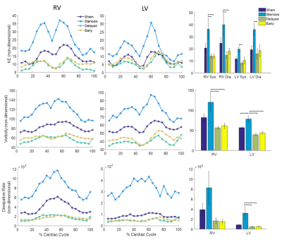 |
Quantifying the impact of pulmonary artery stent interventions on ventricular flow dynamics with 4D Flow MRI
Ryan Pewowaruk1, Cody Johnson2, Chris Francois2, Luke Lamers2, and Alejandro Roldán-Alzate2
1Biomedical Engineering, University of Wisconsin - Madison, Madison, WI, United States, 2University of Wisconsin - Madison, Madison, WI, United States
In a swine model of PAS, this study assessed the effects of early versus delayed stent interventions using 4D Flow MRI. Early and Delayed interventions were both effective but no differences between early and delayed interventions were observed. 4D Flow MRI measured inefficient LV and RV flow in the PAS group and an association was found between EF and vorticity. If confirmed in larger studies, these results provide physiological insight into PAS and congenital heart disease and also highlight the sensitivity of 4D Flow MRI biomarkers to ventricular dysfunction.
|
|
2116.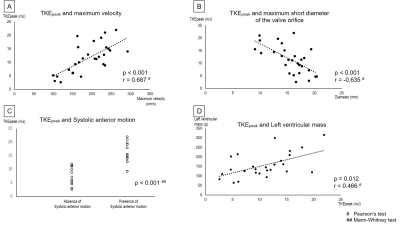 |
Evaluation of Turbulent Kinetic Energy Measurement for hypertrophic cardiomyopathy by Using Triple-VENC 4D Flow MRI
Kotomi Iwata1, Tetsuro Sekine1, Masaki Tachi1, Takahiro Ando1, Izumi Tanaka1, Yoichi Imori2, Junya Matsuda2, Yasuo Amano3, Makoto Obara4, Masashi Ogawa1, Hitoshi Takano2, and Shinichiro Kumita1
1Radiology, Nippon Medical School, Tokyo, Japan, 2Cardiovascular Medicine, Nippon Medical School, Tokyo, Japan, 3Radiology, Nihon University, Tokyo, Japan, 4Philips Electronics Japan Ltd., Tokyo, Japan
The purpose of this study was threefold. First, we validated whether TKE reflects the condition of HOCM and HNCM by comparing them with volunteers. Second, we clarified the factors that increase TKE in HCM. Third, we revealed LV mass which is associated with cardiac load, resulting in a poor outcome. TKE in HOCM was significantly higher than HNCM or volunteers. Maximum velocity, maximum short of the valve orifice, SAM, and LV mass were correlated to TKE. TKE allows to noninvasively depict the flow characteristics and may provide insights into the pathophysiology associated with flow dynamics in HCM.
|
|
2117.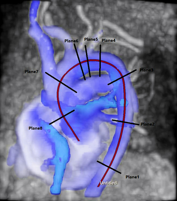 |
Hemodynamic Characteristics in Patients with Plaques in Thoracic Aorta Based on Compressed Sensing 4D Flow MRI
Zhengling Zeng1, Bin Sun1, Yunjing Xue1, Ning Jin2, and Jing An3
1Fujian Medical University union hospital, Fuzhou, China, 2Siemens Medical Solution, Chicago, IL, United States, 3Siemens Shenzhen Magnetic Resonance Ltd, Shenzhen, China
Hemodynamic parameters of patients with and without thoracic aortic atherosclerotic plaques were detected using a compressed sensing (CS) 4d flow sequence. Eight different planes perpendicular to the thoracic aorta were identified to confirm if there were differences in hemodynamic parameters between the plaque and non-plaque groups. The study had shown that the plaque group had lower circumferential wall shear stress, mean velocity and peak velocity compared with that of control groups. Early detection of abnormal blood flow parameters in patients with aortic atherosclerosis may help predict regions prone to plaque, allowing early intervention in patients accordingly.
|
|
2118.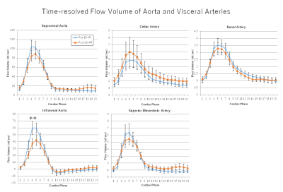 |
EVAR Does Not Affect Mean Blood Flow Volume and Flow Profile of the Visceral Arteries.
Masataka Sugiyama1, Yasuo Takehara1, Ryota Horiguchi2, Takashi Mizuno3, Ryota Hyodo2, Takasuke Ushio4, Tetsuya Wakayama5, Atsushi Nozaki5, Hiroyuki Kabasawa5, Marcus Alley6, Satoshi Goshima4, and Shinji Naganawa2
1Department of Fundamental Development for Advanced Low Invasive Diagnostic Imaging, Nagoya University Graduate School of Medicine, Nagoya, Japan, 2Department of Radiology, Nagoya University, Nagoya, Japan, 3Department of Radiological Technology, Nagoya University Hospital, Nagoya, Japan, 4Department of Radiology, Hamamatsu University School of Medicine, Hamamatsu, Japan, 5Applied Science Laboratory Asia Pacific, GE Healthcare Japan, Hino, Japan, 6Department of Radiology, Stanford University School of Medicine, Palo Alto, CA, United States
We aimed to assess the effect of EVAR treatment to the blood flow profile of visceral arteries using 4D-Flow MRI. 10 patients with AAA underwent 4D-Flow pre- and one month post-EVAR. The flow volume at the sections in suprarenal and infrarenal abdominal aorta, the celiac artery, the superior mesenteric artery and the renal arteries were measured. No significant change in blood flow volume was observed within visceral arteries after EVAR despite the placement of stiff stent in the deformed blood pathway.
|
|
2119. |
4D Flow Analysis of Left Atrium and Appendage in Patients with Atrial Fibrillation
Hana Sheitt1, Andrew G Howard1,2, Steven Wilton1,2, Carmen Lydell1,3, James White1,2, and Julio Garcia1,4
1Stephenson Cardiac Imaging Center, Libin Cardiovascular Institute of Alberta, Calgary, AB, Canada, 2Department of Medicine, University of Calgary, Calgary, AB, Canada, 3Diagnostic Imaging, University of Calgary, Calgary, AB, Canada, 4Department of Cardiac Science, University of Calgary, Calgary, AB, Canada
This study may be of interest for clinicians and researchers who study left atrial arrhythmias. This study demonstrated that left atrial stasis in the appendage is elevated in atrial fibrillation patients.
|
 View the Poster
 Watch the Video
2120. |
Higher plaque burden of middle cerebral artery is associated with recurrent ischemic stroke: a quantitative study by high-resolution MRI
Yuting Wang1, Yuncai Ran2, Ming Zhu3, Xiao Wu4, Ajay Malhotra4, Xiaowen Lei2, Feifei Zhang2, Xiao Wang2, Shanshan Xie2, Jian Zhou2, Jinxia Zhu5, Jingliang Cheng2, and Chengcheng Zhu6
1Department of Radiology, Sichuan Provincial People's Hospital, Chengdu, China, 2Department of Magnetic Resonance, The first Affiliated Hospital of Zhengzhou University, Zhengzhou, China, 34. Interventional department, The first Affiliated Hospital of Zhengzhou University, Zhengzhou, China, 4Yale School of Medicine and Yale University, New Haven, CT, United States, 5MR Collaboration, Siemens Healthcare Ltd., Beijing, China, 63. Department of Radiology and Biomedical Imaging, University of California, San Francisco, San Francisco, CA, United States
Middle cerebral artery plaque burden on high-resolution MR was significantly higher in recurrent stroke group than in the first-onset acute stroke and chronic stroke groups (p=0.002). Patients with acute stroke had higher enhancement ratio than patients with chronic stroke (p=0.014). After adjustment of clinical demographic factors, plaque burden was the only independent imaging feature associated with recurrent stroke (odds ratio=2.26, per 10% increase, 95% CI, 1.03-4.96, p=0.042). In conclusion, higher plaque burden of MCA on MR is independently associated with recurrent ischemic stroke. MR vessel wall imaging may provide unique information for risk stratification of stroke patients.
|
|
2121.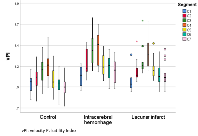 |
Effects of calcification on blood-flow pulsatility variation along the internal carotid artery in small vessel disease patients, a 7T MRI study
Rick J van Tuijl1, Ynte M Ruigrok2, Irene C van der Schaaf1, Gabriël J. E. Rinkel2, Birgitta K Velthuis1, and Jaco J.M. Zwanenburg1
1Radiology, UMC Utrecht, Utrecht, Netherlands, 2Neurology and Neurosurgery, Rudolf Magnus Institute of Neuroscience, UMC Utrecht, Utrecht, Netherlands The influence of intracranial internal carotid artery calcification (iICAC) over the whole internal carotid artery (ICA) trajectory was studied using 4D phase-contrast flow measurements (17 patients with cerebral small vessel disease (CSVD) and 17 healthy controls; 7T MRI). CT images of CSVD patients were used to calculate the iICAC from C3-C7 segments. Results showed a positive correlation between the presence and volume of iICAC with vPI in the CSVD group. iICAC contributes to an increase in vPI over the ICA from the extracranial C1 segment to the C7 segment just proximal to the circle of Willis compared to HC. |
|
2122.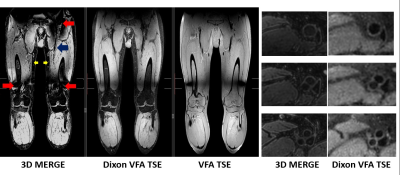 |
Time-efficient large field-of-view fat suppression using SNR priority Dixon turbo spin echo for femoral vessel wall MRI
Niranjan Balu1, Zechen Zhou2, Thomas Hatsukami3, and Chun Yuan1
1Radiology, University of Washington, Seattle, WA, United States, 2Philips Healthcare, Briarcliff Manor, NY, United States, 3Surgery, University of Washington, Seattle, WA, United States
Vessel wall MRI requires good fat suppression to delineate outer vessel boundaries. Large field-of-views required for turbo spin echo femoral vessel wall MRI do not provide good fat suppression with spectral inversion recovery. We developed a time-efficient two-point Dixon turbo spin echo method using a SNR priority variable flip angle schedule and point spread correction reconstruction to allow accelerated acquisition and demonstrate its advantages compared to an existing gradient echo vessel wall MRI method.
|
|
2123.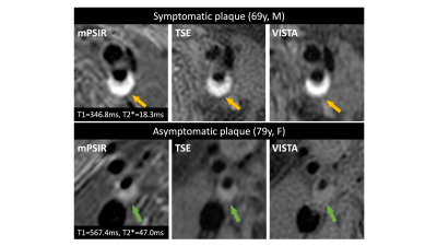 |
Quantitative T1 and T2* mapping for atherosclerotic carotid plaque using multi-echo phase-sensitive inversion recovery sequence
Yasuhiro Fujiwara1 and Motohira Mio2
1Department of Medical Image Sciences, Faculty of Life Sciences, Kumamoto University, Kumamoto, Japan, 2Department of Radiology, Fukuoka University Chikushi Hospital, Chikushi, Japan
The purpose of this study was to investigate the correlation between the T1 value of carotid plaque and signal intensity ratio and to evaluate the clinical usefulness of T1 and T2* values for carotid plaque. Quantitative T1 and T2* mapping using mPSIR have potential to improve the diagnostic accuracy of vulnerability of carotid atherosclerotic plaque.
|
|
2124.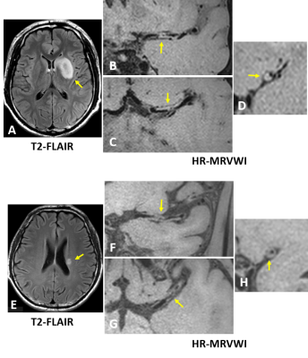 |
Comparison of plaque characteristics between patients with small and large subcortical infarction using high resolution MR vessel wall imaging
Na Zhang1, Tingting Zhu2, Lijie Ren3, Lei Zhang1, Zhangyan Fan3, Liwen Wan1, Hairong Zheng1, and Xin Liu1
1Shenzhen Institutes of Advanced Technology, Chinese Academy of Sciences, Shenzhen, China, 2Department of Radiology, Tongji Hospital, Wuhan, China, 3Department of Neurology, Shenzhen Second People's Hospital, Shenzhen, China
It is generally believed small subcortical infarction (SSI) (also termed lacunar stroke) is caused by intrinsic diseases of penetrating arteries. Recent studies have shown the incidence of atherosclerosis in large arteries which is an important pathogenesis of large subcortical infarction (LSI) is also high in patients with SSI. This indicates the underlying mechanisms of SSI in patients with large artery atherosclerosis may be the same with LSI and inferred from plaque characteristics. However, the plaque characteristics ultimately leading to SSI or LSI remain unclear. This study is to explore the differences of plaque characteristics between patients with SSI and LSI.
|
|
2125.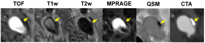 |
Accurate detection of calcified carotid plaques on quantitative susceptibility mapping – A CT validation study
Thanh D Nguyen1, Jingwen Du2, Yan Wen1, Ajay Gupta1, Pascal Spincemaille1, Qi Yang2, and Yi Wang1
1Weill Cornell Medicine, New York, NY, United States, 2Xuanwu Hospital of Capital Medical University, Beijing, China
In this study, we used CT angiography (CTA) as a reference standard to demonstrate that carotid quantitative susceptibility mapping (QSM) provides accurate detection of calcified atherosclerotic plaques in the carotid arteries. Calcification is strongly diamagnetic and appears uniquely hypointense on carotid QSM. Inclusion of QSM in carotid MRI would aid characterization of carotid plaque.
|
|
2126.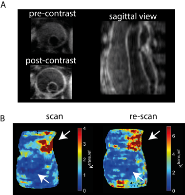 |
Black-blood 3D DCE-MRI of the aortic wall with AIF-free pharmacokinetic modeling: feasibility in abdominal aortic aneurysm patients
Jasper Schoormans1, Claudia Calcagno2, Reza Indrakusuma3, Hamid Jalalzadeh3, Ron Balm3, Stefan Smorenburg4, Gustav J Strijkers1, Aart J Nederveen5, and Bram F Coolen1
1Department of Biomedical Engineering and Physics, Amsterdam University Medical Centers, University of Amsterdam, Amsterdam, Netherlands, 2The BioMedical Engineering and Imaging Institute, Icahn School of Medicine at Mount Sinai, New York, NY, United States, 3Department of Surgery, Amsterdam University Medical Centers, University of Amsterdam, Amsterdam, Netherlands, 4Department of Vascular Surgery, Amsterdam University Medical Centers, University of Amsterdam, Amsterdam, Netherlands, 5Department of Radiology and Nuclear Medicine, Amsterdam University Medical Centers, University of Amsterdam, Amsterdam, Netherlands
We introduced 3D black-blood DCE MRI in combination with AIF-free modeling to facilitate the measurement of pharmacokinetic parameters in the aorta vessel wall of patients with an abdominal aortic aneurysm. Our method enables 3D assessment of microvascularization and permeability which could assist a clinical risk assessment of this condition.
|
|
2127.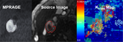 |
Quantitative 3D dynamic contrast‐enhanced of Atherosclerosis by using 3D Stack of Star with Golden-angle radial sparse parallel reconstruction
Seong-Eun Kim1, Ye Tian2, Matthew Alexander1, Dennis L Parker1, Gerald S Treiman 3,4, Adam de Havenon5, and J Scott McNally1
1UCAIR, Department of Radiology and Imaging Sciences, University of Utah, Salt Lake City, UT, United States, 2Department of Physics and Astronomy, University of Utah, Salt Lake City, UT, United States, 3Department of Surgery, University of Utah, Salt Lake City, UT, United States, 4VASLCHCS, Salt Lake City, UT, United States, 5Department of Neurology, University of Utah, Salt Lake City, UT, United States Carotid plaque inflammation can be measured with dynamic contrast enhanced (DCE) MRI and is a marker for plaque instability. Increased vascularity, which is one sign of plaque inflammation, can be detected from kinetic analysis of DCE images However, the trade-off between spatial and temporal resolution limits assessment to a small number of slices and ~15 seconds per time frame. To overcome this limitation, we implemented a short-TR 3D T1w Stack of Stars (SoS) sequence to enable retrospective image formation with arbitrary numbers of radial views by using Golden-angle radial sparse parallel (GRASP) reconstruction.
|
|
2128.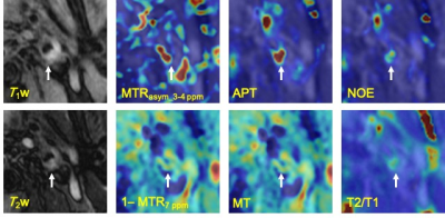 |
Chemical Exchange Saturation Transfer Imaging for Atherosclerotic Plaques
Yuki Kanazawa1, Masafumi Harada1, Tosiaki Miyati2, Takashi Abe1, Mitsuharu Miyoshi3, Yuki Matsumoto1, Hiroaki Hayashi2, Yasuhisa Kanematsu4, and Yasushi Takagi4
1Tokushima University, Tokushima, Japan, 2Faculty of Health Sciences, Institute of Medical, Pharmaceutical and Health Sciences, Kanazawa University, Kanazawa, Japan, 3Global MR Applications and Workflow, GE Healthcare Japan, Hino, Japan, 4Department of Neurosurgery, Tokushima University, Tokushima, Japan
To assess an activity within an atherosclerotic plaque, chemical exchange saturation transfer (CEST) imaging was demonstrated with the multi-pool model Bloch equation. This study was performed with eleven patients with carotid stenosis, was evaluated with each estimated parameters; bulk water, magnetization transfer, amide proton transfer (APT), and nuclear Overhauser effect (NOE). There was no significant difference between mean APT and NOE at 3.5 ppm. This result indicates that CEST plaque imaging should be evaluated with distinguishing APT and NOE. Multi-parametric analysis of CEST imaging may obtain detailed information for component as well as metabolite substances within an atherosclerotic plaque.
|
|
2129.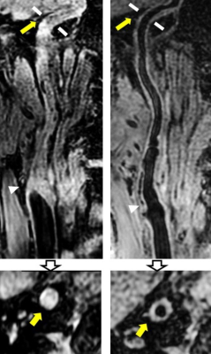 |
Evolution of Diffuse Wall Thickening of Petrous Internal Carotid Artery Peri-Revascularization: A MR Vessel Wall Imaging Follow-up Study
Xiaoyi Chen1, Huimin Xu2, Tao Wang3, Jin Zhang4, Huiyu Qiao5, Hualu Han5, and Xihai Zhao5
1Department of Radiology, Beijing Geriatric Hospital, Beijing, China, 2Department of Radiology, Peking University Third Hospital, Beijing, China, 3Department of Neurosurgery, Peking University Third Hospital, Beijing, China, 4Department of Radiology, Renji Hospital, Shanghai Jiaotong University, Shanghai, China, 5Center for Biomedical Imaging Research, Department of Biomedical Engineering, Tsinghua University School of Medicine, Beijing, China
Carotid artery severe stenosis or occlusion will lead to ischemia within the vessel wall and diffuse wall thickening (DWT) in the downstream arterial segment. The revascularization surgery is an effective treatment for carotid artery stenosis. This study investigated the peri-revascularization change of DWT in petrous internal carotid artery (ICA) among carotid atherosclerotic patients using MR vessel wall imaging. We found that the DWT in ipsilateral petrous ICA recovered after revascularization treatment (1.68±0.68 mm vs. 1.39±0.47 mm, P =0.002) and it began to recover one month later after the revascularization.
|
|
2130.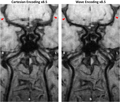 |
4-minute 3D Whole Brain 0.6mm Isotropic T1 Weighted Intracranial Vessel Wall Imaging
Zechen Zhou1, Zhensen Chen2, Jie Sun2, Niranjan Balu2, Baocheng Chu2, Peter Börnert3, Thomas S Hatsukami2, and Chun Yuan2
1Philips Research North America, Cambridge, MA, United States, 2Department of Radiology, University of Washington, Seattle, WA, United States, 3Philips Research Hamburg, Hamburg, Germany
Three-dimensional (3D) T1 weighted (T1w) turbo spin echo (TSE) sequence has demonstrated its value for intracranial vessel wall imaging (IVWI), but its long scan time has hampered wider clinical translation. In this study, a 4-minute 3D T1w TSE sequence was optimized and combined with wave encoded parallel imaging and compressed sensing (PI-CS) reconstruction for whole brain isotropic 0.6mm IVWI. This approach improved the image quality and robustness of IVWI in comparison to the Cartesian encoded PI-CS approach in healthy subjects. It demonstrated promising potential for further clinical evaluation.
|
|
2131.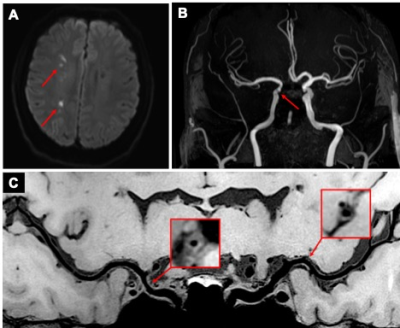 |
Hypertension is Associated with the Diffuseness of Intracranial Atherosclerotic Disease: a Cross-sectional MR Vessel Wall Imaging Study
Jiayu Xiao1, Jae Song2, Fang Wu3, Tao Jiang4, Shlee Song1, Konrad Schlick1, Debiao Li1,5, Qi Yang3, and Zhaoyang Fan1,5
1Cedars-Sinai Medical Center, Los Angeles, CA, United States, 2Hospital of the University of Pennsylvania, Philadelphia, PA, United States, 3Beijing Xuanwu Hospital, Beijing, China, 4Beijing Chaoyang Hospital, Beijing, China, 5University of California, Los Angeles, Los Angeles, CA, United States
Hypertension is associated with intracranial atherosclerosis and cerebrovascular events. However, the correlation between hypertension and atherosclerosis disease burden (the number, distribution
|
|
2132.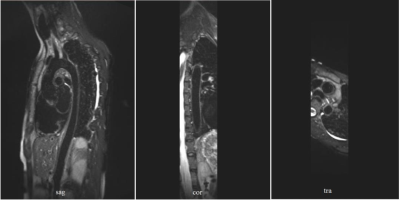 |
Diffusion-Prepared SPACE sequence: Application to Isotropic 3D T2W Black Blood MRI of the Whole Aortic Wall
Fang Dong1 and Jing An1
1Siemens Magnetic Resonance Ltd., Shenzhen, China
T2-weighted MR imaging is a non-invasive way for vasculitis diagnosis. SPACE sequence has been successfully used for T1-weighted aortic wall imaging. While its application in T2w aortic wall imaging is hampered by the long scanning time, blood suppression performance and physiological movement. In this study, three black blood methods with optimized protocols have been tested and evaluated. Diffusion-Prepared SPACE sequence with diastolic acquisition was proved to be a promising way for T2w aortic wall imaging within a clinically acceptable acquisition time.
|
|
2133. |
Wave-CAIPI Highly Accelerated Whole-Brain Intracranial Vessel Wall Imaging
Zhilang Qiu1,2, Sen Jia1,2, Shi Su1, Yanjie Zhu1, Xin Liu1, Hairong Zheng1, Leslie Ying3, Haifeng Wang1, and Dong Liang1
1Shenzhen Institutes of Advanced Technology, Chinese Academy of Sciences, Shenzhen, China, 2Shenzhen College of Advanced Technology, University of Chinese Academy of Sciences, Shenzhen, China, 3University at Buffalo, The State University of New York, Buffalo, NY, United States Whole-brain intracranial vessel wall imaging (VWI) requires long scan time for high resolution and large coverage. In this study, we applied a novel three-dimensional parallel imaging technique, Wave-CAIPI, to accelerate whole-brain intracranial VWI. The highly accelerated (11×) VWI takes 3.5 minutes, at an isotropic resolution of 0.6 mm. Compared to conventional two-dimensional parallel imaging technique (2D CAIPI), Wave-CAIPI is able to achieve higher SNR and better imaging quality on vessel wall depictions. |
|
2134. |
Comparison of high-resolution magnetic resonance imaging and computed tomography for detection of distal vertebral artery calcification
Yina Lan1, Ming Yang2, Xin Zhou3, Ning Ma2, and Xin Lou1
1The First Medical Center of PLA General Hospital, Beijing, China, 2Beijing Tiantan Hospital, Beijing, China, 3xinzhou@wipm.ac.cn, National Center for Magnetic Resonance in Wuhan, Wuhan Institute of Physics and Mathematics, Innovation Academy of Precision Measurement Science and Technology, Chinese Academy of Sciences-Wuhan National Laboratory for Optoelectronics, Beijing, China
In this study, we aim to perform a comparative analysis of high-resolution magnetic resonance imaging (HRMRI) and noncontrast computer tomography (NCCT) in detection of arterial calcifications in V3 horizontal and V4 segment of vertebral artery in patients with posterior circulation ischemic events. We observed that, HRMRI is as accurate as NCCT in the detection of V4 segment calcifications, while HRMRI are superior in detection of V3 horizontal segment calcifications than NCCT.
|
|
2135.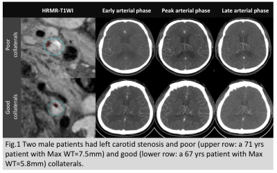 |
Plaque Burden of Carotid Atherosclerotic Stenosis Influences Collateral Circulation
Huimin Xu1, Ran Huo1, Ying Liu1, Lizhi Xie2, Ruijing Xin3, Tao Wang1, Huishu Yuan1, and Xihai Zhao4
1Peking University Third Hospital, Beijing, China, 2GE healthcare, Beijing, China, 3Southeastern University, Nanjing, China, 4Center for Biomedical Imaging Research, Beijing, China
The development of secondary collaterals can be affected by the degree of luminal stenosis, which is not parallel with plaque burden since the positive remodeling effect for elastic arteries. This study aimed to determine the association of carotid plaque burden with status of secondary collaterals in patients with severe carotid stenosis. We found that patients with poor collaterals had significantly larger Max WT than those with good collaterals (7.29 ±1.15 mm vs. 6.59 ± 0.93 mm; P=0.018) when the plaque is larger than 5.45mm.
|
2136. |
Accelerated, free-breathing, contrast-enhanced thoracic MR angiography with XD-GRASP reconstruction
Suvai Gunasekaran1, Hassan Haji-Valizadeh2, Bradley Allen1, Ryan Avery1, and Daniel Kim1
1Northwestern University, Chicago, IL, United States, 2Harvard University, Boston, MA, United States
Current methods for contrast-enhanced thoracic MR angiography (CE-MRA) utilize suboptimal imaging practices such as breath holding or long scan time. Here we developed a 9.6-fold accelerated CE-MRA sequence using stack-of-stars k-space sampling and XD-GRASP reconstruction to produce predictable scan time (< 5 min), without significant loss in image quality. Our sequence was significantly faster than the current clinical free-breathing scan, reducing the scan time by 50%. Additionally, our accelerated CE-MRA scan produced comparable image quality that was clinically acceptable.
|
|
2137.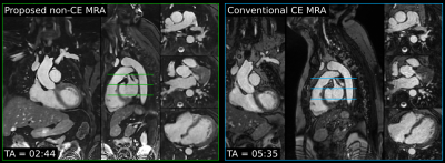 |
Non-contrast Enhanced 3D Cartesian Aortic MR Angiography in 3 minutes
Camila Munoz1, Aurelien Bustin1, Gastao Cruz1, Pier Giorgio Masci1, Rene M Botnar1, and Claudia Prieto1
1Biomedical Engineering Department, School of Biomedical Engineering and Imaging Sciences, King's College London, London, United Kingdom
MR angiography is a useful technique for the diagnosis, monitoring and treatment planning of thoracic aortic disease. However, clinically used contrast-free approaches based on diaphragmatic navigator gating commonly result in long and unpredictable scan times. This study extends our previously introduced technique for non-contrast enhanced (non-CE) accelerated 3D Cartesian non-rigid motion-compensated whole-heart MR angiography for the visualization of the thoracic and suprarenal abdominal aorta from a short and efficient 3-minute scan. The proposed method was tested in five healthy subjects and two patients with cardiovascular disease, resulting in high-quality depiction of the aorta, holding promise for integration in clinical routine.
|
|
2138.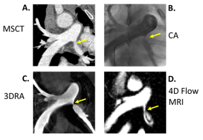 |
Pulmonary artery dimensions from catheter angiography, multi-slice computed tomography, 3D-rotational angiography and PC MR Angiography
Ryan Pewowaruk1, Klarka Mendrisova2, Carolina Larrain2, Chris Francois2, Alejandro Roldán-Alzate2, and Luke Lamers2
1Biomedical Engineering, University of Wisconsin - Madison, Madison, WI, United States, 2University of Wisconsin - Madison, Madison, WI, United States
In a swine congenital heart disease model we show strong agreement between PA dimensions from 3DRA, CA and MSCT. Non contrast-enhanced PC-MRA from PCVIPR showed good agreement with CA and MSCT for the imaging of non-stented proximal PAs.
|
|
2139.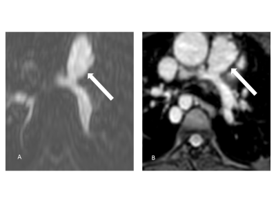 |
Imaging of the Pulmonary Vasculature in Congenital Heart Disease Using Relaxation-Enhanced Angiography Without Contrast and Triggering
Lenhard Pennig1, Anton Wagner1, Kilian Weiss2, Simon Lennartz1, Jan-Peter Grunz3, David Maintz1, Kai Roman Laukamp1, Tilman Hickethier1, Claas Philip Naehle1, Alexander Bunck1, and Jonas Doerner1
Video Permission Withheld
1Institute for Diagnostic and Interventional Radiology, Faculty of Medicine and University Hospital Cologne, University of Cologne, Cologne, Germany, Cologne, Germany, 2Philips GmbH, Hamburg, Germany, 3Department of Diagnostic and Interventional Radiology,, University Hospital Würzburg, Würzburg, Germany, Würzburg, Germany
Patients with Congenital heart disease (CHD) require repetitive imaging of the pulmonary vasculature with CE-MRA showing risks like anaphylactic reactions and uncertain long-term effects of gadolinium deposition in the brain. We adapted a Compressed SENSE (CS) accelerated 3D REACT (navigator- and ECG-triggered) to the pulmonary vessels and compared it to standard 4D CE-MRA in the clinical routine. With REACT providing significant higher image quality and slightly higher interobserver agreement in a reasonable scan time, it may be regarded as a clinically applicable alternative for patients in need of repetitive imaging of the pulmonary vasculature without the use of contrast agents.
|
|
2140.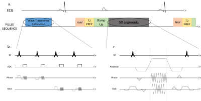 |
Wave-balanced Steady State Free Precession (Wave-bSSFP) for free breathing cardiac MR angiography
Quentin Lebret1,2,3, Pierre Bour1,2,3, Valéry Ozenne1,2,3, and Bruno Quesson1,2,3
1IHU LIRYC, Fondation Bordeaux Université, Pessac, France, 2U1045 CRCTB, Université de Bordeaux, Bordeaux, France, 3INSERM, CRCTB, U1045, Bordeaux, France
Using Wave-CAIPI-like techniques to accelerate Balanced Steady-state Free Precession (bSSFP) sequences is a new parallel imaging technique that reduces the noise levels on the reconstructed image compared to conventional parallel imaging. On the heart, this combination of sequence has not been tested. The experiments show that the movements of the thorax are not an issue, as long as a respiratory and ECG triggering are employed. A retrospective acceleration of 5.4 on a sheep heart image with a 1.4mm isotropic resolution has been achieved.
|
|
2141.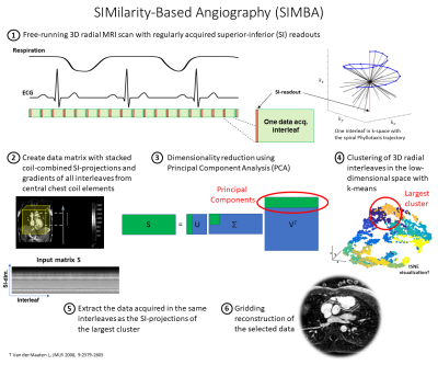 |
Similarity-Based Angiography (SIMBA) for Fast Reconstruction of Static Whole-Heart Coronary MR Images from Free-Running Acquisitions
John Heerfordt1,2, Kevin K. Whitehead3, Jessica A.M. Bastiaansen1, Lorenzo Di Sopra1, Christopher W. Roy1, Jérôme Yerly1,4, Mark A. Fogel3, Matthias Stuber1,4, and Davide Piccini1,2
1Department of Radiology, Lausanne University Hospital and University of Lausanne, Lausanne, Switzerland, 2Advanced Clinical Imaging Technology, Siemens Healthcare, Lausanne, Switzerland, 3Children’s Hospital of Philadelphia, Philadelphia, PA, United States, 4Center for Biomedical Imaging (CIBM), Lausanne, Switzerland
SIMilarity-Based Angiography (SIMBA) is a data-driven method for fast reconstruction of static whole-heart coronary MR angiograms from free-running acquisitions. By assessing the similarity of periodically acquired k-space readouts in the superior-inferior direction, motion-consistent angiograms can be obtained without making stringent assumptions about physiological motion. SIMBA demonstrated potential to reconstruct cardiac and respiratory motion-consistent images with visible coronary arteries both from non-contrast volunteer acquisitions at different field strengths and contrast-enhanced scans of pediatric patients. SIMBA provided improved sharpness and contrast compared to images from all the acquired data and similar vessel conspicuity as end-expiratory mid-diastolic frames of motion-resolved compressed sensing reconstructions.
|
|
2142.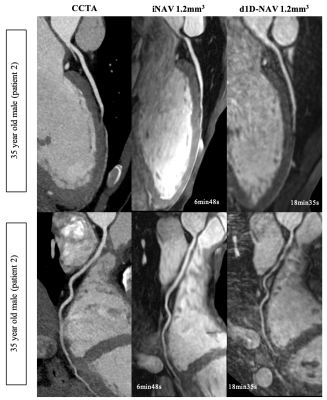 |
3D whole heart CMRA using an image-navigator framework – a clinical comparison study with CCTA and diaphragmatic navigators
Reza Hajhosseiny1, Aurélien Bustin1, Imran Rashid1, Gastao Cruz1, Radhouene Neji1,2, Karl Kunze1,2, Tevfik F. Ismail1, Ronak Rajani1, Pier Giorgio Masci1, Claudia Prieto1, and René M. Botnar1
1Biomedical Engineering Department, School of Biomedical Engineering and Imaging Sciences, King's College London, London, United Kingdom, 2MR Research Collaborations, Siemens Healthcare Limited, Frimley, United Kingdom
Conventional diaphragmatic navigator coronary magnetic resonance angiography (d1D-NAV-CMRA) is limited by long and unpredictable acquisition-times and motion related image quality degradation. To overcome these challenges, we have previously proposed an image-navigated non-rigid motion-compensated, undersampled reconstruction CMRA framework (iNAV-CMRA) with <10min acquisition times. Here we investigated the clinical feasibility of 1.2mm3 iNAV-CMRA, comparing its performance, in patients with suspected coronary artery disease, against 1.2mm3 d1D-NAV-CMRA and coronary computed tomography angiography (CCTA). Compared to the d1D-NAV-CMRA, the iNAV-CMRA had a shorter acquisition-time(7:24±0:33min vs. 21:02±7:27min, p<0.0001), with improved coronary vessel-length, vessel-sharpness and image-quality-score, and visually comparable to CCTA.
|
|
2143.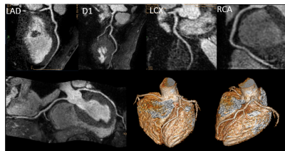 |
Non-contrast enhanced angiography and vessel wall imaging of coronary artery at 3T MR
Li Gao1, Xuchun Yuan1, Weixia Nie1, and Shutian Lai1
1Radiology, Fuwai hospital chinese academy of medical science, shenzhen, Shenzhen, China
Non-contrast enhanced MR angiography of coronary artery is a promising imaging method. In this study, the image quality of two non-contrast enhanced coronary MRA imaging methods were compared, included 3D-mDIXON and 3D-TFE-WHCA. 3D-mDIXON and 3D-TFE-WHCA showed good image quality with high SNR, and 3D-TFE-WHCA showed improved CNR and higher score in vascular display quality. According CTA data, T1-TFE-BB demonstrated the coronary vessel wall as medium high signal with regular margin and good contrast with the fat around the coronary artery. Combination of 3D-TFE-WHCA and T1-TFE-BB can be used as the imaging research method of coronary atherosclerosis.
|
|
2144. |
Improvement in 3.0 T whole-heart coronary MRA using the bSSFP method with a non-selective RF pulse
Takashige Yoshida1, Masami Yoneyama2, Kohei Yuda1, Yuki Furukawa1, Isao Miyazaki1, and Nobuo Kawauchi1
1radiology, Tokyo metropolitan police hospital, Tokyo, Japan, 2Philips Japan, Tokyo, Japan
One of the problems of whole heart coronary MRA using bSSFP sequence at 3.0T is banding artifacts, caused by a long TR due to SAR limitations, resulting in degradation of image quality. A non-selective radio frequency (RF) pulse (e.g. block or hard pulse) enables to shorten the TR that leads to to reduce the banding artifacts. Hence whole heart coronary MRA using the bSSFP with non-selective RF pulse was improved image quality at 3.0T.
|
|
2145.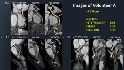 |
Whole heart coronary MRA with 3D non-selective bSSFP-DIXON: comparison with conventional methods
Kazuo Kodaira1, Michinobu Nagao2, Masami Yoneyama3, Yasuhiro Goto1, Isao Shiina1, Yutaka Hamatani1, Mamoru Takeyama1, Isao Tanaka1, and Shuji Sakai2
1Department of Radioligical Services, Tokyo Women's Medical University Hospital, Tokyo, Japan, 2Department of Diagnostic imaging & Nuclear Medicine, Tokyo Women's Medical University Hospital, Tokyo, Japan, 3Philips Japan, Tokyo, Japan
Improvement of fat suppression is important for whole heart coronary magnetic resonance angiography (WHC-MRA), because Unwanted signal arising from fat can compromise vessel delineation and decrease the diagnostic value of WHC-MRA. Balanced DIXON (bDIXON) can reduce fat suppression unevenness than SPIR method while maintaining high signal value. However, it is yet not applied for WHC-MRA. Furthermore, 3D non-selective bDIXON (3D NSbDIXON) with shortening TR can suppress an increase in imaging time. We propose a new sequence of 3D NSbDIXON for WHC-MRA, and examine the image quality in comparison to the conventional methods.
|
|
2146.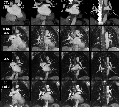 |
Breath-holding and free-breathing radial-based acquisition strategies for non-contrast MR angiography of the pulmonary veins
Pascale Aouad1, Ioannis Koktzoglou2, and Robert Edelman2
1Northwestern University, Chicago, IL, United States, 2Northshore University Healthsystem, chicago, IL, United States
CTA and contrast-enhanced MRA (CEMRA) are routinely used to evaluate vascular anatomy prior to pulmonary vein isolation for atrial fibrillation. As potential non-contrast alternatives, we evaluated three radial techniques (breath-hold 2D radial and stack-of-stars (SOS); free-breathing nonselective SOS). 24 patients were studied using CTA as reference standard. Consistently good image quality was obtained with all three radial techniques. There was good-to-excellent agreement with CTA on breath-holding and free-breathing SOS MRA for PV ostial diameters. Our results indicate that radial MRA may provide a useful non-contrast alternative to CTA and CEMRA, with the flexibility of allowing either breath-holding or free-breathing acquisitions.
|
|
2147. |
A comparative study of non-contrast enhanced 3D TRANCE and 3D Balanced FFE imaging of pulmonary artery with Time-SLIP
Yuan QU1, Yan WANG1, Yuli HUANG2, Li ZHU3, and Haiyang DONG3
1Radiology, People’s Hospital of Xinjiang Uygur Autonomous Region, Urumqi, China, 2MR, Philips Healthcare (Suzhou) Co., Ltd, SUZHOU, China, 3Radiology, Shanghai Chest Hospital, Shanghai JiaoTong University, Shanghai, China
The Time-Spatial Labeling Inversion Pulses (Time-SLIP) technique was used to perform non-contrast enhanced pulmonary artery imaging using 3D TRANCE and 3D Balanced FFE respectively, and the image quality obtained by the two imaging methods and the number of branches of pulmonary artery were compared specifically. Both 3D TRANCE and 3D Balanced FFE imaging methods with Time-SLIP can be used for pulmonary artery imaging. The image quality of pulmonary artery obtained by 3D TRANCE is generally better than 3D Balanced FFE.
|
|
2148.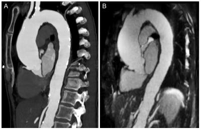 |
Measurement Accuracy of Non-contrast Compressed Sensing Respiratory Motion Resolved Whole Heart MRA in Thoracic Aortic Disease
Akos Varga-Szemes1, Robert E Stroud1, Davide Piccini2, John Heerfordt3, Jerome Yerly3, Lorenzo Di Sopra3, Pal Suranyi1, and U. Joseph Schoepf1
1Medical University of South Carolina, Charleston, SC, United States, 2Siemens Healthcare, Lausanne, Switzerland, 3Lausanne University Hospital and University of Lausanne, Lausanne, Switzerland
Currently, CTA is the only noninvasive method with considerable evidence level for the detection and monitoring of thoracic aortic disease. However, CTA exposes patients to radiation and contrast media. The compressed-sensing whole-heart MRA technique that we evaluated addresses the limitations that prevent conventional MRA techniques to compete with CTA. Our approach eliminates the need for breath-holds or respiratory-navigation, provides a 3D volume of the chest without contrast administration, and allows for accurate anatomical evaluation of the thoracic aorta compared to CTA. Such MRA technique is a potential radiation- and contrast-free alternative for diagnosing and monitoring patients with thoracic aortic disease.
|
2149.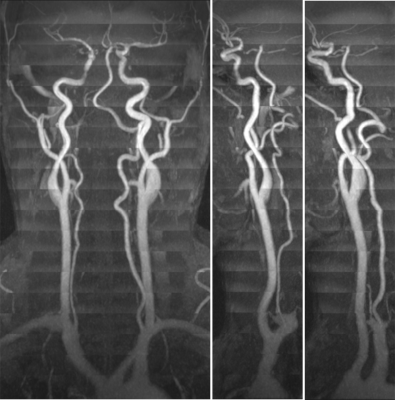 |
Whole-Neck Arterial Evaluation with High Spatial Resolution using 3D Stack-of-Stars Quiescent Interval Slice-Selective (QISS) MRA
Ioannis Koktzoglou1,2 and Robert R Edelman1,3
1Radiology, NorthShore University HealthSystem, Evanston, IL, United States, 2The University of Chicago Pritzker School of Medicine, Chicago, IL, United States, 3Radiology, Northwestern University Feinberg School of Medicine, Chicago, IL, United States
We found that a prototype thin-slab stack-of-stars QISS sequence using a low-flip-angle FLASH readout can be used for whole-neck arterial evaluation in under 7 minutes. The technique minimizes flow-related saturation effects similarly to 2D QISS, while providing greater scan efficiency compared with 3D TOF and much improved image quality and through-plane spatial resolution compared with 2D TOF.
|
|
2150.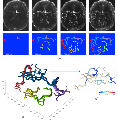 |
Semi-automatic Arterial Territorial Segmentation using ASL-based Dynamic 4D MRA without Vessel-Encoded Labeling
Soroush Heidari Pahlavian1,2, Oren Geri3, Jonathan Russin2, Dafna Ben-Bashat4, Xingfeng Shao1,2, Samantha Ma1,2, Songlin Yu5, Arun Amar2, Danny J.J. Wang1,2, and Lirong Yan1,2
1USC Stevens Neuroimaging and Informatics Institute, Keck School of Medicine, University of Southern California, Los Angeles, CA, United States, 2Department of Neurology, University of Southern California, Los Angeles, CA, United States, 3Razor Labs, Tel Aviv, Israel, 4Medicine & Sagol School of Neuroscience, Tel Aviv University, Tel Aviv, Israel, 5Peking Union Medical College Hospital, Beijing, China
Characterizing vascular territorial structures and hemodynamics from a single artery can provide crucial information for the assessment and treatment of cerebrovascular disorders such as arteriovenous malformations, Moyamoya disease, and aneurysms. In a different approach compared to vessel-selective MR angiography (MRA), here we presented a semi-automatic post-processing technique to segment vascular territories using pulsed arterial spin labeling 4D MRA. Our results demonstrated the feasibility of using 4D MRA in conjunction with arterial territorial segmentation to visualize vascular territories and quantify blood flow supply from individual arteries.
|
|
2151.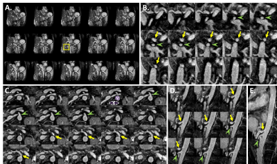 |
Non-Contrast-Enhanced MR Angiography of The Celiac Trunk Using 3D Magnetization-Prepared Dual-Echo Dixon in Free Breathing
Shuo Zhang1,2, Caroline Molavi Tabrizi2, Masami Yoneyama3, Christiane Kuhl2, and Alexandra Barabasch2
1Philips GmbH DACH, Hamburg, Germany, 2Diagnostic and Interventional Radiology, University Hospital RWTH Aachen, Aachen, Germany, 3Philips Japan, Tokyo, Japan
Contrast-enhanced MR angiography (CE-MRA) permits accurate assessment of the abdominal aorta and the splanchnic arteries, including the celiac trunk and its branches. However, it is often restricted for patients undergoing dynamic-contrast-enhanced imaging of the liver in the same exam. In this study, we employ the recently introduced Relaxation-Enhanced Angiography without Contrast and Triggering (REACT) method using magnetization-prepared dual-echo Dixon for 3D free-breathing non-contrast-enhanced imaging of the celiac trunk. Initial results in healthy volunteer and patients are reported in comparison to the other techniques such as the conventional and arterial spin labeling (ASL) based balanced steady-state free precession (bSSFP) sequences.
|
|
2152.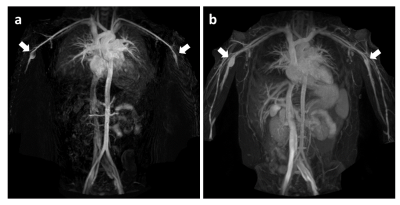 |
Rapid diastolic-phase only TRANCE non-contrast MR angiography in screening of systemic artery aneurysm in patients with Kawasaki disease
Haruki Nonaka1, Masami Yoneyama2, Masahiro Tahara3, Tetsuya Nitta4, Mio Okano1, Yuko Morikawa1, Susumu Kariyama1, Tomohiro Kimura1, Yoriaki Matsumoto1, Takanori Masuda1, Naoyuki Imada5, and Tomoyasu Sato6
1Department of Radiological Technology, Tsuchiya General Hospital, Hiroshima, Japan, 2Philips Japan, Tokyo, Japan, 3Department of Pediatrics, Tsuchiya General Hospital, Hiroshima, Japan, 4Nitta Pediatric Clinic, Hiroshima, Japan, 5Department of Health Care, North Hiroshima Hospital, Hiroshima, Japan, 6Department of Radiology, Tsuchiya General Hospital, Hiroshima, Japan
The purpose of this study was to demonstrate the clinical feasibility and utility of diastolic-phase only ECG-triggered non-contrast-enhanced MR angiography (TRANCE) in screening of systemic artery aneurysms (SAA) in Kawasaki disease patients. Diastolic-phase only TRANCE provided consistent high image quality for evaluating main arteries in children with the average scanning time around 155sec in addition to routine coronary MRA screening. Diastolic-phase only TRANCE could be useful in SAA screening without significant prolongation of examination time.
|
|
2153.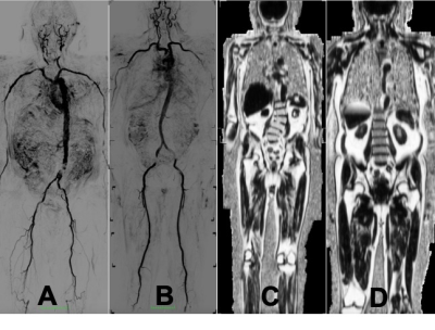 |
Whole-body MRI to assess subclinical cardiovascular disease and frailty development
Bharath Ambale Venkatesh1, Jason Ortman2, Jaclyn Sesso2, Yoko Kato2, Elzbieta Chamera2, Jennifer Wagner3, Yoshimori Kassai4, and Joao Lima2
1Radiology, Johns Hopkins University, Baltimore, MD, United States, 2Johns Hopkins University, Baltimore, MD, United States, 3Canon Medical Research USA, Mayfield Village, OH, United States, 4Canon Medical Systems, Kanagawa, Japan
Whole body non-contrast magnetic resonance angiography and Dixon imaging hold potential for monitoring and quantitative assessment of global plaque burden and cardiometabolic disease in frailty.
|
|
2154.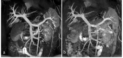 |
A comparative study on TRANCE and bFFE imaging of hepatic portal vein with Time-SLIP
Yan WANG1, Yuli HUANG2, Haiyang DONG3, and Li ZHU3
1Department of Radiology, People’s Hospital of Xinjiang Uygur Autonomous Region, Urumqi, China, 2MR, Philips Healthcare (Suzhou) Co., Ltd, SUZHOU, China, 3Radiology, Shanghai Chest Hospital, Shanghai JiaoTong University, Shanghai, China
The Time-Spatial Labeling Inversion Pulses (Time-SLIP) technique was used to perform non-contrast enhanced hepatic portal vein imaging using 3D TRANCE and 3D Balanced FFE methods respectively, and the image quality obtained by the two imaging methods and the number of branches of hepatic portal vein were compared specifically. Both 3D TRANCE and 3D Balanced FFE imaging methods with Time-SLIP can be used for hepatic portal vein imaging. The image quality of hepatic portal vein obtained by 3D TRANCE was generally better than 3D Balanced FFE.
|
|
2155. |
Optimisation of Poisson-disk Sampling Pattern for Highly Accelerated Femoral NCE-MRA
Hao Li1, Martin John Graves2, David John Lomas1, and Andrew Nicholas Priest2
1Department of Radiology, University of Cambridge, Cambridge, United Kingdom, 2Department of Radiology, Addenbrooke’s Hospital, Cambridge, United Kingdom
NCE-MRA techniques have a long acquisition time but can be accelerated by compressed sensing, parallel imaging and partial Fourier sampling using, for example, a Poisson-disk sampling pattern. In this study, we optimised the parameters in sampling pattern design with different acceleration factors for the 3D accelerated femoral fresh-blood-imaging sequence. The NCE-MRA data were reconstructed using weighted subtraction in k-space combined with phase correction. In a comparison using retrospective acceleration by subsampling of a full dataset, the optimised patterns outperformed the non-optimised patterns.
|
|
2156.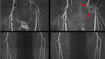 |
Optimized Stack-of-Stars QISS MR Angiography for Near-Isotropic Non-Contrast Evaluation of the Renal and Peripheral Arteries
Robert R Edelman1,2, Jianing Pang3, and Ioannis Koktzoglou1,4
1Radiology, NorthShore University HealthSystem, Evanston, IL, United States, 2Radiology, Feinberg School of Medicine, Northwestern University, Chicago, IL, United States, 3Siemens Medical Solutions USA, Chicago, IL, United States, 4Pritzker School of Medicine, University of Chicago, Chicago, IL, United States
Quiescent-interval slice-selective (QISS) MRA has become established as a simple “push button” nonenhanced alternative to CTA and contrast-enhanced MRA. However, a fundamental drawback of QISS as currently implemented is that it uses a 2D acquisition strategy. The inability to acquire very thin slices along with the non-rectangular slice profile predisposes QISS to artifacts from partial volume averaging and blurring of multiplanar reconstructions. We therefore implemented and optimized a novel thin-slab stack-of-stars QISS (tsSOS-QISS) technique to provide near-isotropic high spatial resolution that rivals CTA, while maintaining the excellent arterial conspicuity, motion insensitivity and breath-hold capability of 2D QISS.
|
|
2157.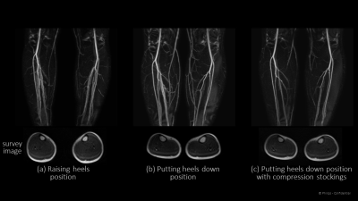 |
Non-triggered, non-contrast lower extremity artery-weighted REACT : Impact of patient positioning for improving artery-vein contrast
Hiroshi Hamano1, Masami Yoneyama1, Yasuhiro Goto2, Takayuki Sakai3, Yasutomo Katsumata4, and Kenji Iinuma1
1Philips Japan, Tokyo, Japan, 2Department of Radiological Services, Tokyo Women's Medical University, Tokyo, Japan, 3Radiology, Eastern Chiba Medical Center, Chiba, Japan, 4Philips Healthcare, Tokyo, Japan
Artery-weighted REACT (REACT-Art), which is optimized the T2prep-time and TFE factor to improve the artery-vein contrast has been proposed, and it provide good artery-dominant contrast compared to conventional REACT. In this study, we focused on the impact of patient positioning to improve the artery-vein contrast with higher robustness, such as raising patient’s heels, putting patient’s heels down, compressing the patient’s legs. we have demonstrated that the patient-positioning like the patient legs to be compressed, and wearing compression stockings might achieve improved artery-vein contrast of REACT-Art with high stability and robustness in the lower extremities without use of contrast-enhancement and ECG-triggering.
|
|
2158.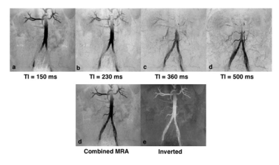 |
Feasibility of Simultaneous Non-contrast Angiography and intraPlaque hemorrhage Imaging (SNAP) in Abdominopelvic Angiography
Dmytro Pylypenko1, Yishi Wang2, Le He1, and Hua Guo1
1Biomedical Engineering, Tsinghua University, Beijing, China, 2Philips Healthcare, Beijing, China
Non-contrast enhanced MRA of abdominopelvic arteries within a large field of view is very challenging due to rapid blood flow and variant flow speed among different subjects. Simultaneous Non-contrast Angiography and intraPlaque imaging (SNAP), a non-contrast enhanced MRA technique, provides reliable imaging for the carotid and intracranial arteries. In this study, we sought to explore the feasibility of SNAP with compressed sensing acceleration in abdominopelvic arteries imaging. The complementary information provided by different scans with different velocity sensitivities can be combined to obtain reliable abdominopelvic MRA with a large coverage.
|
|
2159.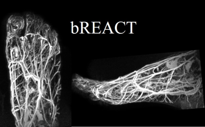 |
Improved visualization of non-contrast-enhanced MRA of the foot using REACT with balanced SSFP-DIXON (bREACT) at 1.5T
Yutaka Hamatani1, Kayoko Abe2, Yasuhiro Goto1, Masami Yoneyama3, Isao Shiina1, Kazuo Kodaira1, Mamoru Takeyama1, Isao Tanaka1, and Shuji Sakai2
1Department of Radiological Services, Tokyo Women's Medical University Hospital, Tokyo, Japan, 2Department of Diagnostic imaging & Nuclear Medicine, Tokyo Women's Medical University Hospital, Tokyo, Japan, 3Philips Japan, Tokyo, Japan
In this study we demonstrated the feasibility of dual-echo balanced SSFP-DIXON sequence with REACT technique for the foot MRA at 1.5T. bREACT dramatically increased the SNR compared to conventional REACT and it could provide excellent contrast between blood vessels and background tissues with high robustness compared with other conventional non-contrast-enhanced MRA methods. This technique would be helpful to assess peripheral blood vessels in the diabetic patients.
|
|
2160.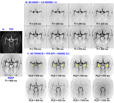 |
Comparison of Non-Contrast Enhanced Dynamic MRA Techniques: 4D-SNAP vs 4D-TRANCE
Hua Guo1, Yuhui Xiong1,2, Sisi Li1, Simin Liu1, and Chun Yuan3
1Center for Biomedical Imaging Research, Department of Biomedical Engineering, School of Medicine, Tsinghua University, Beijing, China, 2Neusoft Medical Systems Co., Ltd., Shanghai, China, 3Vascular Imaging Laboratory, Department of Radiology, University of Washington, Seattle, WA, United States
Simultaneous Non-contrast Angiography and intraPlaque imaging (SNAP), a non-contrast enhanced MRA technique, shows great potentials for either static artery imaging or 4D time-resolved artery imaging. Arterial spin labeling (ASL) based imaging techniques can also be used for dynamic MRA, e.g., 4D time-resolved angiography non-contrast enhanced (4D-TRANCE). Both of them are compatible with parallel imaging thus practical for clinical usage. But they are based on different principles and their performance comparison is desired. This study aims to compare 4D-SNAP and 4D-TRANCE. Results show 4D-SNAP provides 4D dynamic MRA with higher SNR and fewer artifacts than 4D-TRANCE using either TFE or TFE-EPI.
|
|
2161.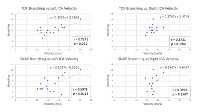 |
An In-Vivo Assessment of Velocity Sensitivity for TOF MRA and SNAP MRA
Anders Gould1, Zhensen Chen1, Zechen Zhou2, Niranjan Balu1, Thomas Hatsukami3, and Chun Yuan1
1Vascular Imaging Lab and Bio-Molecular Imaging Center, Radiology, University of Washington, Seattle, WA, United States, 2Philips Research North America, Cambridge, MA, United States, 3Surgery, University of Washington, Seattle, WA, United States
TOF and SNAP are both useful angiographic imaging sequences. In this study, we compared signal patterns of intracranial arteries depicted by the two techniques and explored their relationship with blood flow velocity. Twenty-four subjects with carotid atherosclerosis were imaged. The number of distal intracranial branches were scored based on the MRA images. SNAP showed more variation across subjects than TOF. TOF MRA score seems more strongly correlated with carotid velocity. This suggests TOF MRA may be more robust, while SNAP MRA may be more sensitive to flow change, although the underlying mechanism needs further clarification in the future.
|
2162. |
Evaluation of Myocardial Strain in Patients With Early Hypertrophic Cardiomyopathy and Hypertensive Heart Disease Using CMR-FT
Xiaoyong Hao1, Jiang Wu1, Chaohui Yang1, Lina Zhu1, Heng Niu1, Xia Zhang1, Xuan Li1, and Kaiyu Wang2
1Shanxi Cardiovascular Hospital, TaiYuan, China, 2GE Healthcare, MR Research China, BeiJing, China
This work assessed the application of cardiovascular magnetic resonance feature tracking (CMR-FT) technique in evaluation of myocardium deformation in patients with early hypertrophic cardiomyopathy and hypertensive heart disease. From the results we can see that the CMR-derived feature tracking technology could detect impairment of left ventricula myocardial deformation in patients with early hypertrophic cardiomyopathy and early hypertensive heart disease, even in patients with normal LVEF.
|
|
2163. |
Mechanical properties from CMR parameters in childhood acute lymphoblastic leukemia survivors.
Delphine Perie1, Egidie Uwase1, Tanguy Artz1, Marianna Gamba1, and Daniel Curnier2
1Mechanical Engineering, Polytechnique Montreal, Montreal, QC, Canada, 2Kinesiology, University of Montreal, Montreal, QC, Canada
Swine myocardium mechanical properties were found related to a combination of CMR relaxations times, magnetization transfer and diffusion parameters. Thus we assessed the relationships between mechanical properties and CMR parameters in childhood acute lymphoblastic leukemia survivors. The results showed that the pressure at diastasis, contractility and stiffness properties of the left ventricle can be partially predicted from the relaxation times T1 and T2 and the partition coefficient, reflecting cardiac health in childhood acute lymphoblastic leukemia. This indirect estimation of the mechanical behavior of the myocardium from multiparametric MRI could solve the challenging early cardiac sequelae detection.
|
|
2164.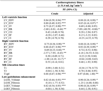 |
Preventive fraction of long-term cardiac function with cardiorespiratory fitness and physical activity in childhood leukemia survivors.
Delphine Perie1, Maxime Caru2, Marianna Gamba1, Louise Leleu1, and Daniel Curnier2
1Mechanical Engineering, Polytechnique Montreal, Montreal, QC, Canada, 2Kinesiology, University of Montreal, Montreal, QC, Canada
In childhood leukemia survivors, doxorubicin leads to dose-dependent cardiotoxicity, despite early diagnosis with both echocardiography and MRI investigations. Physical activity has the potential to reduce the chronic disease risk, but it is currently unknown whether a good cardiorespiratory fitness or the regular practice of physical activity is enough to induce a preventive action on the cardiac function. This study included 81 ALL survivors and found that a good cardiorespiratory fitness was associated with a better preventive fraction, similarly to a good physical activity level. It would be more than 80% of the survivors who could benefit from these long-term effects.
|
|
2165.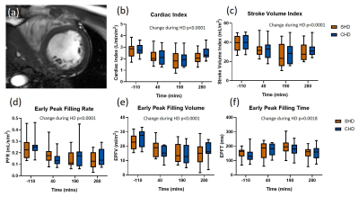 |
Comparing intradialytic effects of standard vs cooled haemodialysis on cerebral blood flow and cardiac function using MRI
Eleanor F Cox1, Latha Gullapudi2,3, Charlotte E Buchanan1, Kelly White3, Sebastian Coleman1, Bernard Canaud4, Maarten W Taal2,3, Nicholas M Selby2,3, and Susan T Francis1
1Sir Peter Mansfield Imaging Centre, University of Nottingham, Nottingham, United Kingdom, 2Centre for Kidney Research and Innovation, University of Nottingham, Derby, United Kingdom, 3Renal Unit, University Hospitals of Derby and Burton NHS Foundation Trust, Derby, United Kingdom, 4Fresenius Medical Care, Bad Homburg, Germany
12 patients received standard (SHD) and cooled (CHD) haemodialysis in a randomized cross-over study. Participants underwent serial cardiac and brain multiparametric MRI before, during and after dialysis. Cerebral blood flow velocities fell significantly during dialysis (carotid artery -19±2%, p<0.001; basilar artery -16±3%, p=0.004). Cardiac index and stroke volume index fell significantly during dialysis (-29±2% and -32±2%, both p<0.001). After two weeks of CHD, pre-dialysis left ventricle (LV) myocardial T1 and wall mass (WM) were lower compared to SHD (T1: 1266(37)ms vs. 1311±18ms, p=0.02; LV WM: 128±12g vs. 137±12g, p=0.003), indicating reduced myocardial tissue oedema with CHD.
|
|
2166. |
CMR Feature tracking: reproducibility in small animals
Hao Li1,2, Steffen Just3, Qinghua Lu2, and Volker Rasche1,3
1Core Facility Small Animal Imaging, Ulm University, ULM, Germany, 2Department of Cardiology, The Second Hospital of Shandong University, Jinan, China, 3Department of Internal Medicine II, Ulm University Medical Center, Ulm, Germany
Cardiac magnetic resonance imaging feature tracking (CMR-FT) is a novel non-invasive imaging technique and has been validated in clinical applications. However, there are only limited studies in small animal research. We performed biventricular strain assessment using CMR-FT in nexiline knock-out mice at rest and dobutamine stress to evaluate its feasibility and intra-observer reproducibility in preclinic research. Excellent intra-observer reproducibility could be observed for rest and stress left ventricular (LV) strain in general and right ventricular (RV) circumferential strain derived from short-axis views. An only fair reproducibility was observed for RV longitudinal strain under stress conditions derived from long-axis views.
|
|
2167.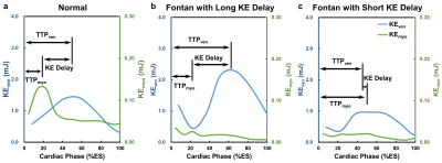 |
Myocardial and Intraventricular Kinetic Energy in Patients with Fontan Circulation
Xue-Zhe Lu1, Ming-Ting Wu2, Ken-Pen Weng3,4, and Hsu-Hsia Peng1
1Department of Biomedical Engineering and Environmental Sciences, National Tsing Hua University, Hsinchu, Taiwan, 2Department of Radiology, Kaohsiung Veterans General Hospital, Kaohsiung, Taiwan, 3Department of Pediatrics, Kaohsiung Veterans General Hospital, Kaohsiung, Taiwan, 4Department of Pediatrics, National Yang-Ming University, Taipei, Taiwan
We aimed to investigate the interaction between systolic myocardial and intraventricular kinetic energy (KEmyo, KEven) in Fontan patients. The tissue phase mapping and 4D flow datasets were acquired for calculation of KEmyo and KEven. The Fontan group showed decreased peak and mean systolic KEven, and decreased peak and mean systolic KEmyo. The KE delay, describing the subtraction of time-to-peak KEmyo from time-to-peak KEven, in Fontan group significantly correlated with peak and mean systolic KEven. In conclusion, the correlation between KE delay and KEven illustrated the adverse
impact of abnormal energy transferring mechanism on the single functional ventricle in Fontan patients.
|
|
2168.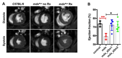 |
MR detects improvement in cardiac function and fibrosis after micro-dystrophin treatment in dystrophic mice
Ravneet Vohra1, Guy Odom2, Jeffrey S Chamberlain2,3, and Donghoon Lee1
1Radiology, University of Washington, Seattle, WA, United States, 2Neurology, University of Washington, Seattle, WA, United States, 3Medicine, University of Washington, Seattle, WA, United States
Cardiomyopathy is an inevitable fate for patients with Duchenne muscular dystrophy (DMD) and is one of the major causes of mortality. Cardiovascular magnetic resonance (CMR) is increasingly being performed at very high magnetic field strength for small animal models of muscular dystrophy. The mdx mouse model is one of the most commonly used animal models for DMD. Recombinant adeno-associated viral vector-mediated gene transfer represents a promising approach for DMD. The aim of this study was to elucidate the functional impact of micro-dystrophin on cardiomyopathy in mdx mice using CMR as a non-invasive biomarker.
|
|
2169. |
An Efficient Sampling Strategy of Fast Multi-slice Cardiac Cine Imaging
Dongchan Kim1, Byungjai Kim2, Kyoung-Nam Kim3, Jun-Young Chung4, HyunWook Park2, and Yeji Han3
1Samsung Electronics, Seoul, Republic of Korea, 2Department of Electrical Engineering, Korea Advanced Institute of Science and Technology, Daejeon, Republic of Korea, 3Department of Biomedical Engineering, Gachon University, Incheon, Republic of Korea, 4Gachon Advanced Institute for Health Sciences and Technology, Gachon University, Incheon, Republic of Korea
Balanced steady-state free precession (bSSFP) with electrocardiogram (ECG) triggering and breath-holding is a common MRI technique for examination of ventricular function. For whole-heart imaging, resting periods are often necessary between breath-holds because many patients find it difficult to perform successive breath-holds and this eventually prolongs the entire examination time. Although self-gating (SG) techniques can relieve the discomfort that comes from multiple breath-holds, the sampling efficiency is limited because the data from inspiration phases are discarded. In this work, we propose a new sampling strategy for efficient multi-slice cardiac cine imaging that acquires data with a reduced number of breath-holds.
|
|
2170.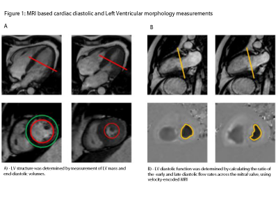 |
Association Between Physical Activity and Left Ventricular Diastolic Function and Morphology assessed by MRI in Obesity
Hugo Klarenberg1, Jeroen H. P. M. van der Velde2, Ilona A. Dekkers3, R. de Mutsert2, J. Wouter Jukema4, Frits R. Rosendaal2, Mark Gosselink5, Martijn Froeling5, Gustav J. Strijkers1, S. Matthijs Boekholdt6, and Hildo J. Lamb3
1Biomedical Engineering and Physics, Amsterdam UMC location AMC, Amsterdam, Netherlands, 2Department of Clinical Epidemiology, Leiden University Medical Center, Leiden, Netherlands, 3Department of Radiology, Leiden University Medical Center, Leiden, Netherlands, 4Department of Cardiology, Leiden University Medical Center, Leiden, Netherlands, 5Department of Radiology, University Medical Center Utrecht, Utrecht, Netherlands, 6Department of Cardiology, Amsterdam UMC location AMC, Amsterdam, Netherlands
Heart failure with preserved ejection fraction is accompanied by diastolic dysfunction. Diastolic dysfunction is associated with metabolic dysregulations, particularly obesity. Physical Activity might reduce this process improving diastolic function and left ventricular (LV) morphology. In this cross-sectional Netherlands Epidemiology of Obesity study, individuals underwent cardiovascular MRI to asses diastolic function and LV Morphology. Physical Activity was determined via the SQUASH questionnaire. Multivariable linear regression was performed adjusting for metabolic covariates. Physical Activity was moderately associated to diastolic function and strongly to LV Morphology. Conclusively, associations of diastolic function and LV morphology with Physical Activity can be determined accurately by MRI.
|
|
2171.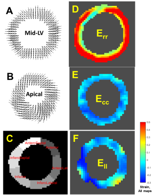 |
Accelerated-x4 Balanced-SSFP Cardiac Cine 2D/3D DENSE with Phase-Cycled Encoding for Improved Performance
Ronald J. Beyers1, Adil Bashir1, and Thomas S. Denney1
1MRI Research Center, Auburn University, Auburn University, AL, United States
Cine frame-rate quantification of myocardial strain has been previously demonstrated with 2D echo-planar and 2D/3D spiral sequence versions of Displacement Encoding with Stimulated Echoes (DENSE). While these non-conventional methods allow fast-acquisition, they are plagued with off-resonance limitations that has hindered their integration into mainstream cardiac MRI application. Here we present a more conventional, accelerated-x4, balanced SSFP (bSSFP) version of Cardiac Cine 2D/3D DENSE with improved phase-cycled encoding for effective artifact suppression. In vivo human scans at 3T demonstrated good agreement of myocardial radial (Err), circumferential (Ecc) and longitudinal (Ell) strains as reported in previous literature along with lower artifact degradation compared to standard complementary-encoded DENSE.
|
|
2172. |
Cardiac magnetic resonance feature-tracking for myocardial strain assessment in real-time cardiac cine MRI
Ashitha Pathrose1, Hassan Haji-Valizadeh1,2, Roberto Sarnari1, James Carr1,2,3, and Daniel Kim1,2
1Radiology, Northwestern University, Chicago, IL, United States, 2Biomedical Engineering, Northwestern University, Chicago, IL, United States, 3Medicine, Northwestern University, Chicago, IL, United States
Cardiac magnetic resonance feature-tracking (CMR-FT) has emerged as a reference standard for the evaluation of cardiac morphology and function. But, the performance of CMR-FT may be affected by data undersampling as done in several real-time cardiac MRI techniques. In our study, we evaluated the performance of CMR-FT for myocardial strain quantification from real-time cardiac cine MRI when compared to standard cardiac cine in cardiac disease patients. We found that even though there is good agreement between the values derived from the real-time and standard cine MRI, care should be taken as measures from the real-time cine can be underestimated.
|
|
2173.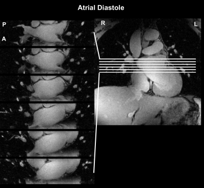 |
Cartesian prospective, duel-gated, cardio-respiratory encoding for 3D cardiac CINE compared to spiral profile retrospective encoding.
Sven HF Jaeschke1 and Aaron T Hess1
1Oxford Centre For Clinical Magnetic Resonance Research, University of Oxford, Oxford, United Kingdom
A number of retrospective Cartesian encoding strategies exist for duel gated cardio-respiratory imaging, including freebreathing cine. If respiration and cardiac motion are known prospectively while scanning, is prospective sampling more efficient? To test this we developed an algorithm to prospectively handle variable acquisition durations for each cardio-respiratory bin while acquiring pre-defined Poisson-Disk variable density sampling masks. We compared this to a retrospective spiral profile Cartesian trajectory and found the prospective method had between 10% and 25% higher efficient for matched scan time and had between 20% and 40% higher peak-to-sidelobe ratio of the point-spread-function, which is beneficial for image reconstruction.
|
|
2174. |
Optimized ultra-high-field cardiac magnetic resonance imaging
El-Sayed H Ibrahim1, V Emre Arpinar1, L Tugan Muftuler1, Andrew Nencka1, and Kevin Koch1
1Medical College of Wisconsin, Milwaukee, WI, United States
Cardiac functional MRI has been established in clinical practice on 1.5T and 3T scanners; nevertheless, the capabilities of ultra-high field (UHF) MRI have not been fully exploited, which are expected to significantly improve image quality and provide information not obtainable at lower-field MRI. Despite its advantages, UHF MRI is challenging due to a number of technical issues, especially B1 and B0 inhomogeneities. In this study, we provide preliminary results of using a multi-channel transceiver modular coil and a dielectric pad towards optimizing UHF cardiac functional MRI by improving image quality and minimizing imaging artifacts.
|
|
2175.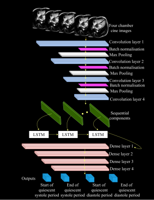 |
Fully automated detection of the quiescent phases of the cardiac cycle from CINE images using deep learning
Naledi Adam1, James Clough1, Ronald Mooiweer1, Phuoc Dong1, Li Huang1, Reza Razavi1, Kuberan Pushparajah1, Amedeo Chiribiri1, Andrew King1, and Sébastien Roujol1
1Biomedical Engineering Department, School of Biomedical Engineering and Imaging Sciences, King's College London, London, United Kingdom
Cardiac magnetic resonance (CMR) imaging is commonly performed using ECG-triggering to restrain acquisition to a quiescent phase of the cardiac cycle (i.e. end-systole or mid-diastole). Identification of the rest periods is commonly performed by visual inspection on CINE images and is thus operator dependent, time consuming and requires a trained operator. In this study, a fully automated method was developed to detect the quiescent cardiac periods of the cardiac cycle from CINE images using an integrated convolutional neural network (CNN) and long short-term memory (CNN-LSTM) network.
|
2176. |
Auto-calibrated simultaneous multi-slice cardiac bSSFP Cine MRI using gradient temporal modulation
Yuan Zheng1, Lele Zhao2, Zhongqi Zhang2, Zhehao Zhang2, Xingxian Shou1, Haojie Li3, Jian Xu1, Weiguo Zhang1, Lu Huang3, and Liming Xia3
1UIH America, Houston, TX, United States, 2United Imaging Healthcare, Shanghai, China, 3Tongji Hospital, Wuhan, China
Simultaneous multi-slice (SMS) imaging is an efficient MRI technique that better preserves SNR than conventional parallel imaging (PI). However, single-band images usually need to be acquired to provide references for slice-unaliasing, causing an untrivial scan time overhead in some scenarios. We extended a recently reported self-calibrated SMS imaging strategy based on RF phase modulation, and propose using gradient temporal modulation to achieve auto-calibrated SMS bSSFP, which avoids the bSSFP banding shift and is preferred in cardiac Cine due to its contrast and intrinsically high SNR. In-plane PI can also be incorporated with shared auto-calibration.
|
|
2177.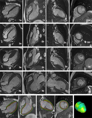 |
Myocardial strain of hypertrophic ventricular noncompaction, isolated ventricular noncompaction and hypertrophic cardiomyopathy
Shenglei Shu1, Jing Wang1, Xiaoyue Zhou2, Chuangsheng Zheng1, and Xiangquan Kong1
1Radiology, Union Hospital, Tongji Medical College, Huazhong University of Scince and Technology, Wuhan, China, 2MR Collaboration, Siemens Healthineers Ltd., Shanghai, China
Understanding of ventricular noncompaction appearance with simultaneous hypertrophic cardiomyopathy remains to be improved, as conventional used markers may not be able to distinguish subtle variation between isolated ventricular noncompaction and hypertrophic cardiomyopathy. We utilize the myocardial strain analysis based on cine images to compare different subgroups in order to find clues of underlying pathophysiology. Remarkable difference of the strain value were found in hypertrophic cardiomyopathy patients compared with the isolated ventricular noncompaction ones.
|
|
2178.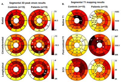 |
Comprehensive assessment of the left ventricle using cardiac magnetic resonance in patients with pre-capillary pulmonary hypertension
Ashitha Pathrose1, Roberto Sarnari1, Sabeth Essl1, Carson Herman1, Daniel Gordon1, Kelvin Chow1,2, Benjamin Freed3, Michael Cuttica3, Michael Markl1,4, and James Carr1,3,4
1Radiology, Northwestern University, Chicago, IL, United States, 2Siemens Medical Solutions USA, Chicago, IL, United States, 3Medicine, Northwestern University, Chicago, IL, United States, 4Biomedical Engineering, Northwestern University, Chicago, IL, United States
Cardiac MR provides valuable information of the cardiac structure-function, but it is underutilized in the diagnosis and monitoring patients with pulmonary hypertension. In this study, we performed a comprehensive evaluation of the left ventricle in patients with pre-capillary pulmonary hypertension including feature-tracking strain, T1 mapping, and tissue phase mapping. We also evaluated the association of these CMR-derived quantitative measures with right heart catheterization derived pressure measurements.
|
|
2179.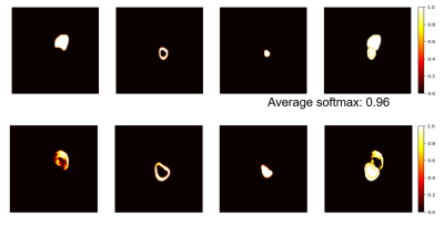 |
Quality prediction score: predicting the CNN-based segmentation performance for cine cardiac MR images
Ping-Chen Wu1, Zheng-Shen Lin1, and Teng-Yi Huang1
1Department of Electrical Engineering, National Taiwan University of Science and Technology, Taipei, Taiwan
In this study, we evaluated the open-source toolbox for heart segmentation by using cross-institutional datasets. Furthermore, we proposed to use a quality prediction socre to provide the estimation of the qualitfy of the cardiac segmentation. The QPS values were highly correlated to the Dice coefficients of the test datasets.
|
|
2180.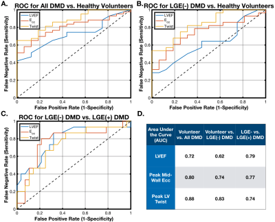 |
Left Ventricular Twist and Circumferential Strain from MRI Tagging Assess Early Cardiovascular Disease in Duchenne Muscular Dystrophy
Patrick Magrath1,2, Nyasha Maforo2,3, Mike Loecher4, Grace Kim5, Holden H. Wu 1,2,3, Ashley Prosper2, Pierangelo Renella2, Nancy Halnon6, and Daniel B. Ennis 4
1Bioengineering, University of California, Los Angeles, CA, United States, 2Radiology, University of California, Los Angeles, CA, United States, 3Physics and Biology in Medicine IDP, University of California, Los Angeles, CA, United States, 4Radiology, Stanford University, Stanford, CA, United States, 5Biostatistics, University of California, Los Angeles, CA, United States, 6Pediatrics, University of California, Los Angeles, CA, United States
Duchenne Muscular Dystrophy is a common fatal inherited genetic disorder impacting 1:3800 male births, and cardiac failure is the primary source of mortality in this cohort. Decreases in LVEF measured by CINE and fibrosis measured by LGE are late and highly variable outcomes. Herein we demonstrate that peak systolic circumferential strain (Ecc) and Twist measured by MR Tagging provide evidence of earlier changes in cardiac function in a substantial DMD cohort, holding promising applications for patient treatment and the evaluation of novel therapeutics.
|
|
2181.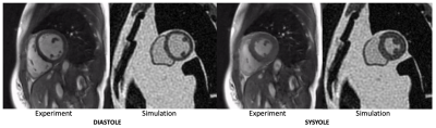 |
Realistic Simulation of High-Performance Low Field Cardiac Cine Imaging
Xitong Wang1, Namgyun Lee2, Adrienne E. Campbell-Washburn3, and Krishna S. Nayak1
1Electrical Engineering, University of Southern California, Los Angeles, CA, United States, 2Biomedical Engineering, University of Southern California, Los Angeles, CA, United States, 3National Heart, Lung, and Blood Insititute, National Institues of Health, Bethesda, MD, United States
There is renewed interest in high-performance low field imaging, especially for applications that are limited by susceptibility and SAR, such as cardiac CINE bSSFP imaging. Here, we demonstrate a framework for realistic simulation of 3D CINE bSSFP cardiac imaging at various field strengths. This is benchmarked against in-vivo data acquired on a high-performance 0.55 Tesla MRI. We utilize the XCAT phantom, 3D stack of spiral sampling, and realistic noise. This setup can be used to predict minimum field strength requirements and to test data sampling and reconstruction techniques.
|
|
2182.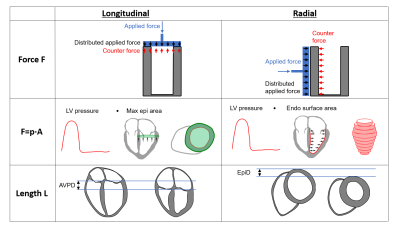 |
Global ventricular force-length loops and the quantification of longitudinal and radial contribution to stroke work
Felicia Seemann1,2, Jonathan Berg1,3, Kristian Solem3, Robert Jablonowski1, Håkan Arheden1, Marcus Carlsson1, and Einar Heiberg1,2,4
1Clinical Physiology, Lund University, Lund, Sweden, 2Biomedical Engineering, Lund University, Lund, Sweden, 3Syntach AB, Lund, Sweden, 4Wallenberg Center for Molecular Medicine, Lund University, Lund, Sweden
The concept of global left ventricular force-length loops is introduced as a method for quantifying the contribution to stroke work resulting from longitudinal and radial pumping mechanics, and validated in an animal model. The force-length loops can be derived noninvasively using cardiovascular magnetic resonance and a brachial cuff pressure. We found that longitudinal and radial pumping contributes equally to stroke work in healthy controls and patients with dilated ischemic cardiomyopathy, but that the longitudinal pumping is more energy efficient in delivering stroke volume compared to radial pumping.
|
|
2183.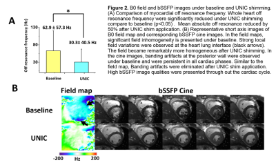 |
Whole Heart High-Order B0 Shimming at 3T Using a UNIfied Coil (UNIC) for RF receive and shimming
Hsin-Jung Yang1, John Stager1, Linda Azab1, Waishing Liu1, Meng Lu1, Yuheng Huang1, Ghazal Yoosefian1, Skyler Selvin1, Richard Handelin1, Yujie Shan1, Fardad Michael Serry1, Yibin Xie1, Anthony Christodoulou1, Xiaoming Bi1,
Rohan Dharmakumar1, Debiao Li1, and Hui Han1
1Cedars-Sinai Medical Center, Los Angeles, CA, United States
B0-field inhomogeneity caused by tissue-air interface has been a long standing challenge in high field(>=3T) cardiac MRI. Although high-order shimming methods has been proposed in recent publications using surface shimming coils, the shimming capability in deep tissue, such as the heart, is still limited due to the restrained current amplitudes and the geometry constraints of the receiving loops. In this study, we developed a cardiac high-order shimming coil based on a novel coil decoupling design to overcome the aforementioned limitations. We demonstrated the developed coil can successfully reduce B0 field variation by 50% in the whole-heart in healthy human volunteers.
|
|
2184.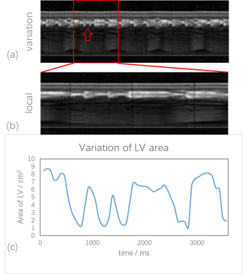 |
Real-time cardiac imaging without ECG gating and respiratory control to monitor arrhythmia
Yichen Zheng1, Ziling Jiao1, Aiqi Sun2, Bo Zhao3,4, and Rui Li1
1Center for Biomedical Imaging Research, Department of Biomedical Engineering, Tsinghua University, Beijing, China, 2Neusoft Medical Systems, Shanghai, China, 3Martinos Center for Biomedical Imaging, Massachusetts General Hospital, Chalestown, MA, United States, 4Department of Radiology, Harvard Medical School, Boston, MA, United States
Non gated real-time cardiac MR imaging with Partial Separable model provides a promising way to overcome the limitation of conventional cine imaging. The new method is able to capture the beat by beat variation, which is especially suitable for arrhythmia. In this study, we have 2 aims. First, to evaluate the consistency of parameters between the conventional cine method and the PS model based real-time method, and second, to capture beat by beat variation in arrhythmia patient.
|
|
2185.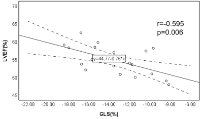 |
Subclinical Myocardial deformation assessment in early stage hypertension patients by Cardiovascular Magnetic Resonance Feature Tracking
Zhaoxia Yang1, Liming Xia2, and Haojie Li2
1Department of Radiology, Tongji Hospital, Tongji Medical College, Huazhong University of Science and Technology, Wuhan,Hubei province, China, 2Tongji Hospital, Tongji Medical College, Huazhong University of Science and Technology, Wuhan,Hubei province, China
Preclinical left ventricular myocardial deformation involvement in the hypertension(HTN) patients could be detected by cardiovascular magnetic resonance feature tracking (CMR-FT). Impaired GRS,GCS,GLS and relevant diastolic longitudinal strain parameters might be associated with alterlation of left ventricular myocardial microstructure and the subtle decrease of diastolic function in the early asymptomatic HTN subjects.CMR-FT derived strain analysis has great potential to predict LVEF change and guide an appropriate treatment.
|
|
2186.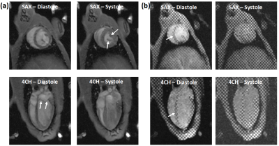 |
Magnetic resonance imaging for assessment of aortic coarctation impact on cardiac function
El-Sayed H Ibrahim1, Pierre Croisille2, and John LaDisa3
1Medical College of Wisconsin, Milwaukee, WI, United States, 2Jean-Monnet University, Lyon, France, 3Marquette University, Milwaukee, WI, United States
Coarctation of the aorta (CoA) is a constriction of the descending thoracic aorta and is one of the most common congenital cardiovascular defects affecting 5,000-8,000 births annually in USA. Patients with CoA can have hyperdynamic and remodeled left ventricle from increased afterload. The mechanisms of morbidity from CoA are difficult to study in clinical setting due to the patients’ heterogeneity from confounding variables and concomitant anomalies. To remove these barriers, we adapted a novel rabbit model of CoA, which we scanned using MRI to study CoA-induced alterations in global and regional cardiac function and compare results to measurements from controls.
|
|
2187.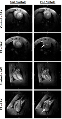 |
Cardiac MRI for identifying radiation therapy effect on the heart
El-Sayed H Ibrahim1, Dhiraj Baruah1, Pierre Croisille2, Jadranka Stojanovska3, Jason Rubenstein1, Rachel Schlaak1, Anne Frei1, Elizabeth Gore1, and Carmen Bergom1
1Medical College of Wisconsin, Milwaukee, WI, United States, 2Jean-Monnet University, Lyon, France, 3University of Michigan, Ann Arbor, MI, United States
Lung cancer is the most frequently diagnosed cancer worldwide. Radiation therapy (RT) is standard-of-care in one-third of the patients, where the incidence of cardiac complications in lung cancer patients after RT reaches 33%. The current paradigm for cardiotoxicity detection and management relies primarily upon assessment of ejection fraction (EF). However, cardiac injury can occur without a clear change in EF. In this study, we investigate the effect of RT on global and regional cardiac function and myocardial T1/T2 values. The results show the capability of regional cardiac MRI for depicting early changes in myocardial contractility pattern and tissue characterization post-RT.
|
|
2188.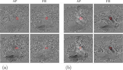 |
Beyond SPAMM-PAV: a Phase Complementary SPAMM (PCSPAMM) acquisition to quantify simultaneously tissue motion and flow velocity
Hernán Mella1,2,3, Julio Sotelo1,2,3, and Sergio Uribe1,2,3,4
1Biomedical Imaging Center, Pontificia Universidad Católica de Chile, Santiago, Chile, 2Millenium Nucleus for Cardiovascular Magnetic Resonance, Santiago, Chile, 3Department of Electrical Engineering, Pontificia Universidad Católica de Chile, Santiago, Chile, 4Department of Radiology, Pontificia Universidad Católica de Chile, Santiago, Chile
In this work, we introduce the Phase Complementary Spatial Modulation of Magnetization (PCSPAMM) acquisition sequence, designed to measure the tissue motion simultaneously with the flow velocity. With a pair of complementary acquisitions, PCSPAMM allows the estimation of a CSPAMM and a Phase-Contrast (PC) MR image, leading to improved tagging contrast and artifacts-free PC images at any cardiac phase.
|
|
2189.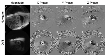 |
Outer volume suppressed 3D-encoded DENSE MRI for quantifying biventricular myocardial strain during a breath-hold
Eric D Carruth1, Samuel W Fielden1, Christopher D Nevius1, Brandon K Fornwalt1, and Christopher M Haggerty1
1Geisinger, Danville, PA, United States
Myocardial strain is a highly sensitive measure of heart function, but techniques to measure strain, such as displacement-encoded (DENSE) MRI, typically focus on the left ventricle, despite known clinical importance of right ventricle mechanics. The high resolution required to measure right ventricular strains is possible using navigator-based approaches; however, these are time-consuming and impractical for broad use. DENSE with outer volume suppression has been applied during a breath hold in the left ventricle, and may also enable resolving the right ventricle. We implemented DENSE with outer volume suppression and achieved similar 3D biventricular strain measurements compared to a navigator-based acquisition.
|
|
2190.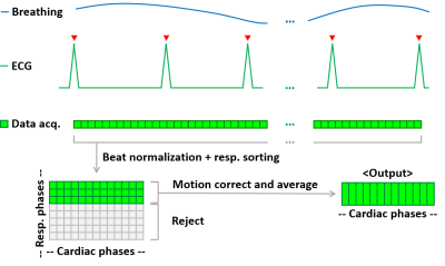 |
Free-Breathing Cardiac Cine MRI with Compressed Sensing Real-Time Imaging and Retrospective Motion Correction
Jianing Pang1, Pedro Itriago Leon2, Xiaoming Bi3, Gary McNeal1, Christoph Forman4, Christianne Leidecker1, and Prakash M. Masand5
1Siemens Medical Solutions USA Inc., Chicago, IL, United States, 2Siemens Medical Solutions USA Inc., Houston, TX, United States, 3Siemens Medical Solutions USA Inc., Los Angeles, CA, United States, 4Siemens Healthcare, Erlangen, Germany, 5Texas Children's Hospital, Houston, TX, United States
Free-breathing cardiac cine imaging is desirable for improving patient experience, expanding CMR eligibility, achieving robust image quality for uncooperative subjects, and simplifying CMR workflow. In this work, we propose a prototype technique, RTCSCineMoCo, that combines highly accelerated real-time acquisition, compressed sensing reconstruction, and retrospective, fully automated respiratory motion correction. In a preliminary evaluation on four volunteers, RTCSCineMoCo achieved similar spatiotemporal resolution, visually comparable image quality, and consistent LV function parameters as the reference breath-hold technique. RTCSCineMoCo is a promising option for free-breathing cardiac cine imaging, and further studies are warranted to evaluate its feasibility and utility in a clinical setting.
|
|
2191.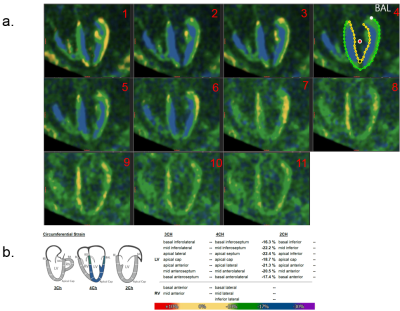 |
Myostrain characterization using fewer SENC cardiac phases – a preliminary study
Xingxian Shou1, Yuan Zheng1, Qi Liu1, Masoud Edalati1, Lele Zhao2, Junpu Hu2, Jian Xu1, Weiguo Zhang1, Donel Tani3, and Nael F. Osman3
1MR, UIH America, Inc., Houston, TX, United States, 2MR, United Imaging Healthcare, Shanghai, China, 3Myocardial Solutions, Inc., Morrisville, NC, United States
SENC MRI is an important technique for myocardium strain quantification. We proposed a modified SENC EPI sequence to only acquire about half of the total cardiac phases in order to get higher SNR strain images. The minimum strains can still be calculated with better SNR from LT/HT images. With the imaging time kept the same at one heartbeat, the SNR of the images is higher and strain calculation is more accurate. The timing of the end-systole stage can be determined by running a CINE scan prior to the SENC protocol.
|