
Digital Poster Session
Cardiovascular: Cardiovascular Techniques
Cardiovascular
2192 -2207 Cardiovascular Techniques - Cardiovascular MRI Techniques & Applications 1
2208 -2223 Cardiovascular Techniques - Cardiovascular MRI Techniques & Application 2
2224 -2238 Cardiovascular Techniques - CMR: Machine Learning & AI
2239 -2254 Cardiovascular Techniques - CMR: Emerging Techniques
2255 -2268 Cardiovascular Techniques - Flow: Cutting-Edge
2269 -2280 Cardiovascular Techniques - 4D Flow & Even Higher
2192.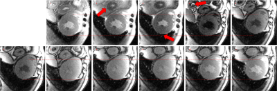 |
Dedicated motion-corrected reconstruction for T1-mapping Modified Look-Locker inversion recovery acquisitions
Gaspar Delso1, Anne Menini2, José T. Ortiz-Pérez3, Susanna Prat3, Adelina Doltra3, Rosario J. Perea3, Teresa M. Caralt3, Daniel Lorenzatti3, Julián Vega3, Marta Sitges3, and Martin A. Janich4
1ASL MR, GE Healthcare, Barcelona, Spain, 2GE Healthcare, San Francisco, CA, United States, 3Hospital Clínic de Barcelona, Barcelona, Spain, 4GE Healthcare, Munich, Germany We present the evaluation results of a new motion correction algorithm, designed specifically to account for the variable image contrast found in T1 mapping MOLLI series. While standard motion correction improves mapping accuracy with respect to uncorrected series, the registration can occasionally diverge, aggravating the problem. The new dedicated algorithm has been shown to yield improved registration performance and reduced probability of divergence. |
|
2193.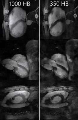 |
Fast 3D Whole Heart Imaging using Seiffert Spirals
Tobias Speidel1, Patrick Metze2, Thomas Hüfken2, and Volker Rasche1,2
1Core Facility Small Animal Imaging (CF-SANI), Ulm University, Ulm, Germany, 2Department of Internal Medicine II, University Ulm Medical Center, Ulm, Germany
The overall duration of acquiring a Nyquist sampled 3D dataset can be significantly shortened by enhancing the efficiency of k-space sampling. This can be achieved by increasing the coverage of k-space for every trajectory interleave. The presented and adapted acquisition scheme using Seiffert Spirals can be a valuable tool for applications in which scan durations are strictly limited e.g. in the case of 3D ECG gated cardiovascular imaging. In this context we apply the Seiffert Spirals to isotropic whole heart imaging with overall scan times of 350 heart beats for a resolution of (1.25 mm)3.
|
|
2194.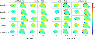 |
High Resolution Simulation and Measurement of Phase-Specific B0 Field Conditions Across the Human Cardiac Cycle
Yun Shang1, Martin Gajdosik1, Sebastian Theilenberg1, Laura M. Schreiber2,3, and Christoph Juchem1,4
1Department of Biomedical Engineering, Columbia University, New York, NY, United States, 2Chair of Cellular and Molecular Imaging, Comprehensive Heart Failure Center, University Hospital Wuerzburg, Wuerzburg, Germany, 3Department of Cardiovascular Imaging, Comprehensive Heart Failure Center, University Hospital Wuerzburg, Wuerzburg, Germany, 4Department of Radiology, Columbia University, New York, NY, United States
B0 inhomogeneity leads to dark band artifacts in cardiac MRI, in particular with the use of SSFP pulse sequences. Limited spatial resolution of MRI-derived B0 maps prevents the systematic analysis of the problem and the development of optimized B0 shim strategies. Here we demonstrate the potential for simulating both overall and cardiac phase-specific B0 field conditions in the human heart at 3 T at high spatial resolution from anatomical MRI. The results are validated by high-resolution B0 field mapping in the same subjects. This approach is expected to develop population-specific B0 shim strategies from readily available anatomical MRI libraries.
|
|
2195.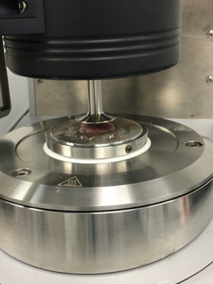 |
Relationships between CMR, rheometry and Raman spectroscopy of doxorubicin induced cardiotoxicity in the swine.
Delphine Perie1, Eric Buzaglo1, Clemence Balosetti1, Helene Heon2, and Daniel Curnier3
1Mechanical Engineering, Polytechnique Montreal, Montreal, QC, Canada, 2Research Center, CHUM, Montreal, QC, Canada, 3Kinesiology, University of Montreal, Montreal, QC, Canada
CMR has already been used to investigate the long term effects of cancer treatments. We investigated relationships between clinical, CMR, mechanical and biochemical properties of the myocardium and their changes due to early doxorubicin induced cardiotoxicity in the swine model. MR relaxation times and CMR function parameters highlighted the importance of regional analysis because of the heterogeneity in the tissue response to doxorubicin treatment. The observation of different remodelling patterns between moving and fixed cardiac walls needs to be translated to personalized diagnosis and medicine approaches for cancer treatment application.
|
|
2196.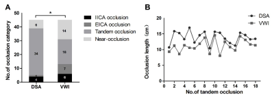 |
Assessment of Apparent Internal Carotid Tandem Occlusion on High-resolution Vessel Wall Imaging Comparison With DSA
shengting chai1,2, Shuang Xia1,2, Zhiguo Sheng3, Weiwei Xie1,2, Chen Wang3, Song Liu1,2, Ruowei Tang1,2, Chen Cao4, Wenqiang Xin5, Zaiyu Guo6, Peng Mi 7, Binge Chang3, and Xinyu Yang5
1Department of Radiology, Tianjin First Central Hospital, Tianjin, China, 2Department of Radiology, First Central Clinical College, Tianjin, China, 3Department of Neurosurgery, Tianjin First Central Hospital, Tianjin, China, 4Department of Radiology, Tianjin Huanhu Hospital, Tianjin, China, 5Department of Neurosurgery, Tianjin Medical University General Hospital, Tianjin, China, 6Department of Neurosurgery, Tianjin TEDA Hospital, Tianjin, China, 7Department of Data statistics, Beijing Hithink Pharmaceutical Technology Service Co.,Ltd, Beijing, China
This study aimed to identify the true ICA tandem occlusions and screen suitability for endovascular recanalization using High-resolution vessel wall imaging (HR-VWI). Patients without blood flow signal in the ICA on MRA who underwent both HR-VWI and DSA was included, and classified into the 4 categories. The suitability for recanalization of occlusion vessels was evaluated. The study showed that about half of patients with an apparent tandem ICA occlusion on DSA, the arteries were, in fact, patent or focal occlusion on VWI. VWI would enable superior identification the true ICA tandem occlusion than DSA.
|
|
2197.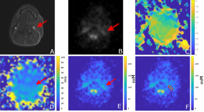 |
Quantitative sodium imaging in the carotid and aorta
Chang Sun1, Mary A McLean2, Titus Lanz3, Frank Riemer4, Rolf F Schulte5, Fiona J Gilbert1, Martin J Graves6, and Joshua D Kaggie1
1Department of Radiology, University of Cambridge, Cambridge, United Kingdom, 2Cancer Research UK, Cambridge, United Kingdom, 3Rapid Biomedical GmbH, Rimpar, Germany, 4Department of Radiology, Haukeland University Hospital, Bergen, Norway, 5GE Healthcare, Munich, Germany, 6Cambridge University Hospital, Cambridge, United Kingdom
Carotid and aortic sodium MR images were acquired from four healthy volunteers, using a 3D cones trajectory and a birdcage sodium transmit/receive coil. Vascular structures such as the carotid bifurcation and aortic arch were observed in the sodium images. T1 maps were estimated via the variable flip angle fitting method. B1 maps were estimated with the dual angle method. Sodium concentration was estimated using a linear model with two fiducials as the reference. The results show sodium MR imaging can provide non-invasive and quantitative measurements of the sodium concentration near possible locations of vascular diseases.
|
|
2198.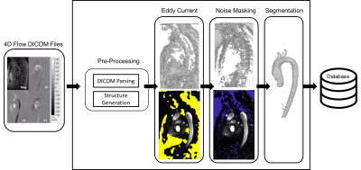 |
SADDLE: A Stand Alone Device for Deep Learning Execution
Justin J Baraboo1, Michael Scott1, Haben Berhane2, and Michael Markl1
1Northwestern Radiology, Chicago, IL, United States, 2Ann & Robert H. Lurie Children's Hospital of Chicago, Chicago, IL, United States
The integration and evaluation of a stand-alone solution for deep learning execution within clinical environments was tested. Using a small GPU device preloaded with a data listener and pre-trained neural networks, bicuspid aortic valve patients having 4D-flow scans were pre-processed and had their aorta’s segmented automatically. Eddy current correction, noise masking, and aortic segmentation were processed sequentially. The device was integrated within the hospital’s network so that data always stayed on-site. The average time for 4D-flow processing and segmentation was roughly 10 minutes. Dice’s coefficients were 0.72±0.17, 0.87±.07, and 0.93±0.03 for eddy current correction, noise masking, and aortic segmentation.
|
|
2199.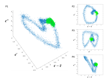 |
Cardiac self-gating signals for free-running imaging: more than just triggers?
Lorenzo Di Sopra1, Jérôme Yerly1,2, Christopher W. Roy1, Juerg Schwitter3, and Matthias Stuber1,2
1Department of Diagnostic and Interventional Radiology, Lausanne University Hospital (CHUV) and University of Lausanne (UNIL), Lausanne, Switzerland, 2Center for Biomedical Imaging (CIBM), Lausanne, Switzerland, 3Division of Cardiology and Cardiac MR Center, Lausanne University Hospital (CHUV) and University of Lausanne (UNIL), Lausanne, Switzerland
Cardiac self-gating (SG) approaches for free-running MR imaging of the heart have proven to be a robust alternative to ECG-gating. However, for both the ECG and SG signals, one feature is typically extracted (ECG: R-wave, SG: zero-crossing) and used for triggering, gating, or data binning. While this often works for constant heart rates, periods of quiescence cannot easily be predicted when heart rates change. Therefore, we have developed an algorithm that, without any prior knowledge, identifies periods of cardiac quiescence from SG signals, and demonstrated that resultant motion-suppressed image quality matches that from conventional approaches.
|
|
2200.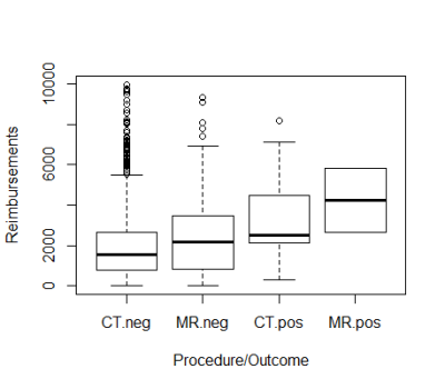 |
Differences in Revenues Associated with Magnetic Resonance Angiography versus Computed Tomographic Angiography for Pulmonary Embolism
Liisa Bergmann1,2, Fabio Gaertner3, and Mark Schiebler1
1Radiology, University of Wisconsin School of Medicine and Public Health, Madison, WI, United States, 2Executive MBA, Wisconsin School of Business at the University of Wisconsin-Madison, Madison, WI, United States, 3Accounting and Information Systems, Wisconsin School of Business at the University of Wisconsin-Madison, Madison, WI, United States
Analysis was performed of hospital revenue generated by inpatient, outpatient and emergency department MR and CT angiographic examinations for suspected pulmonary embolism. Negative examinations generated less revenue than examinations positive for pulmonary embolism. Negative MRAs generated slightly more revenue than negative CTAs. Positive MRAs generated more revenue than positive CTAs, however only two MRAs were positive for pulmonary embolism.
|
|
2201.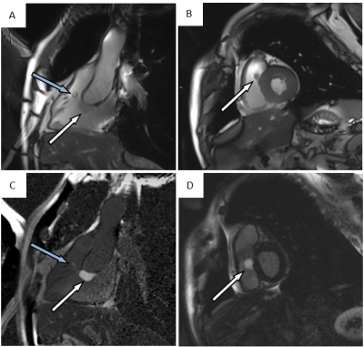 |
Clinical value of Cardiac Magnetic Resonance Imaging in Primary Cardiac tumors
Xiao dan Li1, Yan Chen1, Jiayi Liu1, Lei Xu1, Yu Li1, Dingting Liu1, Qian Qi2, Tianjing Zhang2, and Zhaoying Wen1
1Department of Radiology, Beijing Anzhen Hospital, Capital Medical University, Beijing, China, 2Philips Healthcare, Beijing, China
Cardiac magnetic resonance (CMR) offers superior advantages in cardiac imaging because of greater field of view, excellent soft-tissue imaging, and multiplanar imaging capabilities. CMR imaging can evaluate the characteristics of cardiac tumors by demonstrating the relationship between the tumor and its surrounding tissues,. Moreover, it plays a significant role in assisting the formulation of the surgical plan, in addition to the assessment of tumor progression and the monitoring of postoperative tumor recurrence and metastasis. Our objective is to determine the value of CMR in assessing the likelihood of primary cardiac tumors and guiding patient management.
|
|
2202.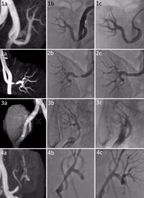 |
Inflow inversion recovery of transplant renal artery stenosis: a validation study
Hai Zhong1, Guangrui Shao1, and Weiqiang Dou2
1radiology, the second hospital of shandong university, Jinan, China, 2GE Healthcare, MR Research China, Beijing, China
Transplant renal artery stenosis(TRAS) was frequently occurred among patients after renal transplantation. The main goal of this study was to evaluate the feasibility of inflow inversion recovery (IFIR) technique in the assessment of TRAS by using digital subtraction angiography(DSA) as a reference. We measured the stenosis degrees of transplant renal arteries with IFIR and DSA, respectively. We obtained a strong correlation between IFIR and DSA, and no significant difference was found between these two techniques. Therefore, IFIR can been demonstrated as a noninvasive, accurate and valuable method in the diagnosis and evaluation of TRAS.
|
|
2203.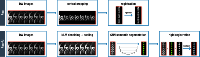 |
In-vivo cardiac diffusion weighted image registration aided by AI semantic segmentation
Pedro F Ferreira1,2, Raquel Martin2, Andrew D Scott1,2, Zohya Khalique1,2, Guang Yang1,2, Sonia Nielles-Vallespin1,2, Dudley Pennell1,2, and David Firmin1,2
1Royal Brompton Hospital, London, United Kingdom, 2Imperial College, London, United Kingdom
In-vivo cardiac diffusion weighted images contain contrast differences that complicate image registration from multiple breath-holds. Additionally, neighbouring structures of the chest wall, liver and stomach do not move rigidly with the heart during the respiratory cycle which further hinders registration. In this work we remove other structures and pre-process the image with the help of a convolutional neural network trained to segment multiple heart structures. These additional steps increase the accuracy of registration of the left ventricular myocardial ring resulting in more accurate diffusion tensors.
|
|
2204.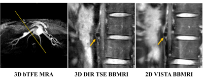 |
Feasibility and Comparison Between Non-enhanced 2D and 3D Free-breathing Black-blood Techniques for Renal Arterial Wall Imaging
Zihan Ning1, Shuo Chen1, Hualu Han1, Huiyu Qiao1, Shasha Deng2, Dandan Yang1, Hao Sun3, and Xihai Zhao1
1Department of Biomedical Engineering, School of Medicine Tsinghua University, Beijing, China, 2Fujian Medical University Union Hospital, Fujian, China, 3Depaetment of Radiology, Peking Union Medical College Hospital, Beijing, China
We optimized non-enhanced 2D and 3D free-breathing black-blood imaging sequences and found excellent agreement in morphological measurements of renal artery (ICC: 0.85-0.95), and the CNR (p=0.17) and image quality score (p=0.60) were comparable between them. 2D imaging had higher SNR for lumen and wall visualization (all p<0.05) whereas 3D imaging had higher CNReff (5.37±1.91 vs. 0.87±0.51, p<0.01). Both sequences showed excellent inter-reader and scan-rescan repeatability (ICC: 0.77-0.99). We concluded that both 2D and 3D sequences are feasible for renal arterial wall imaging, particularly 2D provides high quality images whereas 3D sequence allows large-coverage and more efficient imaging.
|
|
2205.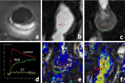 |
Evaluating the Vessel Wall Permeability of Abdominal Aortic Aneurysm using 3D DynamicContrastEnhancedMRI: a feasibility study
Bing Tian1, Qi Liu2, Jianping Lu2, and Chengcheng Zhu3
1Radiology, Changhai hospital, Shanghai, China, 2Changhai hospital of Shanghai, Shanghai, China, 3UCSF, San Francisco, CA, United States
The feasibility of 3D DCE on AAA was demonstrated in our study.
|
|
2206. |
Automatic Detection of Arrhythmia in ECG-free Real-time MRI
Anja Hennemuth1,2, Christina Unterberg3, Sebastian Ulrich Kelle4, Martin Uecker3, Jens Frahm5, and Markus Hüllebrand1,2
1Institute for Imaging Science and Computational Modelling in Cardiovascular Medicine, Charité - Universitätsmedizin Berlin, Berlin, Germany, 2Fraunhofer MEVIS, Bremen, Germany, 3Universitätsmedizin Göttingen, Göttingen, Germany, 4Deutsches Herzzentrum Berlin, Berlin, Germany, 5Max-Planck-Institut fuer biophysikalische Chemie, Göttingen, Germany
The analysis of cardiac function in patients suffering from arrhythmia poses a problem for conventional ECG-synchronized imaging and the patients' ability to hold their breath. Real-time imaging approaches provide ungated image data, which contains the information about the motion variation induced by breathing and arrhythmia but require a high effort in post-processing and interpretation. The goal of the presented work is to enable an automatic analysis of cardiac real-time image sequences of patients suffering from arrhythmia. To this end, we combine a fast CNN-based segmentation of the myocardium with a curve pattern analysis of the blood volume changes over time.
|
|
2207.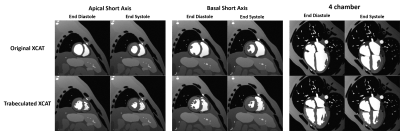 |
Towards Realistic Cardiac MR Image Simulation; Inclusion of the Endocardial Trabeculae in the XCAT Heart Anatomy
Sina Amirrajab1, William Paul Segars2, Cristian Lorenz3, Juergen Weese3, and Marcel Breeuwer1,4
1Biomedical Engineering Department, Eindhoven University of Technology, Eindhoven, Netherlands, 2Carl E. Ravin Advanced Imaging Laboratories, Duke University, Durham, NC, United States, 3Philips Research Laboratories, Hamburg, Germany, 4MR R&D - Clinical Science, Philips Healthcare, Best, Netherlands
The importance of realistic cardiac MR simulation has been realized over the past decade. For this application, XCAT phantom provides realistic highly detailed whole body anatomical models including the heart and respiratory motion. Although the current XCAT heart model is complete in terms of substructures, the trabeculae structure of the endocardium, which is geometrically complex, is lacking. Based on a high-resolution ex-vivo cardiac image data, we modeled and incorporated the irregularity of the trabeculae into the existing XCAT model. We demonstrated that greater realism in cardiac MRI simulation can be achieved by including the trabeculae anatomy into the heart.
|
 Watch the Video
 View the Poster
2208.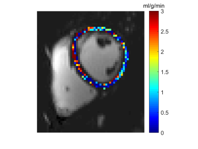 |
Comparison of Myocardial Blood Flow Measurements with Arterial Spin Labeling in Breathhold and Synchronized Breathing Acquisitions
Verónica Aramendía-Vidaurreta1, Pedro Macías-Gordaliza2,3, Marta Vidorreta4, Rebeca Echeverria-Chasco1, Gorka Bastarrika1, Arrate Muñoz-Barrutia2,3, and María Fernández-Seara1
1Radiology, Clínica Universidad de Navarra, Pamplona, Spain, 2Universidad Carlos III de Madrid, Madrid, Spain, 3Instituto de Investigación Sanitaria Gregorio Marañón, Madrid, Spain, 4Siemens Healthineers, Madrid, Spain
Arterial spin labeling enables non-invasive quantification of myocardial perfusion. However, there is a need to improve its reproducibility by reducing subtraction errors due to motion and thus, avoiding signal blurring in the acquired low-resolution images. Here, we demonstrate the feasibility of quantifying MBF employing synchronized breathing techniques with FAIR-ASL in healthy subjects. Visually, motion is significantly reduced, MBF values are consistent with those reported in the literature and coefficient of variation for intrasession reproducibility is 13%.
|
|
2209.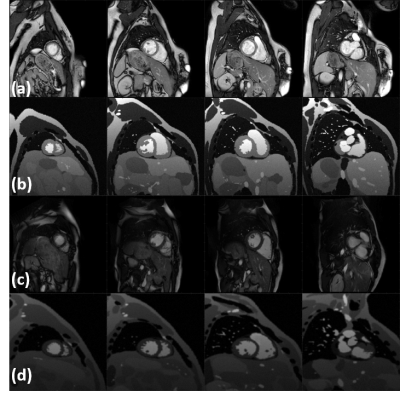 |
Generation of realistic and heterogeneous virtual population of cardiovascular magnetic resonance simulated images
Sina Amirrajab1, Yasmina Al Khalil1, Cristian Lorenz2, Juergen Weese2, and Marcel Breeuwer1,3
1Biomedical Engineering Department, Eindhoven University of Technology, Eindhoven, Netherlands, 2Philips Research Laboratories, Hamburg, Germany, 3MR R&D - Clinical Science, Philips Healthcare, Best, Netherlands
This study investigates an approach to generate a realistic, heterogeneous database of simulated cardiac MR images to aid the development of fully automated and generalizable deep learning based segmentation algorithms, less sensitive to variability in CMR image appearance. XCAT phantoms were used to create the virtual population by altering the heart position and geometry and MRXCAT approach was improved to simulate more organs. Images simulated in this study were quantitatively and qualitatively comparable to real CMR images acquired by two different sites and vendors. Initial experiments using such a heterogeneous image dataset show a positive impact on the segmentation performance.
|
|
2210.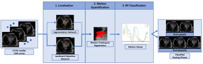 |
A Robust Deep-Learning-based Automated Cardiac Resting Phase Detection: Validation in a Prospective Study
Seung Su Yoon1,2, Elisabeth Hoppe1, Michaela Schmidt2, Christoph Forman2, Teodora Chitiboi3, Puneet Sharma3, Christoph Tillmanns4, Andreas Maier1, and Jens Wetzl2
1Department of Computer Science, Pattern Recognition Lab, Friedrich-Alexander-Universität Erlangen-Nürnberg, Erlangen, Germany, 2Magnetic Resonance, Siemens Healthcare GmbH, Erlangen, Germany, 3Siemens Medical Solutions USA, Inc, Princeton, NJ, United States, 4Diagnostikum Berlin, Berlin, Germany
The detection of a window with the least motion, e.g. end-systolic or end-diastolic resting phases (RPs) within the cardiac cycle is necessary for data acquisition for static cardiac imaging. In the current workflow, it is manually performed on CINE images by visual inspection. To automate the workflow, we propose an improved Deep-Learning-based automated localized RP detection. While the first step is responsible to localize the anatomy of interest, the second is to quantify motion within the target, and third is to classify RPs. We validated the system with a prospective volunteer study and achieved accuracy in the range of 28ms.
|
|
2211.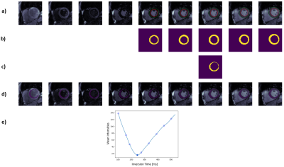 |
Automated Deep-Learning-based Inversion Time Selection for Cardiac Late Gadolinium Enhancement Imaging
Seung Su Yoon1,2, Michaela Schmidt2, Bernd J Wintersperger3,4, Teodora Chitiboi5, Puneet Sharma5, Christoph Tillmanns6, Andreas Maier1, and Jens Wetzl2
1Department of Computer Science, Pattern Recognition Lab, Friedrich-Alexander-Universität Erlangen-Nürnberg, Erlangen, Germany, 2Magnetic Resonance, Siemens Healthcare GmbH, Erlangen, Germany, 3Department of Medical Imaging, University Health Network, Toronto, ON, Canada, 4Department of Medical Imaging, University of Toronto, Toronto, ON, Canada, 5Siemens Medical Solutions USA, Inc, Princeton, NJ, United States, 6Diagnostikum Berlin, Berlin, Germany
In Cardiac MRI, Late gadolinium enhancement (LGE) imaging is generally performed for the assessment of myocardial viability. As LGE is based on inversion recovery techniques, the correct myocardial nulling is necessary for image contrast optimization. In current clinical practice, it is done by visual evaluation. As it required user expertise and interaction, an automated inversion time selection is proposed. The Deep-Learnig-based system to detect the null point of inversion time was successfully demonstrated in all datasets comparing with two expert annotations.
|
|
2212. |
Automated Segmentation of Right Ventricle in CMR Images based on Dense and Multi-scale U-net Network
Peng Liu1 and Lijia Wang1
1University of Shanghai for Science and Technology, Shanghai, China
It is essential to segment right ventricle (RV) for evaluating cardiac functional parameters of cardiac diseases in clinical diagnosis and prognosis. However, the complex structure of RV makes traditional segmentation methods not so effective in right ventricular segmentation. A new Dense and Multi-scale U-net deep learning method is proposed to segment right ventricle in cine cardiac magnetic resonance (CMR) short-axis images automatically, which shows high coincidence and small difference with manual segmentation and is promising for diagnosis and analysis of clinical cardiac diseases.
|
|
2213.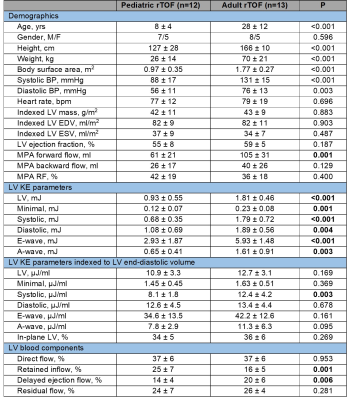 |
Intra-cardiac flow kinetic energy assessment from 4D flow CMR imaging in pediatric and adult repaired Tetralogy of Fallot
XIAODAN ZHAO1, LIWEI HU2, RONG ZHEN OUYANG2, RU SAN TAN1,3, PING CHAI4, MARIELLE FORTIER3,5, SHUO ZHANG6, WEN RUAN1, SHUANG LENG1, JUN-MEI ZHANG1,3, BRYANT JENNIFER1, LYNETTE LS TEO4, ROB VAN DER GEEST7, TENG HONG TAN3,5,
JAMES YIP4, JU LE TAN1,3, YUMIN ZHONG2, and LIANG ZHONG1,3
1National Heart Centre Singapore, Singapore, Singapore, 2Shanghai Children’s Medical Centre, Shanghai Jiaotong University School of Medicine, ShangHai, China, 3Duke-NUS Medical School, Singapore, Singapore, 4National University Hospital Singapore, Singapore, Singapore, 5KK Women’s and Children’s Hospital, Singapore, Singapore, 6Philips Germany, Humburg, Germany, 7Department of Radiology, Leiden University Medical Center, Leiden, Netherlands
Whole-heart 4D cardiovascular magnetic resonance (CMR) phase-contrast flow measurement enables qualitative and quantitative assessment of intra-cardiac flow. Its feasibility was investigated in 12 pediatric and 13 adult patients with repaired Tetralogy of Fallot (rTOF). Left ventricular (LV) kinetic energy (KE) and pathline-derived flow components were analyzed. KE parameters were significantly correlated with age, but only LV systolic KE remained significantly different after indexing to LV end-diastolic volume. Pediatric rTOF patients had similar LV ejection fraction, indexed LV volumes and mass, but significantly reduced indexed LV systolic KE, retained inflow and increased delayed ejection flow compared with adults.
|
|
2214.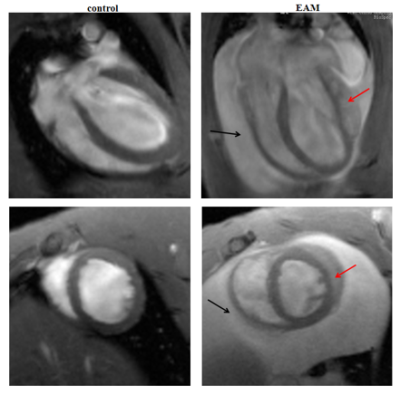 |
CMR Tissue-Tracking myocardial strain analysis in acute myocarditis in a rat model: diagnostic value and association with LGE
jing zhu1, lei wang2, and fabao gao2
1sichuan university, chengdu city, China, 2sichuan university, chengdu, China
To assess the diagnostic value of CMR Tissue-Tracking(TT) for strain analysis in a rat model of acute myocarditis and the strain’s association with myocardial impairment. Experimental autoimmune myocarditis(EAM) was induced in 16 male rats, 10 rats served as control. Rats were scanned at 7T MRI 21 days after model induction, using cine-FLASH sequence and late gadolinium enhancement imaging. Myocardial strain values significantly reduced in EAM rats, compared with the controls. The area under the curve(AUC) of myocardial strain was excellent. Quantitative analysis of late gadolinium enhancement(LGE) showed a significant correlation with myocardial strain.
|
|
2215.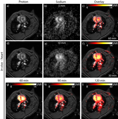 |
Sodium MRI of the Rat Heart at 9.4 T using a Quadrature Birdcage
Laura Boehmert1, Helmar Waiczies2, Andre Kuehne2, Celal Oezerdem1, Sonia Waiczies1, Ludger Starke1, Min-Chi Ku1, Andreas Pohlmann1, Paula Ramos Delgado1, Erdmann Seeliger3, and Thoralf Niendorf1,2,4,5
1Berlin Ultrahigh Field Facility (B.U.F.F.), Max Delbrück Center for Molecular Medicine in the Helmholtz Association, Berlin, Germany, 2MRI.TOOLS GmbH, Berlin, Germany, 3Institute of Vegetative Physiology, Charité University Medicine, Berlin, Germany, 4DZHK (German Centre for Cardiovascular Research), partner site Berlin, Berlin, Germany, 5Experimental and Clinical Research Center, a joint cooperation between the Charité Medical Faculty and the Max Delbrück Center for Molecular Medicine, Berlin, Germany
We propose probing of tissue sodium concentrations in the heart using 23Na MRI for adding a very useful dimension to our understanding of cardiac disorders. Changes in the sodium concentration might be indicative of early pathophysiological changes in such diseases. This work focuses on the development, evaluation and application of a quadrature birdcage RF resonator tailored for cardiac 23Na MRI at 9.4T with the goal to provide a uniform excitation profile.
|
|
2216.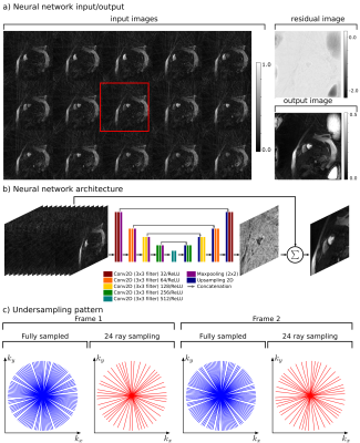 |
Rapid myocardial perfusion MRI reconstruction using deep learning networks
Eric Kenneth Gibbons1,2, Ye Tian3, Qi Huang4, Akshay Chaudhari5, and Edward DiBella2,4
1Electrical and Computer Engineering, Weber State University, Ogden, UT, United States, 2Radiology and Imaging Sciences, University of Utah, Salt Lake City, UT, United States, 3Physics, University of Utah, Salt Lake City, UT, United States, 4Biomedical Engineering, University of Utah, Salt Lake City, UT, United States, 5Radiology, Stanford University, Stanford, CA, United States
Current acquisition strategies in cardiac perfusion MRI rely on non-uniform sampling that is highly undersampled in spatial and temporal domains. While iterative reconstruction methods are able to reconstruct such data reasonably well, reconstruction speeds are prohibitively long. This abstract applies novel deep learning approaches to accelerate reconstruction speeds relative to iterative algorithms with comparable image quality. Validation is performed through the calculation of a perfusion index.
|
|
2217.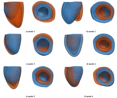 |
An MR image-guided left-ventricular shape model embedding local physiological coordinates and directions
Stefano Buoso1, Christian T Stoeck1, Johanna Stimm1, and Sebastian Kozerke1
1Institute for Biomedical Engineering, ETH Zurich, Zurich, Switzerland
We show the feasibility of embedding physiological coordinates and directions into a left ventricle anatomical shape model using Proper Orthogonal Decomposition. The volumetric anatomical mesh and the physiological parametrization can be personalized directly from the selection of control points on MR cardiac images. This approach provides a consistent way of augmenting low-resolution data using features from high-resolution datasets. Additionally, the physiological parametrization is automatically adapted to each specific case without any additional calculation steps. This simplifies the processing of clinical images and, particularly, strain calculations and microstructural analysis that require the definition of the physiological parametrization.
|
|
2218.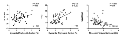 |
Metabolic Syndrome and Myocardium Steatosis in Subclinical Type 2 Diabetes Mellitus: A 1H-Magnetic Resonance Spectroscopy study
Yue Gao1, Zhi-gang Yang1, Rui Shi1, and Xi Liu1
1radiology, west china hospital of sichuan university, chengdu, China
Myocardium steatosis was positively associated with decreased myocardial deformation and perfusion dysfunction.
|
|
2219.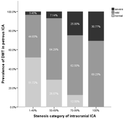 |
Association between intracranial internal carotid artery steno-occlusive disease and diffuse wall thickening in its petrous segment
Jin Zhang1, Xiaoyi Chen2, Huilin Zhao1, Bin Cui3, Xiao Li1, Beibei Sun1, Xiaosheng Liu1, Zhongshuai Zhang4, Xihai Zhao5, and Jianrong Xu1
1Renji Hospital, Shanghai Jiaotong University, Shanghai, China, 2Beijing Geriatric Hospital, Beijing, China, 3Aerospace Center Hospital, Beijing, China, 4Siemens Healthcare, Shanghai, China, 5Biomedical Engineering & Center for Biomedical Imaging Research, Tsinghua University, Beijing, China
This study employed isotropic high-resolution SPACE or VISTA sequence to explore the association between intracranial ICA steno-occlusive disease caused by atherosclerosis and diffuse wall thickening (DWT) in ipsilateral petrous ICA. The results indicated that the diffuse wall thickening of petrous ICA is commonly seen among patients with steno-occlusive disease causing by atherosclerotic plaque in intracranial ICA, especially in occluded intracranial ICA.
|
|
2220.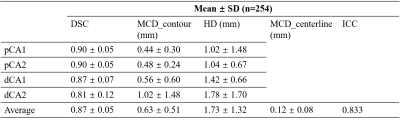 |
Evaluation of carotid lumen segmentation on bifurcation geometry quantification
lian luo1, shuai liu2, Peirong Jiang3, Tan Gong1, Yuqian Mei1, Fei Shang1, and Xihai Zhao2
1Department of Biomedical Engineering, Beijing Institute of Technology, Beijing, China, 2Tsinghua University, Beijing, China, 3Fujian Medical University Union Hospital, Fujian, China
To evaluate carotid lumen segmentation results on bifurcation geometry quantification, bifurcation angle and mean centerline distance were calculated. The intra-class correlation coefficient (ICC), linear regression and Bland-Altman plot of bifurcation angle, as well as the mean centerline distance indicated that the segmentation results had strong correlation with golden standard. The segmentation results could be used on geometry quantification.
|
|
2221.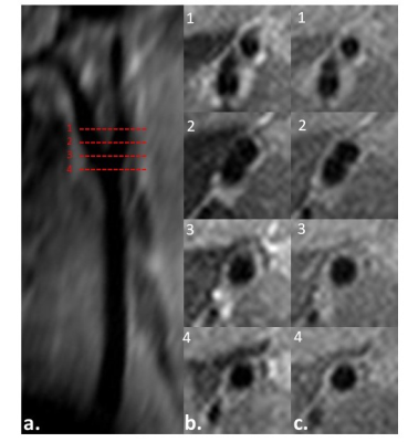 |
Carotid Arterial Wall MRI Of ApoE-/- Mouse at 7T using 3D DANTE-Prepared Variable-Flip-Angle Rapid Acquisition with Relaxation Enhancement
Yuanbo Yang1, Zhonghao Li2, Yihao Guo1, Yingjie Mei3, Ming Zhao2, Guoxi Xie4, and Yanqiu Feng1
1Guangdong Provincial Key Laboratory of Medical Image Processing & Key Laboratory of Mental Health of the Ministry of Education, School of Biomedical Engineering, Southern Medical University, Guangzhou, China, 2Department of Pathophysiology, Key Lab for Shock and Microcirculation Research of Guangdong, School of Basic Medical Sciences, Southern Medical University, Guangzhou, 510515, China, Guangzhou, China, 3philips healthcare, Guangzhou, China, 4Department of Biomedical Engineering, School of Basic Sciences, Guangzhou Medical University, Guangzhou, China
Carotid atherosclerosis is a degenerative disease of the arterial wall, which can result in predisposition to cerebral thrombo-embolic stroke1. The combination of DANTE and VF-RARE (DANTE-VF-RARE) can suppress flow signal effectively. It was used for vessel wall imaging of human in the previous studies4. However, the feasibility of DANTE-VF-RARE sequence remains unclear for imaging of carotid for apoE-/- mouse. This work aims to develop 3D DANTE-VF-RARE on 7Tesla (T) and investigate its feasibility for vessel wall imaging of apoE-/- mouse.
|
|
2222. |
Longitudinal assessment of global longitudinal strain and hemodynamic forces in the diabetic mouse heart
Mariah RR Daal1, Bram F Coolen1, Dorita THPM Dekkers1, David Hauteman2, Rob CI Wüst3, and Gustav J Strijkers1
1Department of Biomedical Engineering, Amsterdam University Medical Center, University of Amsterdam, Amsterdam, Netherlands, 2Medis medical imaging systems B.V., Leiden, Netherlands, 3Faculty of Behavioural and Movement Sciences, Vrije Universiteit, Amsterdam, Netherlands
The current classification of heart failure based on diastolic and systolic dysfunction limits the understanding of the time course of heart failure with preserved left ventricular ejection fraction (HFpEF), and new parameters are warranted to assess cardiac function. Here, we assessed global longitudinal strain (GLS), as well as hemodynamic forces (HDF) in a mouse model of diabetes (db/db) at two time points, with the aim to study the prognostic value of these new parameters for cardiac dysfunction.
|
|
2223.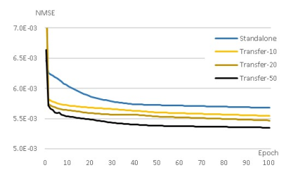 |
Transfer Learning for Compressed-sensing Cardiac CINE MRI
Seong-Jae Park1, Jong-Hyun Yoon2, and Chang-Beom Ahn1
1Kwangwoon University, Seoul, Republic of Korea, 2Gachon University, Incheon, Republic of Korea
Compressed-sensing cardiovascular CINE MRI was performed using deep artificial neural network and transfer learning. Transfer learning is a method to use weights obtained from previous learning as initial weights for current learning to improve generality and performance of the neural network. When learning data is limited, it is useful for generalization by using previous learning along with other data. It also reduces learning time by 80 to 98 percent. And to prevent modification of measurement data by Deep learning, K-space correction was added as a post-processing process.
|
2224.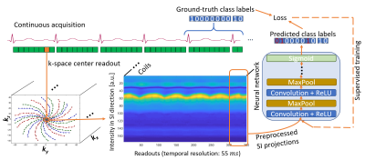 |
DeepECG: Towards 3-D Continuous Cardiac MRI without ECG-Gating - Deep Learning-based R-Wave Classification for Automated Cardiac Phase Binning
Elisabeth Hoppe1, Jens Wetzl2, Seung Su Yoon1, Manuel Schneider2, Bernhard Stimpel1, Alexander Preuhs1, and Andreas Maier1
1Department of Computer Science, Pattern Recognition Lab, Friedrich-Alexander-Universität Erlangen-Nürnberg, Erlangen, Germany, 2Magnetic Resonance, Siemens Healthcare, Erlangen, Germany
For continuous cardiac CINE acquisitions, cardiac binning of the data is necessary, which is done either using ECG-gating or hand-crafted postprocessing methods. To overcome these limitations, we propose a deep learning classifier to detect R-waves from repeated 1-D superior-inferior projections of the imaged data. After training with R-wave positions from the ECG signal as ground-truth data, detection of R-waves is possible without additional ECG-gating or hand-crafted features and can be used for retrospective cardiac binning. Our first proof-of-concept achieves a high accuracy of over 91% on previously unseen cardiac CINE data.
|
|
2225. |
Myocardial Segmentation and Phase Unwrapping for Automatic Analysis of DENSE Cardiac MRI using Deep Learning
Sona Ghadimi1, Xue Feng1, Craig H. Meyer1, and Frederick H. Epstein1
1Biomedical Engineering, University of Virginia, Charlottesville, VA, United States
DENSE myocardial strain imaging is a method wherein tissue displacement is encoded in the image phase. Myocardial segmentation and phase unwrapping are two key steps in quantitative displacement and strain analysis of DENSE images. Prior DENSE analysis methods for segmentation and phase unwrapping were semi-automated techniques, requiring user intervention. In this study, we developed deep learning (DL) methods for fully automated myocardial segmentation and phase unwrapping for short-axis DENSE images. Quantitative and qualitative evaluations show promising results for the proposed DL-based segmentation and phase unwrapping methods, eliminating all manual steps needed for fully automatic DENSE strain analysis.
|
|
2226. |
Real-time Cine Cardiac Magnetic Resonance Imaging Using a Convolutional Neural Network
Yan-Chi Ivy Chan1,2, Jay B. Patel3, Wai-Yan Ryana Fok1,2, Kwannapas Saengsin1,4, Andrew J. Powell1, and Mehdi H. Moghari1
1Department of Cardiology, Boston Children's Hospital and the Department of Pediatrics, Harvard Medical School, Boston, MA, United States, 2Department of Informatics, Technical University of Munich, Garching, Germany, 3Massachusetts Institute of Technology, Cambridge, MA, United States, 4Department of Pediatrics, Chiang Mai University, Chiang Mai, Thailand
We have developed and evaluated a new real-time acquisition and reconstruction technique for 2D steady-state free precession cine cardiac magnetic resonance imaging (MRI). Patient study is presented to validate the proposed technique.
|
|
2227.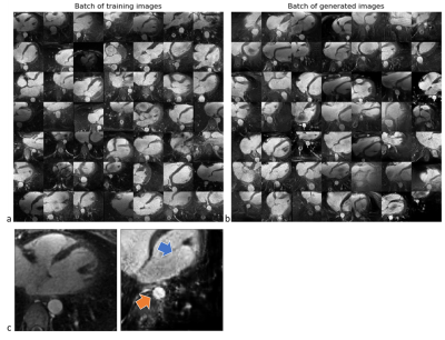 |
Learning cardiac morphology from MR images using a generative adversarial network: a proof of concept study
Davide Piccini1,2,3, Aurélien Maillot1,2, John Heerfordt1,2, Dimitri Van De Ville4,5, Juerg Schwitter6, Matthias Stuber2,7, Jonas Richiardi1,2, and Tobias Kober1,2,3
1Advanced Clinical Imaging Technology, Siemens Healthcare, Lausanne, Switzerland, 2Department of Radiology, Lausanne University Hospital and University of Lausanne, Lausanne, Switzerland, 3LTS5, École Polytechnique Fédérale de Lausanne (EPFL), Lausanne, Switzerland, 4Institute of Bioengineering/Center for Neuroprosthetics, École Polytechnique Fédérale de Lausanne (EPFL), Lausanne, Switzerland, 5Department of Radiology and Medical Informatics, University Hospital of Geneva (HUG), Geneva, Switzerland, 6Division of Cardiology and Cardiac MR Center, University Hospital of Lausanne (CHUV), Lausanne, Switzerland, 7Center for Biomedical Imaging (CIBM), Lausanne, Switzerland
Learning anatomical characteristics from large databases of radiological data could be leveraged to create realistic representations of a specific subject’s anatomy and to provide a personalized clinical assessment by comparison to the acquired data. Here, we extracted 2D patches containing the descending aorta from 297 3D whole-heart MRI acquisitions and trained a Wasserstein generative adversarial network with a gradient penalty term (WGAN-GP). We used the same network to generate realistic versions of the aortic region on masked real images using a loss function that combines a contextual and a perceptual term. Results were qualitatively assessed by an expert reader.
|
|
2228.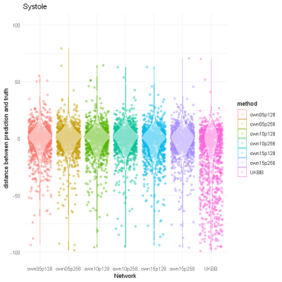 |
The Apprentice Surpasses the Master: Training a Neural Network for Cardiac Segmentation Using a Specialized Network and Indirectly Labeled Data
Markus J. Ankenbrand1, David Lohr1, Tobias Wech2, and Laura M. Schreiber1
1Chair of Cellular and Molecular Imaging, Comprehensive Heart Failure Center (CHFC), University Hospital Würzburg, Würzburg, Germany, 2Department of Diagnostic and Interventional Radiology, University Hospital Würzburg, Würzburg, Germany
Training of neural networks for segmentation of CMR images requires large amounts of labeled data and network generalization is biased by training data characteristics. We used a specialized network to label a heterogeneous, publicly available dataset of 1140 cine images with known left-ventricular volumes. We evaluated the performance of this network using true and predicted volumes and trained another neural network on subjects with high prediction accuracy using extensive data augmentation. The resulting network outperforms the original one on the full dataset, even on subgroups where the original network fails, indicating great generalization and thus suitability for transfer learning applications.
|
|
2229.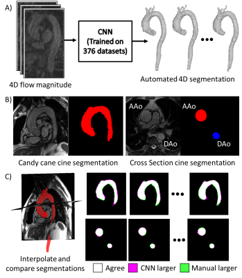 |
Verification of fully automated deep learning-based 4D segmentation of the thoracic aorta from 4D flow MRI
Michael Baran Scott1, Haben Baran Berhane2, Kevin Ryan Kalisz1, Tugce Agirlar Trabzonlu1, Jeesoo Lee1, Marci Messina1, Chris Malaisrie1, Patrick McCarthy1, James Carr1, Alexander J Barker3, Ryan Avery1, and Michael Markl1
1Northwestern University, Chicago, IL, United States, 2Lurie Children's Hospital of Chicago, Chicago, IL, United States, 3University of Colorado, Anschutz Medical Campus, Aurora, CO, United States
A convolutional neural network (CNN) originally implemented for time-averaged 3D segmentation of the thoracic aorta from 4D flow MRI was retrained to generate time-resolved segmentations without generating additional reference data. To validate the segmentations, automatically generated time-resolved segmentations were compared against two 2D cine acquisitions in 20 patients. The CNN achieved average Dice scores 0.87±0.04 and 0.88±0.04 for candy-cane and cross-section views of the aorta across all patients and timepoints. Automated time-resolved segmentation of 4D flow MRI data will enable calculation of metrics such as wall shear stress and aortic compliance that are sensitive to wall location.
|
|
2230.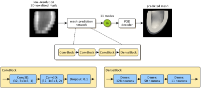 |
A Machine Learning Approach to Left Ventricle Mesh Prediction from Multi-Slice MR Images
Thomas Joyce1, Stefano Buoso1, Yuerong Xu1, and Sebastian Kozerke1
1Institute for Biomedical Engineering, University and ETH Zurich, Zurich, Switzerland
There has been considerable success applying deep learning to cardiac MRI segmentation. However, segmentation masks have several shortcomings. In particular, they are discrete (voxel-based) representations of continuous anatomy, and are hence not suitable for use in biomechanical simulation. Mesh representations can potentially overcome both of these drawbacks. We demonstrate an approach to predicting left-ventricular (LV) meshes from short-axis MR images. The proposed approach: (i) works robustly on data with differing slice numbers, slice thicknesses, and left-ventricular coverage, (ii) has no test-time optimisation loop, but rather directly predicts the mesh from a mask, and, (iii) generalises to real data with pathology.
|
|
2231.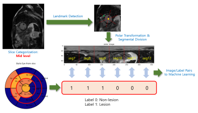 |
Segmental assessment of myocardial late gadolinium enhancement based on weakly supervised learning
Yoon-Chul Kim1 and Yeon Hyeon Choe2
1Clinical Research Institute, Samsung Medical Center, Sungkyunkwan Univ. School of Medicine, Seoul, Korea, Republic of, 2Department of Radiology, Samsung Medical Center, Sungkyunkwan Univ. School of Medicine, Seoul, Korea, Republic of
Radiologic diagnosis of myocardial late gadolinium enhancement (LGE) is often represented as a description of lesion characteristics and distributions in the standard 16- (or 17-) segment myocardial model. In this study, we used short-axis LGE images and 16-segment labeled results from 66 patients with coronary artery disease and non-ischemic heart disease and trained a deep convolutional neural network (CNN) model. Short-axis images were transformed to polar coordinates after identification of the LV center point and anterior RV insertion point. The proposed method does not require manual delineation of the lesions and potentially enables automatic diagnosis of myocardial viability.
|
|
2232.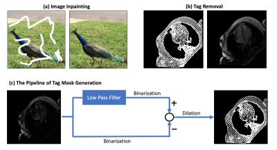 |
Tag Removal in Cardiac Tagged MRI using Edge-guided Adversarial Learning
Botian Xu1, Yaqiong Chai1, and John C. Wood1,2
1Department of Biomedical Engineering, University of Southern California, Los Angeles, CA, United States, 2Division of Cardiology, Children's Hospital Los Angeles, Los Angeles, CA, United States
Tagging in cardiac MRI could cause great challenges to subsequent downstream analysis. Modeling tag removal task as an image recovery problem, we propose a GAN-based method constrained by edge prior, termed EG-GAN, to remove tags progressively without model collapse, and it outperforms conventional approaches.
|
|
2233.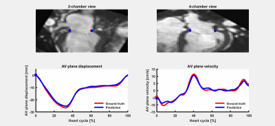 |
Time-resolved tracking of the atrioventricular plane displacement in long-axis cine images with residual neural networks
Ricardo A Gonzales1,2, John Onofrey1, Jérôme Lamy1, Felicia Seemann3,4, Einar Heiberg3,4,5, and Dana C Peters1
1Department of Radiology and Biomedical Imaging, Yale University, New Haven, CT, United States, 2Department of Electrical Engineering, Universidad de Ingenieria y Tecnologia, Lima, Peru, 3Department of Clinical Physiology, Lund University, Lund, Sweden, 4Department of Biomedical Engineering, Lund University, Lund, Sweden, 5Wallenberg Center for Molecular Medicine, Lund University, Lund, Sweden
Diastolic dysfunction is assessed by measurement of mitral annular (MA) early diastolic velocity (e’), commonly performed in echocardiography. Similar measurements can be obtained with valvular plane tracking in MRI long-axis cines. These measurements have been validated and have good reproducibility, yet manual MA points annotations are required. In this work we present a machine learning convolutional neural network with a residual architecture for automatic annotation of MA points in MRI long-axis cine images of the 2 and 4-chamber views. The landmark tracking allowed a fast and accurate evaluation of diastolic parameters improving the clinical applicability of MRI for diastolic assessment.
|
|
2234.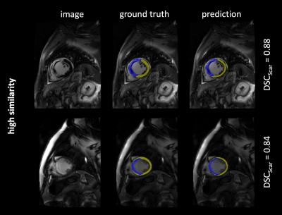 |
Deep Learning based Semantic Segmentation of Scar Tissue in Late-Gadolinium Enhancement CMR – First Results
Julius Frederik Heidenreich1, Tobias Gassenmaier1, Markus Ankenbrand2, David Lohr2, Thorsten Alexander Bley1, and Tobias Wech1
1Department of Diagnostic and Interventional Radiology, University Hospital Würzburg, Würzburg, Germany, 2Congestive Heart Failure Center, University Hospital Würzburg, Würzburg, Germany
Quantitative analysis of scar tissue in late gadolinium enhancement (LGE) cardiac magnetic resonance imaging (CMR) typically requires manual or at best semi-automatic segmentation by a trained physician. To supersede this time-consuming and tedious task, a convolutional neural network with a U-Net architecture and a ResNet34 backbone was trained for semantic segmentation of scar tissue in LGE CMR. The predictions of the proposed model yielded high performance for the detection of focal scar tissue and bears thus potential for fully automated and consequently time-efficient post-processing.
|
|
2235.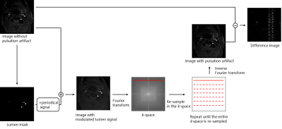 |
Reducing pulsatile artefact in TOF sequence using CNN
Qiaoyi Xue1, Jianmin Yuan1, Haodong Qin2, Hui Liu1, Ran Huo3, and Huishu Yuan3
1Central Research Institute, United Imaging Healthcare Group, United Imaging Healthcare, Shanghai, China, 2Magnetic Resonance Business Unit, United Imaging Healthcare, Shanghai, China, 3Department of Radiology, Peking University Third Hospital, Beijing, China
Time-of-flight (TOF) sequence is a non-contrast enhancing blood vessel imaging technique critical for vessel wall analysis. However, the periodical pulsation in the vessels, especially arteries, often causes pulsatile artifact along the phase encoding direction in the image. This degrades image quality and brings difficulty for diagnosis. The purpose of this work is to develop a convolutional neural network (CNN) method to reduce the pulsatile artefact in TOF sequence.
|
|
2236.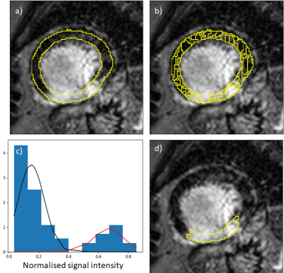 |
Fully automated quantification of left ventricular scar in patients with ischemic heart disease using deep learning and Gaussian mixture models
Cian Michael Scannell1, Adriana Villa1, Stefano Figliozzi1, Jack Lee1, Mikto Veta2, Marcel Breeuwer2,3, and Amedeo Chiribiri1
1King's College London, London, United Kingdom, 2Eindhoven University of Technology, Eindhoven, Netherlands, 3Philips Healthcare, Best, Netherlands
Late gadolinium enhancement MRI is crucial tool for guiding the management of patients with known/suspected myocardial infarction. Currently, clinical practice relies on the visual inspection of these images but there has been extensive work on semi-automated approaches for quantifying scar. Their utility is however limited by the time-intensive manual interactions involved, including the drawing of the myocardial contours. In this work, deep learning methodology is employed to automatically achieve the previously manual steps and these segmentations are used as input to a completely unsupervised scar quantification algorithm. This allows automatic, fast and reproducible quantification of regions of scar.
|
|
2237.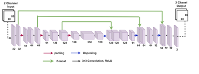 |
Accelerated Phase Contrast MRI Reconstruction with Deep U-NET Convolutional Neural Networks
Ruponti Nath1, Sean Callahan1, Narayana Singam2, Marcus Stoddard2, and Amir Amini1
1ECE, University of Louisville, Louisville, KY, United States, 2Cardiovascular Medicine, University of Louisville, Louisville, KY, United States
We propose a framework for accelerated reconstruction of 2D phase contrast MRI from undersampled K space by using deep convolutional neural networks. The reconstruction problem is considered as a de-aliasing problem in complex spatial domain. A U-net architecture was trained and tested on 4D flow MRI data in 10 patients with aortic stenosis and 4 healthy volunteers. The reconstructed complex two channel image showed that the U-net is able to unaliase the undersampled flow images with resulting magnitude and phase difference images showing good agreement with the fully sampled magnitude and phase images.
|
|
2238.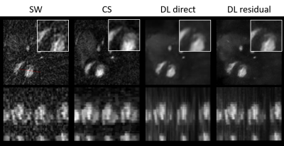 |
Deep Learning Artifact Removal for Real-Time Rodent Cardiac MRI
Patrick Metze1, Hao Li2, Tobias Speidel2, Ina Vernikouskaya1, and Volker Rasche1,2
1Department of Internal Medicine II, University Ulm Medical Center, Ulm, Germany, 2Core Facility Small Animal Imaging (CF-SANI), Ulm University, Ulm, Germany
Reconstruction methods incorporating Deep Learning have gained a lot of traction in the recent past. However, most of these methods have been evaluated in human MRI. In this work, we show the feasibility of deep-learning artifact removal for the tiny golden angle radial trajectory in rodent cardiac MRI and validate two approaches against a Compressed Sensing reconstruction and the gated reference standard. The deep-learning based methods achieve acceptable visual image quality and exhibit only slight, but for one method significant, differences in the functional analysis.
|
2239.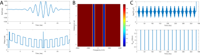 |
ATP Degradation Rates are altered in the Diabetic Myocardium: A 31P Saturation Transfer Study (with Dual-band, quasi-Adiabatic Optimal Control)
Jack JJJ Miller1,2,3, Matthew Kerr2, Ladislav Valkovic3,4, Kerstin Timm2, Justin Lau2,3, Paul A. Bottomley5, Lisa Heather2, and Damian Tyler3
1Department of Physics, University of Oxford, Oxford, United Kingdom, 2Department of Physiology, Anatomy and Genetics, University of Oxford, Oxford, United Kingdom, 3OCMR, Cardiovascular Medicine, University of Oxford, Oxford, United Kingdom, 4Department of Imaging Methods, Institute of Measurement Science, Slovak Academy of Sciences, Bratislava, Slovakia, 5Radiology and Radiological Science, Johns Hopkins, Baltimore, MD, United States
Saturation Transfer 31P MRS enables the quantification of the rate of ATP synthesis and degradation. In this work, we use quasi-adiabatic optimal-control designed single-band (PCr) and dual-band (Gamma ATP + Pi) saturation schemes to separately estimate $$$k_f$$$ and $$$k_r$$$ in the control and diabetic perfused rat heart at 12T. This scheme is rapid, B1 insensitive, and suitable for use at ultrahigh field. We demonstrate profound metabolic alterations in the diabetic myocardium, with a reduction in $$$k_r$$$ and ATP concentration.
|
|
2240.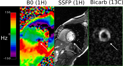 |
RF Coil and Gradient Shim Considerations for Hyperpolarized 13C Imaging of the Human Myocardium
Galen Durant Reed1, JaeMo Park2, Junjie Ma2, Crystal Harrison2, Rolf Schulte3, Albert Chen 4, Salvador Pena2, Jeannie Baxter2, Kelley Derner2, Maida Tai2, Dean Sherry2, Vlad G Zaha2, and Craig R Malloy2
1GE Healthcare, Dallas, TX, United States, 2University of Texas Southwestern Medical Center, Dallas, TX, United States, 3GE Healthcare, Munich, Germany, 4GE Healthcare, Toronto, ON, Canada
Initial human hyperpolarized 13C cardiac imaging experiments utilized a frequency-selective, single-shot spiral pulse sequence for encoding, and a rigid 8 channel receiver array paired with a Helmholtz clamshell transmitter. In phantom studies, we evaluate this RF hardware along with flexible transmit / receiver designs and test these in healthy volunteers. Furthermore, as pointed out previously, the detected circumferential distribution of [13C]bicarbonate around the myocardium is often non-uniform in pigs, thus confounding the translation of this technique for regional metabolism evaluation. We observed this artifact in human volunteers and explore its possible sources.
|
|
2241.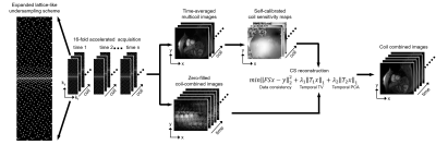 |
16-fold accelerated real-time free-breathing cardiac cine MRI in patients with a cardiac implantable electronic device
Sungtak Hong1, Kyungpyo Hong2, Austin E Culver1, Bradley D Allen1, Daniel C Lee3, and Daniel Kim1
1Radiology, Northwestern University, Chicago, IL, United States, 2Translational and Molecular Imaging Institute and Department of Radiology, Icahn School of Medicine at Mount Sinai, New York, NY, United States, 3Division of Cardiology, Internal Medicine, Northwestern University, Chicago, IL, United States
CMR measurements in CIED patients are critical but challenging due to susceptibility artifacts. We described the development of highly accelerated, real-time, free-breathing cine pulse sequence using compressed sensing with variable density “lattice-like” k-space sampling scheme. Comprehensive comparison was done to estimate performance against clinical standard breath-hold cine pulse sequence.
|
|
2242.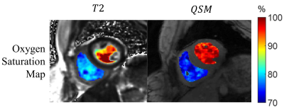 |
Noninvasive Cardiac Chamber Blood Oxygenation Measurements: T2 and QSM approaches.
Yan Wen1,2, Jonathan W. Weinsaft3, Thanh D. Nguyen2, Jiwon Kim3, Yi Wang4,5, and Pascal Spincemaille2
1Meinig School of Biomedical Engineering, Cornell University, new york, NY, United States, 2Radiology, Weill Cornell Medicine, New York, NY, United States, 3Medicine, Weill Cornell Medicine, New York, NY, United States, 4Meinig School of Biomedical Engineering, Cornell University, New York, NY, United States, 5Radiology, Weill Cornell Medicine, new york, NY, United States
Previous studies have demonstrated noninvasive cardiac chamber blood oxygenation measurement using T2 and QSM. This work shows the initial comparison between the two approaches for such measurements in healthy volunteers.
|
|
2243.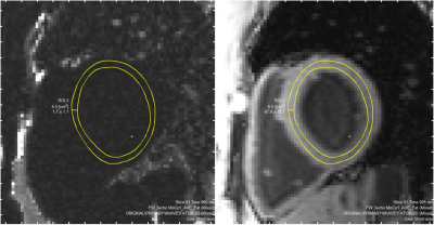 |
Using Novel Fat Water Separation Sequence to Quantify Intramyocardial Fat-Fraction
Xin Dong1,2, Olumide Awelewa 1,2, Graham Galloway2, Viral Chikani3, Rob Robergs1, and Arnold Ng4,5
1Queensland University of Technology, Brisbane, Australia, 2Translational Research Institute, Woolloongabba, Australia, 3Endocrinology, Princess Alexandra Hospital, Woolloongabba, Australia, 4Cardiology, Princess Alexandra Hospital, Woolloongabba, Australia, 5The University of New South Wales, Sydney, Australia
VARPRO is a novel fat water separation sequence that shows promise for myocardial lipid characterization. Two questions were addressed in this study: 1. Does VARPRO provide the same myocardial fat-fraction result compared to established magnetic resonance spectroscopy (MRS)? 2. Is the myocardial fat-fraction obtained by VARPRO reproducible? The results suggest the two methods are strongly associated but agreed poorly. The VARPRO estimation is proportionally biased, however, the bias can be easily accounted for using a linear equation. The temporal reproducibility of VARPRO is satisfactory. VARPRO could be a potential, alternative modality to MRS in myocardial lipid quantification.
|
|
2244.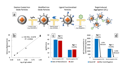 |
Magnetic Nanoparticle Sensor for Longitudinal Biomarker Monitoring
Richard Joshua Murdock1,2 and Michael Cima2,3
1Health Sciences & Technology, Massachusetts Institute of Technology, Cambridge, MA, United States, 2Koch Institute For Integrative Cancer Research, Cambridge, MA, United States, 3Materials Science & Engineering, Massachusetts Institute of Technology, Cambridge, MA, United States
Continuous biochemical monitoring remains a critical challenge in the biosensing community as longevity, biocompatibility, and signal transduction limit multi-month implantation. We propose a novel design and evaluation of the sensing capability of a magnetic nanoparticle dosimeter assay for in vivo implementation, readable by magnetic relaxation. This local diagnostic device offers continuous sentinel evaluation and longitudinal tracking of critical biomarkers found to correlate with cardiovascular disease states. The current design exhibits an order of magnitude improvement in performance compared to prior studies, translating into a predicted 29 week lifetime.
|
|
2245.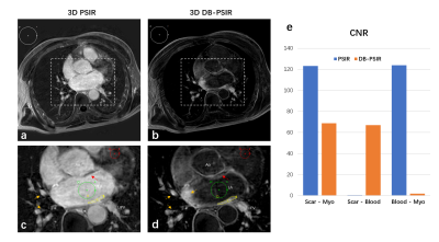 |
Detection of atrial scar using 3D high-resolution dark-blood phase sensitive inversion recovery imaging
Dongyue Si1, Yanfang Wu2, Jie Yin2, Rui Guo3, Jingjing Xiao4, Bowei Liu1, Xue Lin2, Peng Gao2, Deyan Yang2, Quan Fang2, Jianwen Luo1, Daniel A. Herzka5,6, and Haiyan Ding1
1Center for Biomedical Imaging Research (CBIR), Department of Biomedical Engineering, School of Medicine, Tsinghua University, Beijing, China, 2Department of Cardiology, Peking Union Medical College Hospital, Chinese Academy of Medical Sciences & Peking Union Medical College, Beijing, China, 3Department of Medicine, Beth Israel Deaconess Medical Center and Harvard Medical School, Boston, MA, United States, 4Department of Medical Engineering, Xinqiao Hosptial, Army Medical University, Chongqing, China, 5Department of Biomedical Engineering, Johns Hopkins School of Medicine, Baltimore, MD, United States, 6Cardiovascular Interventional Program, National Heart, Lung, and Blood Institute, National Institutes of Health, Bethesda, MD, United States
Radiofrequency ablation (RFA) is a first-line treatment for controlling atrial fibrillation (AF). Visualizing fibrotic tissues as well as the surrounding anatomical structure could improve the delivery of therapy. In this work, an independent navigator-gated three-dimensional dark-blood phase-sensitive inversion recovery sequence was developed for atrial visualization. Dark-blood was achieved by optimizing inversion recovery and T2-preparation timing. Preliminary results from in vivo imaging in patients with AF post RFA demonstrate that the proposed technique can clearly visualize the atrial wall and scar formation. Both dark blood contrast and high resolution (1.25×1.25×3 mm3) were achieved.
|
|
2246.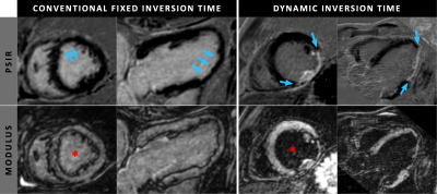 |
The effect of a dynamic inversion time in high-resolution isotropic 3D dark-blood LGE without additional magnetization preparation
Robert J Holtackers1,2,3, Suzanne Gommers1,2, Caroline M van de Heyning3,4, Jouke Smink5, Amedeo Chiribiri3, Joachim E Wildberger1,2, and Rachel ter Bekke2,6
1Department of Radiology & Nuclear Medicine, Maastricht University Medical Centre, Maastricht, Netherlands, 2CARIM School of Cardiovascular Diseases, Maastricht University, Maastricht, Netherlands, 3School of Biomedical Engineering and Imaging Sciences, King’s College London, London, United Kingdom, 4Department of Cardiology, Antwerp University Hospital, Antwerp, Belgium, 5Philips Healthcare, Best, Netherlands, 6Department of Cardiology, Maastricht University Medical Centre, Maastricht, Netherlands Conventional 2D LGE often suffers from poor scar-to-blood contrast and limited spatial resolution. While 3D methods are known for their superior spatial resolution, our earlier proposed dark-blood LGE without additional magnetization preparation enables improved scar-to-blood contrast. In the present study we sought to assess the effect of a slowly increasing, dynamic inversion time on left-ventricular blood-pool suppression in a high-resolution isotropic 3D dark-blood LGE acquisition, and compare it against a conventional, fixed inversion time. Our findings show that such a dynamic inversion time significantly improves blood-pool suppression and thereby maximizes the dark-blood contrast mechanism for improved detection of myocardial scarring. |
|
2247.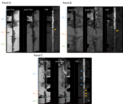 |
Multiplanar reconstruction of intracranial arteries: luminal morphological characterization of atherosclerosis
Gador Canton1, Hiroko Watase2, Josh Liu3, Li Chen4, Yin Guo4, Yongjun Wang5, and Chun Yuan1
1Radiology, University of Washington, Seattle, WA, United States, 2Surgery, University of Washington, Seattle, WA, United States, 3University of Washington, Seattle, WA, United States, 4Electrical and Computer Engineering, University of Washington, Seattle, WA, United States, 5Beijing Tiantan Hospital, Capital Medical University, Beijing, China
This study aims to characterize the luminal morphology of middle cerebral arteries affected by intracranial atherosclerotic disease in a cohort of ischemic stroke patients. We used a visualization method (MOCHA) based on multiplanar reconstruction of the intracranial vasculature. We identified three distinct morphological patterns based on the effect of the atherosclerotic plaque presence on the downstream geometry. Together with the location of the plaque with respect to the middle cerebral artery branching point from the internal carotid artery, these distinct morphological patterns might be the differential factor in recurrent stroke risk stratification due to the resulting local hemodynamics.
|
|
2248.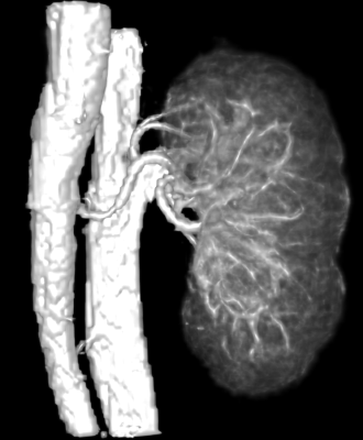 |
Vascular Imaging of the Human Kidney with Positive Contrast via SPION and UTE
Liam Timms1, Tianyi Zhou1, Ju Qiao2, Vishala Mishra3, Gayatri Veeramani4,5, Andrew Allegretti4, Thomas Benkert6, John Kirsch2, Ravi Seethamraju7, Mukesh Harisinghani3, and Srinivas Sridhar1,8
Video Permission Withheld
1Physics, Northeastern University, Boston, MA, United States, 2Massachusetts General Hospital, Boston, MA, United States, 3Radiology, Massachusetts General Hospital, Boston, MA, United States, 4Nephrology, Massachusetts General Hospital, Boston, MA, United States, 5Nephrology, Brigham and Women’s Hospital, Boston, MA, United States, 6Siemens Healthcare, Erlangen, Germany, 7Siemens Healthcare, Boston, MA, United States, 8Theranano LLC, Newton, MA, United States
A contrast-enhanced MRA technique using Ultra-short Time-to-Echo (UTE) pulse sequence with super-paramagnetic iron oxide nanoparticle (SPION) contrast agent is demonstrated for abdominal vascular imaging in human patients. It successfully depicts the kidney anatomy, as well as kidney cysts and kidney vascular structure in excellent clarity and detail. With manual segmentation, total kidney volume, cyst volume and renal artery diameter were estimated. This initial study in humans demonstrated the basic feasibility of abdominal QUTE-CE MRA as an alternative to Gd based CEMRA.
|
|
2249. |
Rapid Whole-Heart 3D Cine Using a Golden Ratio Stack of Spirals Trajectory
Javier Montalt-Tordera1, Grzegorz Kowalik1, Alexander Gotschy2,3, Jennifer Steeden1, and Vivek Muthurangu1
1Institute of Cardiovascular Science, UCL, London, United Kingdom, 2Great Ormond Street Hospital, London, United Kingdom, 3Institute for Biomedical Engineering, ETH Zurich, Zurich, Switzerland
A free-breathing three-dimensional cine imaging sequence was developed using a stack of spirals trajectory with golden angle rotations for efficient k-space sampling. Parallel imaging and compressed sensing were used in reconstruction for data acceleration. Respiratory motion artefact was avoided by binning k-space data into multiple respiratory phases. The proposed imaging protocol was tested on 10 patients and compared with a standard 2D breath-held short-axis stack. The proposed method was faster to acquire and generally able to provide correct quantitative measurements, but image quality needs to be improved and reconstruction time is too long for clinical practice.
|
|
2250.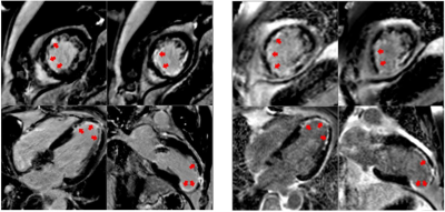 |
A pilot investigation into the use of a high-performance 0.55T scanner in CMR late gadolinium enhancement imaging of myocardial infarction
W. Patricia Bandettini1, Sujata M. Shanbhag1, Christine M. Mancini1, Delaney R. McGuirt1, Jennifer L. Henry1, Margaret M. Lowery1, Marcus Y. Chen1, Hui Xue1, Peter M. Kellman1, and Adrienne E. Campbell-Washburn1
1NATIONAL INSTITUTES OF HEALTH/NHLBI, BETHESDA, MD, United States
We demonstrate the diagnostic capabilities of a high-performance, low-field (0.55 Tesla) scanner in the acquisition and interpretation of late gadolinium enhancement (LGE) in patients referred for assessment of myocardial infarction (MI). Patients underwent paired comparison exams with breath-held gradient echo LGE imaging at 1.5T and breath-held bSSFP LGE imaging at 0.55T. The number of enhancing segments identified between each field strength was similar (59 segments at 0.55T vs 63 segments at 1.5T), and assessment of epicardial coronary artery distribution matched exactly between the two field strengths; included were two multi-vessel disease cases.
|
|
2251.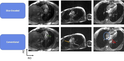 |
Slice Encoding for the Reduction of Outflow Signal Corruption in Cine Balanced Steady State Free Precession Imaging
Fadil Ali1, Mark Bydder1, Da Wang2, Vahid K Ghodrati1, Chang Gao1, Ashley Propser1, Kim-Lien Nguyen1, and Peng Hu1
1Radiology, University of California, Los Angeles, Los Angeles, CA, United States, 2Radiology, The Unversity of Chicago, Chicago, IL, United States
Balanced steady-state free precession (bSSFP) is susceptible to outflowing signal because of it features no spoiling. This results in misregistrated artifacts that hamper the cardiac evaluations. We propose a method that tackles this issue in the acquisition process by spatially encoding for this outflowing signal to prevent it from projecting onto the imaged slice.
|
|
2252. |
Stabilized decompositions for improved cardiac self-gating: A proof-of-concept in single breath-hold 3D cine imaging
Anna Padée1, Lorenzo Di Sopra2, John Heerfordt2,3, Jérôme Yerly2,4, Marco Merlo1, Tobias Kober2,3,5, Davide Piccini2,3,5, Matthias Stuber2,4, Christopher Roy2, and Jonas Richiardi2,3
1Laboratory for Psychiatric Neuroscience and Psychotherapy, University of Fribourg, Fribourg, Switzerland, 2Department of Radiology, Lausanne University Hospital and University of Lausanne, Lausanne, Switzerland, 3Advanced Clinical Imaging Technology, Siemens Healthcare, Lausanne, Switzerland, 4Center for Biomedical Imaging, Lausanne, Switzerland, 5LTS5, Ecole Polytechnique Fédérale de Lausanne, Lausanne, Switzerland
ECG recording during whole heart MR imaging requires extra setup time and is not always reliable. Recently, self-gated (SG) acquisition techniques have been developed, which extract the cardiac signal from k-space center amplitude modulations. Here, we investigated whether different single- (Empirical Mode Decomposition (EMD), Ensemble EMD (EEMD)) and multi-coil (PCA, ICA with novel stabilisation) decomposition methods yielded SG triggers with minimal variability to ECG R-wave location. EEMD yielded median variability similar to PCA. Compared to PCA, our proposed stabilised ICA approach yielded SG triggers with 66% lower variability (median over 5 subjects), although not for all subjects.
|
|
2253. |
Identification of hyperelastic properties of the myocardium from CMR patient-specific 3D geometry and the Virtual Field Method.
Delphine Perie1, Mehdi Ghafari1, and Daniel Curnier2
1Mechanical Engineering, Polytechnique Montreal, Montreal, QC, Canada, 2Kinesiology, University of Montreal, Montreal, QC, Canada
Due to the complexity of the heart’s geometry, the characterization of the material properties of the myocardium remains challenging. Using kinematically admissible hyperboloid virtual fields and a finite element mesh of the left ventricle, we successfully solved the virtual field method equations to determine the stiffness of the myocardium from a reverse identification approach. The distribution of the elastic stiffness presented same patterns between the 3 volunteers. However, the standard risk volunteer presented higher stiffness within the mobile wall than the high risk volunteers. This approach would be an efficient tool to characterize early cardiac dysfunction.
|
|
2254. |
Compressed sensing reconstruction of cardiac CEST-MRI preserves accuracy, sensitivity and specificity of endogenous metabolites
Bonnie Lam1, Barry Fung1, and Moriel Vandsburger1
1University of California, Berkeley, Berkeley, CA, United States
While accelerated imaging via compressed sensing reconstruction has been explored in several dynamic contrast settings, incorporation of compressed sensing for accelerated image acquisition has not been fully explored in the setting of cardiac CEST. Here we test the impact of various compressed sensing methods and acceleration factors upon the accuracy of endogenous cardiac CEST in mice, specifically measures of specificity, accuracy, and sensitivity.
|
2255.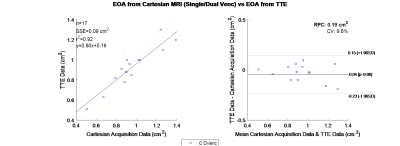 |
Effective Orifice Area (EOA) for Evaluation of Aortic Stenosis from 4D Flow MRI
Samuel Edward Bibelhauser1, Sean P Callahan1, Narayana Singam2, Marcus Stoddard2,3, and Amir Amini1,3
1Electrical and Computer Engineering, University of Louisville, Louisville, KY, United States, 2Division of Cardiovascular Medicine, University of Louisville, Louisville, KY, United States, 3Radiology, VA Medical Center, Louisville, KY, United States
Effective Orifice Area (EOA) is a valuable diagnostic parameter for the determination of the severity of Aortic Stenosis(AS). A 4D Flow based measurement of this parameter was developed, and tested. The method was validated on a rigid phantom, applied to 4 healthy volunteers and 17 patient subjects with moderate to severe AS. The resultant EOA, for patients, was compared to their transthoracic echocardiography (TTE) with Doppler result. The comparison used Bland Altman analysis and linear regression which revealed a bias of 0.04 cm2 and correlation coefficient of 0.92. Showing a high level of concordance between the two modalities.
|
|
2256.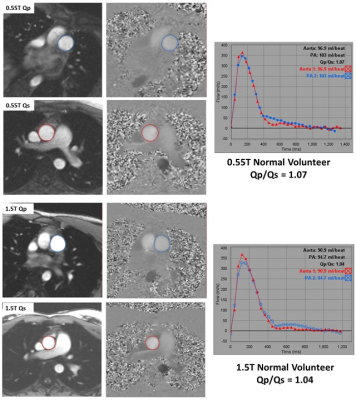 |
Comparison of Phase-Contrast Flow Imaging at 0.55T and 1.5T
Sujata M. Shanbhag1, Rajiv M. Ramasawmy1, W. Patricia Bandettini1, Christine M. Mancini1, Delaney R. McGuirt1, Jennifer L. Henry1, Margaret M. Lowery1, Marcus Y. Chen1, and Adrienne Campbell-Washburn1
1NATIONAL INSTITUTES OF HEALTH/NHLBI, BETHESDA, MD, United States, Bethesda, MD, United States
Low-field MRI equipped with high-performance technology and modern imaging methods offers the potential to make technically demanding cardiac imaging more accessible. This study demonstrates that a low-field (0.55T) system can provide accurate phase-contrast flow measurements in both healthy volunteers and patients who were clinically referred for assessment of intracardiac shunt and valvular disease. Comparative studies between contemporary high-performance 0.55T and 1.5T systems generated well-correlated quantitative flow measurements (r>0.87).
|
|
2257.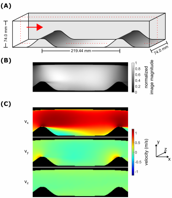 |
On the dynamic range of Reynolds stress tensor quantification
Simon Schmidt1, Kristine John2, Martin Bruschewski2, Sebastian Flassbeck1, Mark E. Ladd1, Sven Grundmann2, and Sebastian Schmitter1,3
1Medical Physics in Radiology, German Cancer Research Center (DKFZ), Heidelberg, Germany, 2Institute of Fluid Mechanics, University of Rostock, Rostock, Germany, 3Physikalisch-Technische Bundesanstalt (PTB), Braunschweig and Berlin, Germany
In this work, we successfully quantified the Reynolds stress tensor (RST) in a well-known fluid-dynamic test case: the flow over periodic hills. This test case was chosen because the turbulence in this kind of flow is strongly inhomogeneous and anisotropic, representing a challenging measurement task. The results indicate, in analogy to intravoxel velocity standard deviation (IVSD) mapping, that RST quantification is highly susceptible to the applied $$$m_1^{enc}~$$$value. Furthermore, RST mapping inherently requires a higher dynamic range compared to IVSD mapping, since the shear stresses are typically much lower than the normal stresses.
|
|
2258.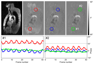 |
Spoke-wise Maxwell Correction for Real-time Phase-contrast Flow MRI with Highly Undersampled Radial Trajectories
Jost Michael Kollmeier1, Dirk Voit1, and Jens Frahm1
1Biomedizinische NMR, Max-Planck-Institut für biophysikalische Chemie, Göttingen, Germany
Real-time phase-contrast flow MRI based on highly undersampled radial FLASH employs fast gradients that make concomitant field contributions relevant even at 3 T. In order to correct for phase errors a spoke-wise Maxwell correction has been developed and successfully integrated into a model-based reconstruction. For radial trajectories corrections for individual k-space lines are required as concomitant fields may change from spoke to spoke. The proposed method significantly improves phase errors in velocity maps as shown for three-directional real-time phase-contrast flow MRI of the human aortic arch.
|
|
2259.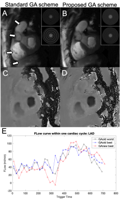 |
Reducing k-space gaps of golden angle rotated spiral MRI binned into cardiac frames applied to flow measurements of the coronary arteries
Dan Zhu1,2 and Michael Schär2
1Department of Biomedical Engineering, Johns Hopkins University School of Medicine, Baltimore, MD, United States, 2Russell H. Morgan Department of Radiology and Radiological Science, Johns Hopkins University School of Medicine, Baltimore, MD, United States
Velocity encoded MRI has clinical significance in the diagnosis of coronary artery disease. High spatial and temporal resolution increase the accuracy and reproducibility and preserve reliable flow pattern. By using rotated golden angle with k-t space reconstruction and retrospective cardiac gating, high quality flow CINE could be achieved with high spatial and temporal resolution during a single breath-hold. However, binning to cardiac frames may lead to large gaps in k-space. In this work, we propose a new scheme of rotated golden angle and optimized retrospective gating by shifting the binning window. Improved image quality is demonstrated with the proposed methods.
|
|
2260.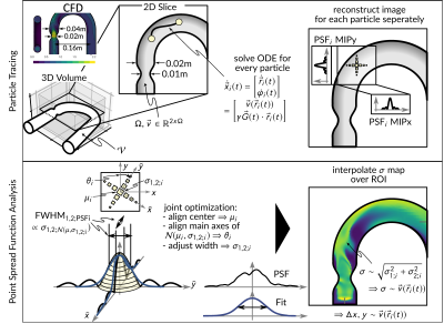 |
Limitations of Echo Planar Imaging in PC-MRI of High Flow Regimes
Hannes Dillinger1, Jonas Walheim1, Robbert J.H. van Gorkum1, and Sebastian Kozerke1
1University and ETH Zurich, Zurich, Switzerland
An evaluation of velocity field reconstructions from echo-planar trajectories in PC-MRI relative to ground truth computational fluid dynamics (CFD) data in stenotic high flow regimes is presented. The reconstructed velocity images and variations in spatial resolution resulting from modulation of the Point Spread Function (PSF) are investigated. Significant artifacts and resolution loss of up to factor 8 for EPI in high-flow regimes of 220cm/s are found. In-vitro experiments confirm that depending on the orientation of the main flow direction the reconstructed velocity images differ significantly in regions of high shear or acceleration with implications for in-vivo applications.
|
|
2261. |
Magnitude difference regularized reconstruction for phase contrast flow MRI
Taehoon Shin1,2 and Wanyong Shin3
1Ewha Womans University, Seoul, Korea, Republic of, 2Case Western Reserve University, Cleaveland, OH, United States, 3Cleveland Clinic, Cleveland, OH, United States
Based on the magnitude similarity between bipolar-encoded k-space data for PC flow imaging, magnitude-difference regularization was incorporated into the conventional compressed sensing (CS) reconstruction. The gradient of the magnitude regularization was derived so the reconstruction problem can be solved iteratively. Retrospecitve in-vivo studies show that the addition of magnitude-difference regularization into conventional CS reconstruction improves the accuracy of image reconstruction using highly undersampled phase-contrast flow MR data.
|
|
2262. |
Feasibility of a Free-Breathing 2D Phase Contrast Sequence for Aortic Pulse Wave Velocity Measurements
Grant S Roberts1, Kevin M Johnson1,2, Steven R Kecskemeti2, Ozioma Okonkwo3, Sarah Lose3, Laura Eisenmenger2, and Oliver Wieben1,2
1Medical Physics, University of Wisconsin - Madison, Madison, WI, United States, 2Radiology, University of Wisconsin - Madison, Madison, WI, United States, 3Medicine, University of Wisconsin - Madison, Madison, WI, United States
Pulse wave velocity (PWV) is a biomarker that indirectly relates to arterial stiffness, an early indicator of cardiovascular disease. Breath-hold phase contrast (PC) MRI can be used to assess PWV in the aorta, however in certain populations, breath-holds may be difficult. We present a method to measure aortic PWV using a free-breathing radially-undersampled PC sequence. Initial results show that the free-breathing PC-derived PWV measures are comparable to the measures obtained from breath-hold Cartesian PC scans. Larger cohorts are warranted to verify these findings.
|
|
2263.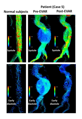 |
Energy loss assessed with 4D-Flow for the normal subjects and patients with an abdominal aortic aneurysm pre and post endovascular aortic repair
Takashi Mizuno1, Yasuo Takehara2, Masataka Sugiyama2, Ryota Horiguchi3, Shinji Naganawa3, Yasuo Sakurai1, Yutaka Kato1, Shinji Abe1, Haruo Isoda4, Tomohiro Sato5, Tsuneo Ishiguchi5, Masanori Tadokoro5, and Atushi Nozaki6
1Department of Radiological Technology, Nagoya University Hospital, Nagoya, Japan, 2Department of Fundamental Development of Low Invasive Diagnostic Imaging, Nagoya University Graduate School of Medicine, Nagoya, Japan, 3Department of Radiology, Nagoya University Graduate School of Medicine, Nagoya, Japan, 4Brain & Mind Research Center, Nagoya University, Nagoya, Japan, 5Trust Clinic, Nagoya, Japan, 6GE Healthcare Japan, Tokyo, Japan
We analyzed energy loss in normal subjects, patients in pre- and post- endovascular aortic repair. Patients had vortices in dilated flow path in pretreatment and tortuous high-velocity streamlines in posttreatment. Energy loss of posttreatment was higher than that of pretreatment. Energy loss would be affected most effectively by the change of the flow direction.
|
|
2264.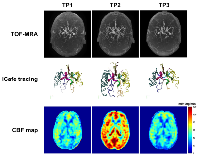 |
Flow measurement with time of flight (TOF) in patients undergoing carotid revascularization: A comparison with arterial spin labeling (ASL)
Zhensen Chen1, Li Chen2, Manabu Shirakawa1, Wenjin Liu1, Dakota Ortega1, Jinmei Chen1, Niranjan Balu1, Theodore Trouard3, Thomas S Hatsukami4, Wei Zhou5, and Chun Yuan1
1Radiology, University of Washington, Seattle, WA, United States, 2Electrical and computer engineering, University of Washington, Seattle, WA, United States, 3Biomedical Engineering, University of Arizona, Tuscon, AZ, United States, 4Surgery, University of Washington, Seattle, WA, United States, 5Surgery, University of Arizona, Tuscon, AZ, United States
In this study, we assessed the potential of using vasculature features extracted from TOF images as surrogate markers of intracranial blood flow in patients undergoing carotid revascularization surgery and MR imaging prior to, within 48 hours after and 6 months after surgery. The intracranial vasculature features, including total volume, total length and total number of branches etc., on TOF images were extracted using a novel tool named iCafe, and then compared with arterial spin labeling (ASL) cerebral blood flow (CBF) measurement. The results show that TOF-iCafe vasculature features have similar behaviors as ASL CBF over the time points.
|
|
2265.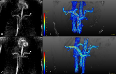 |
Comparison between 4D Flow in abdominal magnetic resonance imaging for portal vein using SENSE and Compressed SENSE
Yuli HUANG1, Li ZHU2, Haiyang DONG2, Yuan QU3, and Yan WANG3
1MR, Philips Healthcare (Suzhou) Co., Ltd, SUZHOU, China, 2Radiology, Shanghai Chest Hospital, Shanghai JiaoTong University, Shanghai, China, 3Radiology, People’s Hospital of Xinjiang Uygur Autonomous Region, Urumqi, China
4D Flow MRI is a technique that can characterize vascular structure and hemodynamics. Compared with SENSE, Compressed SENSE can further reduce scanning time using the sparse characteristics of MR images. This study aims to investigate the feasibility of 4D Flow MRI with Compressed SENSE by comparing with 4D Flow MRI with SENSE. 4D Flow dynamic angiography of hepatic portal vein using SENSE and Compressed SENSE could be completed within 5 minutes with optimized parameters. The scanning time of Compressed SENSE 4D Flow was shorter than that of SENSE 4D Flow.
|
|
2266.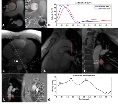 |
Higher frame-rate phase-contrast with temporally under-sampled phase-reference acquisition: preliminary data in two vascular regions
Jerome Lamy1, Gigi Galiana2, and Dana Peters2
1Department of Radiology and Biomedical Imaging, Yale University, New Haven, CT, United States, 2Department of Radiology and Biomedical Imaging, Yale, New Haven, CT, United States
Phase-contrast (PC) MRI requires at least doubled scan time compared to anatomical imaging. This has required careful trade-offs regarding spatial and temporal resolution, and breath-holding times, and often the temporal or spatial resolution is insufficient. Here we introduce a simple novel method which temporally under-samples acquisition of the phase-reference images, providing increased temporal resolution. This method is evaluated both in the aortic and pulmonary vein flow, which exhibits several rapid flow peaks requiring good spatial resolution. This method allowed to accurately identify velocity peaks for the aorta and pulmonary veins, providing higher temporal resolution as compared to the standard PC-MRI.
|
|
2267.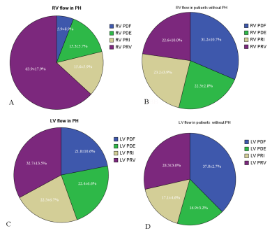 |
Biventricular flow components assessed with 4-dimensional flow cardiac magnetic resonance in patients with pulmonary hypertension
Min Liu1, Xincao Tao2, Jing An3, Ning Jin4, and Fan Lin5
1Radiology, China-Japan Friendship Hospital, Beijing, China, 2Department of Pulmonary and Critical Care Medicine, China-Japan Friendship Hospital, Beijing, China, 3Siemens Shenzhen Magnetic Resonance Ltd, Siemens Shenzhen Magnetic Resonance Ltd, Shen Zhen, China, 4Siemens Medical Solution, Siemens Medical Solution, Chicago, IL, United States, 5Department of Cardiovascular surgery, China-Japan Friendship Hospital, Beijing, China
Using 4D FLOW CMR, flow components including percent of direct flow (PDF), retained inflow (PRI), delayed ejection flow (PDE) and residual volume (PRV) of bi-ventricles in patients with PH were quantified. Compared with non-PH patients, flow components had significantly alterations. RV PDF and PRV significantly correlated with RVEDV, RSV, RVEF and RV mass as well as NT-proBNP. Moreover, RV PDF and PVR correlated with pulmonary artery pressure and PVR. We think that 4D flow CMR may become a powerful surrogate for the future PH studies
|
|
2268.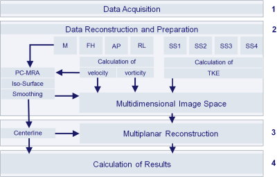 |
Method for Comprehensive Analysis of Vascular Geometry and Fluid Dynamics Using MRI
Dominik Daniel Gabbert1, Arash Kheradvar2, Michael Jerosch-Herold3, Thekla Oechtering4, Felix Wadle1, Anselm Sebastian Uebing1, Hans-Heiner Kramer1, Inga Voges1, and Carsten Rickers1
1Department of Congenital Heart Disease and Pediatric Cardiology, University Hospital Schleswig-Holstein, Kiel, Germany, 2The Edwards Lifesciences Center for Advanced Cardiovascular Technology, University of California, Irvine, CA, United States, 3Harvard Medical School, Brigham and Women’s Hospital, Boston, MA, United States, 4Department of Radiology and Nuclear Medicine, University Hospital Schleswig-Holstein, Lübeck, Germany
Many secondary flow patterns in the large vessels have been shown to represent significant pathophysiological phenomena. However, the computational cost to analyze a large number of quantities usually discourage the use of these parameters for clinical decision making given the limited computing resources in clinical settings. We describe a novel postprocessing method for comprehensive analysis of vascular anatomy and fluid dynamics based on 4D Flow MRI. The method defines a multi-dimensional feature space built from a few complementary fluid dynamics building blocks to efficiently determine a large number of anatomic and fluid dynamics parameters in the course of a vessel.
|
2269.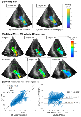 |
4D flow MRI vs. Doppler Echocardiography: Data Integration Framework for Direct Comparison of Intra-cardiac Flow Dynamics
Jeesoo Lee1,2, Nadia El Hangouche2, Alex J Barker3, James D Thomas2, and Michael Markl1,4
1Radiology, Northwestern University, Chicago, IL, United States, 2Cardiology, Northwestern Memorial Hospital, Chicago, IL, United States, 3Radiology, University of Colorado Denver, Denver, CO, United States, 4Biomedical Engineering, Northwestern University, Chicago, IL, United States
We developed a framework to integrate 4D flow MRI with color Doppler echocardiography (CDE) for direct localized inter-modal velocity comparison. The framework combines CDE and 4D flow MRI data by using cine-MRI and a two-step image registration; 1) registration of CDE to cine-MRI and 2) registration of cine-MRI to 4D flow MRI. As a result, a voxel-wise inter-modal velocity difference map overlaid to a tissue image was constructed. Six healthy subjects with a same-day CDE and 4D flow MRI were utilized and a strong correlation with moderate agreement was found in the LVOT.
|
|
2270.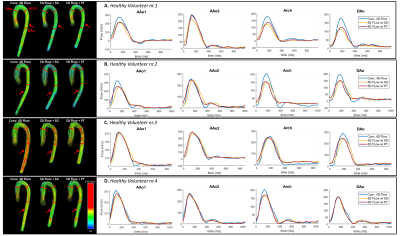 |
5D Flow – A quantitative in vivo comparison between Self-Gating and Pilot Tone Gating
Mariana B. L. Falcão1, Lorenzo Di Sopra1, Liliana Ma2,3, Mario Bacher1,4, Davide Piccini1,5, Jérôme Yerly1,6, Peter Speier4, Tobias Rutz7, Milan Prša7, Michael Markl2,3, Matthias Stuber1,6, and Christopher Roy1
1Department of Diagnostic and Interventional Radiology, Lausanne University Hospital (CHUV) and University of Lausanne (UNIL), Lausanne, Switzerland, 2Department of Radiology, Feinberg School of Medicine, Northwestern University, Chicago, IL, United States, 3Department of Biomedical Engineering, Northwestern University, Chicago, IL, United States, 4Siemens Healthcare GmbH, Erlangen, Germany, 5Advanced Clinical Imaging Technology, Siemens Healthcare AG, Lausanne, Switzerland, 6Center for Biomedical Imaging, Lausanne, Switzerland, 7Department of Cardiology, Lausanne University Hospital (CHUV), Lausanne, Switzerland
Conventional 4D flow MRI techniques often have prolonged and unpredictable scan times, due to the use of respiratory navigation. To address this, a fully self-gated cardiac and respiratory motion-resolved whole-heart 5D flow protocol with a fixed scan time was recently developed using a free-running framework. This protocol extracts cardiac and respiratory signals from periodic readouts (self-gating). In this study, we explore the use of Pilot Tone signals as an alternative method for cardiac and respiratory signal extraction to reconstruct 5D flow data and compare reconstructions to those using the previously established self-gating method and the conventional 4D flow sequence.
|
|
2271.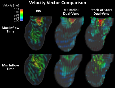 |
Comparison of Radial Acceleration Techniques for Dual-Venc 4D Flow MRI of the Left Ventricle: Validation with Particle Imaging Velocimetry
Philip A Corrado1, Rafael Medero2, Kevin M. Johnson1,3, Christopher J François 3, Alejandro Roldán-Alzate2,4, and Oliver Wieben1,3
1Medical Physics, University of Wisconsin-Madison, Madison, WI, United States, 2Mechanical Engineering, University of Wisconsin-Madison, Madison, WI, United States, 3Radiology, University of Wisconsin-Madison, Madison, WI, United States, 4Biomedical Engineering, University of Wisconsin-Madison, Madison, WI, United States
Dual-Venc 4D flow MRI offers higher velocity sensitivity than single-Venc imaging but requires increased scan time. Radial sampling, either with a 3D-radial or hybrid stack-of-stars (SOS) trajectory, allows for acceleration that can offset the increase, but the optimal technique depends on anatomical region and is not apparent for imaging the left ventricle (LV). We compared 3D-radial and SOS velocity images in an in-vitro LV model versus a reference dataset acquired with particle imaging velocimetry (PIV), an experimental optical imaging technique. 3D-radial matched PIV velocities better than SOS, suggesting it is better for accelerated, dual-Venc imaging of the LV.
|
|
2272.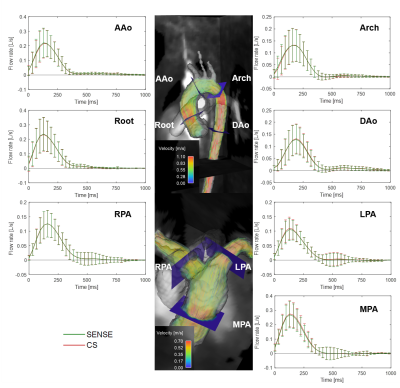 |
In-vitro and in-vivo validation of a pediatric whole heart 4D flow acquisition in 4 minutes with compressed sensing
Takashi Fujiwara1, Lorna Browne1, Ladonna Malone1, Quin Lu2, Brian Fonseca3, Michael DiMaria3, and Alex J Barker1,4
1Department of Radiology, Section of Pediatric Radiology, Children's Hospital Colorado, University of Colorado Anschutz Medical Campus, Aurora, CO, United States, 2Philips Healthcare NA, San Francisco, CA, United States, 3Department of Pediatrics, Section of Pediatric Cardiology, Children's Hospital Colorado, University of Colorado Anschutz Medical Campus, Aurora, CO, United States, 4Department of Bioengineering, University of Colorado Anschutz Medical Campus, Aurora, CO, United States
We investigate the feasibility of the accelerating 4D flow with compressed sensing and online reconstruction in pediatric patients. In-vitro models and imaging in pediatric patients was performed and hemodynamic quantification was compared to conventional approaches. The results demonstrated flow metrics and clinically relevant indices measured by 6-fold accelerated CS were in good agreement with those measured by conventional acceleration techniques, with scan time savings of 30.2%. This suggests the feasibility of using CS 4D flow for pediatric patients in the clinical setting.
|
|
2273.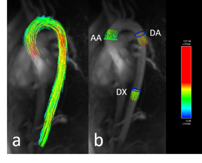 |
Respiratory-Controlled Adaptive 4D Flow of the Aorta: Reproducibility and Comparison to 2D Phase Contrast Imaging
Qingdi Wang1, Xiaojing Guo1, Emma Hornsey2, Lucy McKenna2, Leonid Churilov1,3, Mark Brooks1,2, George Matalanis1,2, Jason Chuen1,2, Eric Poon1, Daniel Staeb4, Ning Jin5, Andrew Ooi1, and Ruth P Lim1,2
1The University of Melbourne, Melbourne, Australia, 2Austin Health, Heidelberg, Australia, 3The Florey Institute of Neuroscience and Mental Health, Melbourne, Australia, 4Siemens Healthcare Pty Ltd, Melbourne, Australia, 5Siemens Medical Solutions, Chicago, IL, United States
A four-dimensional phase-contrast magnetic resonance imaging sequence with respiratory-controlled adaptive k-space reordering (ReCAR-4DPC) offers potential benefits of improved scan efficiency and motion robustness. Imaging was performed on 15 volunteers and 2 patients with aortic dissection. Inter-scan repeatability and inter-reader agreement of flow metrics derived from ReCAR-4DPC was assessed and compared to metrics derived from 2-dimensional phase contrast imaging (2DPC). There was almost perfect inter-scan and inter-reader concordance (Lin's concordance correlation coefficient for all metrics > 0.91 and >0.98 respectively). Concordance with 2DPC was also high (LCCC all > 0.873). RC-4DFlow is reproducible and repeatable, with high concordance with 2DPC metrics.
|
|
2274.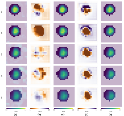 |
Combining variational methods and particle tracking in 4D Flow MRI for displacement artifact compensation
Stefano Buoso1, Hannes Dillinger1, and Sebastian Kozerke1
1Institute for Biomedical Engineering, ETH Zurich, Zurich, Switzerland
We propose a variational method for reconstructing divergence-free velocity fields from 4D Flow MRI data with compensation of spatial displacement artifacts. A standard 4D Flow MRI sequence to measure flow velocities in an expanding flow phantom with Reynolds number of 4000 was used. A variational formulation allows to reconstruct a divergence-free velocity field used to estimate the displacement artefact associated with spin motion between the time point of motion encoding and echo time. The field was used to apply a spatial shift of the original MRI data and compute new divergence-free flow velocities corrected for spatial motion artifacts.
|
|
2275.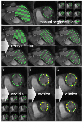 |
Automated mitral valve vortex ring extraction from 4D flow MRI: impact of segmentation
Corina Kräuter1,2, Ursula Reiter1, Clemens Reiter1, Volha Nizhnikava1, Marc Masana3, Albrecht Schmidt4, Michael Fuchsjäger1, Rudolf Stollberger2, and Gert Reiter5
1Department of Radiology, Medical University of Graz, Graz, Austria, 2Institute of Medical Engineering, Graz University of Technology, Graz, Austria, 3Computer Vision Center, Universitat Autònoma de Barcelona, Barcelona, Spain, 4Department of Internal Medicine, Medical University of Graz, Graz, Austria, 5Research and Development, Siemens Healthcare Diagnostics GmbH, Graz, Austria
Automated mitral valve vortex ring evolution analysis from magnetic resonance 4D flow data is feasible. However, time-consuming manual segmentation of the left ventricular blood pool represents a bottleneck. We simulated speed-up and variability of manual segmentation and analyzed the impact on vortex ring parameters. Automated mitral valve vortex ring extraction and analysis yielded robust results, even when applying the end-diastolic segmentation mask to all cardiac phases. Error analysis of vortex ring parameters showed that under-segmentation of the ventricular blood pool should be avoided. Speeding up segmentation by using only the end-diastolic mask enables clinical mitral valve vortex ring analysis studies.
|
|
2276.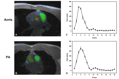 |
Investigation of multi-site reliability and reproducibility study of non-respiratory gated cardiovascular 4D flow MRI
Wen Chen1, Song Yang1, Qian Lu1, Lin Xu1, Yang Fan2, Yong Zhang3, and Changjie Pan3
1Radiology department, Hubei Shiyan Taihe Hospital, Shiyan, China, 2GE healthcare China, Beijing, China, 3Radiology department, Changzhou No2 people hospital, Changzhou, China
Respiratory gating or navigator technique is used in 4D flow imaging to reduce the effect of respiratory motion. Non-respiratory gated 4D flow MRI reduces the overall scan time but may face uncertainty in reproducibility and reliability. In this work, the reliability and reproducibility of non-respiratory gated 4D flow was investigated in 10 patients and 3 different sites in two separate scans. Better measurement consistency was observed intra-site than inter-site. The intra-correlation coefficient results showed excellent to good reproducibility between sites were observed ( ICC > 0.95 for blood flow, ICC > 0.85 for peak). Bland–Altman analysis also showed great test-retest reliability.
|
|
2277.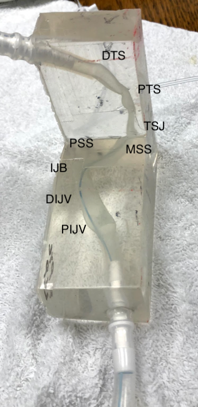 |
Noninvasive Pressure Measurements in the Transverse Sinuses using ICOSA6 4DFlow – an In Vitro Validation with Catheter-based Manometry
Henrik Haraldsson1, Keerthi Valluru1, Megan Ballweber1, Ning Jin2, Sinyeob Ahn3, Evan Kao1, David Saloner1,4, and Matthew Amans1
1University of California, San Francisco, San Francisco, CA, United States, 2Siemens Healthcare, Chicago, IL, United States, 3Siemens Healthcare, San Francisco, CA, United States, 4Veterans Affairs Medical Center, San Francisco, CA, United States
Idiopathic intracranial hypertension is a condition of elevated intracranial cerebrospinal fluid pressure. Transverse sinus stenoses are clearly related to the pathophysiology, and catheter-based manometry is the “gold standard” to assess pressure gradients across these stenoses. The aim of this work is to evaluate the feasibility of assessing these pressure gradients in the intracranial veins using 4DFlow with ICOSA6 motion encoding – which also account for turbulence - and to compare these pressure gradients with catheter-based manometry. Pressure gradient were measured in patient specific phantoms using both 4DFlow and catheter-based manometry. The pressure gradient assessed showed good agreement when accounting for turbulence.
|
|
2278.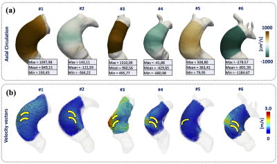 |
A novel, accurate and robust three-dimensional quantification of circulation using finite-element methods in 4D Flow MRI data of thoracic aorta
Julio Sotelo1,2,3, Malenka M Bissell4, Yaxin Jiang4, Hernan Mella1,2,3, Joaquin Mura3,5, and Sergio Uribe1,3,6,7
1Biomedical Imaging Center, Pontificia Universidad Catolica de Chile, Santiago, Chile, 2Department of Electrical Engineering, Pontificia Universidad Catolica de Chile, Santiago, Chile, 3Millennium Nucleus for Cardiovascular Magnetic Resonance, Santiago, Chile, 4Department of Biomedical Imaging Science, Leeds Institute to Cardiovascular and Metabolic Medicine, University of Leeds, Leeds, United Kingdom, 5Department of Mechanical Engineering, Universidad Técnica Federico Santa María, Santiago, Chile, 6Department of Radiology, Pontificia Universidad Catolica de Chile, Santiago, Chile, 7Institute for Biological and Medical Engineering, Schools of Engineering, Medicine and Biological Sciences, Pontificia Universidad Catolica de Chile, Santiago, Chile
The circulation is normally analyzed in a 2D cross-section of the aorta, manually placed. To avoid this problem, evaluate and validate a new methodology based on Finite Elements (FE) to calculate the circulation in three-dimensions, in in-silico models, and in the ascending aorta of a group of volunteers and patients. In in-silico experiment we obtain an RMSE of the circulation values less than 1.6e-6. We also found that significant differences exist between volunteers and patients with a p-value of 0.0283. Our method is straight forward to calculate, accurate and robust throughout different resolutions and noise levels.
|
|
2279.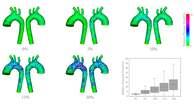 |
A Direct Variational Pressure Estimation Approach for Velocity Data in 4D Flow MRI
Lixing Ren1,2, Xiaowei He2, Dandan Zheng3, and Enhua Wu2,4
1University of Chinese Academy of Sciences, Beijing, China, 2The State Key Lab. of CS, Institute of Software, Chinese Academy of Sciences, Beijing, China, 3Clinical Science, Philips Healthcare, Beijing, China, 4University of Macau, Macau, China
Understanding the nature of the pressure changes is crucial for diagnosis and therapy strategy optimization for aortic aneurysm. 4D flow MRI had offered the opportunity to assess 3D blood flow characteristics. However, the pressure quantification is still a challenge due to the low signal-to-noise ratio and the partial volume effects near the wall. The focus of this work is to address this deficiency by proposing a novel workflow of direct pressure estimation based on simulated noisy 4D flow MRI data.
|
|
2280. |
Impact of Vessel Wall Characteristics on Quantitative 4D-Flow Dynamics in Compliant 3D-Printed Thoracic Aorta Models.
Judith Zimmermann1,2,3, Kathrin Bäumler1, Michael Loecher1,3, Alison Marsden4,5, and Daniel Ennis1,3,5
1Radiology, Stanford University, Stanford, CA, United States, 2Computer Science, Technical University of Munich, Munich, Germany, 3Radiology, Veterans Administration Health Care System, Palo Alto, CA, United States, 4Pediatrics, Stanford University, Stanford, CA, United States, 5Cardiovascular Institute, Stanford, CA, United States
A detailed understanding of flow dynamics in the aorta is of clinical interest and enabled by 4D-flow MRI. This work presents an in vitro flow circuit that embeds a subject-specific 3D-printed compliant aorta model to demonstrate feasibility while also exploring the impact of vessel wall characteristics (thickness, stiffness) and heart rate on quantitative flow dynamics. The experimental setup will be a valuable tool for assessing 4D flow MRI sampling requirements, generating high-quality ground truth data for CFD and FSI validation, as well as studying flow dynamics in different vascular pathologies.
|