
Digital Poster Session
Body: Thoracic and Breast MRI
Body
2281 -2296 Thoracic and Breast MRI - Hyperpolarized Gas Lung MRI
2297 -2312 Thoracic and Breast MRI - Lung & Mediastinum
2313 -2327 Thoracic and Breast MRI - Breast (Machine Learning, Radiomics & Texture Analysis)
2328 -2341 Thoracic and Breast MRI - Breast (DWI)
2342 -2356 Thoracic and Breast MRI - Hyperpolarized, Lung & Mediastinum
2357 -2371 Thoracic and Breast MRI - Lung, Mediastinum & Hyperpolarized
2372 -2386 Thoracic and Breast MRI - Breast (Perfusion & Other Techniques & Topics)
2281.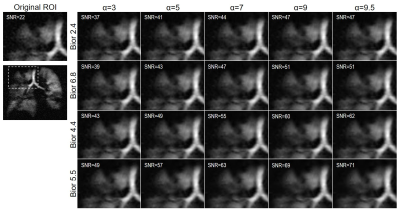 |
Discrete Wavelet Transform (DWT) Denoising of Hyperpolarized 129Xe MR Images
Norman B Konyer1, Nayana Menon1, Paul Polak1,2, Parameswaran Nair3,4, Michael D Noseworthy1,2, and Sarah Svenningsen1,3,4
1Imaging Research Centre, St. Joseph’s Healthcare Hamilton, Hamilton, ON, Canada, 2Department of Electrical and Computer Engineering, McMaster University, Hamilton, ON, Canada, 3Department of Medicine, McMaster University, Hamilton, ON, Canada, 4Firestone Institute for Respiratory Health, St. Joseph's Healthcare Hamilton, Hamilton, ON, Canada
Hyperpolarized 129Xe MRI is an attractive approach to regionally quantify ventilation, however, it often suffers from low SNR. The use of a wavelet transform algorithm was explored as a way to improve SNR while not compromising image detail. Various wavelets and sparsity parameters were explored, all of which provided a significant improvement in SNR while minimally impacting image detail.
|
|
2282.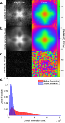 |
Simple Method for Off-Resonance Gas Artifact Removal in 129Xe Gas-Transfer MRI
Matthew Willmering1, Peter Niedbalski1, Laura Walkup1,2, Zackary Cleveland1,2, and Jason Woods1,2
1Cincinnati Children's Hospital Medical Center, Cincinnati, OH, United States, 2University of Cincinnati, Cincinnati, OH, United States
Hyperpolarized dissolved-phase 129Xe imaging allows pulmonary gas transfer processes to be quantified at multiple stages and physical abnormalities in gas-exchange to be identified in subjects with pulmonary disease. However, the technique is hindered by gas-phase contamination due to imperfect frequency-selective excitation of the 50-fold larger pool of gaseous 129Xe. The previous method to remove this contamination required additional echoes. We present a simple post-acquisition method to remove gas-phase contamination with >90% efficiency. This method can be applied to the standard gas-transfer MRI sequence, thus permitting contamination removal for existing data, facilitating more accurate and consistent hyperpolarized 129Xe gas-transfer imaging.
|
|
2283.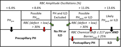 |
Noninvasive Diagnosis of Pulmonary Hypertension with Hyperpolarized 129Xe Magnetic Resonance Imaging and Spectroscopy
Elianna Ada Bier1, Ziyi Wang1, Hamid Chalian2, Joseph Mammarappallil2, John C Nouls2, Leith Rankine3, Junlan Lu3, Bastiaan Driehuys2, and Sudarshan Rajagopal4
1Biomedical Engineering, Duke University, Durham, NC, United States, 2Radiology, Duke University Medical Center, Durham, NC, United States, 3Medical Physics Graduate Program, Duke University, Durham, NC, United States, 4Division of Cardiology, Department of Medicine, Duke University Medical Center, Durham, NC, United States
Using unique signatures in 129Xe MR 3D gas exchange imaging and dynamic spectroscopy, we tested an algorithm to detect pulmonary hypertension (PH). The algorithm was developed using a training cohort (n=105) and tested on 25 subjects whose PH status was determined via right heart catheterization. Two expert readers rated scan quality and interpreted each subject’s 129Xe MRI/MRS to determine PH status. Using the 16 patients for which readers rated scan diagnostic quality as 4-5/5, the diagnostic accuracy was 0.84 (sensitivity=0.78, specificity=0.93). This model demonstrates the ability to distinguish subjects with pre-capillary PH from those without PH or with post-capillary PH.
|
|
2284.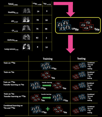 |
Automatic Segmentation of Hyperpolarized Gas MRI via Deep Learning
Joshua R Astley1,2, Alberto M Biancardi1, Paul JC Hughes1, Laurie J Smith1, Helen Marshall1, Grace T Mussell1, James Eaden1, Nicholas D Weatherley1, Guilhem J Collier1, Jim M Wild1, and Bilal A Tahir1,2
1POLARIS, Department of Infection, Immunity & Cardiovascular Disease, University of Sheffield, Sheffield, United Kingdom, 2Department of Oncology and Metabolism, University of Sheffield, Sheffield, United Kingdom
Deep learning (DL)-based segmentation was conducted on a total of 431 3He and 129Xe 3D ventilation images using several training paradigms. Combined 3He and 129Xe training showed a significant improvement over all other DL methods. In the majority of DL models, no significant difference was observed between 3He and 129Xe testing data. Results suggest that 3He and 129Xe images share important features that allow combined 3He and 129Xe DL models to provide superior segmentations to singular gas models. In addition, it was shown that DL generates faster segmentations without the requirement of proton MRI compared to
state-of-the-art model-based solutions.
|
|
2285.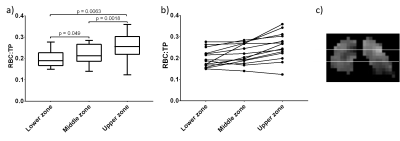 |
Regional hyperpolarised 129-xenon MR spectroscopy and diffusion-weighted MRI in patients with idiopathic pulmonary fibrosis
James A Eaden1,2, Ho-Fung Chan1, Guilhem J Collier1, Nicholas D Weatherley1, Jim Lithgow1, Oliver Rodgers1, Jody FD Bray1, Graham Norquay1, Stephen M Bianchi2, and Jim M Wild1,3
1POLARIS, MRI unit, Department of Infection Immunity and Cardiovascular Disease, University of Sheffield, Sheffield, United Kingdom, 2Academic Directorate of Respiratory Medicine, Sheffield Teaching Hospitals NHS Foundation Trust, Sheffield, United Kingdom, 3Insigneo institute, University of Sheffield, Sheffield, United Kingdom
Preliminary findings are presented from a prospective, longitudinal hyperpolarised 129Xe MRI study of idiopathic pulmonary fibrosis patients. To date, 28 patients have undergone baseline 129Xe DW-MRI and 10 patients have returned for a six-month scan. 14 patients have undergone baseline dissolved 129Xe imaging using IDEAL CSI method. Regional values were produced by separating the lung into three zones. We demonstrate regional differences in 129Xe gas transfer and 129Xe DW-MRI measurements. LmD was found to be more sensitive to longitudinal change than ADC. Over a six-month period, the significant change in mean 129Xe LmD was observed in the lower zone only.
|
|
2286.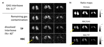 |
Dissolved 129Xe lung MRI with 4-echo IDEAL 3D radial CSI: quantification of regional gas transfer in idiopathic pulmonary fibrosis
Guilhem Jean Collier1, James A Eaden1, Paul J Hughes1, Stephen M Bianchi2, Neil J Stewart1, Nick D Weatherley1, Graham Norquay1, Rolf F Schulte3, and Jim M Wild1
1Department of Infection, Immunity & Cardiovascular Disease, University of Sheffield, Sheffield, United Kingdom, 2Academic directorate of respiratory medicine, Sheffield Teaching Hospitals NHS Foundation Trust, Sheffield, United Kingdom, 3GE Healthcare, Munich, Germany Imaging of the different resonances of dissolved hyperpolarized xenon in the lung is performed using a 4-echo flyback 3D radial implementation of the IDEAL technique and is evaluated in subjects with idiopathic pulmonary fibrosis. Results show: - excellent chemical shift separation of the dissolved peaks and gas contamination removal, - agreement with global spectroscopic and pulmonary function measurements of gas transfer, - reduced imaging gas transfer, especially in the basal lung, when compared to healthy volunteers, - increased cardiogenic modulation of dissolved xenon signal in the blood. Clinical results are in line with previously-published data. |
|
2287.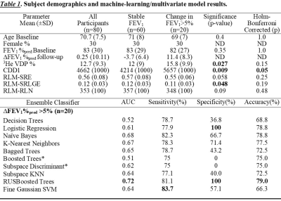 |
Can Hyperpolarized Gas MRI and Machine Learning Predict Longitudinal Changes in Airflow Limitation in Ex-smokers?
Maksym Sharma1, Andrew R. Westcott1, Aaron Fenster1, David G. McCormack2, and Grace Parraga3
1Department of Medical Biophysics, Western University, London, ON, Canada, 2Department of Medicine, Western University, London, ON, Canada, 3Department of Medical Biophysics, Department of Medicine, Western University, London, ON, Canada
Hyperpolarized-gas-MRI provides a way to measure lung ventilation in patients with chronic obstructive pulmonary disease (COPD) in whom progressive worsening of expiratory airflow occurs over time. Progression of COPD is believed to stem from airway wall and lumen changes, airway remodeling or obliteration and emphysema. Our objective was to test machine-learning algorithms trained on hyperpolarized 3He MRI for predicting clinically-relevant FEV1 changes. Novel 3-dimensional adaptations of gray-level run-length-matrices, gap-length-matrices, zone-size-matrices and co-occurrence-matrices were used for feature extraction which lead to the identification of features that predicted changes in airflow limitation (∆FEV1%pred>5%) over a 2.5-year time-period in at-risk and COPD ex-smokers.
|
|
2288.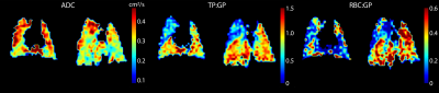 |
Assessment of Gas Exchange for Lung Volume Reduction Surgery in an Emphysema Rat Model Using Xenon Polarization Transfer Contrast Imaging
Faraz Amzajerdian 1,2, Luis Loza1, Tahmina Achekzai1, Federico Sertic1,3, Yi Xin1,2, Harrilla Profka1, Hooman Hamedani1,2, Ian Duncan1, Stephen J Kadlecek1, Kai Ruppert1, and Rahim R. Rizi1
1Radiology, University of Pennsylvania, Philadelphia, PA, United States, 2Bioengineering, University of Pennsylvania, Philadelphia, PA, United States, 3Surgery, University of Pennsylvania, Philadelphia, PA, United States
Lung volume reduction surgery (LVRS) treats emphysema by removing severely emphysematous regions of the lung. Techniques that can better identify which regions of the lung to remove as well as assess the resultant changes in lung function could lead to better surgical outcomes. Here, we explored the feasibility of using hyperpolarized xenon-129 (HXe) MRI for this purpose. Xenon polarization Transfer Contrast (XTC) imaging was used to quantify gas exchange as a metric for assessing the severity and distribution of emphysematous lung regions before and after LVRS.
|
|
2289.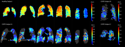 |
Quantifying Gas Exchange Rates Between Red Blood Cells and Airways Using Hyperpolarized Xenon-129
Faraz Amzajerdian 1,2, Stephen J Kadlecek1, Hooman Hamedani1,2, Yi Xin1,2, Ryan Baron1, Ian Duncan1, Maurizio Cereda1,3, Kai Ruppert1, and Rahim R. Rizi1
1Radiology, University of Pennsylvania, Philadelphia, PA, United States, 2Bioengineering, University of Pennsylvania, Philadelphia, PA, United States, 3Anesthesiology and Critical Care, University of Pennsylvania, Philadelphia, PA, United States
Xenon Polarization Transfer Contrast (XTC) imaging is useful for quantifying the exchange rates between gas-phase and dissolved-phase xenon, particularly in emphysematic lungs which experience reduced local tissue and blood volumes. However, this technique does not account for local gas volumes, which can provide important context for interpreting these exchange rates. In this work, a method of incorporating both gas exchange rates and local gas volumes is described.
|
|
2290.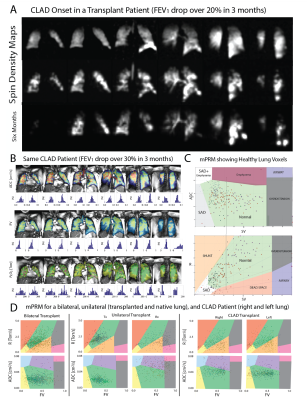 |
A Multiparametric Functional Map for Monitoring and Predicting Chronic Lung Transplant Rejection
Hooman Hamedani1,2, Stephen J Kadlecek1, Ryan Baron1, Faraz Amzajerdian 1,2, Kai Ruppert1, Ian Duncan1, Yi Xin1,2, Mehrdad Pourfathi1, Sarmad Siddiqui1, Luis Loza1, Tahmina Achekzai1, Maurizio Cereda1,3, and Rahim R. Rizi1
1Radiology, University of Pennsylvania, Philadelphia, PA, United States, 2Bioengineering, University of Pennsylvania, Philadelphia, PA, United States, 3Anesthesiology and Critical Care, University of Pennsylvania, Philadelphia, PA, United States
mPRM analysis allowed us to distinguish otherwise healthy transplanted lungs and to classify functional abnormalities in lungs compromised by CLAD.
|
|
2291.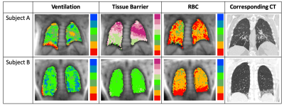 |
Hyperpolarized 129Xe MRI Measures of Gas Exchange in Non-specific Interstitial Pneumonia
David Mummy1, Ziyi Wang2, Elianna Bier2, Robert Tighe3, Bastiaan Driehuys1, and Joseph Mammarappallil1
1Radiology, Duke University, Durham, NC, United States, 2Biomedical Engineering, Duke University, Durham, NC, United States, 3Medicine, Duke University, Durham, NC, United States
Hyperpolarized 129Xe gas exchange MRI was used to compare patients with non-specific interstitial pneumonia (NSIP) and healthy controls. NSIP patients had significantly increased interstitial barrier uptake and decreased red blood cell transfer despite preserved ventilation; in some cases, these abnormalities were present without spatially associated CT findings. These results suggest that 129Xe gas exchange MRI is sensitive to disease activity that is not reflected on 129Xe ventilation alone, or on CT. Ongoing analyses will test the ability of this technique to assess response to targeted therapies and guide clinical decision making in NSIP and other interstitial lung diseases.
|
|
2292.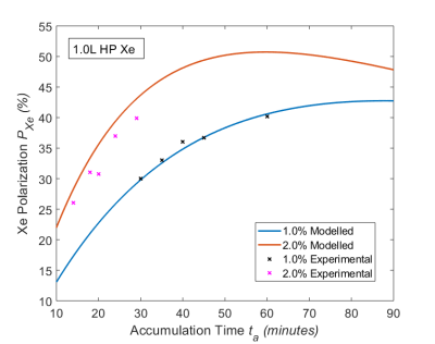 |
Optimizing Hyperpolarized 129Xe Production under Practical Cryogenic Accumulation Conditions
Joseph W Plummer1, Zackary I Cleveland1, Laura Walkup1, Jason W Woods1, Kiarash Emami2, and Andrew Dummer2
1Center for Pulmonary Imaging, Cincinnati Children's Hospital Medical Center, Cincinnati, OH, United States, 2Polarean Imaging PLC, Durham, NC, United States
Following improvements in continuous-flow hyperpolarization technology, 129Xe has shown ever-increasing utility as a pulmonary contrast agent. However, polarization remains constrained by conflicting demands of spin-exchange efficiency and T1 relaxation during cryogenic collection. By collecting 129Xe across a range of practical conditions, we developed model to predict conditions for optimal 129Xe polarization. In many regimes, the benefits of optimal spin exchange efficiency were outweighed by polarization lost to rapid relaxation in polycrystalline 129Xe snow (T1snow = 84 minutes). For 1-L, clinical 129Xe doses, our model suggests optimal polarizations are obtained using accumulation times <30 minutes and relatively rich (2–2.3%) xenon mixtures.
|
|
2293.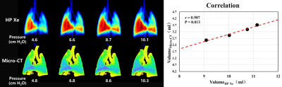 |
Quantitative Assessment of Lung Compliance using both Hyperpolarized 129Xe MRI and Micro-CT
Hongchuang Li1, Ming Zhang1, Haidong Li1, Xiuchao Zhao1, Qiuchen Rao1, Xiaoling Liu1, Yeqing Han1, Xianping Sun1, Xinrui Wang2, Chaohui Ye1, Xin Lou2, and Xin Zhou1
1Wuhan Institute of Physics and Mathematics, National Center for Magnetic Resonance in Wuhan, Wuhan Institute of Physics and Mathematics, Innovation Academy of Precision Measurement Science and Technology, Chinese Academy of Sciences-Wuhan National Laboratory for Optoelectronics, Wuhan, China, 2Department of Radiology, Chinese PLA General Hospital, Beijing, P. R. China, Beijing, China
Lung compliance is commonly used in clinical diagnosis of pulmonary fibrosis and emphysema, and it is generally measured using pulmonary function tests (PFTs) in clinic. In this study, the imaging methods including both hyperpolarized 129Xe MRI and micro-CT were used to quantify lung compliance in rats, and the significant correlation was found between these two imaging methods. Our study also demonstrates the feasibility of measuring lung compliance using hyperpolarized 129Xe MRI, which would contribute to fully understanding the lung function changes caused by the pulmonary diseases.
|
|
2294.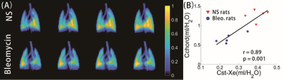 |
Quantitative Evaluation of Changes in Lung Compliance Caused by Fibrosis using Hyperpolarized Gas MRI
Ming Zhang1, Haidong Li1, Hongchuang Li1, Xiuchao Zhao1, Yeqing Han1, Xianping Sun1, Chaohui Ye1, and Xin Zhou1
1National Center for Magnetic Resonance in Wuhan, Wuhan Institute of Physics and Mathematics, Innovation Academy of Precision Measurement Science and Technology, Chinese Academy of Sciences-Wuhan National Laboratory for Optoelectronics, Wuhan, China
Pulmonary compliance is generally measured using pulmonary function tests (PFTs) in clinical practice. In this study, we proposed a strategy of quantifying pulmonary compliance (Cst-Xe) using hyperpolarized (HP) 129Xe MRI. The Cst-Xe reliably distinguishes the healthy and bleomycin rats, and was correlated well with pulmonary compliance via PFTs and microstructure parameters via HP 129Xe DWI, respectively. The results have demonstrated the feasibility of HP 129Xe MRI in quantitatively and non-invasively assessing the pulmonary compliance, and also have improved the understanding of the lung compliance changes in fibrosis.
|
|
2295. |
Imaging Hyperpolarized Xe Changes in the Lung Over a Breathing Cycle Using a Dynamic CSI
Tahmina Achekzai1, Stephen J Kadlecek1, Kai Ruppert1, Luis Loza1, Faraz Amzajerdian 1,2, Yi Xin1,2, Ian Duncan1, and Rahim R. Rizi1
1Radiology, University of Pennsylvania, Philadelphia, PA, United States, 2Bioengineering, University of Pennsylvania, Philadelphia, PA, United States
We demonstrated a new imaging technique capable of quantifying Xe gas uptake into airways and parenchyma over the course of a breathing cycle. Current imaging techniques acquire entire images at a single breathing point. These methods may not replicate organ function during tidal breathing, and could potentially miss diagnostically important dynamic information depending on the time of acquisition. This sequence traverses the k-space over a series of 576 breaths to produce 12 images at different time points. Three healthy rats were imaged, and Xe signal was found to fluctuate at different times and amplitudes depending on respective lung compartment.
|
|
2296. |
Hyperpolarised 129Xe Imaging Using A 0.5T Upright Paramed MRI Scanner.
James Harkin1, Robert Irwin1, Joshua McAteer1, Penny Gowland2, Michael Barlow3, and Ian Hall3
1University of Nottingham, Nottingham, United Kingdom, 2Science, University of Nottingham, Nottingham, United Kingdom, 3Medicine & Health Sciences, University of Nottingham, Nottingham, United Kingdom
Using a multinuclear 0.5T Paramed Medical Systems MROpen Upright MRI scanner, 129Xe images have been acquired within the duration of a breath hold. An active SAR monitoring system has been implemented to allow for the safe collection of data within SAR limits of chest imaging. There is the intention of applying compressed sensing to the current 129Xe sequence. The upright and open design of the scanner makes imaging more comfortable for patients and allows for imaging in later stages of respiratory diseases.
|
 View the Poster
 Watch the Video
2297.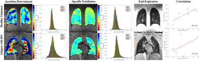 |
Pulmonary Ventilation Analysis Using Motion-Resolved Ultrashort Echo (UTE) MRI
Fei Tan1, Xucheng Zhu1, and Peder E. Z. Larson1,2
1Graduate Program in Bioengineering, UC Berkeley - UCSF, San Francisco, CA, United States, 2Department of Radiology and Biomedical Imaging, University of California, San Francisco, San Francisco, CA, United States
Pulmonary ventilation analysis using motion-resolved 1H UTE MRI with tissue deformation-based and signal intensity-based methods was conducted for the first time. The ventilation maps derived from both approaches are consistent with each other on healthy volunteers and a pediatric subject with lung abnormality.
|
|
2298.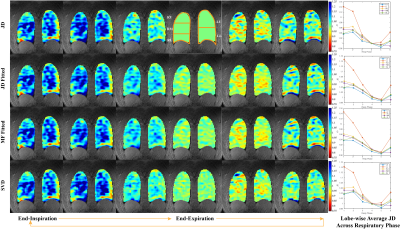 |
Dynamic 3D Ventilation Maps with Phase-Resolved Ultrashort Echo Time (UTE) MRI
Fei Tan1, Xucheng Zhu1, and Peder E. Z. Larson1,2
1Graduate Program in Bioengineering, UC Berkeley - UCSF, San Francisco, CA, United States, 2Department of Radiology and Biomedical Imaging, University of California, San Francisco, San Francisco, CA, United States
In this abstract, we propose a novel respiratory phase-resolved reconstruction for free-breathing pulmonary 1H UTE MRI. We compute the tissue deformation-based ventilation maps and further use the parametric and non-parametric fitting methods to reduce the noise and registration errors.
|
|
2299. |
Feasibility study of nodule conspicuity for lung screening using ultrashort echo time MRI
Saori Koshino1,2, Takeharu Yoshikawa3, Yukihiro Nomura3, Soichiro Miki3, Shouhei Hanaoka2, Takeyuki Watadani2, Naoto Hayashi3, and Osamu Abe1,2
1Department of Radiology, Graduate School of Medicine, The University of Tokyo, Tokyo, Japan, 2Department of Radiology, The University of Tokyo Hospital, Tokyo, Japan, 3Department of Computational Diagnostic Radiology and Preventive Medicine, The University of Tokyo Hospital, Tokyo, Japan
Pulmonary MRI with ultrashort echo time (UTE) is expected to be an alternative to low-dose CT for lung screening. In this study, the feasibility of breath-hold (BH) and respiratory triggered (RT) scans with UTE was investigated. In total, 229 lung MRI examinations that were composed of 136 examinations with a solid nodule (SN) and 93 examinations with a ground-glass nodule (GGN) were evaluated. Both results of qualitative and quantitative assessment suggested that SN was more clearly depicted than GGN. BH scan in pulmonary MRI should be more suitable for lung screening than RT scan.
|
|
2300. |
Imaging of lung parenchyma: comparison of UTE3D, ZTE3D vnav, and ZTE4D acquisitions
Dorottya Papp1, Piotr A. Wielopolski1, Bernadette B.L.J. Elders2, Pierluigi Ciet1, Mika W. Vogel3, Gyula Kotek1, and Juan Antonio Hernandez-Tamames1
1Radiology and Nuclear Medicine, Erasmus Medical Center, Rotterdam, Netherlands, 2Pediatrics, Erasmus Medical Center, Rotterdam, Netherlands, 3GE Healthcare, Hoevelaken, Netherlands
MRI has recently emerged as a potential clinical tool that can produce high resolution images of structural changes in the lung thanks to the use of ultrashort TE readouts without the ionizing radiation of CT scans [3-6]. Thanks to this developments, pediatric cystic fibrosis (CF) patients can undergo routine monitoring with CT like quality [1]. In this study we investigate three different MRI sequences (UTE, ZTE3D vnav and ZTE 4D). In general, all sequences perform reasonably well though ZTE3D vnav seems to provide the best performance for CF monitoring.
|
|
2301.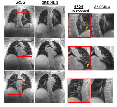 |
Motion-Corrected Pulmonary Imaging Using Spiral UTE Acquisition and Accelerated GROG-XD-GRASP Reconstruction
Eddy Solomon1, Li Feng2, Benkert Thomas 3, Moritz Schneider 4, Kai Tobias Block1, Daniel K Sodickson1, and Hersh Chandarana1
1Radiology, New York University School of Medicine, New York, NY, United States, 2Radiology, Icahn School of Medicine at Mount Sinai, New York, NY, United States, 3Siemens Healthcare GmbH, Erlangen, Germany, 4Radiology, University Hospital LMU Munich, Munich, Germany
Despite recent advances in imaging techniques, respiratory motion remains a major challenge in lung MRI. This work explores ultrashort echo time (UTE) technique based on a stack-of-spirals trajectory for lung MRI. To achieve motion-resolved and high-resolution pulmonary MRI during free breathing, spiral arms were grouped into different respiratory states based on self‐navigator signals. These data were reconstructed with an eXtra-Dimensional (XD) compressed-sensing algorithm that use self-calibrating GRAPPA operator gridding (GROG). The proposed reconstruction pipeline enabled motion-compensated imaging revealing fine details of pulmonary parenchymal anatomy.
|
|
2302.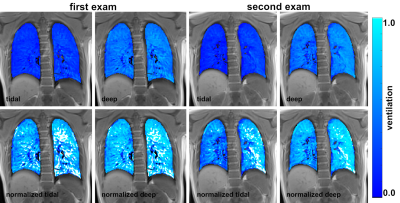 |
Reproducibility of functional lung MRI using non-contrast-enhanced 3D-UTE MRI in breath-hold technique
Corona Metz1, Julius Frederik Heidenreich1, Andreas Max Weng 1, Thomas Benkert2, Herbert Köstler1, Thorsten Alexander Bley1, and Simon Veldhoen1
1Department of Diagnostic and Interventional Radiology, University Hospital, Würzburg, Germany, 2Application Development, Siemens Healthcare GmbH, Erlangen, Germany
Functional and morphologic assessment of lung disease is to date performed separately using pulmonary function testing and mostly radiation-based imaging. UTE MRI is a promising tool for radiation-free pulmonary imaging. Pre-existing invasive functional MRI techniques have been transferred to UTE MRI. For non-invasive combined functional and morphologic imaging, radiation-free 3D-UTE MRI acquired in breath-hold and using stack-of-spirals trajectories enables ventilation measurements with high reproducibility for both tidal and deep fractional ventilation.
|
|
2303.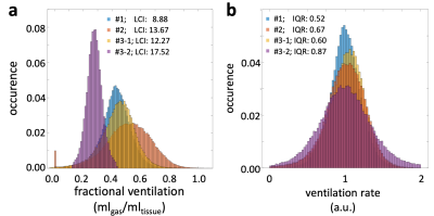 |
Assessment of the Ventilation Rate allows for Interindividual Comparability of Ventilation Inhomogeneity in Functional 3D-UTE Lung MRI
Julius Frederik Heidenreich1, Andreas Max Weng1, Corona Metz1, Thomas Benkert2, Helge Hebestreit3, Thorsten Alexander Bley1, Herbert Köstler1, and Simon Veldhoen1
1Department of Diagnostic and Interventional Radiology, University Hospital Wurzburg, Wurzburg, Germany, 2Application Development, Siemens Healthcare GmbH, Würzburg, Germany, 3Department of Pediatrics, University Hospital Wurzburg, Wurzburg, Germany
Lung imaging with single-breath-hold 3D-UTE MRI allows for lung function analyses such as fractional ventilation (FV). Despite being a promising technique for unenhanced functional imaging, its limited interindividual comparability due to variable breathing depths has been a major drawback. Referencing the voxelwise FV to the mean FV (entire lung) provides the ventilation rate, which can be used for inter-/intraindividual comparison. We demonstrate that the distribution width of the ventilation rate strongly correlates to the ventilation inhomogeneity in patients with cystic fibrosis as measured by the lung clearance index, which makes it a promising parameter for analysis of lung ventilation.
|
|
2304. |
Comparison of Capability for Therapeutic Effect Prediction between CEST Imaging and FDG-PET/CT in NSCLC Patients with Chemoradiotherapy
Yoshiharu Ohno1,2, Masao Yui3, Daisuke Takenaka4, Yoshimori Kassai3, Kazuhiro Murayama1, and Takeshi Yoshikawa2
1Radiology, Fujita Health University School of Medicine, Toyoake, Japan, 2Radiology, Kobe University Graduate School of Medicine, Kobe, Japan, 3Canon Medical Systems Corporation, Otawara, Japan, 4Diagnostic Radiology, Hyogo Cancer Center, Akashi, Japan
No major repots have been evaluated the capability for therapeutic effect evaluation or prediction by chemical exchange saturation transfer (CEST) imaging and compared with FDG-PET/CT. We hypothesized that CEST imaging had equal or better potential for therapeutic effect evaluation and prediction in non-small cell lung cancer (NSCLC) patients with conservative therapy, when compared with FDG-PET/CT. The purpose of this study was to directly and prospectively compare the capability for prediction of therapeutic effect for chemoradiotherapy between CEST imaging and FDG-PET/CT in NSCLC patients.
|
|
2305.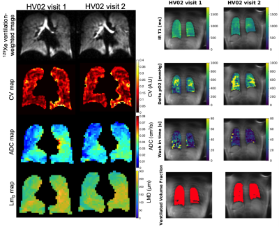 |
Repeatability and correlation of hyperpolarized xenon-129 and oxygen enhanced MRI parameters in healthy volunteers
Paul J.C. Hughes1, Marta Tibiletti2, Matthew J. Heaton2, Ho-Fung Chan1, Guilhem J. Collier1, Matthew Austin1, Laurie J. Smith1,3, Jim Lithgow1, Josephine H. Naish2,4, Jim M. Wild1,5, and Geoff J.M. Parker2,6
1POLARIS, Department of Infection, Immunity and Cardiovascular Disease, The University of Sheffield, Sheffield, United Kingdom, 2Bioxydyn, Manchester, United Kingdom, 3Sheffield Children's Hospital NHS Foundation Trust, Sheffield, United Kingdom, 4Division of Cardiovascular Sciences, University of Manchester, Manchester, United Kingdom, 5Insigneo Institute for in silico Medicine, The University of Sheffield, Sheffield, United Kingdom, 6Centre for Medical Image Computing, University College London, London, United Kingdom
Hyperpolarised xenon-129 MRI has shown utility in longitudinal assessment of lung structure and function, and has been shown to be a repeatable method. Oxygen enhanced MRI is a cheaper method of imaging different aspects of lung function. This work aimed to assess a single-centre repeatability of multiple metrics from both imaging methods in volunteers, and assess any correlations between xenon-129 and oxygen enhanced metrics of lung function.
|
|
2306.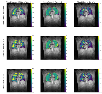 |
On gamma variate fitting for perfusion quantification in the lung.
Marta Tibiletti1, Jo Naish1,2, Matthew J. Heaton1, Paul JC Hughes3, Jim Wild3, and Geoff JM Parker1,4
1Bioxydyn Ltd, Manchester, United Kingdom, 2Division of Cardiovascular Sciences, University of Manchester, Manchester, United Kingdom, 3POLARIS, , Academic Radiology, Department of Infection, Immunity and Cardiovascular Disease, The University of Sheffield, Sheffield, United Kingdom, 4Centre for Medical Image Computing, University College London, London, United Kingdom
Application of first pass imaging to the lung is a promising technique. The majority of works apply a Singular Value Decomposition (SVD) with or without a prior fit to a gamma variate function to the concentration-time curve. In this study we compare lung perfusion quantification with and without 2 versions of the gamma-variate fitting in data from 66 patients with Interstitial lung disease. We found that the presence and choice of gamma variate fitting has an influence on perfusion parameters, but this is generally very small, except for mean transit time, suggesting that these approaches are broadly equivalent in practice.
|
|
2307.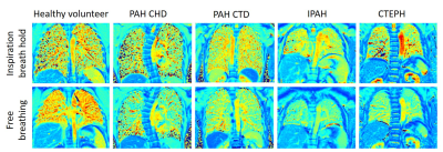 |
Free breathing and breath hold lung T1 is significantly different in pulmonary hypertension subgroups.
Laura Saunders1, Andy Swift1, and Jim Wild1
1University of Sheffield, Sheffield, United Kingdom
This work explores lung T1 mapping as a contrast-free, radiation-free method of differentiating subgroups in pulmonary hypertension, measured during both free breathing and inspiration breath hold acquisitions. Inspiration lung T1 mapping is significantly different between patients with pulmonary arterial hypertension (PAH) and chronic thromboembolic PH (CTEPH), as well as between patients with idiopathic PAH and PAH due to connective tissue disease (CTD) and congenital heart disease (CHD). Free breathing lung T1 mapping was also significantly different between patients with IPAH and PAH CTD and CHD and may be useful in patients who struggle to maintain breath hold due to dyspnoea.
|
|
2308.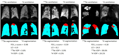 |
129Xe and free-breathing 1H ventilation MRI in patients with cystic fibrosis
Helen Marshall1, Laurie J Smith1, Alberto M Biancardi1, Guilhem J Collier1, Andreas Voskrebenzev2, Jens Vogel-Claussen2, and Jim M Wild1
1POLARIS, Academic Radiology, University of Sheffield, Sheffield, United Kingdom, 2Institute for Diagnostic and Interventional Radiology, Hannover Medical School, Hannover, Germany
Free-breathing 1H MRI offers a means of producing surrogate ventilation images without use of a contrast agent but more validation against direct ventilation imaging methods such as hyperpolarised gas MRI is required. 24 patients with cystic fibrosis were scanned with 1H and 129Xe ventilation MRI. There were strong correlations between 1H ventilation defect percentage (VDP), 129Xe VDP, lung clearance index and FEV1. 1H VDP was typically smaller than 129Xe VDP with wide limits of agreement (LOA) (bias 2.8%, LOA -13.7%, 19.3%). Both similarities and differences between 129Xe and 1H ventilation images were observed
across the range of disease severity.
|
|
2309.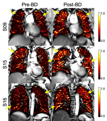 |
Free breathing 1H MRI Specific Ventilation: Sensitivity to Bronchodilator and Airway Dysfunction in Severe Asthma
Dante PI Capaldi1, Norman B Konyer2, Melanie Kjarsgaard3, Michael D Noseworthy2,4, Parameswaran Nair3,5, and Sarah Svenningsen2,3,5
1Department of Radiation Oncology, Stanford University School of Medicine, Stanford, CA, United States, 2Imaging Research Centre, St. Joseph’s Healthcare Hamilton, Hamilton, ON, Canada, 3Firestone Institute for Respiratory Health, St. Joseph's Healthcare Hamilton, Hamilton, ON, Canada, 4Department of Electrical and Computer Engineering, McMaster University, Hamilton, ON, Canada, 5Department of Medicine, McMaster University, Hamilton, ON, Canada
Free-breathing 1H MRI–derived specific ventilation (SV) provides a way to visualize regional ventilation, which has been shown to be reduced in asthmatics compared to healthy volunteers. We hypothesized that free breathing MRI–derived SV in severe asthmatics would be sensitive to airway bronchodilation and airway dysfunction, and hence our objective was to measure SV using 1H MRI in healthy volunteers and severe asthmatics pre- and post-bronchodilator and to evaluate the relationship with airway oscillometry measurements. Preliminary results in severe-asthmatics showed that free-breathing 1H MRI–derived SV is sensitive to bronchodilator therapy and may reflect regional small airway pathology.
|
|
2310.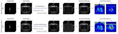 |
Low-rank and sparse matrix decomposition for accelerated non-contrast-enhanced functional lung MRI
Efe Ilicak1, Jascha Zapp1, Lothar R. Schad1, and Frank Zoellner1
1Computer Assisted Clinical Medicine, Heidelberg University, Mannheim, Germany
Lung functions have significant clinical value for diagnosis of pulmonary diseases. Fourier Decomposition is a non-contrast-enhanced method for assessing pulmonary functions from time-resolved images. However, its performance depends on temporal resolution. Here we propose two compressed sensing reconstruction strategies based on low-rank and sparse matrix decomposition. Retrospective demonstrations on in vivo acquisitions demonstrate the performance of these techniques, enabling improved scan efficiency without degrading image quality.
|
|
2311.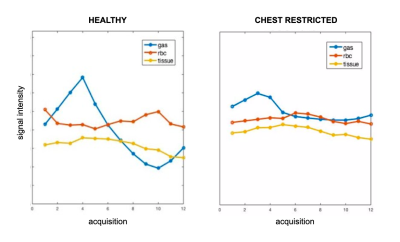 |
Quantification of Hyperpolarized 129-Xe signal fluctuations in a rodent chest restriction model
Tahmina S Achekzai1, Steve J Kadlecek1, Yi Xin2,3, Federico Sertic2,4, Luis Loza2, Faraz Amzajerdian 2,3, Kai Ruppert2, Harrilla Profka2, Ian Duncan2, and Rahim R. Rizi2
1UPenn, Philadelphia, PA, United States, 2Radiology, University of Pennsylvania, Philadelphia, PA, United States, 3Bioengineering, University of Pennsylvania, Philadelphia, PA, United States, 4Surgery, University of Pennsylvania, Philadelphia, PA, United States
We demonstrated a new imaging technique capable of quantifying Xe gas uptake into airways and parenchyma over the course of a breathing cycle. Most current imaging techniques acquire entire images at a single breathing point. These methods may not replicate organ function during tidal breathing, and could potentially miss diagnostically important dynamic information depending on the time of acquisition. This sequence traverses the k-space over a series of 576 breaths to produce 12 images at different time points. Three healthy rats were imaged, and Xe signal was found to fluctuate at different times and amplitudes depending on respective lung compartment.
|
|
2312. |
Applicability of automated cardiac phase sorting for phase resolved functional lung (PREFUL) MRI
Lea Behrendt1,2, Andreas Voskrebenzev1,2, Filip Klimeš1,2, Marcel Gutberlet1,2, Hinrich Winther1, Till Kaireit1,2, Tawfik Moher Alsady1,2, Gesa Pöhler1,2, Frank Wacker1,2, and Jens Vogel-Claussen1,2
1Institute of Diagnostic and Interventional Radiology, Hannover Medical School, Hannover, Germany, 2Biomedical Research in Endstage and Obstructive Lung Disease Hannover (BREATH), German Center for Lung Research (DZL), Hannover, Germany
Phase-resolved functional lung (PREFUL) MRI is a promising contrast agent-free 1H method to derive information about perfusion dynamics in free breathing. The analysis includes the segmentation of a main vessel to determine the perfusion phase for reconstruction of a full cardiac cycle. Since manual segmentation can be challenging and time-consuming, an algorithm for automated phase sorting is desirable. In this study such an algorithm is proposed, tested in 34 patients with CF and CTEPH and compared to manual phase sorting. The algorithm was able to perform segmentation in all cases and showed improved phase sorting compared to manual segmentation.
|
2313.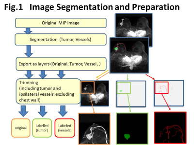 |
Automatic Segmentation of Tumor-related Vessels of Breast Cancer on Ultrafast DCE MRI using U-Net
Masako Kataoka1, Takuto Fukutome2, Tomohiro Takemura2, Kango Kawase2, Kojiro Yano3, Maya Honda4, Mami Iima4, Akane Ohashi4, Masakazu Toi5, and Kaori Togashi4
1Radiology (Diagnostic Imaging and Nuclear Medicine), Kyoto Univ. Hospital, Kyoto, Japan, 2Faculty of Medicine, Kyoto University, Kyoto, Japan, 3Osaka Institute of Technology, Osaka, Japan, 4Kyoto University Graduate School of Medicine, Kyoto, Japan, 5Kyoto University, Kyoto, Japan
We aimed to develop a system for automatic segmentation of tumor-related vessels on ultrafast dynamic contrast (UF-DCE) MRI using U-net. Training set consisted of image dataset of 20 MIP images obtained from -15 to +60 sec after contrast injection from 59 patients. Exclusion criteria were those with poor image quality. The dice similarity coefficient was above 0.8 for training set, 0.6 for validation set. Careful analysis of failed cases revealed that inaccurate segmentation of the vessel were caused by low-contrast images, noisy-images, artifact, bright skin line due to incomplete fat suppression, and non-mass enhancement that may mimic vasculature.
|
|
2314.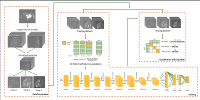 |
Classification of Breast Magnetic Resonance Imaging Using 3D Convolution Neural Network: A Pilot Study
Cindy Xue1, Gladys Lo1, Victor Ai1, Oilei O.L Wong1, Max W.K Law1, and Jing Yuan1
1Hong Kong Sanatorium and Hospital, Hong Kong, Hong Kong
BIRADS classification is one of the standard of reporting breast MRI. It reveals the information about the likelihood of cancer and management recommendation for the patients. In this study, we aimed to develop and evaluate a 3D convolution neural network (CNN) for breast MRI BIRADS classification. This 3D CNN network was evaluated and it achieved overall 90% classification accuracy. In particular, the network also has high sensitivity (100%) of highly suspicious of malignancy findings. The results suggested that a deep learning-based computerized tool might be useful in BIRADS-MRI classification.
|
|
2315.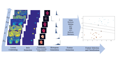 |
Diffusion-Weighted MRI-Based Quantitative Markers for Characterizing Breast Cancer Lesions Using Machine Learning
Rahul Mehta1,2, Muge Karaman1,2, Yangyang Bu3,4, Zheng Zhong1,2, Guangyu Dan1,2, Shiwei Wang3,4, Changyu Zhou3,4, Weihong Hu3,4, X. Joe Zhou1,2,5, and Maosheng Xu3,4
1Center for Magnetic Resonance Research, University of Illinois at Chicago, Chicago, IL, United States, 2Department of Bioengineering, University of Illinois at Chicago, Chicago, IL, United States, 3The First Clinical Medical College, Zhejiang Chinese Medical University, Hangzhou, China, 4The First Affiliated Hospital of Zhejiang Chinese Medical University, Hangzhou, China, 5Departments of Radiology and Neurosurgery, University of Illinois at Chicago, Chicago, IL, United States
We investigate the quantitative markers obtained from the parameters of two diffusion-weighted imaging (DWI) models, continuous-time random-walk (CTRW) and intravoxel incoherent motion (IVIM) models, for differentiating malignant and benign breast lesions. The quantitative markers are extracted from the histograms of each parameter, and then the statistical importance of each marker is determined using a feature importance algorithm. Our results show the Gradient Boosted Classifier (GBC) achieves optimal performance using the top quantitative markers. The statistical histogram features from the parameters of CTRW and IVIM models can be used in a GBC to provide a new avenue in breast cancer diagnosis.
|
|
2316.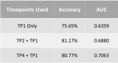 |
Artificial Intelligence Prediction of Breast Cancer Pathologic Complete Response from Axillary Lymph Node MRIs
Janice Yang1,2, Thomas Ren1, Hongyi Duanmu3, Pauline Huang1, Renee Cattell1, Haifang Li1, Fusheng Wang3, and Tim Q Duong1
1Radiology, Stony Brook University, Stony Brook, NY, United States, 2Dougherty Valley High School, San Ramon, CA, United States, 3Computer Science, Stony Brook University, Stony Brook, NY, United States
Breast cancer patient response to neoadjuvant chemotherapy cannot be accurately predicted or monitored through imaging, leading to unnecessary treatment and sentinel lymph node biopsies. We developed convolutional neural networks to predict pathologic complete response utilizing a combination of axillary lymph node MRIs from before and during treatment. 3-fold cross validation reveals that the model trained on scans before and after the first cycle of neoadjuvant chemotherapy performed best with an accuracy of 81.17%. These results point to improved predictive performance of early imaging markers in axillary lymph nodes and encourages its implementation to aid treatment planning and improve prognosis.
|
|
2317.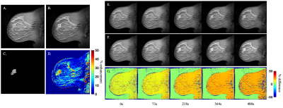 |
Implementing compressed sensing with deep image prior to reconstruct undersampled dynamic contrast-enhanced MRI data of the breast
Kalina P Slavkova1, Julie C DiCarlo2,3, David M Van Veen4, Anum K Syed5, Ajil Jalal4, John Virostko6, Anna G Sorace7, Alexandros G Dimakis4,8, and Thomas E Yankeelov2,5,6,9
1Physics, University of Texas at Austin, Austin, TX, United States, 2Oden Institute for Computational Engineering and Sciences, University of Texas at Austin, Austin, TX, United States, 3Livestrong Cancer Institutes, University of Texas at Austin, Austin, TX, United States, 4Electrical and Computer Engineering, University of Texas at Austin, Austin, TX, United States, 5Biomedical Engineering, University of Texas at Austin, Austin, TX, United States, 6Medicine, University of Texas at Austin, Austin, TX, United States, 7Biomedical Engineering, University of Alabama at Birmingham, Birmingham, AL, United States, 8Wireless Networking and Communications Group, University of Texas at Austin, Austin, TX, United States, 9Oncology, University of Texas at Austin, Austin, TX, United States
We evaluate the ability of the compressed sensing with deep image prior (CS-DIP) algorithm to reconstruct undersampled dynamic contrast-enhanced MRI data of the breast. The performance of the reconstruction is evaluated by comparing quantitative parameters computed from the reconstructed data to the original parameter values computed from fully-sampled data. We hypothesize that CS-DIP will enable dramatically fewer k-space measurements, thereby allowing for higher temporal (while maintaining spatial) resolution of breast MRI scans.
|
|
2318.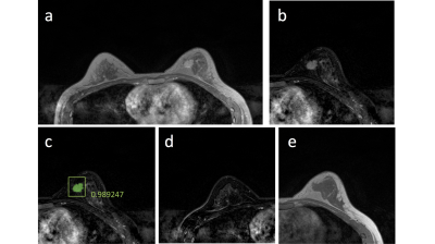 |
Automatic Detection and Segmentation of Breast Cancer on MRI Using Mask R-CNN Trained on Non-Fat-Sat Images and Tested on Fat-Sat Images
Yang Zhang1, Jiejie Zhou2, Youngjean Park3, Siwa Chan4, Meihao Wang2, Min Jung Kim3, Kai-Ting Chang1, Peter Chang1, Daniel Chow1, Jeon-Hor Chen1,5, and Min-Ying Su1
1Department of Radiological Science, University of California, Irvine, CA, United States, 2Department of Radiology, First Affiliate Hospital of Wenzhou Medical University, Wenzhou, China, 3Department of Radiology and Research Institute of Radiological Science, Severance Hospital, Yonsei University College of Medicine, Seoul, Korea, Republic of, 4Department of Medical Imaging, Taichung Tzu-Chi Hospital, Taichung, Taiwan, 5Department of Radiology, E-Da Hospital and I-Shou University, Kaohsiung, Taiwan
The mask R-CNN algorithm was implemented to search entire images to identify suspicious lesions for further evaluation of malignancy probability. One training (N=98, Siemens 1.5T, non-fat-sat) and two independent testing (N=241, Siemens 3T, non-fat-sat; and N=91, GE 3T, fat-sat) datasets were used. The pre-contrast and subtraction image, and the subtraction image of the contralateral breast, were used as three inputs. The training set had a total of 1353 positive slices (containing lesion), 8055 negative slices without lesion. The 10-fold cross-validation showed accuracy=0.80 and mean DSC= 0.82. The accuracy was 0.73 and 0.62 for two testing datasets, lower for fat-sat images.
|
|
2319.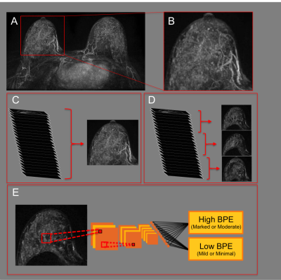 |
Background Parenchymal Enhancement (BPE) classification on Breast MRI using Deep Learning
Sarah Eskreis-Winkler1, Katja Pinker1, Donna D'Alessio1, Katherine Gallagher1, Nicole Saphier1, Danny Martinez1, Elizabeth Sutton1, and Elizabeth Morris1
1Memorial Sloan Kettering Cancer Center, New York, NY, United States
Breast cancer risk in high risk women is significantly increased for those with high background parenchymal enhancement (BPE) compared to low BPE, yet there is only fair to moderate inter-rater agreement for BPE assessment among radiologists, limiting the use of BPE as a marker of cancer risk. We developed a deep learning algorithm that classifies BPE with high accuracy. The algorithm works best when sub-MIPs, not MIPs, are used as network inputs. The algorithm has potential to autopopulate breast MRI reports in our breast imaging clinic, and ultimately to standardize BPE as a marker of breast cancer risk.
|
|
2320.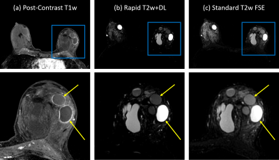 |
Shortening Diagnostic T2w Breast Protocols to Capitalize on the Benefits of a Deep Learning Reconstruction
Timothy J Allen1, Leah C Henze Bancroft2, Roberta M Strigel1,2,3, Ty Cashen4, Orhan Unal2, Frank R Korosec1,2, Ping Wang1, Lloyd Estkowski4, Fred Kelcz2, Amy M Fowler1,2,3, R Marc Lebel4, and James Holmes2
1Medical Physics, University of Wisconsin-Madison, Madison, WI, United States, 2Radiology, University of Wisconsin-Madison, Madison, WI, United States, 3Carbone Cancer Center, University of Wisconsin-Madison, Madison, WI, United States, 4Global MR Applications and Workflow, GE Healthcare, Madison, WI, United States
A SSFSE-based fast acquisition was used in conjunction with a vendor-supplied, deep learning-based reconstruction to obtain T2-weighted images (Rapid T2w+DL) for diagnostic breast MRI. Radiologists compared the Rapid T2w+DL images to standard-of-care T2w FSE images assessing both image quality and utility in completing a typical clinical task. The fast acquisition provided a reduction in average scan time from 3:35 (min:sec) to 1:16 (64%). Deep learning was used to alleviate issues associated with the fast protocol (e.g. blurring and reduced SNR). Rapid T2w+DL images allowed for completion of the clinical task with results comparable to those obtained with standard T2w images.
|
|
2321.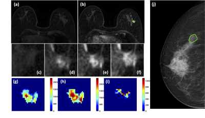 |
Comparison of Breast Cancer Diagnostic Performance Using Radiomics Models Built Based on Features Extracted from DCE-MRI and Mammography
Jiejie Zhou1, Yang Zhang2, Kyoung Eun Lee3, Jeon-Hor Chen2, Xiaxia He1, Nina Xu1, Shuxin Ye1, Ouchen Wang1, Jiance Li1, Yezhi Lin4, Meihao Wang1, and Min-Ying Su2
1First Affiliate Hospital of Wenzhou Medical University, Wenzhou, China, 2University of California, Irvine, CA, United States, 3Inje University Seoul Paik Hospital, Seoul, Korea, Republic of, 4Wenzhou Medical University, Wenzhou, China
A total of 89 patients receiving both DCE-MRI and mammography were analyzed, including 56 malignant and 33 benign lesions. The 3D tumor mask on MRI was generated using computer algorithms. A total of 99 texture and histogram features were extracted from three DCE parameters maps. The suspicious area on mammography was outlined using MRI findings as guidance, and a similar radiomics method was applied to extract features from the mass and the margin. Random forest was applied to select features for building diagnostic models. The overall accuracy was 0.80 for MRI, 0.75 for mammography, and improved to 0.85 when combined.
|
|
2322.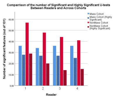 |
Inter-reader variability in Breast MRI Radiomics
Michael James Fox1, Roberto Lo Gullo2, Isaac Daimiel Naranjo2, Carolina Saccarelli2, Almir Bitencourt2, Katja Pinker-Domenig2,3, and Peter Gibbs2
1Sloan Kettering Institute, Memorial Sloan Kettering Cancer Center, New York, NY, United States, 2Department of Radiology, Memorial Sloan Kettering Cancer Center, New York, NY, United States, 3Department of Biomedical Imaging and Image-guided Therapy, Medical University of Vienna, Wien, Austria
The purpose of this study was to investigate the effect of choice of radiologist on radiomics in breast MRI. Two cohorts of 100 patients each had breast lesions segmented by 4 independent radiologists, and these segmented images used to produce 101 radiomic features. over 90% of all features were found to have good or excellent agreement across readers, and a similar number of highly significant U-test were found for all readers. Differences between reader interpretation exist, however, they would not appear to effect the ability to distinguish between benign or malignant lesions.
|
|
2323.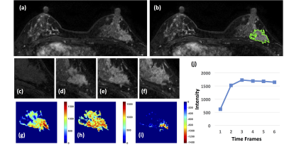 |
Diagnosis of Non-Mass-Like Enhancement Lesions on DCE-MRI by Using Quantitative Radiomics and Radiologists’ BI-RADS Reading
Meihao Wang1, Yang Zhang2, Jiejie Zhou1, Haiwei Miu1, Nina Xu1, Xiaxia He1, Shuxin Ye1, Huiru Liu1, Ouchen Wang1, Jiance Li1, Yezhi Lin3, and Min-Ying Su2
1First Affiliate Hospital of Wenzhou Medical University, Wenzhou, China, 2University of California, Irvine, Irvine, CA, United States, 3Wenzhou Medical University, Wenzhou, China
A total of 105 lesions, 70 malignant and 35 benign, presenting as non-mass-like enhancements were analyzed. Two radiologists gave the BI-RADS reading for the morphological distribution and the internal enhancement pattern. For each case, the 3D tumor mask was generated using FCM clustering algorithm with connective labeling and hole filling. Three DCE parameters maps were generated from the images, and PyRadiomics was applied to extract a total of 321 features for each case. The diagnostic model was built using SVM with 10-fold cross-validation. The accuracy of the radiomics model was 82%, higher compared to 72% built with the BI-RADS reading.
|
|
2324.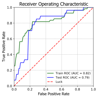 |
Prediction axillary lymph node status of Breast Cancer by MRI Radiomics
Lan Li1, Tao YU1, ShiXi Jiang1, YouXi Yuan1, JiuQuan Zhang1, and Jianqing SUN2
1Chongqing University Cancer Hospital & Chongqing Cancer Institute & Chongqing Cancer Hospital, ChongQing, China, 2Philips Healthcare, Shanghai, China
To establish a radiomic model based on dynamic contrast enhanced (DCE) magnetic resonance imaging predicting ALN status noninvasively before operation.
|
|
2325.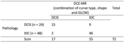 |
Combination of curve types, shape and gray level co-occurrence matrix features on breast MR to differentiate mass-like DCIS from invasive cancer
Kun Cao1, Ying li1, and Ying-Shi Sun1
1Peking University Cancer Hospital & Institute, Beijing, China
For radiologists, mass-like DCIS on breast MRI is unable to be distinguished from invasive ductal cancer by bare eyes. However, different treatment strategies for these two entities pushed us to do the differentiation using innovative approaches. By combining usage of tumor dynamic curve type on DCE, whole tumor shape features and features from GLCM to reflecting heterogeneity inside certain areas on MR images, we were able to achieve diagnostic accuracy 84.7% in telling DCIS from IDC in mass-like lesions, by using only one feature from each categories.
|
|
2326.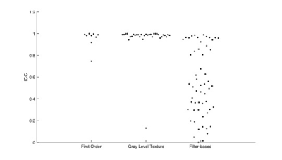 |
Improvement of Radiomics Prediction by Robustness Preselection
Renee Cattell1, Shenglan Chen2, Jie Ding2,3, and Chuan Huang2,4,5
1Stony Brook University, Stony Brook, NY, United States, 2Biomedical Engineering, Stony Brook University, Stony Brook, NY, United States, 3Diagnostic Radiology, Li Ka Shing Faculty of Medicine, The University of Hong Kong, Hong Kong, Hong Kong, 4Radiology, Stony Brook University, Stony Brook, NY, United States, 5Psychiatry, Stony Brook University, Stony Brook, NY, United States
Radiomic analysis has exponentially increased the amount of quantitative data extractable from a single medical image. However, the effect of various image acquisition conditions on the reproducibility or robustness of these features is understudied. Specifically, when generating a predictive model to be used in a multi-institutional setting, it must be robust to voxel size changes. This study aims to develop a task-specific robustness preselection step for incorporation into radiomics pipeline to improve the generalizability of a model applied to a testing set of dissimilar resolution.
|
|
2327. |
Textural kinetics of suspicious breast lesions on ultrafast DCE-MRI as a lesion classifier
Federico Pineda1, Ty Easley1, Deepa Sheth1, Hiroyuki Abe1, Milica Medved1, and Gregory Karczmar1
1University of Chicago, Chicago, IL, United States
MRI will likely take on a greater role in breast cancer screening. However, one of the main concerns with MRI's expanded role is that it will lead to many false positives. This work aims to alleviate this problem by leveraging the advantages of ultrafast imaging of initial enhancement. Here we calculated parameters descriptive of the texture of enhancement and its changes throughout the ultrafast series. The results show that 4-D texture parameters may be useful in classifying suspicious lesions (AUC=0.75), the resulting model could have ruled out malignancy in 18% of the benign lesions analyzed, while maintaining 100% sensitivity.
|
2328. |
Multi-exponential model of diffusion signal in healthy and cancerous breast tissue with fixed ADCs
Ana Elvira Rodríguez-Soto1, Maren M. Sjaastad Andreassen2, Christopher Charles Conlin1, Helen Park3, Igor Vidic4, Neil Peter Jerome4,5, Agnes Østlie5, Tone Frost Bathen2,5, Pål Erik Goa4,5, Tyler Seibert6, Michael Hahn1, Anders Martin Dale7, and Rebecca Rakow-Penner1
1Department of Radiology, University of California, San Diego, La Jolla, CA, United States, 2Department of Circulation and Medical Imaging, Norwegian University of Science and Technology, Trondheim, Norway, 3School of Medicine, University of California, San Diego, La Jolla, CA, United States, 4Department of Physics, Norwegian University of Science and Technology, Trondheim, Norway, 5Department of Radiology and Nuclear Medicine, St. Olav's University Hospital, Trondheim, Norway, 6Radiation Medicine and Applied Sciences, University of California, San Diego, La Jolla, CA, United States, 7Department of Neurosciences, University of California, San Diego, La Jolla, CA, United States
Breast DWI has shown great potential to become a contrast-free diagnostic tool for breast cancer. The purpose of this work was to determine a unified model to describe the diffusion signal of cancer and healthy breast tissues, and compare tumor conspicuity of model components to DCE-MRI and DWI estimates. Multi-exponential models with fixed ADCs were determined and the weights of each component estimated. Tumor conspicuity, defined as contrast-to-noise ratio, was found to be ~3 times higher in DCE-MRI than in the weights of multi-exponential components. This model may increase the sensitivity and specificity of DWI for breast cancer diagnosis.
|
|
2329.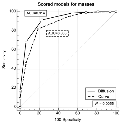 |
DWI or dynamic contrast-enhanced curve: a retrospective analysis of MRI-based differential diagnosis of benign and malignant breast lesions
Xiaoping Yang1, Lina Zhang1, Shu Li1, Ruimei Chai1, Zheng Zhang1, Nan Li1, and Mengshi Dong1
1Department of Radiology, the First Affiliated Hospital of China Medical University, Shenyang, China
Combined with morphological and enhanced information, DWI model is superior or equal to TIC model in differentiating benign and malignant breast lesions.
|
|
2330.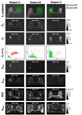 |
Discrimination of breast cancer from healthy breast tissues using a tri-exponential model
Maren Marie Sjaastad Andreassen1, Ana Elvira Rodriguez-Zoto2, Cristopher Charles Conlin2, Igor Vidic3, Grace Sora Ahn4, Neil Peter Jerome1,5, Agnes Østlie5, Tone Frost Bathen1,5, Pål Erik Goa3,5, Rebecca Rakow-Penner2, and Anders Martin Dale2,6
1Department of Circulation and Medical Imaging, Norwegian University of Science and Technology, Trondheim, Norway, 2Department of Radiology, University of California, San Diego, La Jolla, CA, United States, 3Department of Physics, Norwegian University of Science and Technology, Trondheim, Norway, 4School of Medicine, University of California, San Diego, La Jolla, CA, United States, 5Department of Radiology and Nuclear Medicine, Norwegian University of Science and Technology, Trondheim, Norway, 6Department of Neurosciences, University of California, San Diego, La Jolla, CA, United States
The purpose of this work was to develop a method to discriminate breast cancer from healthy breast tissues using signal contribution maps from multi-b diffusion MRI acquisitions. Signal contributions were estimated using a tri-exponential model, with the ADC values for three distinct compartments assumed fixed across voxels and subjects. A linear discriminant function was constructed using the estimated signal contributions from the two lowest ADC components. Average ROC AUC for discriminating cancer from healthy breast tissues was 0.99 (CI95% = 0.98-1.00), superior to that of independent signal contributions, maximum b-value volume and conventional DWI estimates (ADC and apparent diffusion kurtosis).
|
|
2331.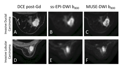 |
Diffusion-weighted imaging using multiplexed sensitivity encoding (MUSE) in breast cancer: comparison with single-shot echo-planar imaging
Gabrielle C Baxter1, Andrew J Patterson2, Ramona Woitek1, Amy J Frary1, Gavin C Houston3, Martin J Graves2, and Fiona J Gilbert1
1Department of Radiology, University of Cambridge, Cambridge, United Kingdom, 2Department of Radiology, Addenbrooke's Hospital, Cambridge, United Kingdom, 3GE Healthcare, Buckinghamshire, United Kingdom
Diffusion-weighted images acquired using single-shot echo-planar imaging (ss-EPI) often have a low resolution and are limited by blurring and distortion. This study investigated the use of MUSE (multiplexed sensitivity encoding), a multi-shot segmented-EPI technique, in the detection and characterisation of breast cancer. MUSE showed an improvement in image quality and an increased separation of malignant from benign lesions using the normalised apparent diffusion coefficient (ADC).
|
|
2332. |
Multiparametric approach of diagnosing breast lesions using DWI and ultrafast DCE MRI: compared with conventional DCE MRI
Akane Ohashi1, Masako Kataoka1, Mami Iima1, Maya Honda1, Ayami Ohno Kishimoto1, Rie Ota1, Kanae Kawai Miyake1, Masakazu Toi2, and Kaori Togashi1
1Diagnostic Imaging and Nuclear Medicine, Kyoto University Graduate School of Medicine, Kyoto, Japan, 2Breast Surgery, Kyoto University Graduate School of Medicine, Kyoto, Japan
Ultrafast dynamic contrast-enhanced (UF-DCE) MRI allows us to obtain kinetic information based on early inflow of contrast medium. A prediction model of diagnosing malignancy using maximum slope (MS) derived from UF-DCE MRI, ADC from DWI, lesion size and patient’s age was constructed. The second model using washout index (WI) derived from conventional DCE MRI instead of MS was constructed for comparison. The two multiparametric prediction models achieved almost the same AUC (AUC 0.91 vs 0.89, p=0.20). The multiparametric approach based on UF-DCE MRI, DWI and patient’s age provided excellent diagnostic performance with decreased scanning time.
|
|
2333.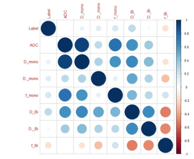 |
A Pilot Evaluation of IVIM Imaging Combined with DCE-MRI Characterize Architectural Distortion Detected on Mammography
deshuo dong1, lina zhang1, ailian liu1, qingwei song1, nan zhang1, anliang chen1, yan guo2, and lizhi xie2
1Radiology Department, The First Affiliated Hospital of Dalian Medical University, DaLian, China, 2GE Healthcare, MR Research China, Beijing, Beijing, China
Intravoxel incoherent motion (IVIM) imaging provides quantitative measurement of cellularity and vascularity. The parameters derived from IVIM can distinguish malignant and benign breast tumors. This study concerned clinical and conventional MRI features, perfusion as well as diffusion parameters using IVIM imaging, and then compared these parameters to conventional MRI images on the classification of Architectural Distortion lesions detected on Mammography.
|
|
2334.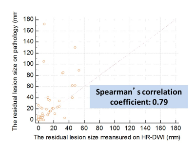 |
Pre-surgical evaluation of residual cancer by breast MRI after neoadjuvant systemic treatment with High-resolution Diffusion Weighted Imaging
Rie Ota1, Masako Kataoka1, Maya Honda1, Ayami Ohno Kishimoto1, Akane Ohashi1, Mami Iima1, Kanae Miyake Kawai1, Tatsuki R Kataoka2, Masakazu Toi3, and Kaori Togashi1
1Department of Diagnostic Imaging and Nuclear Medicine, Kyoto University graduate school of medicine, Kyoto, Japan, 2Department of Pathology, Kyoto University Hospital, Kyoto, Japan, 3Department of Breast Surgery, Kyoto University Hospital, Kyoto, Japan Poster Permission Withheld
We aimed to investigate the accuracy of high resolution-DWI (HR-DWI) using rs-EPI in estimating residual lesion size after neoadjuvant systemic treatment (NST) using pathological information as a reference. Diagnosis of pCR by two readers were accurate in 83/79 % with HR-DWI, while 50/46 % on HR-CE MRI. Spearman’s correlation coefficient between size on pathology and that on HR-DWI were 0.79 (p<0.0001), indicating strong correlations. Spearman’s correlation coefficient between size on pathology and that on HR-CE-MRI were 0.68 (p<0.0001). Using HR-DWI, residual lesions were depicted with good agreement with the pathology without using contrast agent.
|
|
2335. |
Approach for building a DWI dedicated lexicon for breast cancer
Aika Okazawa1, Mami Iima1,2, Ryosuke Okumura1, Masako Kataoka2, Taro Nishi1, Tomotaka Noda1, Sachiko Takahara3, and Kaori Togashi2
1Radiology, Kitano Hospital, The Tazuke Kofukai Medical Research Institute, Osaka, Japan, 2Diagnostic Imaging and Nuclear Medicine, Kyoto University Graduate School of Medicine, Kyoto, Japan, 3Breast Surgery, Kitano Hospital, The Tazuke Kofukai Medical Research Institute, Osaka, Japan
Our proposed DWI reading method based on the BI-RADS lexicons from multiple b-value images had comparable diagnostic performance to the standard BI-RADS. DWI reading method tended to show with higher specificity, and might increase diagnostic confidence in differentiating malignant and benign breast tumors. Excellent to substantial agreement was observed for DWI reading, and substantial agreement was found for mass type classifications between DWI and BI-RADS. Significant difference found in lesion conspicuity between malignant and benign tumors and ADC values assessmet may also provide confidence in tumor characterization.
|
|
2336. |
Diffusion-Weighted Breast MRI in the Supine Position
Catherine J. Moran1, Matthew J. Middione1,2, Valentina Mazzoli1, Bruce L. Daniel1, Daniel B. Ennis1,2, and Brian A. Hargreaves1
1Radiology, Stanford University, Stanford, CA, United States, 2Veterans Administration Health Care System, Palo Alto, CA, United States Diffusion-Weighted Imaging has shown strong potential to be the centerpiece of a breast MRI screening protocol that does not require a contrast injection. Breast MRI is conventionally performed in the prone position but supine positioning could increase patient comfort and alignment of MRI findings with other interventional procedures. Here we investigate six multi-shot DWI breast protocols with supine positioning in assymptomatic volunteers. Over all images quality and trade-offs between motion artifacts, distortion and images sharpness are characterized. |
|
2337.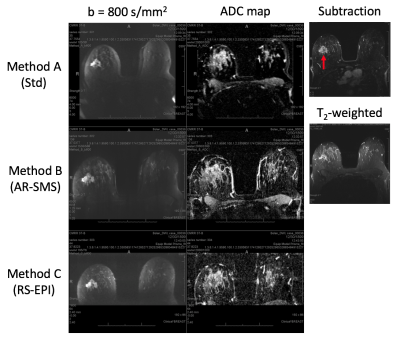 |
A Reader Study Comparing the Quality of High-Resolution Diffusion Weighted Imaging Methods for Breast MRI
Jessica A McKay1,2, An L Church3, Nathan Rubin4, Tim H Emory3, Noelle F Hoven3, Jessica E Kuehn-Hajder3, Michael T Nelson3, and Patrick J Bolan2,3
1Department of Biomedical Engineering, University of Minnesota, Minneapolis, MN, United States, 2Center for Magnetic Resonance Research, University of Minnesota, Minneapolis, MN, United States, 3Department of Radiology, University of Minnesota, Minneapolis, MN, United States, 4Masonic Cancer Center, University of Minnesota, Minneapolis, MN, United States
Axially reformatted (AR) SMS can provide full-coverage breast DWI with high quality and 1.25 mm isotropic nominal resolution. In this work we assess the image quality of AR-SMS in a clinical setting and compare it with a standard clinical sequence and RS-EPI. Three radiologists scored DWI and ADC maps from 30 patients with enhancing lesions. All three readers scored AR-SMS with the highest quality and ranked it as superior most frequently. Accounting for reader and subject effects, a linear mixed model found that AR-SMS outperformed RS-EPI, which both outperformed the Standard with statistical significance in both quality score and rank.
|
|
2338. |
Comparative Analysis of the Value of APTWI and IVIM in Evaluating the Benign and Malignant Breast Lesions: A pilot study
Nan Meng1, Xuejia Wang2, Dongming Han2, Jing Sun3, Meng Zhang2, Kaiyu Wang4, and Meiyun Wang*5
1Department of Radiology, Zhengzhou University People’s Hospital & Henan Provincial People’s Hospital, Academy of Medical Sciences, Zhengzhou University, Zhengzhou, China, 2Department of MRI, the First Affiliated Hospital of Xinxiang Medical University, Weihui, China, 3Department of Pediatrics, Zhengzhou Central Hospital, Zhengzhou University, Zhengzhou, China, 4GE Healthcare, MR Research China, Beijing, China, 5Department of Radiology, Zhengzhou University People’s Hospital & Henan Provincial People’s Hospital, Zhengzhou, China
Amide proton transfer-weighted imaging (APTWI) has unique advantages in displaying the metabolism of diseased proteins. Intravoxel incoherent motion (IVIM) method is able to obtain a variety of quantitative parameters, including D, D* and f values, which reflect the perfusion and diffusion information of the tissues. Our results show that APTWI and IVIM have similar diagnostic performance in the diagnosis of benign and malignant breast tumors.
|
|
2339. |
Accelerating Acquisition of RESOLVE-DWI with Simultaneous Multi-slice (SMS) Technique in Diagnosing Breast Lesions
TAO AI1 and Liming Xia1
1Radiology, Tongji Hospital, Tongji Medical College, HUST, Wuhan, China
Readout-segmented echo planar imaging (RESOLVE) has demonstrated its high diagnostic performance in breast with sharper Images, better lesion-background contrast and higher spatial resolution. However, long scan time (multi-shot strategy) may significantly decrease its clinical application due to possible motion artifacts and limitation of more complex diffusion approaches. Simultaneous multi-slice (SMS) technique allows for rapid realization of breast MR imaging, which may serve as a superior alternative for the diagnosis of breast lesions. In study , SMS was implemented into DWI to reduce the scan time by factor of 2-4, while maintaining the same anatomic coverage and basic image quality.
|
|
2340. |
Simultaneously acquired DWI and PET for early prediction of pathological response to neoadjuvant chemotherapy in breast cancer
Yee-Fan Lee1, Jo-Yu Chen1, Ning Chien1, Emi Niisato2, Robert Grimm3, Chiao Lo4, Ming-Yang Wang4, Chiun-Sheng Huang4, and Yeun-Chung Chang1
1Department of Medical Imaging, National Taiwan University Hospital and College of Medicine, Taipei, Taiwan, 2Siemens Healthcare Limited, Taipei, Taiwan, 3Siemens Healthcare, Erlangen, Germany, 4Department of Surgery, National Taiwan University Hospital and College of Medicine, Taipei, Taiwan Neoadjuvant chemotherapy (NAC) for treatment of breast cancer has the potential benefit of reducing tumor size before surgery. The response to NAC may also provide prognostic information which may lead to more effective chemotherapy. Combining PET and MRI information is useful for assessing the tumor response to NAC in a comprehensive manner. In this study, we performed texture analysis which indicated that the entropy of ADC and difference entropy of SUV were related and can be used to predict pCR with high areas under the receiver operating characteristic curve (AUC).
|
|
2341. |
Rapid 3D B1+-Compensated, Simultaneous Whole Breast T1, T2, and Apparent Diffusion Coefficient Quantification with MR Multitasking
Sen Ma1,2, Anthony G. Christodoulou2, Nan Wang1,2, and Debiao Li1,2
1Department of Bioengineering, University of California, Los Angeles, Los Angeles, CA, United States, 2Biomedical Imaging Research Institute, Cedars-Sinai Medical Center, Los Angeles, CA, United States
Quantitative relaxation and diffusion mapping are desirable in clinical breast MRI as biomarkers for tissue characterization and prediction of treatment response. Conventional multi-parametric mappings are typically acquired in separate scans. We simultaneously generate high quality, co-registered T1/T2/ADC maps without image distortion for 3D whole breast coverage in 8.5min, incorporating B1+ compensation into the Multitasking framework to improve T1 accuracy. We demonstrate excellent agreement of T1/T2/ADC measurements between the proposed Multitasking approach and available reference methods, as well as good repeatability. We also show that all T1/T2/ADC measurements produced by the Multitasking framework agree with literature range.
|
2342.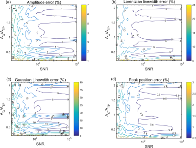 |
Evaluating the accuracy of time-domain Voigt fitting of dissolved-phase 129Xe spectra in the lungs
Graham Norquay1 and Jim M Wild1
1POLARIS, Infection, Immunity and Cardiovascular Disease, University of Sheffield, Sheffield, United Kingdom
This work quantifies the accuracy of time-domain Voigt fitting of dissolved-phase 129Xe peak parameters (xenon in red blood cells and tissue/blood plasma). It is shown that time-domain Voigt fitting of dissolved-phase 129Xe resonances in the lungs is accurate to within 5% when the SNR values of the spectra are >100 and the gas phase resonance amplitude is < factor of 2 higher than each of the dissolved-phase 129Xe resonances.
|
|
2343.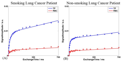 |
Quantify the Influence of Smoking on Pulmonary Structure and Function in Lung Cancer Patients using Hyperpolarized 129Xe MR
Qian Zhou1, Haidong Li1, Qiuchen Rao1, Junshuai Xie1, Xiuchao Zhao1, He Deng1, Lei Shi1, Xianping Sun1, Yeqing Han1, Dongshan Han2, Xin Lou2, Chaohui Ye1, and Xin Zhou1
1National Center for Magnetic Resonance in Wuhan, Wuhan Institute of Physics and Mathematics, Innovation Academy of Precision Measurement Science and Technology, Chinese Academy of Sciences-Wuhan National Laboratory for Optoelectronics, Wuhan, China, 2Department of Radiology, Chinese PLA General Hospital, Beijing, P. R. China, Beijing, China
Pulmonary diseases, such as lung cancer, usually result in changes of pulmonary structure and function. To assess these changes, gas exchange across the respiratory membrane and gas diffusion in the alveoli can be quantified using hyperpolarized 129Xe MR via CSSR and DWI in lung cancer patients, The significant differences were found in mean exchange time constant(T) and septal wall thickness(d) between smoking and non-smoking lung cancer patients, indicating the impact of smoking. The results manifested that the hyperpolarized 129Xe MR is a promising method for optimizing radiation therapy in the future.
|
|
2344.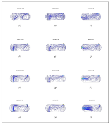 |
Resolving the discrepancy between theoretical and experimental polarization of HP 129Xe using numerical simulations and optical spectroscopy
Michele Kelley1, Alex Burant1, and Rosa Tamara Branca1
1Physics and Astronomy, University of North Carolina - Chapel Hill, Chapel Hill, NC, United States
For emerging biomedical applications of hyperpolarized xenon, the ability to obtain high nuclear spin polarization levels is imperative. Yet, experimental nuclear spin polarization levels of xenon obtained by continuous flow spin-exchange optical pumping are highly variable and often well below theoretical predictions. Identifying possible depolarization mechanisms has been the focus of those trying to rectify this discrepancy. Instead, we revisit assumptions made about the physical system. By using a combination of numerical simulations and in situ optical spectroscopy measurements, we found that lower Rb densities and shorter residence times than typically assumed lead to lower, not higher, theoretical polarization values.
|
|
2345.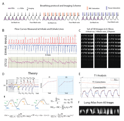 |
Measuring Ventilation and Gas Exchange Rates with Hyperpolarized 129Xe During Tidal Breathing in Human Subjects
Hooman Hamedani1,2, Stephen J Kadlecek1, Faraz Amzajerdian 1,2, Ryan Baron1, Kai Ruppert1, Ian Duncan1, Yi Xin1,2, Mehrdad Pourfathi1, Sarmad Siddiqui1, Luis Loza1, Tahmina Achekzai1, Maurizio Cereda1,3, Rahim R. Rizi1, and
fmig upenn4
1Radiology, University of Pennsylvania, Philadelphia, PA, United States, 2Bioengineering, University of Pennsylvania, Philadelphia, PA, United States, 3Anesthesiology and Critical Care, University of Pennsylvania, Philadelphia, PA, United States, 4University of Pennsylvania, Philadelphia, PA, United States
A super multibreath maneuver to measure ventilation and gas exchange during tidal breathing in human subjects.
|
|
2346.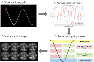 |
Evaluation of Ventilation Function Using 3D Ultrashort Echo-Time Imaging Without Exogeneous Gases
Seokwon Lee1, Jinil Park1, Hyonha Kim2, Ho Yun Lee3, and Jang-Yeon Park1,4
1Department of Biomedical Engineering, Sungkyunkwan University, Suwon, Republic of Korea, 2Department of Biomedical Engineering, Hankuk University of Foreign Studies, Yongin, Republic of Korea, 3Department of Radiology and Center for Imaging Science, Samsung Medical Center, Seoul, Republic of Korea, 4Center for Neuroscience Imaging Research, Institute for Basic Science, Suwon, Republic of Korea
Lung MRI is widely used for diagnosis pulmonary disease, there has been continuing interest in the possibility of using MRI to detect the pulmonary lesion. Recent works acquired ventilation defect region using ventilation map. Here we proposed another method, which evaluates ventilation flow map and regional fractional ventilation flow-volume loop using 3D UTE. These methods provide valuable information to evaluate pulmonary ventilation function as well as ventilation map. The potential of methods was demonstrated by different ventilation flow map and regional fractional ventilation flow volume loops in healthy subjects.
|
|
2347.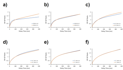 |
The Impact of Acquisition Parameters on Alveolar Septal Wall Thickness Measurements with Hyperpolarized Xenon-129 MRI
Kai Ruppert1, Faraz Amzajerdian1, Yi Xin1, Hooman Hamedani1, Luis Loza1, Tahmina S. Achekzai1, Ryan J. Baron1, Ian F. Duncan1, Harrilla Profka1, Yiwen Qian1, Mehrdad Pourfathi1, Federico Sertic1, Stephen Kadlecek1, and Rahim
R. Rizi1
1Radiology, University of Pennsylvania, Philadelhia, PA, United States
Measurements of the apparent alveolar septal wall thickness (SWT) with hyperpolarized xenon-129 (HXe) MRI are sensitive to inflammatory or fibrotic pathologies in the lung parenchyma but are also affected by lung inflation level. Here, we investigated the dependence of such measurements on the choice of acquisition parameters in a rabbit model. We found the SWT measurements to be strongly affected by the imaging parameters: in particular, the number and flip angle of the applied RF pulses centered at the dissolved-phase resonances. If this bias is minimized, the previously reported dependence of septal thickness measurements on lung inflation disappears.
|
|
2348.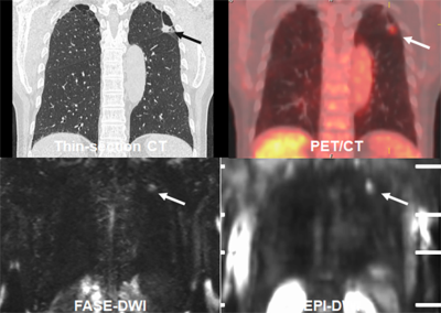 |
Fast Advanced Spin-Echo vs. Echo-Planar Imaging: DWI at 3T MR System for Postoperative Clinical Outcome Prediction in NSCLC Patients
Yoshiharu Ohno1,2, Masao Yui3, Daisuke Takenaka4, Yoshimori Kassai3, Kazuhiro Murayama1, and Takeshi Yoshikawa2
1Radiology, Fujita Health University School of Medicine, Toyoake, Japan, 2Radiology, Kobe University Graduate School of Medicine, Kobe, Japan, 3Canon Medical Systems Corporation, Otawara, Japan, 4Diagnostic Radiology, Hyogo Cancer Center, Akashi, Japan
No one has compared the capability among DWIs obtained by EPI and FASE at a 3T MR system and PET/CT for prediction of postoperative clinical outcome. We hypothesize that DWI obtained by FASE sequence are more useful than that by EPI sequence at 3T MR system and PET/CT for predicting postoperative recurrence and treatment outcome in NSCLC patients with surgical treatment. The purpose of this study was to directly and prospectively compare the quantitative capability for prediction of postoperative recurrence and treatment outcome among DWI with FASE and EPI sequences at 3T system and FDG-PET/CT in NSCLC patients.
|
|
2349.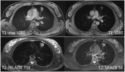 |
3D and 2D MRI sequences of lung nodules: comparison of capability for nodule detection and image quality evaluation
Shuyi Yang1, Qinqin Yan1, Fei Shan1, Zhiyong Zhang2, Yuxin Shi1, and Mengxiao Liu3
1Shanghai public health clinical center, Shanghai, China, 2Fudan University, Shanghai, China, 3Scientific Marketing, Diagnositic Imaging, Siemens Healthcare Ltd., Shanghai, China
This study aimed to evaluate the capability of different non-enhanced MRI sequences for lung nodules assessment. Sensitivity and specificity from MR images were calculated, with CT regarded as golden standard. The sensitivities by T1-starVIBE, T1-VIBE, T2-fBLADE TSE and T2-SPACE were 79.79%, 75%, 88.3%, 89.89%, while the specificity were 100%, 83.33%, 33.33%, 53.33% respectively. In conclusion, for uncooperative patients who can’t hold their breath, T1-starVIBE and T2-fBLADE were advised because of the excellent ability to remove artifacts. T1-VIBE can applied to those who coordinate well. All those three sequences can be used for pulmonary nodules assessment and long-term follow-up in clinical.
|
|
2350. |
Native T1-mapping of focal pulmonary lesions in 3.0-T magnetic resonance imaging: lesions display and accuracy of size estimation
Shuyi Yang1, Qinqin Yan1, Fei Shan1, Zhiyong Zhang1, and Yuxin Shi1
1Shanghai public health clinical center, Shanghai, China
This study aimed to investigate the diagnosis efficacy of native T1-mapping in focal pulmonary lesion, focused on lesions display and accuracy of size estimation. All lesions were clearlly demonstrated by CT, native T1-mapping, T1-star VIBE, and T2-fBLADE TSE. The tumor and distal obstructive pulmonary atelectasis can also be detected clearly by native T1-mapping. T1-mapping-based diameter measurements yielded excellent intra-observer and inter-methods consistent.In conclusion, we considered that native T1-mapping displays focal pulmonary lesions clearly (particularly when accompanying obstructive atelectasis and/or pneumonia) and enable accurate and reliable diameter measurement.
|
|
2351.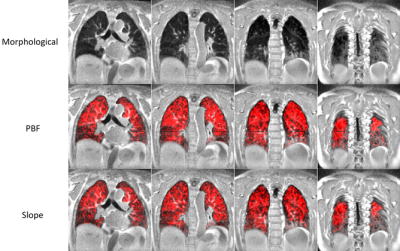 |
Comparison of Quantitative and Semi-Quantitative Metrics from Dynamic Contrast Enhanced MRI: Evaluation in Idiopathic Pulmonary Fibrosis
Luis A Torres1, Greg Barton1, Nathan Sandbo2, Mark L Shiebler2,3, and Sean B Fain1,3,4
1Dept. of Medical Physics, University of Wisconsin - Madison, Madison, WI, United States, 2Dept. of Medicine, University of Wisconsin - Madison, Madison, WI, United States, 3Dept. of Radiology, University of Wisconsin - Madison, Madison, WI, United States, 4Dept. of Biomedical Engineering, University of Wisconsin - Madison, Madison, WI, United States
Fully quantitative pulmonary perfusion metrics for the evaluation of lung disease can be difficult and time consuming to compute. We compare fully quantitative pulmonary blood flow (PBF) and mean transit time (MTT) to simpler semi-quantitative metrics slope and first moment transit time (FMTT) in idiopathic pulmonary fibrosis (IPF). We found strong correlations between the slope and PBF, as well as MTT and FMTT. A decrease in slope values was found in subjects with IPF when compared to healthy subjects. Slope could potentially be used as a surrogate for PBF to evaluate IPF.
|
|
2352.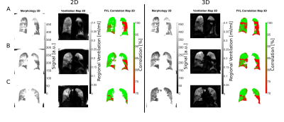 |
Feasibility of 3D flow-volume loop analysis using phase-resolved functional lung imaging (PREFUL) in comparison with 2D PREFUL
Filip Klimeš1,2, Andreas Voskrebenzev1,2, Marcel Gutberlet1,2, Robert Grimm3, Cristian Crisosto1,2, Frank Wacker1,2, and Jens Vogel-Claussen1,2
1Institute of Diagnostic and Interventional Radiology, Hanover Medical School, Hanover, Germany, 2Biomedical Research in Endstage and Obstructive Lung Disease Hannover (BREATH), German Center for Lung Research (DZL), Hanover, Germany, 3Siemens Healthcare GmbH, Erlangen, Germany
The measurement of ventilation dynamics with 2D proton MRI (PREFUL) might offer a sensitive lung function test but lacks a high spatial resolution and whole-lung acquisition is time consuming. Recently, the feasibility of 3D PREFUL was demonstrated. In this work, ventilation dynamics assessment using flow-volume loop (FVL) analysis is performed on 2D and 3D data of a healthy volunteer and an asthma patient. Full respiratory cycle was reconstructed to calculate regional ventilation and cross-correlation metric of FVL. Similar results suggest that 3D PREFUL might be an interesting alternative to 2D PREFUL due to its improved spatial resolution and acquisition efficiency.
|
|
2353.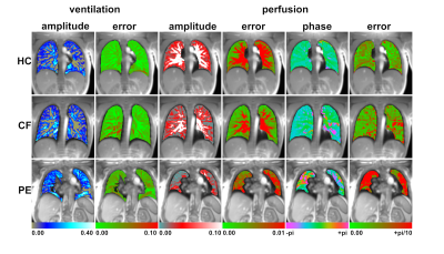 |
Assessing the Error in Functional Lung Parameters obtained via SENCEFUL MRI
Andreas Max Weng1, Simon Veldhoen1, Christian Kestler1, and Herbert Köstler1
1Department of Diagnostic and Interventional Radiology, University Hospital of Würzburg, Würzburg, Germany
Until now, no study assessed the error that arises during calculation of quantitative functional lung parameters obtained from MRI. Using a bootstrapping approach with shuffling residuals the error of ventilation and perfusion (amplitude and phase) of the SENCEFUL technique was investigated in this study. Obtained error values were in an acceptable range rendering quantitative functional lung MRI a promising technique for the assessment of regional alterations in lung function.
|
|
2354.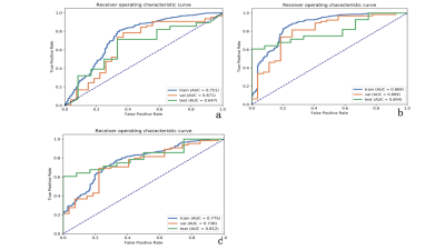 |
Development and Validation of T2WI-Based Radiomics Classifier in Patients with solid solitary pulmonary lesion
Wan Qi1, Yuze Wang1, Xinchun Li1, Tianjing Zhang2, Jianqing Sun2, and Yingjie Mei2
1The First Affiliated Hospital of Guangzhou Medical University, Guangzhou, China, 2Philips Healthcare, Guangzhou, China
The solitary pulmonary lesion (SPL) is one of the most common imaging findings. In this study, lung MR using T2 weighted imaging were acquired and analyzed using radiomics. Both 2D and 3D features combined with machine learning approach were compared to determinate an optimal model for differentiation of SPLs. We found that the radiomics signatures derived from 3D features outperformed that from 2D features. The best 3D radiomics model for the classification was a combination of principal component analysis(PCA), analysis of variance(ANOVA) and linear discriminant analysis (LDA). The T2WI-based radiomics model shows potential in differentiating malignancy from benign SPLs.
|
|
2355.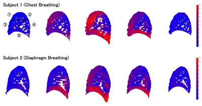 |
Detecting regional differences of lung motion by optical flow analysis combined with rapid cine MRI using compressed sensing
Koji Sagiyama1, Masateru Kawakubo2, Ryohei Funatsu3, Takeshi Kamitani1, Yuzo Yamasaki1, Tomoyuki Hida1, Yuko Matsuura1, Yuriko Murayama1, and Hidetake Yabuuchi4
1Department of Clinical Radiology, Graduate School of Medical Sciences, Kyushu University, Fukuoka, Japan, 2Department of Health Sciences, Faculty of Medical Sciences, Kyushu University, Fukuoka, Japan, 3Department of Medical Technology, Division of Radiology, Kyushu University, Fukuoka, Japan, 4Departmant of Health Sciences, Graduate School of Medical Sciences, Kyushu University, Fukuoka, Japan
The application of cine MRI to lung disease has been technically challenging. In this study, we exploited optical flow analysis combined with rapid acquisition of cine MRI using compressed sensing to detect differences in regional lung motion. The high spatial and temporal resolution and higher signal-to-noise ratio of cine MRI enabled optical flow analysis to visualize regional differences between five lung segments. It also differentiated between chest and diaphragm breathing patterns in volunteers. In the future, this non-invasive method could be applied to assess and follow lung diseases, such as interstitial pneumonia or chronic obstructive pulmonary disease.
|
|
2356.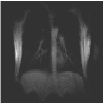 |
High Time Resolution 2D Chest Imaging Using A 0.5T Upright Paramed MRI Scanner.
James Harkin1, Michael Barlow2, Alan Smyth3, Ian Hall1, and Andrew Prayle3
1University of Nottingham, Nottingham, United Kingdom, 2Faculty of Medicine & Health Sciences, University of Nottingham, Nottingham, United Kingdom, 3Medicine and Health Sciences, University of Nottingham, Nottingham, United Kingdom
Using a 0.5T Paramed MROpen Upright MRI scanner, adult and paediatric cohorts as young as 3 can be scanned without sedation. 2D images can be acquired every <0.5s allowing for diaphragm movement to be characterised. Lung density variation through the breathing cycle can also be extracted.
|
2357.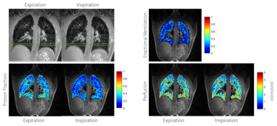 |
Functional Lung Investigation with Tiny Golden Angle Ultra-Short Echo-Time Stack of Stars (UTE STyGAS)
Anke Balasch1 and Volker Rasche1
1Department of Internal Medicine II, Ulm University Medical Center, Ulm, Germany
Deriving lung data with MRI is challenging due to several reasons, including short T2*, cardiac and respiratory motion, and low proton density. In this study, a tiny golden angle UTE stack-of-stars (UTE STyGAS) sequence was applied for quantification of lung parenchymal functional parameters, including proton fraction, fractional ventilation and perfusion. Data were acquired during free breathing and reconstructed applying an image-based self-gating technique.
|
|
2358.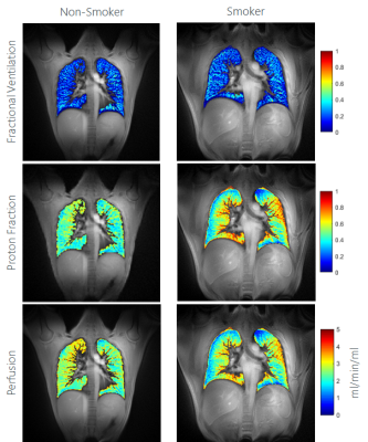 |
Lung Function Assessment by Self-Gated 2D UTE – Comparison of Smoker and Non-Smoker
Anke Balasch1, Patrick Metze1, Tobias Speidel2, and Volker Rasche1
1Department of Internal Medicine II, Ulm University Medical Center, Ulm, Germany, 2Core-Facility Small Animal Imaging, Ulm University, Ulm, Germany
In this study, a two-dimensional ultra-short TE protocol was used to acquire free-breathing data of the lung. The signal intensity in expiration and inspiration were used to derive the fractional ventilation, proton fraction and perfusion of the lung parenchyma.
|
|
2359.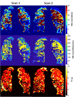 |
Same-day reproducibility of quantitative lung perfusion metrics in patients with IPF
Paul J.C. Hughes 1, Nicholas D. Weatherley1, Helen Marshall1, Stephen M. Bianchi2, and Jim M. Wild1,3
1POLARIS, Department of Infection, Immunity and Cardiovascular Disease,, The University of Sheffield, Sheffield, United Kingdom, 2Sheffield Teaching Hospitals Foundation Trust, Academic Directorate of Respiratory Medicine, Sheffield, United Kingdom, 3Insigneo Institute for in silico Medicine, The University of Sheffield, Sheffield, United Kingdom
Recent work in assessing patients with idiopathic pulmonary fibrosis suggests that vascular changes, as assessed by dynamic contrast enhanced lung MRI, are a useful marker to assess disease progression. Therefore this work aimed to develop an image processing pipeline to assess same-day intra-participant reproducibility of quantitative perfusion metrics in a cohort of patients with idiopathic pulmonary fibrosis. From the data here it suggests that these quantitative metrics are not reproducible, even when patients are scanned on the same day.
|
|
2360.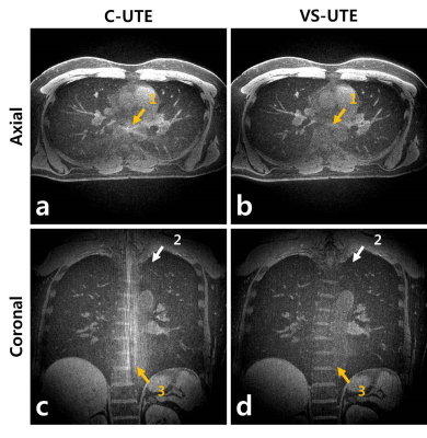 |
Reducing Streak Artifacts in 3D Radial Imaging Using Volume-Selective Signal Acquisition
Jinil Park1 and Jang-Yeon Park1,2
1Biomedical Engineering, Sungkyunkwan University, Suwon, Korea, Republic of, 2Center for Neuroscience Imaging Research, Institute for Basic Science, Suwon, Korea, Republic of
The non-Cartesian sampling can obtain the image even for the number of data that does not satisfy Nyquist, but the under-sampled image is accompanied by streak artifact. Since the non-selective pulse used in conventional 3D radial UTE(C-UTE) is excite unwanted areas out of FOV, streak artifacts may appear in the image when under-sampling. Volume-Select UTE(VS-UTE) performs spin excitation using frequency-selective SINC pulses. Accordingly, the proposed method effectively suppresses the signal outside the FOV, which reduces streak artifacts. The purpose of this study is to introduce a VS-UTE sequence and to confirm that the streak artifacts are reduced effectively.
|
|
2361.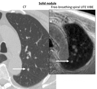 |
Pulmonary nodule detection using ultra-short TE (UTE) with a 3D variable-TE stack-of-spirals sequence in a 3T MR-PET scanner
Yu-Sen Huang1, Mao-Yuan Marine Su1, Ning Chien1, Jin-Shing Chen2, and Yeun-Chung Chang1
1Department of Medical Imaging, National Taiwan University Hospital and National Taiwan University College of Medicine, Taipei, Taiwan, 2Department of Surgery, National Taiwan University Hospital and National Taiwan University College of Medicine, Taipei, Taiwan Although the 3T MR-PET system has been used clinically for the whole body, PET/CT still has advantages over this technique when evaluating the pulmonary region. Ultrashort TE with 3D variable-TE stack-of-spirals sampling has been used for pulmonary nodule detection in MR examination1, which allows for shorter scanning times using an undersampling technique in combination with iterative self-consistent parallel imaging reconstruction (SPIRiT). The goal of this study was to investigate the feasibility of this technique for detecting pulmonary nodules in the 3T MR-PET scanner, which has fewer coil elements compared with most other MR-only scanners, to achieve minimum PET attenuation.
|
|
2362. |
Reducing the breath hold time required to image the diaphragm in an upright position
Joshua McAteer1, James Harkin2, Olivier Mougin1, Christoph Arthofer2, Paul Glover1, and Penny Gowland1
1Physics, University of Nottingham, Nottingham, United Kingdom, 2Medicine, University of Nottingham, Nottingham, United Kingdom
We investigated accelerating two paradigms of in vivo diaphragm imaging using a 0.5T upright scanner. These two paradigms, high resolution and low resolution, were optimised to deliver minimal artefacts and maximal information about the diaphragm surface. These scans delivered sufficient image quality for the tissue boundary of the diaphragm to be located. This will allow for future work to study the diaphragm in the upright position of participants with COPD who are only able to hold their breath for ~5 sec.
|
|
2363.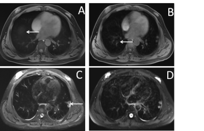 |
3.0T Magnetic resonance for adult pulmonary tuberculosis
Qinqin Yan1, Shuyi Yang2, Jie Shen2, Shuihua Lu2, Fei Shan2, and Yuxin Shi2
1Radiology of Departemnt, Shanghai Public Health Clinical Center, Shanghai, China, 2Shanghai Public Health Clinical Center, Shanghai, China
Pulmonary tuberculosis is common chronic infectious disease, with a wide range of patients from infancy to the elderly. Currently, CT is the a commonly used method of examination, but radiation is not negligible. MRI as a non-radiative lesion, and functional measurement, can be used for tuberculosis examination and follow-up. This article provides a feasible examination plan for routine examination of tuberculosis via comparisons of T1-weighted Star vibe and standad vibe, and T2-weighted fBLADE and 3D SPACE.
|
|
2364.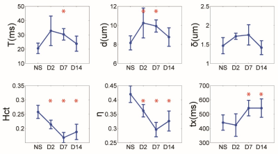 |
Quantify the Progression of Acute Lung Injury Induced by Lipopolysaccharide with Hyperpolarized 129Xe MR
Haidong Li1, Ming Zhang1, Hongchuang Li1, Xiuchao Zhao1, Yeqing Han1, Xianping Sun1, Chaohui Ye1, and Xin Zhou1
1National Center for Magnetic Resonance in Wuhan, Wuhan Institute of Physics and Mathematics, Innovation Academy of Precision Measurement Science and Technology, Chinese Academy of Sciences-Wuhan National Laboratory for Optoelectronics, Wuhan, China
CT and PET should not be performed on patient repeatedly in short-term for monitoring the progression of diseases since the ionizing radiation and radioactivity. Herein, we tried to demonstrate the feasibility of hyperpolarized 129Xe MR for monitoring the progression of acute lung injury (ALI) in rats. Our results indicated hyperpolarized 129Xe MR has the potential to monitor the progression of ALI in rats.
|
|
2365.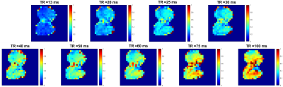 |
A Technique for Quantitative Measurement of Pulmonary Gas Uptake using Hyperpolarized 129Xe MRI in Free-Breathing Mice
Luis A Loza1, Stephen J Kadlecek1, Mehrdad Pourfathi1, Kai Ruppert1, Tahmina S Achekzai1, Ian F Duncan1, and Rahim R Rizi1
1Radiology, University of Pennsylvania, Philadelphia, PA, United States
Here we present an imaging technique for measuring regional gas uptake in a free-breathing mouse non-invasively. Gas- and dissolved-phase images were acquired simultaneously for a wide range of repetition times (13 ms up to 100 ms), and used to derive septal wall thickness measurements by fitting to a theoretical gas uptake model. The work shown here sets the groundwork for non-terminal longitudinal studies in rodent models of pulmonary radiation and fibrosis.
|
|
2366.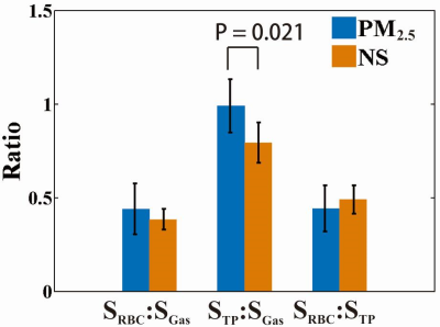 |
Investigation of Lung Injury Caused by Air pollutants PM2.5 with both Pulmonary Function Tests and Hyperpolarized 129Xe MR
Ming Zhang1, Haidong Li1, Hongchuang Li1, Xiuchao Zhao1, Qian Zhou1, Qiuchen Rao1, Yeqing Han1, Yina Lan2, He Deng1, Xianping Sun1, Xin Lou2, Chaohui Ye1, and Xin Zhou1
1National Center for Magnetic Resonance in Wuhan, Wuhan Institute of Physics and Mathematics, Innovation Academy of Precision Measurement Science and Technology, Chinese Academy of Sciences-Wuhan National Laboratory for Optoelectronics, Wuhan, China, 2Department of Radiology, Chinese PLA General Hospital, Beijing, China
In this study, hyperpolarized 129Xe MR and pulmonary function tests were utilized to quantitatively evaluate the pulmonary physiological changes caused by air pollutants (PM2.5), which are difficult to be non-invasively assessed using the conventional methods including bronchoalveolar lavage fluid analysis and histopathological sections. Significant differences were found in mean exchange time constant, septal wall thickness and TP/GAS ratio using 129Xe dynamic spectra. The results from our study indicated hyperpolarized 129Xe MR may be a promising method for quantifying lung injury caused by air pollution in clinic.
|
|
2367.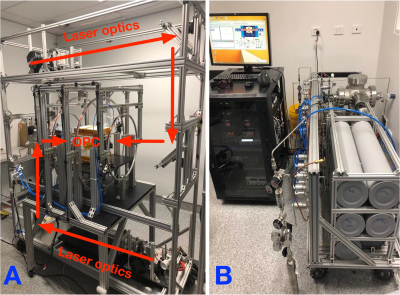 |
Phase imaging of hyperpolarized 129Xe gas in a human lung
Gang Zheng1, Wai Tung Lee2,3, Xin Tong4, Tim D’Adam2, Steven Parnell5, Michael de Veer1, Shenpeng Li1, Hongxin Wang1, Richard Mcintyre1, Francis Thien6, and Gary Egan1
1Monash Biomedical Imaging, Monash University, Clayton, Australia, 2Australian Nuclear Science and Technology Organisation, Lucas Heights, Australia, 3Instrument Technologies Division, European Spallation Source ERIC, Lund, Sweden, 4China Spallation Neutron Source, Dongguan, China, 5Delft University of Technical, Delft, Netherlands, 6Eastern Clinical School, Monash University, Box Hill, Australia
We describe a custom-designed spin-exchange-optical pumping (SEOP) polarizer which can reliably produce hyperpolarized 129-Xe (HP-Xe) gas. Imaging of a HP-Xe phantom showed phase images were less sensitive to depolarization and could provide on/off images of HP-Xe. In a human lung, phase image provided higher imaging contrast in low HP-Xe polarization states.
|
|
2368.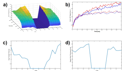 |
Does Pulmonary Gas Transport Affect Spatially-Resolved Alveolar Septal Wall Thickness Measurements with Hyperpolarized Xenon-129 MRI?
Kai Ruppert1, Faraz Amzajerdian1, Yi Xin1, Hooman Hamedani1, Luis Loza1, Tahmina S. Achekzai1, Ryan J. Baron1, Ian F. Duncan1, Harrilla Profka1, Yiwen Qian1, Mehrdad Pourfathi1, Federico Sertic1, Stephen Kadlecek1, and Rahim
R. Rizi1
1Radiology, University of Pennsylvania, Philadelhia, PA, United States
Septal wall thickness (SWT) measurements with hyperpolarized xenon-129 (HXe) MRI are typically conducted as spectroscopic acquisitions that lack spatial information. Recently, efforts have been made to obtain spatial maps of SWT. Here, we investigated differences in apparent lung physiology between peripheral and central lung regions and found an implausible change in observed capillary transit time and SWT from the periphery to the center of the lungs. This effect is likely caused by the transport of xenon-saturated blood towards the heart, and is not based on physiological differences, thus requiring a fundamental revision of analytical gas uptake models.
|
|
2369.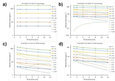 |
The Impact of T2* And Pulmonary Gas Exchange on The Accuracy Of The Applied Dissolved-phase Flip Angle in Hyperpolarized Xenon-129 MRI?
Yiwen Qian1, Kai Ruppert1, Faraz Amzajerdian1, Yi Xin1, Hooman Hamedani1, Luis Loza1, Tahmina S. Achekzai1, Ryan J. Baron1, Ian F. Duncan1, Harrilla Profka1, Mehrdad Pourfathi1, Federico Sertic1, Stephen Kadlecek1, and Rahim
R. Rizi1
1Radiology, University of Pennsylvania, Philadelhia, PA, United States
The fleeting nature of hyperpolarized xenon-129 magnetization necessitates a separate flip angle calibration procedure based on the decay of the gas-phase signal. This calibration is subsequently assumed to be valid for imaging of the xenon dissolved in the lung tissue despite the rapid exchange between the parenchyma and the alveolar gas volume and despite that the T2* is much shorter in the former than in the latter. In this work we implemented a 1-dimensional gas exchange simulation and investigated to what extent the effective flip angle at the dissolved-phase frequencies is affected by exchange and acquisition parameters.
|
|
2370.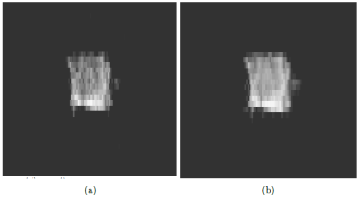 |
Feasibility of 19F Lung Imaging On A 0.5T Multi-Nuclear Upright Paramed MRI Scanner By Imaging Proton on HFC Gas.
James Harkin1, Robert Irwin1, Peter Thelwall2, and Michael Barlow1
1Respiratory Medicine, University of Nottingham, Nottingham, United Kingdom, 2Newcastle University, Newcastle upon Tyne, United Kingdom
Using a multinuclear 0.5T Paramed Medical Systems MROpen Upright MRI scanner the feasibility of 19F imaging was assessed. The upright and open design of the scanner makes imaging more comfortable for patients and allows for imaging in later stages of respiratory diseases. It proved to be possible to image 1H of a fluorinated gas within a single repetition and there is an expectation of at least a 2.5 fold improvement in signal when moving to 19F.
|
|
2371.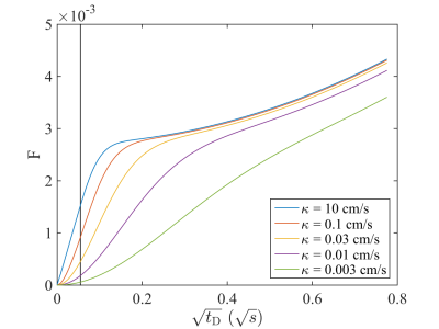 |
Assessment of interstitial membrane permeability using a generalized model of 129Xe septal uptake in the lung
Agilo L. Kern1,2, Karen M. Olsson2,3, Till F. Kaireit1,2, Frank K. Wacker1,2, Jens M. Hohlfeld2,3,4, Marius M. Hoeper2,3, and Jens Vogel-Claussen1,2
1Institute of Diagnostic and Interventional Radiology, Hannover Medical School, Hannover, Germany, 2Biomedical Research in Endstage and Obstructive Lung Disease Hannover (BREATH), German Center for Lung Research (DZL), Hannover, Germany, 3Department of Respiratory Medicine, Hannover Medical School, Hannover, Germany, 4Clinical Airway Research, Fraunhofer Institute for Toxicology and Experimental Medicine, Hannover, Germany
The hyperpolarized 129Xe chemical shift saturation recovery (CSSR) method combined with mathematical models of gas uptake provides unique insights into lung microstructure and function. Here, we propose a generalization of the Patz model for assessing interstitial membrane permeability. We test the model’s potential for in-vivo determination of membrane permeability by fitting to CSSR data from healthy volunteers and pulmonary hypertension (PH) patients. The obtained permeabilities lie between those estimated for O2 and CO2 in healthy volunteers and tend to be lower in PH. The proposed model enables the non-invasive assessment of interstitial barrier permeability and shows potential as diagnostic tool.
|
2372.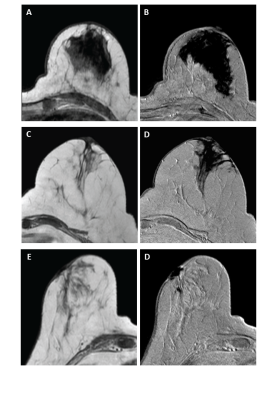 |
Assessing void artifacts in follow-up breast MRI scans after sentinel node biopsy using superparamagnetic tracer
Anke Christenhusz1, Lejla Alic1, Frank F.J. Simonis1, Margreet C Van der Schaaf2, Caroline A.H. Klazen2, Joost M Klaase3, Anneriet E Dassen3, and Bennie ten Haken1
1Techmed Centre, University of Twente, Enschede, Netherlands, 2Department of Radiology, Medisch Spectrum Twente, Enschede, Netherlands, 3Department of Surgery, Medisch Spectrum Twente, Enschede, Netherlands
Sentinel lymph node biopsy using a magnetometer and a superparamagnetic iron oxide (SPIO) tracer is currently being introduced for breast cancer patients eligible for breast conserving surgery. However, the residual SPIOs potentially result in void artifacts in follow-up MRI examinations. This study assesses the influence of injection dose on the severity of the void artifacts in follow-up MRI by means of qualitative grading by radiologists.
|
|
2373.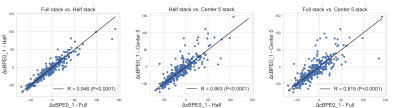 |
Comparison of breast BPE segmentation methods for early prediction of response to treatment
Alex Nguyen1, Fredrik Strand2, Vignesh Arasu1, Wen Li1, Natsuko Onishi1, Jessica Gibbs1, Bonnie N Joe1, Laura J Esserman3, The I-SPY2 Investigator Network4, David C Newitt1, and Nola M Hylton1
1Department of Radiology and Biomedical Imaging, University of California, San Francisco, San Francisco, CA, United States, 2Department of Oncology and Pathology, Karolinska Institutet, Stockholm, Sweden, 3Department of Surgery, University of California, San Francisco, San Francisco, CA, United States, 4Quantum Leap Healthcare Collaborative, San Francisco, CA, United States
Breast parenchymal enhancement (BPE) has shown association with breast cancer risk and response to neoadjuvant treatment. However, BPE quantification is challenging and there is no agreed upon standard. This study compares the results of three fully automated segmentation methods for early prediction of pathologic complete response (pCR) following neoadjuvant treatment. We evaluated three different sub-volumes of interest segmented from DCE-MRI: full stack, half stack, and center 5 slices. The differences between methods were assessed and a univariate logistic regression model was implemented to determine predictive performance of each segmentation method.
|
|
2374.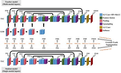 |
Breast Lesion Segmentation in MR Images through Knowledge Distillation-based Modality Speculation
Cheng Li1, Hui Sun1, Taohui Xiao1, Zaiyi Liu2, Qiegen Liu3, Xin Liu1, Hairong Zheng1, and Shanshan Wang1
1Paul C. Lauterbur Research Center for Biomedical Imaging, Shenzhen Institutes of Advanced Technology, Chinese Academy of Sciences, Shenzhen, China, 2Department of Radiology, Guangdong General Hospital, Guangdong Academy of Medical Sciences, Guangzhou, China, 3Department of Electronic Information Engineering, Nanchang University, Nanchang, China
Multi-modal MR images are widely utilized to overcome the shortcomings of single modalities and pursue accurate image-based diagnoses. However, multi-modal MR imaging takes a longer time. For automated diagnosis, misalignment between modalities brings extra problems. To address these issues, we propose a new strategy to speculate the modality information by distillation-based knowledge transfer. Experiments on breast lesion segmentation confirm the feasibility of the proposed method. Networks trained with our method and single-modal MR image inputs can partially recover the breast lesion segmentation performance of models trained using two-modal MR images.
|
|
2375.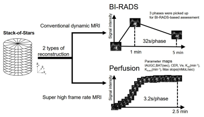 |
Perfusion analyses of breast benign and malignant lesions using super high frame rate reconstruction with stack-of-stars acquisition
Yoshie Omiya1, Tetsuya Wakayama2, Daiki Tamada1, Sagar Mandava3, Ty A Cashen4, Hiroshi Onishi1, and Utaroh Motosugi1
1University of Yamanashi, Chuo, Japan, 2MR Collaboration and Development, GE Healthcare, Hino, Japan, 3MR Collaboration and Development, GE Healthcare, Tucson, AZ, United States, 4MR Collaboration and Development, GE Healthcare, Madison, WI, United States
Dynamic contrast-enhanced MRI (DCE-MRI) is used to diagnose breast cancer. Although 3-phase DCE-MRI is used in clinical settings according to BI-RADS, higher temporal resolution with perfusion analyses may be essential to precisely distinguish between benign and malignant breast lesions. Stack-of-stars (SOS) acquisition enables motion-robust DCE-MRI with arbitrary high temporal resolution. We performed perfusion analyses of breast lesions using super-high frame rate reconstruction (3.2 s/phase) with SOS acquisition, compared with BI-RADS-based enhancement category using typical temporal resolution (32 s/phase), and found that two more lesions were accurately diagnosed via perfusion-based analyses using super-high frame reconstruction compared to BI-RADS-based evaluation.
|
|
2376.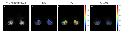 |
Multi-nuclear MRI to disentangle intracellular sodium concentration and extracellular volume fraction in breast
Carlotta Ianniello1,2, Ryan Brown1,2, Linda Moy1,2, and Guillaume Madelin1,2
1Radiology, Center for Advanced Imaging Innovation and Research (CAI2R) and Center for Biomedical Imaging, New York University School of Medicine, NEW YORK, NY, United States, 2The Sackler Institute of Graduate Biomedical Science, New York University School of Medicine, NEW YORK, NY, United States
The intracellular sodium concentration (CIC) and extracellular volume fraction (ECV) are promising biomarkers in breast lesions. However, separating the contribution of the intracellular (IC) and extracellular (EC) compartments only relying on differences in 23Na relaxation properties is challenging due to close similarity between the two compartments and the inability to measure IC and EC relaxation times in vivo. In this study we demonstrate the feasibility to quantify CIC and ECV in three healthy subjects by using complementary information from 23Na and 1H MRI measurements. The mean CIC and ECV between three healthy subjects were 27.7±4.1 mM and 0.29±0.07, respectively.
|
|
2377.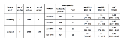 |
A meta-analysis comparing the diagnostic performance of abbreviated MRI (ABB-MRI) and a full diagnostic protocol (FDP-MRI) in breast cancer
Gabrielle C Baxter1, Ayla Selamoglu2, James W Mackay1, Simon Bond3, Ewan Gray4, and Fiona J Gilbert1
1Department of Radiology, University of Cambridge, Cambridge, United Kingdom, 2Department of Psychiatry, University of Cambridge, Cambridge, United Kingdom, 3National Institute of Health Research, Cambridge Clinical Trials Unit, Cambridge, United Kingdom, 4University of Edinburgh, Edinburgh, United Kingdom
The diagnostic performance of abbreviated MRI (ABB-MRI) and full diagnostic protocol MRI (FDP-MRI) in the detection and diagnosis of breast cancer was compared using meta-analysis. 13 eligible studies were included (5 screening studies and 8 enriched cohort studies) and pooled estimates of sensitivity, specificity and area under the curve were calculated for ABB-MRI and FDP-MRI for both types of study. The diagnostic performances of ABB-MRI and FDP-MRI were comparable.
|
|
2378.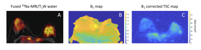 |
Quantitative Bilateral Sodium and Proton Breast Imaging at 3T
Gabrielle C Baxter1, Mary A McLean2, Joshua D Kaggie1, Frank Riemer3, Ferdia A Gallagher1, Andrew D James4,5, Aneurin J Kennerley5,6, Rolf F Schulte7, William J Brackenbury4,5, and Fiona J Gilbert1
1Department of Radiology, University of Cambridge, Cambridge, United Kingdom, 2Cancer Research UK, Cambridge, United Kingdom, 3Department of Radiology, Haukeland University Hospital, Bergen, Norway, 4Department of Biology, University of York, York, United Kingdom, 5York Biomedical Research Institute, University of York, York, United Kingdom, 6Department of Chemistry, University of York, York, United Kingdom, 7GE Healthcare, Munich, Germany
Sodium (23Na)-MRI was performed using a dual-tuned bilateral 23Na/1H breast coil on four healthy volunteers. Images acquired using 23Na-MRI show a relationship between sodium concentration and diffusion properties of breast tissue. Sodium signal variations due to mild flip angle non-uniformity were observed between left and right breasts. After correction using B1 mapping, tissue sodium concentration (TSC) maps were more closely matched between breasts.
|
|
2379.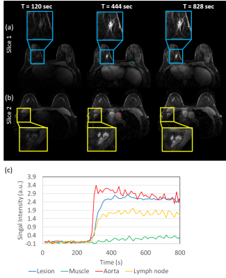 |
Use of a Digital Reference Object for Validation of Advanced Reconstruction Methods in High Spatial-temporal Resolution Breast DCE-MRI
Ping N Wang1, Julia V Velikina1, Roberta M Strigel1,2,3, Leah C Henze Bancroft2, Ty A Cashen4, Kevin M Johnson1, Alexey A Samsonov2, Ali Ersoz5, Edward F Jackson1,2,3, and James H Holmes2
1Department of Medical Physics, University of Wisconsin Madison, Madison, WI, United States, 2Department of Radiology, University of Wisconsin Madison, Madison, WI, United States, 3Carbone Cancer Center, University of Wisconsin Madison, Madison, WI, United States, 4Global MR Applications & Workflow, GE Healthcare, Madison, WI, United States, 5MR Engineering, GE Healthcare, Waukesha, WI, United States
Advanced data acquisition and reconstruction methods have been proposed to improve temporal and spatial resolution DCE imaging for breast. However, validation and comparison of these methods against a known truth is challenging. In this work we propose a digital reference object for breast pharmacokinetic simulation to evaluate different advanced reconstructions including MOCCO and iGRASP. The approach allowed comparison of the reconstructed temporal characteristics including pharmacokinetic analysis using different reconstruction parameters against the assigned ground truth. Spatial sharpness was also measured to compare with reference fully sampled images.
|
|
2380.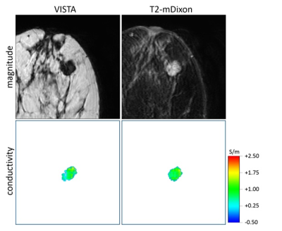 |
Non-contrast method for breast tumor detection using EPT based tissue conductivity maps reconstructed from routine T2-mDixon TSE images
Rakesh Kumar Gupta1, Ulrich Katscher2, Rupsa Bhattacharjee3, Mamta Gupta1, and Indrajit Saha 3
1Department of Radiology, Fortis Memorial Research Institute, Gurugram, India, 2Philips GmbH / Innovative Technologies / Research Laboratories, Hamburg, Germany, 3Philips Health Systems, Philips India Limited, Gurugram, India
Breast tissue conductivity map based on EPT MRI is one of the non-contrast methods that can differentiate malignant from benign breast lesions; however, the need for DCE based tumor “mask” to generate conductivity maps from the phase of VISTA images in breast challenges EPT’s role as a non-contrast method. This study demonstrates an alternative to reconstruct breast tissue conductivity maps using T2-w mDixon TSE sequence that is comparable with DCE/VISTA method, and demonstrates similarities in detecting cancerous tissues. EPT maps of breast using T2-w mDixon TSE method has the potential to help in developing non-contrast MR breast cancer screening protocol.
|
|
2381.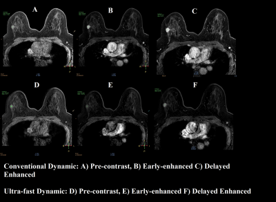 |
Ultra-fast DCE breast MRI protocol using Compressed-SENSE: A quantitative comparison study with standard protocol in Breast Cancer detection
Shikha Panwar1, Rupsa Bhattacharjee2, Vivek Pawar3, Stanley PK2, and Harsh Mahajan1
1Mahajan Imaging, Gurugram, India, 2Philips Health Systems, Philips India Limited, Gurugram, India, 3Philips Health Systems, Philips India Limited, Pune, India
Main challenge associated with conventional DCE-MRI of breast is compromise between spatial and temporal-resolution. In this study our objective is to develop an ultra-fast breast dynamic protocol keeping both spatial and temporal resolution sufficiently high. This is enabled by using compressed SENSE acceleration technique. Proposed Compressed-SENSE enabled ultra-fast dynamic breast DCE protocol design has the potential to be used as alternative of conventional DCE protocol. It gives contrast wash-in information from arteries and veins within very short time, and can significantly differentiate between malignant and benign breast lesions quantitatively (validated on proven 10 malignant and 10 benign cases).
|
|
2382.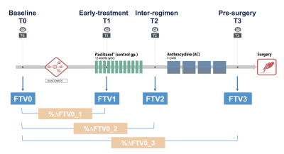 |
Impact of MRI protocol compliance on the prediction of pCR in the I-SPY 2 neoadjuvant breast cancer trial
Natsuko Onishi1, Li Wen1, Jessica Gibbs1, Ella F. Jones1, Lisa J. Wilmes1, Alex Nguyen1, Vignesh Arasu1, Bonnie N. Joe1, Laura J. Esserman2, The I-SPY 2 Investigator Network3, David C. Newitt1, and Nola M. Hylton1
1Department of Radiology & Biomedical Imaging, University of California, San Francisco, San Francisco, CA, United States, 2Department of Surgery, University of California, San Francisco, San Francisco, CA, United States, 3Quantum Leap Healthcare Collaborative, San Francisco, CA, United States
In the I-SPY2 breast cancer trial, functional tumor volume (FTV, a quantitative measure of tumor burden derived from dynamic contrast-enhanced MRI) serves as a marker used for patient randomization and estimation of predictive probabilities for pathologic complete response (pCR). This retrospective study investigated the impact of MRI protocol compliance on prediction of pCR using FTV at four time points during neoadjuvant chemotherapy. FTV with compliant image quality factors tended to show higher AUC than that with non-compliant factors. This tendency was most prominent in percent change of FTV from baseline at early-treatment (3 weeks after treatment initiation) and inter-regimen.
|
|
2383.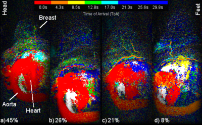 |
The impact of aorta contrast enhancement patterns on ultrafast time of arrival measurements when screening for breast cancer with MRI
Keith S Cover1
1Cancer Center Amsterdam, Amsterdam University Medical Center, VUmc, Amsterdam, Netherlands
A growing number of MRI screening protocols to detect breast cancer are using ultrafast imaging to improve specificity by distinguishing between malignant and benign for small lesions by their time of arrival (ToA) of contrast relative to the aorta. Sagittal MIP of color intensity projections (CIP) of the middle quarter of the chest were used to display the DISCO ultrafast enhancement patterns of the aorta for 556 scans. While 71% of the scans presented simple aorta enhancement patterns, the remaining 29% were more complex. To ensure accurate ultrafast ToA measurements, the aorta enhancement pattern of each scan should be reviewed.
|
|
2384.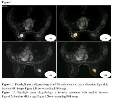 |
A feasibility study of radiomics based on dynamic contrast enhanced magnetic resonance imaging in identifying benign and malignant breast mass.
Weibo Gao1, Xin Chen1, Quanxin Yang1, and Xiaohui Li1
1The second affiliated hospital of xi 'an jiaotong university, xi an, China
The current study aim to investigate the value of radiomics’ features based on dynamiccontrast-enhanced magnetic resonance imaging (DCE-MRI) in the identification of benign andmalignant breast tumors. It was concluded that the radiomics’ features based on dynamicenhanced magnetic resonance imaging have certain value in identifying benign and malignantbreast tumors, among them, the superior radiomics’ features is Gabor, gray level co-occurrencematrix variance and variance, which worth further study.
|
|
2385.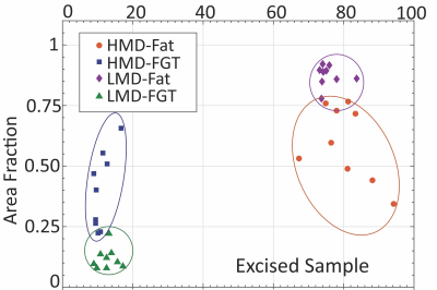 |
Portable NMR for quantification of breast density and tissue composition: from proof-of-concept in vitro to trials in vivo
Konstantin I. Momot1, Honor J. Hugo 2, Xuan Huang2, Tony Blick2, Tonima S. Ali3, Monique C. Tourell3, Thomas Lloyd4, and Erik W. Thompson2
1School of Chemistry and Physics, Queensland University of Technology, Brisbane, Australia, 2Queensland University of Technology, Brisbane, Australia, 3University of Queensland, Brisbane, Australia, 4Princess Alexandra Hospital, Brisbane, Australia
Elevated mammographic density (MD) is a significant independent risk factor for breast cancer and a source of masking in X-ray mammography. Availability of a low-cost, non-ionising measurement technique for quantitative assessment of MD would be valuable both in therapeutic and research contexts. We demonstrate that Portable NMR, a low-cost technique based on the same physics as MRI, represents a reliable approach to quantification of MD in excised breast tissue samples in vitro. We also present the initial results of an observational trial in healthy volunteers, which show that Portable NMR has a great potential for quantification of MD in vivo.
|
|
2386.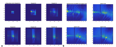 |
Advances in 3D High-Speed MR Spectroscopic Imaging for Monitoring Neoadjuvant Chemotherapy in Breast Cancer
Stefan Posse1, Ingrid Lane2, Bruno Sa De La Rocque Guimaraes1, Bernard Twafik3, and Ursa Brown-Glaberman3
1Neurology, University of New Mexico, Albuquerque, NM, United States, 2Mind Research Network, Albuquerque, NM, United States, 3Division of Hematology / Oncology, University of New Mexico Comprehensive Cancer Center, Albuquerque, NM, United States
Three patients with biopsy-confirmed, infiltrating ductal carcinoma were studied at 3 Tesla to monitor changes in total Choline during neoadjuvant chemotherapy in comparison with dynamic contrast enhanced MRI. A novel bilateral high-speed 3D Proton-Echo-Planar-Spectroscopic-Imaging (PEPSI) protocol with integrated water reference acquisition mapped decreases in total Choline early during treatment in 2 HER2-negative patients with partial pathologic response, but not in the third HER2-positive patient. Surgical clip related B0-inhomogeneity impacted both PEPSI and DCE-MRI measurements. To address this limitation, we developed a novel expanded k-space encoding approach for PEPSI and demonstrated signal recovery in tissue regions affected by dephasing.
|