
Digital Poster Session
Body: Abdominopelvic MRI - Cancer
Body
2387 -2400 Abdominopelvic MRI - Cancer - Deep Prostatomics
2401 -2415 Abdominopelvic MRI - Cancer - Hepatocellular Carcinoma
2416 -2428 Abdominopelvic MRI - Cancer - Shallow Teaching Prostate (Not Deep Learning)
2429 -2439 Abdominopelvic MRI - Cancer - Rectal Cancer
2440 -2452 Abdominopelvic MRI - Cancer - Prostate
2453 -2465 Abdominopelvic MRI - Cancer - Genitourinary Oncology & Function
2466 -2477 Abdominopelvic MRI - Cancer - Hepatobiliary & Pancreas
2387.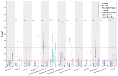 |
The repeatability of deep learning-based segmentation of the prostate on T2-weighted MR images
Mohammed R. S. Sunoqrot1, Sandra Kucharczak2, Magdalena Grajek2, Kirsten M. Selnæs1,3, Tone F. Bathen1,3, and Mattijs Elschot1,3
1Department of Circulation and Medical Imaging, NTNU, Norwegian University of Science and Technolog, Trondheim, Norway, 2Department of quantum electronics, Adam Mickiewicz University, Poznań, Poland, 3Department of Radiology and Nuclear Medicine, St. Olavs Hospital, Trondheim University Hospital, Trondheim, Norway
Inter- and intra-observer variability are current limitations of radiological reading of multiparametric MR images of the prostate. Deep learning (DL)-based segmentation has proven to provide good performance, but little is known about the repeatability of these methods. In this work, we investigated the intra-patient repeatability of shape features for DL segmentation methods of the prostate on T2-weighted MR images and compared it to manual segmentations. We found that the repeatability of the investigated methods is excellent for most of the investigated shape features.
|
|
2388.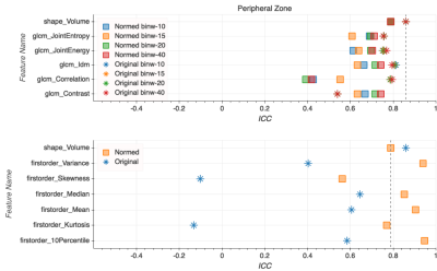 |
Improving the repeatability of radiomic analysis of the prostate through deep normalization of T2w MRI inputs
Stephanie Alley1, Andrey Fedorov2,3, Cynthia Menard4, and Samuel Kadoury1,4
1Polytechnique Montréal, Montréal, QC, Canada, 2Brigham and Women’s Hospital, Boston, MA, United States, 3Harvard Medical School, Boston, MA, United States, 4Centre Hospitalier de l’Université de Montréal, Montréal, QC, Canada
Radiomics analyses are being increasingly employed to investigate tissue heterogeneity present within the prostate gland. We present a method for improving the repeatability of radiomics features extracted from T2-weighted images using a deep normalization technique based on fully convolutional networks (FCNs). We test the repeatability of select radiomics features on a previously published test-retest prostate dataset. We demonstrate that the intraclass correlation coefficient of first-order statistics features extracted from images normalized using the FCN-based pre-processor is consistently higher than for features extracted from non-normalized images.
|
|
2389.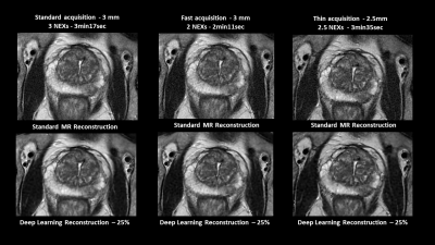 |
Learning how to adapt T2 PROPELLER MR prostate imaging: going beyond PIRADS requirements with MR Deep Learning reconstruction
Julie Poujol1, Charline Henry1, Vincent Barrau2, François Legou2, Eric Pessis2, Xinzeng Wang3, and Daniel Litwiller4
1Clinical Research & Development, GE Healthcare, Buc, France, 2Centre Cardiologique du Nord, Saint-Denis, France, 3Global MR Applications & Workflow, GE Healthcare, Houston, TX, United States, 4Global MR Applications & Workflow, GE Healthcare, New York, NY, United States
To give a confident image-based prostate cancer diagnosis, PIRADS recommends using multiparametric MR (mp-MRI) exam composed of DWI and T2w sequences. By using a new deep learning-based image reconstruction algorithm, we aim to improve the utility of T2w PROPELLER images by reducing acquisition time and/or increasing spatial resolution beyond PIRADS requirements. We present quantitative analysis based on signal-to-noise ratio estimates and qualitative analysis based on delineation of anatomical structures, overall image quality, vision of thin structures.
|
|
2390.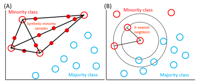 |
Augmented ensemble learning is effective strategy for imbalanced small dataset: improve differentiation of low from high grade prostate cancer
Yuta Akamine1, Yoshiko Ueno2, Keitaro Sofue2, Takamichi Murakami2, Yu Ueda1, Ahsan Budrul1, Masami Yoneyama1, Makoto Obara1, and Marc Van Cauteren3
1Philips Japan, Tokyo, Japan, 2Department of Radiology, Kobe University Graduate School of Medicine, Hyogo, Japan, 3Asia Pacific, Philips Healthcare, Tokyo, Japan
Machine learning (ML) techniques have gained more attention to distinguish low from high grade prostate cancer. However, obtaining big training data is difficult. Moreover, ML models created by imbalanced dataset have a high accuracy for majority, but a low accuracy for minority. For this problem, data augmentation is widely studied. Recently, ensemble learning, which merges different classifiers, has shown great potential. Combinations of data augmentation and ensemble learning were investigated, using multi-parametric MR. We demonstrated that synthetic-minority-over-sampling-technique (SMOTE) with ensemble learning showed increased F1 (0.831) and AUC (0.762) and is effective strategy to improve diagnosis performance for imbalanced small dataset.
|
|
2391. |
Applying Radiomics and Convolutional Neural Network Analysis to distinguish Lymph Node Invasion in Prostate Cancer using Multi-parametric MRI
Yang Song1, Ying Hou2, Min-xiong Zhou3, Xu Yan4, Ye-feng Yao1, Yu-dong Zhang2, and Guang Yang1
1Shanghai Key Laboratory of Magnetic Resonance, East China Normal Univeristy, Shanghai, China, 2Department of Radiology, the First Affiliated Hospital with Nanjing Medical University, Nanjing, China, 3Shanghai University of Medicine & Health Sciences, Shanghai, China, 4MR Scientific Marketing, Siemens Healthcare, Shanghai, China
We compared a radiomics model and a convolutional neural network (CNN) model to distinguish lymph node invasion (LNI) in prostate cancer (PCa) using multi-parametric magnetic resonance images (mp-MRI). We trained the models in 281 patients and evaluated them in another 71 cases. The radiomics/CNN model produced an AUC 0.741/0.722, sensitivity 0.769/0.692, specificity 0.690/0.845 for the differentiation of LNI, which showed potential for the diagnosis of LNI in PCa.
|
|
2392.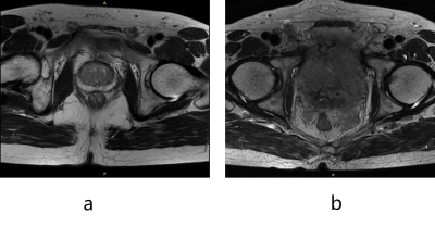 |
Prostate MRI Image Quality Control using Deep Learning
Jing Zhang1, Yang Song1, Ying Hou2, Yu-dong Zhang2, Xu Yan3, Yefeng Yao1, and Guang Yang1
1Shanghai Key Laboratory of Magnetic Resonance, Department of Physics, East China Normal University, shanghai, China, 2Department of Radiology, The First Affiliated Hospital with Nanjing Medical University,, Nanjing, China, 3MR Scientific Marketing, Siemens Healthcare, shanghai, China
Deep learning-based computer aided diagnosis (CAD) has been proposed to detect and classify prostate cancer lesions in multi-parametric Magnetic Resonance Imaging (mp-MRI) images. CAD requires their input images meet certain quality standards. In this work, we proposed a ResNet50-based model to filter out images not suitable as the input to the following lesion detection network. Taking unqualified images as positive cases, we obtained an area under ROC curve (AUC) of 0.8526 in test cohort, which helped to improve the performance of detection model and increased the interpretability by rejecting unqualified images with a reason instead of giving wrong results.
|
|
2393.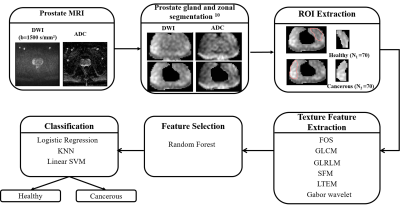 |
Prostate Tumor Characterization using Texture Analysis of Diffusion MRI
Dharmesh Singh1, Virendra Kumar2, Chandan J Das3, Anup Singh1,4, and Amit Mehndiratta1,4
1Centre for Biomedical Engineering, Indian Institute of Technology (IIT) Delhi, New Delhi, India, 2Department of NMR, All India Institute of Medical Sciences Delhi, New Delhi, India, 3Department of Radiology, All India Institute of Medical Sciences Delhi, New Delhi, India, 4Department of Biomedical Engineering, All India Institute of Medical Sciences Delhi, New Delhi, India
Accurate diagnosis of prostate-cancer(PCa) remains challenging due to high false-negative rate of biopsy and low-specificity of the screening test. Computer-aided diagnosis(CAD) systems are increasingly being used for detection and diagnosis of PCa. Texture analysis has been proved to be a significant CAD tool in medical applications. The aim of this research was to investigate the role of texture parameters extracted from diffusion-weighted MRI and machine-learning classifiers in distinguishing PCa from normal peripheral-zone(PZ). The proposed methodology has achieved 93% accuracy using support-vector machine classifier. Experiments showed that the application of texture-analysis could improve the accuracy of identifying healthy and cancerous prostate-regions.
|
|
2394.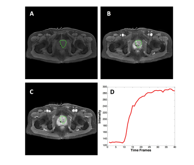 |
Artificial Intelligence Analysis on Prostate DCE-MRI to Distinguish Prostate Cancer and Benign Prostatic Hyperplasia
Yang Zhang1, Weikang Li2, Zhao Zhang2, Yingnan Xue2, Peter Chang1, Daniel Chow1, Min-Ying Su1, and Qiong Ye2,3
1Department of Radiological Sciences, University of California, Irvine, Irvine, CA, United States, 2The Department of Radiology, The First Affiliated Hospital of Wenzhou Medical University, Wenzhou, China, 3High Magnetic Field Laboratory, Hefei Institutes of Physical Science, Chinese Academy of Sciences, Hefei, China
Three convolutional neural network architectures were applied to differentiate prostate cancer from benign prostate hyperplasia based on DCE-MRI: (1) VGG serial convolutional neural network; (2) one-directional Convolutional Long Short Term Memory (CLSTM) network; (3) bi-directional CLSTM network. A total of 104 patients were analyzed, including 67 prostate cancer and 37 benign prostatic hyperplasia. Upon 10-fold cross-validation, the differentiation accuracy was 0.64-0.77 (mean 0.68) using VGG, 0.75-0.87 (mean 0.81) using the CLSTM, and 0.73-0.89 (mean 0.84) using bi-directional CLSTM. The radiomics model built by SVM using histogram and texture features extracted from the manually-drawn tumor ROI yielded accuracy of 0.81.
|
|
2395.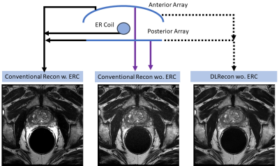 |
High Resolution Prostate T2-weighted MRI with Deep Learning and without an Endorectal Coil
Xinzeng Wang1, Jong Bum Son2, Priya Bhosale3, Aliya Qayyum3, Juan Jose Ibarra-Rovira3, Ken-Ping Hwang2, Jason Stafford2, Mark David Pagel4, Marc Lebel5, Ersin Bayram1, Jingfei Ma2, and Janio Szklaruk3
1Global MR Applications & Workflow, GE Healthcare, Houston, TX, United States, 2Department of Imaging Physics, MD Anderson Cancer Center, Houston, TX, United States, 3Department of Abdominal Imaging, MD Anderson Cancer Center, Houston, TX, United States, 4Department of Cancer Systems Imaging, MD Anderson Cancer Center, Houston, TX, United States, 5Global MR Applications & Workflow, GE Healthcare, Calgary, AB, Canada
Endorectal coil (ERC) offers high SNR that is needed for high-resolution prostate MRI, which is essential for accurate visualization of prostate and detection of prostate cancer. Unfortunately, use of an ERC is cumbersome, costly, and discomforting or even intolerable to some patients. Further, ERC often has exacerbated motion artifacts due to its proximity to the regions of interests. In this work, we applied a novel deep learning-based MR reconstruction method to clinical prostate T2-weighted imaging without an ERC (non-ERC). DL Recon improved non-ERC image SNR, reduced artifacts, and improved overall image quality compared to conventional reconstruction with/without endorectal coil.
|
|
2396.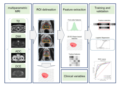 |
Radiomics based on multiparametric magnetic resonance imaging to predict extraprostatic extension of prostate cancer
Lili Xu1, Gumuyang Zhang1, Lun Zhao2, Li Mao2, Xiuli Li2, Weigang Yan3, Yu Xiao4, Jing Lei1, Zhengyu Jin1, and Hao Sun1
1Department of Radiology, Peking Union Medical College Hospital, Peking Union Medical College, Chinese Academy of Medical Sciences, Beijing, China, 2Deepwise AI Lab, Beijing, China, 3Department of Urology, Peking Union Medical College Hospital, Peking Union Medical College, Chinese Academy of Medical Sciences, Beijing, China, 4Department of Pathology, Peking Union Medical College Hospital, Peking Union Medical College, Chinese Academy of Medical Sciences, Beijing, China
The preoperative prediction of EPE has a profound impact on treatment decision making, however, it still remains challenging presently. In this study, we compared the radiomics signatures extracted from different MR sequences to diagnose EPE. The radiomics signature based on DWI showed better performance for EPE prediction among mpMRI sequences. The radiomics model based on DWI, T2WI and DCE images was demonstrated feasible for the prediction of EPE. But the added value of clinical variables to the radiomics model was not prominent.
|
|
2397. |
Differential diagnosis of prostate cancer and benign prostatic hyperplasia using texture features based on multiple high b-values DWI
Jingjun Wu1, Ailian Liu1, Jiazheng Wang2, Zhiwei Shen2, Lihua Chen1, Qingwei Song1, Renwang Pu1, and Bingbing Gao1
1Department of Radiology, the First Affiliated Hospital of Dalian Medical University, Dalian, China, 2Philips Healthcare, Beijing, China
This work aimed for multiple high b-values DWI texture features based strategy to identify prostate cancer and benign prostatic hyperplasia, which may provide more abundant and comprehensive quantitative information, and promote clinical decision-making. The results showed that the diagnostic value was improved with AUC of 0.832 (sensitivity: 0.800, specificity: 0.762) based on combined texture features from ultra-high b value (b = 800, 1200, 2000s/mm2) DWI images, which is a valuable strategy for clinical practice.
|
|
2398.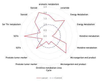 |
Characterization of the Metabolomic Signature of Prostate Cancer by Mass Spectrometry and Magnetic Resonance Spectroscopy of Urine
Leo L. Cheng1, Andrew Gusev1, Alex Buko2, Takushi Oga2, and Adam S. Feldman1
1MGH/Harvard, Boston, MA, United States, 2Human Metabolome Technologies, Boston, MA, United States
We used Capillary Electrophoresis Time of Flight Mass Spectrometry and Magnetic Resonance Spectroscopy to analyze urine from men undergoing prostate biopsy to investigate their metabolomic profiles and look for potential biomarkers. CE-MS analysis identified 60 metabolites that were statistically different between urine samples of men with PCa and PCa-free men and had overlap with MRS. Pathway analysis of these showed high activity in ceramide, short chain fatty acid (SCFA), branched chain amino acid, serine, threonine and tryptophan metabolism. Targeted studies of these metabolites are underway in our lab; if validated, they have potential to serve as non-invasive biomarkers for PCa.
|
|
2399.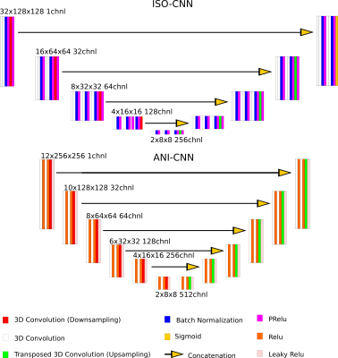 |
Evaluation of convolution strategies for 3D-CNN for MR Prostate segmentation
Yanlu Wang1,2, Patrick Masaba3, Fredrik Strand4,5, and Fredrik Järderling6,7
1Department of Clinical Sciences, Intervention and Technology, Karolinska Institute, Stockholm, Sweden, 2Medical Radiation Physics and Nuclear Medicine, Karolinska University Hospital, Solna, Sweden, 3Imaging and Function, Karolinska University Hospital, Stockholm, Sweden, 4Breast Radiology, Karolinska University Hospital, Solna, Sweden, 5Department of Oncology-Pathology, Karolinska Institute, Stockholm, Sweden, 6Department of Molecular Medicine and Surgery, Karolinska Institute, Stockholm, Sweden, 7Department of Radiology, Capio S:t Göran Hospital, Stockholm, Sweden
Typical prostate MR dataset are 2D with few slices and high in-plane resolution. These dimensions do not conform naturally to traditional 3D convolution neural network up/down-sampling schemes, which customarily double/halves the input dimensions in all directions. We compared two strategies for up/down-sampling applied to prostate segmentation in MRI datasets: 1) Transform the datasets and applying orthodox convolution layers. 2) Adapt the up/down-sampling convolution layers in order to use more native resolution input datasets. Our results show that, in general, the former strategy works best, both in terms training loss convergence and overall segmentation results.
|
|
2400.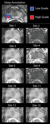 |
Post processing decisions, independent of institution and fit, influence the perceived diagnostic utility of prostate cancer diffusion maps
Sean D McGarry1, John D Bukowy2, Kenneth Iczkowski3, Allison K Lowman2, Anjishnu Banerjee4, Kenneth Jacobsohn5, Petar Duvnjak2, Michael Griffin2, Dariya Malyarenko6, Tom Chenevert6, Yuan Li7,8, DaeKeun You7, Yue Cao6,7,
Andrey Fedorov9, Laura C Bell10, Chad Quarles10, Melissa A Prah1, Kathleen M Schmainda1, Stefanie Hectors11, Bachir Taouli11, Eve LoCastro12, Yousef Mazaheri12,13, Amita Shukla-Dave12,13, Thomas Yankeelov14, David Hormuth II14, Ananth J Madhuranthakam15,
Keith Hulsey15, Mark Muzi16, Michael Jacobs17, Meiyappan Solaiyappan17, William See5, Mark Hohenwalter2, and Peter S LaViolette18
1Biophysics, Medical College of Wisconsin, Milwaukee, WI, United States, 2Radiology, Medical College of Wisconsin, Milwaukee, WI, United States, 3Pathology, Medical College of Wisconsin, Milwaukee, WI, United States, 4Biostatistics, Medical College of Wisconsin, Milwaukee, WI, United States, 5Urological Surgery, Medical College of Wisconsin, Milwaukee, WI, United States, 6Radiology, University of Michigan, Ann Arbor, MI, United States, 7Radiation Oncology, University of Michigan, Ann Arbor, MI, United States, 8Biomedical Engineering, University of Michigan, Ann Arbor, MI, United States, 9Radiology, Brigham and Womens Hospital, Boston, MA, United States, 10Neuroimaging Research, Barrow Neurological Institute, Phoenix, AZ, United States, 11Translational and Molecular Imaging Institute, Icahn School of Medicine at Mount Sinai, New York, NY, United States, 12Medical Physics, Memorial Sloan Kettering Cancer Center, New York, NY, United States, 13Radiology, Memorial Sloan Kettering Cancer Center, New York, NY, United States, 14Institute for Computer Engineering and Sciences, University of Texas, Austin, TX, United States, 15Radiology, University of Texas Southwestern Medical Center, Dallas, TX, United States, 16Radiology and Neurology, University of Washington, Seattle, WA, United States, 17Radiology, Johns Hopkins University, Baltimore, MD, United States, 18Radiology, Medical College of Wisconsin, wauwatosa, WI, United States
Diffusion data from 33 prospectively recruited patients was distributed to 14 sites, who returned monoexponential, biexponential, and diffusion kurtosis fits of the data. Receiver operator characteristic curve AUC was evaluated while varying the positive condition in the ROC analysis, minimum lesion size included, and method of extracting values from the ROI. Cluster size and stratification technique have a significant impact on the AUC and should be explicitly stated in future rad-path studies.
|
 View the Poster
 Watch the Video
2401.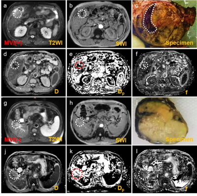 |
Visualize the micro-hemorrhage of HCC through SWI and intravoxel incoherent motions to predict the microvascular invasion
Zhijun Geng1, Yunfei Zhang2, Hui Li1, Jing Zhao1, Sihui Zeng1, Cheng Zhang1, Chuanmiao Xie1, and Yongming Dai2
1Sun Yat-sen University Cancer Center, Guangzhou, China, 2United Imaging Healthcare, Shanghai, China
Tightly correlated with the prognosis, microvascular invasion (MVI) serves as one significant predictor determining the clinical management. Abnormal angiogenesis, one typical character during carcinogenesis, can lead to the abnormal micro-vessel manifestations including micro-hemorrhage and more, which may correlate with another abnormal vascular manifestations—MVI. Hence, this research aims to predict the MVI via visualizing the intra-tumor hemorrhage with susceptibility weighted imaging (SWI) widely proven to be powerful for imaging hemorrhage and IVIM hypothesized to be effective for visualizing the hemorrhage via quantifying the abnormal perfusion. The results displayed that visualizing the micro-hemorrhage holds great potential for predicting MVI of HCC.
|
|
2402.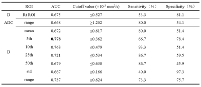 |
3D ROI Histogram Based on Intravoxel Incoherent Motion for Preoperative Evaluation of Pathological Differentiation of the Hepatocellular Carcinoma
Zhe Zhang1, Ailian Liu1, Ying Zhao1, Jingjun Wu1, Nan Wang1, Dahua Cui1, Tao Lin1, Qingwei Song1, Xin Li1, Tingfan Wu1, and Yan Guo1
1Department of Radiology, the First Affiliated Hospital of Dalian Medical University, Dalian, China, Dalian, China
The purpose of this study was to preoperatively evaluate pathological differentiation of the whole hepatocellular carcinoma (HCC) by 3D ROI histogram of ADC, D, D* and f. The results showed that 3D ROI histogram of ADC, D can identify poor differentiated HCC. The D-5th achieved the highest AUC values (AUC: 0.778; sensitivity: 66.7%; specificity: 78.4%). Although there was no significant difference between AUC of 3D ROI histogram and routine ROI, 3D ROI histogram can reflect the heterogeneity to evaluate the differentiation of the whole tumor.
|
|
2403.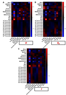 |
Evaluating the feasibility of predicting the multiple pathological indexes of hepatocellular carcinoma with a single MR scan
Xue Han1, Yunfei Zhang2, Qi Wang1, Yan Ding1, Hui Liu1, Xiaojing Ma1, Gaofeng Shi1, and Yongming Dai2
1The Fourth Affiliated Hospital of Hebei Medical University, Shijiazhuang, China, 2United Imaging Healthcare, Shanghai, China
Imaging manifestations and pathological findings are tightly correlated to each other as they all provide the portraying of the tumor from different perspectives. We hypothesized that exploiting the quantitative markers from MR images would be a feasible way for simultaneously predicting the multiple pathological indexes. With regard to conventional “mean value” of ROI, histogram metrics, taking the tumor heterogeneity into consideration, can give full play of quantitative indicators. This research aims to preliminarily evaluate the feasibility of simultaneously predicting the multiple pathological indexes with a single Intravoxel incoherent motion (IVIM) scan by extracting multiple histogram metrics of parametric images.
|
|
2404. |
Predicting therapeutic response using dynamic enhanced MRI radiomics for patients with HCC treated with TACE: comparison of radiomics models
Ying Zhao1, Ailian Liu1, Jingjun Wu1, Nan Wang1, Dahua Cui1, Tao Lin1, Qingwei Song1, Xin Li2, Tingfan Wu2, and Yan Guo3
1The First Affiliated Hospital of Dalian Medical University, Dalian, China, 2Translational Medicine Team, GE Healthcare, Shanghai, China, 3GE Healthcare, Beijing, China
In the current study, dynamic enhanced MRI-based radiomics model was applied to predict therapeutic response in hepatocellular carcinoma treated with transcatheter arterial chemoembolization. In order to obtain the optimal radiomics model, we compared the diagnostic performance of the model built by different classifiers.
|
|
2405.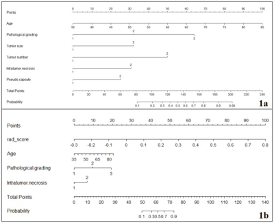 |
Multi-sequence MRI-based radiomics nomogram to predict two-year recurrence in hepatocellular carcinoma after partial hepatectomy
Ying Zhao1, Ailian Liu1, Jingjun Wu1, Dahua Cui1, Tao Lin1, Qingwei Song1, Xin Li2, Tingfan Wu2, Yan Guo3, Lizhi Xie3, and Jingjing Cui4
1The First Affiliated Hospital of Dalian Medical University, Dalian, China, 2Translational Medicine Team, GE Healthcare, Shanghai, China, 3GE Healthcare, Beijing, China, 4Huiying Medical Technology Inc., Beijing, China
In the current study, multi-sequence MRI radiomics nomogram was demonstrated to be capable to predict two-year recurrence in hepatocellular carcinoma after partial hepatectomy, which will provide more prognostic information and facilitate clinical management.
|
|
2406. |
Analysis of liver function after molecular targeting therapy of HCC with hepatocyte fraction index obtained from gadoxetic acid-enhanced MRI.
Yudai Syogan1, Satoshi Kobayashi1, Yu Ueda2, Noboru Taniguchi1, Naoki Ohno1, Tosiaki Miyati1, Toshifumi Gabata3, Takeshi Terashima4, Kuniaki Arai4, and Tatsuya Yamashita4
1Quantum Medical Technology, Kanazawa University Graduate School of Medical Sciences, Kanazawa, Japan, 2Philips.Japan, Tokyo, Japan, 3Radiology, Kanazawa University Graduate School of Medical Sciences, Kanazawa, Japan, 4Gastroenterology, Kanazawa University Graduate School of Medical Sciences, Kanazawa, Japan Poster Permission Withheld
Hepatocyte fraction index (HeFra index) obtained from gadoxetic acid-enhanced MR imaging might be possible surrogate marker of liver function after molecular targeting therapy to advanced HCC, and the molecular targeting therapy to advanced HCC do not affect the function of background liver parenchyma.
|
|
2407. |
Longitudinal study of liver imaging reporting and Data system LR-2, LR-3 and LR-4 observations in cirrhosis on MRI
Fei Xing1, Xue qin Zhang1, Jian Lu1, and Xiao fen Miao 1
1The Third Hospital Affiliated of Nantong University, Nantong, China
Using LI-RADS v2018 on MRI, LR-2, LR-3 and LR-4 lesions in cirrhosis have different natural outcomes, LI-RADS lesions demonstrate increasing risk of progression to HCC with increasing category. About two-fifths of LR-4 observations progressed to a malignant category. LR-3 observations with APHE or threshold/subthreshold growth upgraded to LR-5 were significantly higher. Most LR-2 observations that remain stable in category for at least a year.
|
|
2408.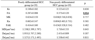 |
The value of multi-quantitative parameters of DKI to evaluate the pathological grade of hepatocellular carcinoma (HCC): a preliminary study
Liuji Sheng1, Ailian Liu1, Ying Zhao 1, Jingjun Wu1, Nan Wang1, Dahua Cui1, Tao Lin1, Qingwei Song1, Xin Li2, Tingfan Wu2, and Yan Guo3
1Department of Radiology, the First Affiliated Hospital of Dalian Medical University, Dalian, China, 2Translational Medicine Team, GE Healthcare, Shanghai, China, 3GE Healthcare, Beijing, China
The main purpose of this work was to use multi-quantitative parameters of DKI to evaluate the pathological grade of HCC before surgery. The results showed that FA have a powerful value in preoperative assessment of pathological grade of HCC (AUC:0.716; sensitivity:73.7%; specificity: 65.0%).
|
|
2409. |
Can IVIM Be used for Preoperative Assessment of Microvascular Invasion in Hepatocellular Carcinoma ?
Yi Wei1, Zheng Ye1, Hehan Tang1, Bin Song1, Xiaocheng Wei2, Lisha Nie2, and Hancheng Yang2
1West China Hospital, Sichuan University, Chengdu, China, 2GE Healthcare, MR Research, Beijing, China
Microvascular invasion (MVI) is one of the most important factors for the recurrence of hepatocellular carcinoma (HCC), however, accurate preoperative evaluation of MVI is quietly difficult because of the controversy results caused by the conventional imaging features. Compared with diffusion-weighted imaging (DWI), Intravoxel incoherent motion (IVIM) diffusion-weighted MR imaging could better characterize heterogeneity and irregularity of tissue components, and thus may have the potential to better evaluate MVI. In this study, we prospectively determine the usefulness of IVIM parameters and conventional radiologic features for preoperative prediction of MVI in patients with HCC.
|
|
2410.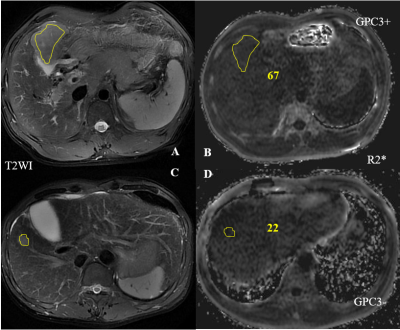 |
R2* yield by IDEAL IQ MRI in Evaluating the Expression Status of Glypican-3 in Hepatocellular Carcinoma
Rushi Chen1, Yan Bai2, Ge Zhang2, Kaiyu Wang3, and Meiyun Wang2
1Henan provincial people's hospital& Zhengzhou University People’s Hospital, Zhengzhou, China, 2Henan provincial people's hospital, Zhengzhou, China, 3GE Healthcare, MR Research, Beijing, China
Accurate diagnosis and evaluation of HCC is important for improving the efficiency of HCC treatment and patient prognosis. Glypican-3 (GPC3) has been considered as a promising diagnostic and prognostic biomarker and a target for immune-therapeutic in HCC. Our study aimed to investigate the utility of IDEAL IQ MRI to prospectively evaluate the expression status of GPC3. Our results showed that the intratumoral R2* value was significantly higher in the positive-GPC3 HCC patients than in the negative-GPC3 HCC patients (P=0.003). The IDEAL IQ MRI may have potential for non-invasively predict GPC3 expression status.
|
|
2411.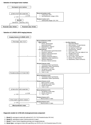 |
Diagnosis of LR-M in at risk patients: performances of serological tumor markers and LI-RADS version 2018 features
Hanyu Jiang1,2, Bin Song1, and Mustafa Shadi Rifaat Bashir2
1Department of Radiology, West China Hospital, Sichuan University, Chengdu, China, 2Department of Radiology, Duke University Medical Center, Durham, NC, United States
The aim of this study was to develop diagnostic models comprising serological tumor markers and gadoxetate disodium-enhanced magnetic resonance imaging (EOB-MRI) features for patients with LR-M lesions by LI-RADS version 2018 (v2018). We retrospectively analyzed prospectively collected data from 45 consecutive at risk patients with LR-M observations and generated diagnostic models for LR-M to predict tumors with a cholangiocarcinoma component (M-CC). Among these models, carbohydrate antigen (CA) 19-9 alone demonstrated excellent diagnostic specificity, while the model integrating both CA 19-9 and the EOB-MRI feature “blood products in mass” achieved optimal overall performance.
|
|
2412.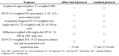 |
Optimized Colorectal Liver Metastasis Screening: Diagnostic Performance of an Abbreviated Gadoxetic Acid-enhanced MR Imaging Protocol.
Nobuyuki Kawai1, Yoshifumi Noda1, Satoshi Goshima2, Keita Fujimoto1, Kimihiro Kajita3, Hiroshi Kawada1, Yukichi Tanahashi1, and Masayuki Matsuo1
1Radiology, Gifu University, Gifu, Japan, 2Diagnostic Radiology and Nuclear Medicine, Hamamatsu University School of Medicine, Hamamatsu, Japan, 3Radiology Services, Gifu University Hospital, Gifu, Japan
Gadoxetic acid-enhanced MR imaging is an essential modality for the screening and assessment of hepatic diseases, which has an incremental value when combined with contrast-enhanced computed tomography for detecting colorectal liver metastasis (CLM). We assessed the diagnostic performance of an abbreviated protocol (axial heavily T2-weighted + axial and reconstructed-coronal hepatobiliary phase + axial diffusion-weighted imaging) compared with the standard routine clinical one for CLM screening. Our results demonstrated excellent performance for the detection of CLM comparable to standard one less than one third of acquisition time.
|
|
2413.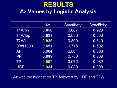 |
Texture Analysis in MRI Diagnosis of Hepatocellular Carcinoma
Takeshi Yoshikawa1, Yoshiharu Ohno2, Ryo Shiroishi3, Masao Yui3, Yoshimori Kassai3, Shinichiro Seki4, Katsusuke Kyotani5, and Yuji Kishida6
1Kobe University Graduate School of Medicine, Kobe, Japan, 2Fujita Health University School of Medicine, Toyoake, Japan, 3Canon Medical Systems Corporation, Otawara, Japan, 4Hyogo Prefectural Tamba Medical Center, Tamba, Japan, 5Kobe University Hospital, Kobe, Japan, 6Konan Medical Center, Kobe, Japan
Texture analysis can characterize spatial variations of gray levels on an image. Our results showed texture analysis has a potential to improve MR ability in diagnosis of hepatocellular carcinoma. Optimal imaging techniques might be different from ones for visual assessment. Capsule, hemorrhage, and cellularity in HCC are possible influential factors.
|
|
2414.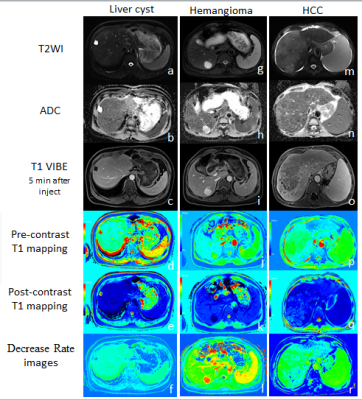 |
Quantitative evaluation liver cyst, hemangioma and hepatocellular carcinoma by using T1 mapping and DWI
Fei Wang1, Yupei Zhang1, Mengxiao Liu2, and Juan Zhu1
1Department of MRI,AnQing Municipal Hospital, Anqing, China, 2MR scientific Marketing, Diagnostic Imaging, Siemens Healthcare Ltd, Shanghai, China
In this study, pre- and post-contrast T1-mapping technique and DWI were used for differential diagnosis of hepatic cyst, hemangioma and HCC. pre- and post-contrast T1 value, the decrease rate of T1 value and ADC value of the three types of lesions were compared and analyzed.T1-mapping technology for differential diagnosis of hepatic cyst,hemangioma and HCC provides a new quantitative method, especially for no-enhanced T1-mapping, have promise of clinical application.
|
|
2415. |
RADIOLOGICAL COURSE OF SUB-CENTIMETER ARTERIALLY ENHANCING NODULES DETECTED DURING SURVEILLANCE FOR HEPATOCELLULAR CARCINOMA
Sonal Krishan1 and Prabhat Kumar2
1Medanta Hospital, Gurgaon, India, 2Medanta Hospital, GURGAON, India
Clinical data on the course of sub-centimeter-sized nodules (SCSNs) detected during surveillance for HCC is limited. Through this study we wished to evaluate arterially enhancing SCSN evolving into HCC. We tried identify specific size cut off, rate of growth, enhancement features which can predict which SCSN will turn into HCC. 47.5% of SCSN have the potential to turn into HCC. 8.55 mm is the optimal size cut off above which risk of developing HCC increases with a sensitivity of 71.4%. Diffusion restriction was seen in 50% and non uptake of hepatobiliary contrast in 91% of SCSN which developed into HCC.
|
2416.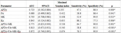 |
Differentiate benign prostatic hyperplasia and prostatic cancer using APTw imaging and DKI Imaging
Yunsong Liu1, Lihua Chen1, Weiping Yang1, Xinmiao Bu1, Jianqing Sun2, Jiazheng Wang2, Zhiwei Shen2, and Ailian Liu1
1The First Affiliated Hospital of Dalian Medical University, Dalian, China, 2Philips Healthcare, Beijing, China
The present study aimed to explore the value of amide proton transfer-weighted (APTw) imaging combined with diffusional kurtosis imaging (DKI) in the differential diagnosis between benign prostatic hyperplasia (BPH) and prostatic cancer. The results showed that the combined technique had a good diagnostic effect. When APT value was combined with 3 DKI parameters (FA, MK, Ka), the diagnostic efficacy significantly enhanced (AUC: 0.872; sensitivity: 76.5%; specificity: 90.9%).
|
|
2417.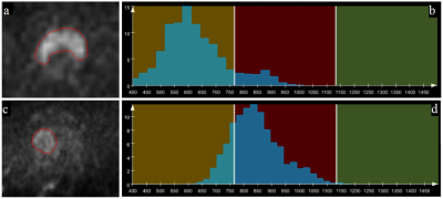 |
Differentiation of Prostate Cancer from Benign Prostatic Hyperplasia using Whole-lesion Histogram Analysis on Diffusion-weighted Imaging
Luguang Chen1, Pengyi Xin1, Qingsong Yang1, Tao Song1, Chao Ma1, Robert Grimm2, Caixia Fu3, and Jianping Lu1
1Radiology, Changhai Hospital of Shanghai, Shanghai, China, 2Application Predevelopment, Siemens Healthcare, Erlangen, Germany, 3Application Development, Siemens Shenzhen Magnetic Resonace Ltd, Shenzhen, China
Histogram and texture analysis have the potential in evaluating the heterogeneous features of tumors. This study aimed to assess the usefulness of histogram and texture analysis of ADC maps for differentiating PCa from BPH with pathology as the reference. 90 PCa and 112 BPH patients were enrolled and analyzed using histogram and texture analyses. Significant differences were observed in age, PSA, lesion volume and histogram parameters (except kurtosis) of ADC map between the PCa and BPH patients. The whole-lesion histogram and texture analysis-based parameters from the quantitative ADC map may serve as a useful biological characterization of prostate cancer.
|
|
2418.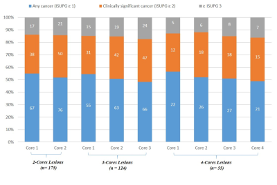 |
The Value of Obtaining Incremental Cores in MRI Guided Prostate Biopsy
Nicole Seyfried1, Amr Mahran1,2, Ananya Panda3, Verena Obmann4, Christina Buzzy1,2, Yun Jiang5, Katie Wright5, Dean Nakamoto6, Indravadan Patel7, Britt Conroy1,2, Lee Ponsky1,2, and Vikas Gulani5
1School of Medicine, Case Western Reserve University, Cleveland, OH, United States, 2Urology Institute, University Hospitals, Cleveland, OH, United States, 3Radiology, Mayo Clinic, Rochester, MN, United States, 4Department of Diagnostic, Interventional and Pediatric Radiology, Bern University Hospital, Bern, Switzerland, 5Department of Radiology, University of Michigan, Ann Arbor, MI, United States, 6Radiology, Louis Stokes Cleveland VA Medical Center, Cleveland, OH, United States, 7Radiology, Mayo Clinic, Scottsdale, AZ, United States
The value of incremental biopsy cores during in-gantry MRI-guided targeted prostate biopsy for prostate cancer diagnosis was examined. 135 patients with 175 lesions underwent in-gantry targeted prostate biopsy and the rates of pathology upgrading for sequential cores were determined. Significant upgrading was observed when increasing from 1 to 2 cores and from 2 to 3 cores, but not from core 3 to 4. Higher Prostate Imaging Reporting and Data System (PI-RADS) Version 2.0 score and anteriorly located lesions were more likely to result in an upgrade.
|
|
2419.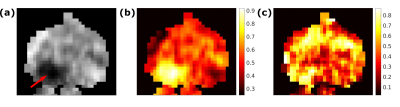 |
Two-component model of prostate tissue using hybrid multidimensional T2 and diffusion-weighted imaging
Ingrid Framås Syversen1, Mattijs Elschot2, Tone Frost Bathen2, and Pål Erik Goa3
1Kavli Institute for Systems Neuroscience, Norwegian University of Science and Technology, Trondheim, Norway, 2Department of Circulation and Medical Imaging, Norwegian University of Science and Technology, Trondheim, Norway, 3Department of Physics, Norwegian University of Science and Technology, Trondheim, Norway
T2 and diffusion-weighted imaging (DWI) are increasingly used in the detection and staging of prostate cancer, however, usually under the assumption that the T2 values and apparent diffusion coefficients (ADC) are independent of each other. Using hybrid multidimensional imaging, where images are acquired at two echo times and two b-values, we estimate volume fractions of slow and fast diffusion compartments in the prostate with a two-compartment model. The results suggest that the volume fractions can be used to discriminate between tumor and normal prostate tissue.
|
|
2420.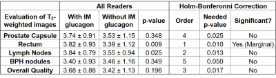 |
Does intramuscular injection of glucagon improve prostate MR image quality?
Karthik Meenakshi Sundaram1, Ali B Syed1, Stephanie Tzu-Ying Chang1,2, and Andreas Markus Loening1
1Radiology, Stanford University School of Medicine, Stanford, CA, United States, 2Department of Radiology, Veterans Affair Palo Alto Healthcare System, Palo Alto, CA, United States
ACR PI-RADs guidelines suggest anti-spasmodic agents may be beneficial for some patients during multi-parametric prostate MRI (mpMRI). Recent evidence suggests that the anti-spasmodic scopolamine butylbromide improves image quality on mpMRI. However, no studies have evaluated intramuscular (IM) injected glucagon. This retrospective IRB approved single-center study performed data-mining of 2595 mpMRI radiology reports (693 with glucagon) and subjective image quality assessment of 63 mpMRI exams (32 with glucagon). No significant benefit of IM glucagon for prostate evaluation was found on mpMRI. Our findings suggest that the risks associated with IM glucagon (medication side effects, patient discomfort, cost) may outweigh any benefits.
|
|
2421.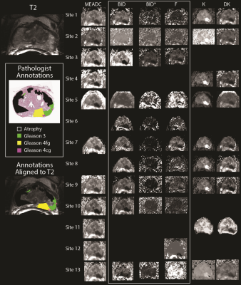 |
Multi-site analysis of DWI metrics for differentiating pathologically confirmed prostate cancer: patient and digital reference object results
Peter S LaViolette1, Sean D McGarry2, John D Bukowy1, Allison K Lowman1, Anjishnu Banerjee3, Dariya Malyarenko4, Tom Chenevert4, Yue Cao4,5, Andrey Fedorov6, Laura C Bell7, Chad Quarles7, Melissa A Prah2, Kathleen M Schmainda2,
Stefanie Hectors8, Bachir Taouli8, Eve LoCastro9, Yousef Mazaheri9,10, Amita Shukla-Dave9,10, Thomas Yankeelov11, David Hormuth II11, Ananth J Madhuranthakam12, Keith Hulsey12, Wei Huang13, Mark Muzi14, Michael Jacobs15, Meiyappan Solaiyappan15,
Kenneth Jacobsohn16, Mark Hohenwalter1, Petar Duvnjak1, Michael Griffin1, Watchareepohn Palanghmonthip17,18, William See16, Marja Nevalainen17, and Kenneth Iczkowski17
1Radiology, Medical College of Wisconsin, Milwaukee, WI, United States, 2Biophysics, Medical College of Wisconsin, Milwaukee, WI, United States, 3Biostatistics, Medical College of Wisconsin, Milwaukee, WI, United States, 4Radiology, University of Michigan, Ann Arbor, MI, United States, 5Radiation Oncology, University of Michigan, Ann Arbor, MI, United States, 6Radiology, Brigham and Womens Hospital, Boston, MA, United States, 7Neuroimaging Research, Barrow Neurological Institute, Phoenix, AZ, United States, 8Translational and Molecular Imaging Institute, Icahn School of Medicine at Mount Sinai, New York, NY, United States, 9Medical Physics, Memorial Sloan Kettering Cancer Center, New York, NY, United States, 10Radiology, Memorial Sloan Kettering Cancer Center, New York, NY, United States, 11Institute for Computer Engineering and Sciences, University of Texas, Austin, TX, United States, 12radiology, University of Texas Southwestern Medical Center, Dallas, TX, United States, 13Advanced Imaging Research Center, Oregon Health Sciences University, Portland, OR, United States, 14Radiology and Neurology, University of Washington, Seattle, WA, United States, 15Radiology, Johns Hopkins University, Baltimore, MD, United States, 16Urological Surgery, Medical College of Wisconsin, Milwaukee, WI, United States, 17Pathology, Medical College of Wisconsin, Milwaukee, WI, United States, 18Pathology, Chiang Mai University, Chiang Mai, Thailand
This study presents a multi-site study measuring the ability of site-specific diffusion weighted imaging fitting algorithms for differentiating prostate cancer. A dataset of DWI collected from 33 patients and a simulated digital reference object was distributed to thirteen sites who fit the multi-b DWI models with onsite-implemented software. Derived parametric maps were then submitted for central analysis. Each map was aligned to the T2-weighted image, and DWI metrics were extracted from aligned pathologist annotations. A statistical analysis was performed to determine the ability of each metric to differentiate PCA and to determine how fits of simulated data differed between sites.
|
|
2422.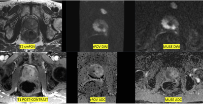 |
Comparing multi-shot DWI with multiplexed sensitivity encoding (MUSE) with reduced field of view DWI in prostate MRI by visual grading analysis
Anugayathri Jawahar1, Ali B Syed 1, Peter Wei 1, Lloyd Estkowski2, Arnaud Guidon3, Perla Subbaiah4, Bruce Lewis Daniel1, and Andreas M Loening 1
1Body MRI, Stanford University, PALO ALTO, CA, United States, 2Global MR Applications & Workflow, GE Healthcare, Waukesha, WI, United States, 3MR Body Applications & Workflow, GE Healthcare, Boston, MA, United States, 4Mathematics, Oakland University, Rochester, MI, United States
Diffusion-weighted imaging (DWI) using echo-planar technique is highly sensitive to susceptibility artifacts, in prostate MRI this is mainly due to rectal gas. In the hopes of identifying a sequence with better robustness to rectal gas, we compared multi-shot DWI using MUSE (with 3 shots) with a rFOV-DWI acquisition. In this IRB approved prospective study, 3 readers performed visual grading of MUSE and rFOV-DWI on 50 consecutive prostate MRI studies. MUSE-DWI and ADC maps showed statistically significant improvements in image quality for anatomic contour, lesion conspicuity, and overall image quality, as well as significantly reduced susceptibility artifacts related to rectal gas.
|
|
2423.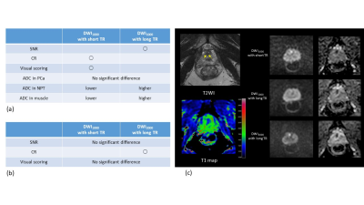 |
Short TR DWI in prostate
Yu Ueda1, Tsutomu Tamada2, Koji Yoshida2, Ayumu Kido2, Makoto Obara1, Masami Yoneyama1, Yuta Akamine1, and Marc Van Cauteren3
1Philips Japan, Tokyo, Japan, 2Kawasaki medical school, Kurashiki, Japan, 3Philips Healthcare, Asia Pacific, Tokyo, Japan
DWI is a key component of mp-MRI in prostate and the usefulness of high b-value DWI has been reported. However, the quality might depend on magnetic field strength and scanner performance. It would be beneficial to acquire sufficient diffusion contrast and ADC using standard b-value of 1000 s/mm2 without an additional higher b-value acquisition. We exploited the difference of T1 between cancer and normal to improve contrast. The contrast of T1 enhanced DWI1000 using short TR was better compared to that of DWI1000 with normal long TR but it was less than that of DWI2000 with normal long TR.
|
|
2424.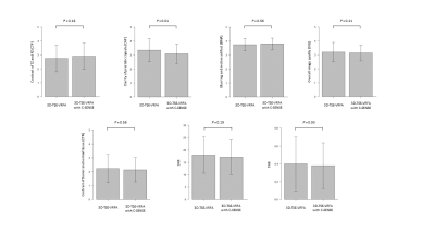 |
Clinical value of accelerated three-dimensional T2-weighted turbo spin-echo imaging with compressed SENSE in prostate MRI
Tustomu Tamada1, Yu Ueda2, Kosuke Ito3, Takeshi Fukunaga1, Ayumu Kido1, Tomohiro Mochizuki4, and Akira Yamamoto1
1Radiology, Kawasaki Medical School, Kurashiki City, Japan, 2Philips Japan, Minato-ku, Japan, 3Kawasaki Medical School, Kurashiki City, Japan, 4Philips Japan, Osaka, Japan Poster Permission Withheld
T2WI is a key component of mpMRI in prostate. 2D T2WI obtained in multi plane is recommend in PI-RADS v2, resulting in prolonged examination time. The usefulness of 3D-VRFA-TSE, which has a potential to reduce scan time by multiplanar postprocessing reconstruction of images into any desired plane, has been reported. As an further effective way to accelerate scan time is provided by C-SENSE, which uses undersampling of the k-space, we compared 3D-VRFA-TSE with 3D-VRFA-TSE with C-SENSE. C-SENSE enables a reduction in acquisition time of 40% in 3D-VRFA-TSE, maintaining image quality and lesion conspicuity except for clarity of prostatic capsule.
|
|
2425.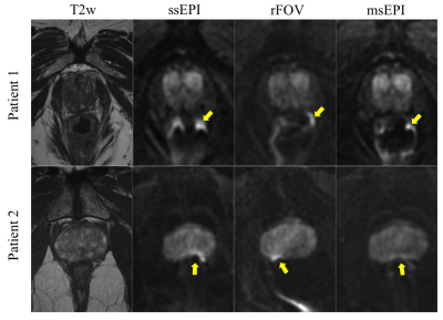 |
Reduced field-of-view and multi-shot DWI acquisition techniques: prospective evaluation in prostate cancer imaging
Edward M Lawrence1, Yuxin Zhang1, Jitka Starekova1, Zihan Wang2, and Diego Hernando1
1Radiology, University of Wisconsin-Madison, Madison, WI, United States, 2Biomedical Engineering, University of Wisconsin-Madison, Madison, WI, United States
Diffusion weighted imaging (DWI) non-invasively evaluates tissue microstructure, relevant in the setting of prostate cancer. Distortion from susceptibility effects can confound standard single-shot echo planar imaging (ssEPI). Reduced-distortion techniques, including reduced field of view (rFOV) and multi-shot EPI (msEPI), may improve prostate imaging quality but further evaluation is warranted. Therefore, a prospective comparison of rFOV and msEPI to ssEPI, in either biopsy proven or suspected prostate cancer patients, was performed. Our results demonstrate that both rFOV and msEPI reduce distortion artifacts, and improve image quality, in comparison to ssEPI. Furthermore, ADC quantification was reproducible across these three techniques.
|
|
2426.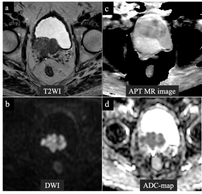 |
Amide proton transfer MR imaging of prostate cancer: a preliminary study using 3D acquisition
Ayumu Kido1, Tsutomu Tamada1, Yu Ueda2, Tomohiro Mochizuki3, Takeshi Fukunaga1, and Akira Yamamoto1
1Radiology, Kawasaki Medical School, Kurashiki, Japan, 2Philips Japan, Minato-ku, Japan, 3Philips Japan, Osaka, Japan
APT-MRI is a contrast-agent-free MRI technique that exploits signal from the amide protons (NH groups) contained in proteins and peptides. No studies have reported the use of 3DAPT. Quantitative analysis (ADC and APT SI (%)) were calculated for PC lesions and for benign regions that included normal PZ and benign prostatic lesions. APT SI was comparable among normal regions, benign lesions, and PC. However, APT SI was significantly higher in large PC than in small PC. In addition, the APT SIs of PC lesions correlated well with PSA. APT SI may represent tumor volume, PC staging, and prognosis.
|
|
2427.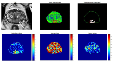 |
Developing Hybrid Multidimensional MRI without an endorectal coil for the detection of prostate cancer: a feasibility study
Durgesh Kumar Dwivedi1,2, Aritrick Chatterjee1, Ajit Devaraj3, Ambereen Yousuf1, Gregory S Karczmar1, Tatjana Antic4, Scott Eggener5, and Aytekin Oto1
1Department of Radiology, University of Chicago, Chicago, IL, United States, 2Department of Radiodiagnosis, King George's Medical University, Lucknow, India, 3Philips Research North America, Cambridge, MA, United States, 4Department of Pathology, University of Chicago, Chicago, IL, United States, 5Department of Urology, University of Chicago, Chicago, IL, United States
There are conflicting reports on the use of endorectal coil in the detection of prostate cancer at 3T due to increased patient discomfort and susceptibility artifacts. The purpose of the present study was to evaluate the feasibility of Hybrid Multidimensional MRI (HM-MRI) without an endorectal coil (using pelvic phased-array coil only) in the detection of prostate cancer (PCa) and to calculate fractional volumes of prostatic tissue composition, non-invasively. A non-endorectal coil HM-MRI is feasible. We obtained a significant difference in epithelium volume between normal and PCa (p = 0.02), however stromal and luminal volume did not reach a significance level.
|
|
2428.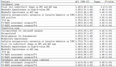 |
Interobserver agreement of the PI-RADS Version 2.1 lexicon: A Multicenter Study of Six Radiologists with different levels of experience
Li Zhang1, Longchao Li1, Min Tang1, Yinzhong Wang2, and Xiaoyan Wang2
1Department of MRI, Shaanxi Provincial People’s hospital, Xi’an, China, 2Lanzhou university First Hospital, Lanzhou, China
PI-RADS Version 2.1 makes several minor modifications aimed at addressing these issues and simplifying the scoring system without changing the overall framework for acquisition or interpretation using the principles of the dominant sequence paradigm. Formal investigations of the Interobserver agreement of the PI-RADS version 2.1 scoring system are critical to confirm that it is attaining its primary aim of standardization.
|
2429. |
A preliminary study of 3D-amide proton transfer MR imaging in rectal adenocarcinoma: comparison with diffusion kurtosis imaging
Weicui Chen1, Ling Li2, Yingjie Mei3, Jilei Zhang3, Weikang Huang1, Bo Liu1, and Xian Liu1
1Radiology, The second Affiliated hospital of Guangzhou University of Chinese Medicine, Guangzhou, China, 2Guangzhou University of Chinese Medicine; The second Affiliated hospital of Guangzhou University of Chinese Medicine;, Guangzhou, China, 3Clinical Science,Philips Healthcare;, Shanghai, China
We explore the utility of 3D APTw imaging and DKI in assessing the pathological factors of rectal adenocarcinoma. The mean APTSI and MK was higher with the advanced staging and grading in rectal adenocarcinoma. In addition, the mean APTw SI have a significantly positive correlation with WHO grades. Therefore, the mean APTw SI is promising to be a non-invasive biomarker in clinical practice and to become a potential predictor for estimating the prognosis of rectal adenocarcinoma.
|
|
2430. |
Relationship between ADC values of MRI diffusion-weighted imaging and the pathological prognostic factors of rectal cancer
rui qi1 and ying wan2
1radiology, Sichuan university, Chengdu, China, 2pathology, Sichuan university, Chengdu, China To investigate the correlation between the ADC value of MRI-DWI and the pathological prognostic factors of rectal cancer. 87 patients with rectal cancer were grouped according to the pathological prognostic factors. The mean ADC values of tumor tissues were measured, and the relationship between ADC values of different groups was analyzed. The differences of ADC values of the different differentiation groups, pN stage groups and P53 expression groups had statistical significance. The P53 expression level and ADC value showed a moderate negative correlation. It is expected the biological behavior of tumors can be predicted by quantitative analysis of ADC values. |
|
2431.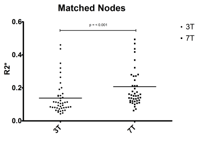 |
R2* relaxation rates of pelvic lymph nodes in USPIO-enhanced MRI of rectal cancer patients at 3 and 7T
Tijmen Koëter1, Rutger CH Stijns1, Bart JW Philips2, Jurgen J Futterer2, Patrick Zamecnik2, Sjaak JA van Asten2, Marnix C Maas2, and Tom WJ Scheenen2
1Radiology and Nuclear Medicine, Department of Surgery, Radboudumc, Nijmegen, Netherlands, 2Radiology and Nuclear Medicine, Radboudumc, Nijmegen, Netherlands
Six patients with rectal cancer underwent USPIO-enhanced MRI at 3 and 7T. The presence and appearance of lymph nodes was annotated by two trained radiologists. Lymph nodes were matched between 3 and 7T, and ROIs were drawn inside the nodes to calculate mean R2* relaxation rates in order to assess the possibilities of quantitative thresholds in R2* between suspicious and benign lymph nodes at both field strengths.
|
|
2432.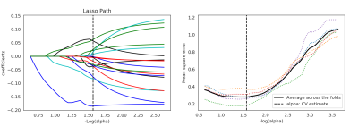 |
Using radiomics to build prediction model for diagnosing metastatic lymph node in rectal cancer basing on HR-T2 imaging
Gesheng Song1, Aiyin Li1, Jingjing Cui2, and Yan Jia2
1Shandong Provincial Qianfoshan Hospital,the First Hospital Affiliated with Shandong First Medical, Jinan, China, 2Huiying Medical Technology Co., Ltd., Beijing, China
MRI based radiomics machine learning model could differentiating metastatic lymph node in rectal cancer.
|
|
2433.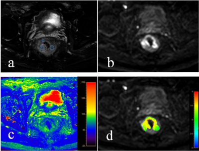 |
Comparative study of APTw combined with T2 mapping quantitative imaging in patients of rectal cancer with and without chemotherapy
Anliang Chen1, Ailian Liu1, Xuedong Wang1, Yunsong Liu1, Jingjun Wu1, Qingwei Song1, Renwang Pu1, Jiazheng Wang2, Zhiwei Shen2, and Liangjie Lin2
1Radiology, The First Affiliated Hospital of Dalian Medical University, Dalian, China, 2Philips Healthcare, Beijing, China
Amide proton transfer-weighted (APTw) imaging can be used to assess changes of intracellular protein concentration and the pH value.And the T2 mapping allows non-invasive visualization and quantification of tissue composition. Here, weaimsto explore the value of APTw imaging in combination with T2 mapping in comparative analysesof rental cancer with and without chemotherapy. Results indicated that a ultrahigh diagnostic efficacy (AUC: 1.000; sensitivity: 100%; specificity: 100%) was achieved with combination of APT and T2 values.
|
|
2434.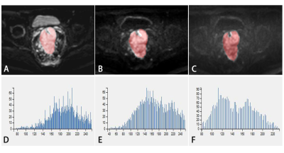 |
Value of texture analysis based on DWI with multiple b-values for evaluation of tumor heterogeneity in rectal cancer patients
Jingjun Wu1, Ailian Liu1, Jiazheng Wang2, Zhongping Zhang2, Anliang Chen1, Xuedong Wang1, Yunsong Liu1, Lihua Chen1, Qingwei Song1, Renwang Pu1, and Bingbing Gao1
1Department of Radiology, the First Affiliated Hospital of Dalian Medical University, Dalian, China, 2Philips Healthcare, Beijing, China
Rectal cancer is the third most common cancer and the second cause of cancer-related death worldwide. DWI is now recommended as a routine addition to the MRI protocol for the assessment of rectal cancer. However, the effect of the b-value on the diagnostic performance has not yet been explored. The current study aims to evaluate the value of texture analysis based on DWI with different B values to evaluate the heterogeneity of rectal cancer patients. The skewness and kurtosis were all significantly different in DWI with different B values, and they were obviously changed when B reached 800.
|
|
2435.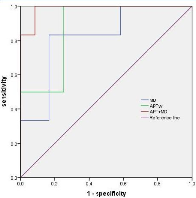 |
Comparative study of APT combined with DKI in post-chemotherapy and non-chemotherapy rectal cancer
Xuedong Wang1, Anliang Chen1, Qingwei Song1, Jiazheng Wang2, and Ailian Liu1
1The first affiliated hospital of dalian medical university, DaLian, China, 2Philips Healthcare, China, Beijing, China
We aimed to explore the difference of amide proton transfer-weighted (APTw) imaging combined with DKI between the chemotherapy group and non-chemotherapy group of rectal cancer. The result showed that the highest diagnostic efficacy(AUC: 0.986; sensitivity: 91.8%; specificity: 100%) was acquired using APT combined with DKI, rather than APT and DKI value respectively.
|
|
| 2436. | Comparative study of APTw quantitative imaging in normal intestinal wall and rectal cancer
Xuedong Wang1, Ailian Liu1, Jiazheng Wang2, Yishi Wang2, Anliang Chen1, Lihua Chen1, and Qingwei Song1
1The first affiliated hospital of dalian medical university, DaLian, China, 2Philips Healthcare, China, Beijing, China
The purpose of this study was to investigate the feasibility of using APTw for the diagnosis of rectal cancer.
|
|
2437. |
Amide Proton Transfer Imaging of Rectal Cancer: Which Duration and Power Level of Saturation Pulse is better?
Lan Zhang1 and Xin Li1
1Union Hospital, Tongji Medical College, Huazhong University of Science and Technology, Wuhan, China
We speculate that APT value may be a useful biomarker for assessing rectal pathological characteristics, which could have a potential impact on the clinical therapeutic strategies for patients. A baseline of APT value needs to be established for rectal cancer, which remains challenging due to the susceptibility effects. So we need to optimize the sequence by adjusting duration and power level of saturation pulse
|
|
2438. |
Reduced field-of-view versus conventional pelvic diffusion-weighted imaging in rectal cancer
Ali B Syed1, Anugayathri Jawahar1, and Vipul R Sheth1
1Department of Radiology, Stanford University, Stanford, CA, United States
There is growing literature supporting the use of diffusion weighted imaging (DWI) for staging and re-staging of rectal cancer. We investigated the performance of reduced FOV (rFOV) DWI in comparison with conventional whole pelvis DWI. Two readers graded rFOV and conventional DWI for anatomic detail, lesion conspicuity, tumor margin delineation, artifact, and overall image quality. rFOV DWI showed statistically significant improvement in anatomic detail, lesion conspicuity, tumor margin delineation, and overall image quality. No significant difference in artifact/distortion or lymph nodes was detected. rFOV DWI is a promising technique in rectal cancer evaluation and improves tumor visualization.
|
|
2439.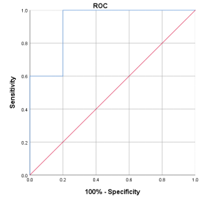 |
T2 mapping-based the change trend of retrospective analysis of rectal cancer before and after chemotherapy
Yunsong Liu1, Anliang Chen1, Jingjun Wu1, Xuedong Wang1, Lihua Chen1, Yishi Wang2, Jiazheng Wang2, Zhiwei Shen2, and Ailian Liu1
1The First Affiliated Hospital of Dalian Medical University, Dalian, China, 2Philips Healthcare, Beijing, China
The current study aimed to investigate the value of T2 mapping in evaluating the difference of rectal cancer before and after chemotherapy. The results showed that T2 mapping had high diagnostic value in the two groups (AUC: 0.92; sensitivity: 100%; specificity: 80%).
|
2440.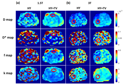 |
IVIM-DKI analysis using Hybrid model with parameter reconstruction method in prostate cancer at 1.5T vs 3T MRI
Archana Vadiraj Malagi1, Arjun Lokesh2, Esha Baidya Kayal1, Kedar Khare3, Virendra Kumar4, Chandan J. Das2, and Amit Mehndiratta1,5
1Centre for Biomedical Engineering, Indian Institute of Technology Delhi, New Delhi, India, 2Department of Radiodiagnosis, All India Institute of Medical Sciences Delhi, New Delhi, India, 3Department of Physics, Indian Institute of Technology Delhi, New Delhi, India, 4Department of Nuclear Magnetic Resonance (NMR), All India Institute of Medical Sciences Delhi, New Delhi, India, 5Department of Biomedical Engineering, All India Institute of Medical Sciences Delhi, New Delhi, India
Currently in countries with poor resources availability of higher magnetic strength 3T MRI is low. Objective is to analyze whether with lower magnetic strength 1.5T MRI with advanced parameter reconstruction method can perform better or equivalent to 3T MRI. IVIM-DKI signal was modelled using hybrid (HY) model which produces non-physiological inhomogeneity in parameter map. This inhomogeneity can be corrected by using total variation penalty function (TV) with HY model. IVIM-DKI maps obtained from HY with TV produced more clinically reliable than HY model. Even with low magnetic strength i.e. 1.5T, overall HY+TV model outperformed qualitatively & quantitatively against 3T MRI results.
|
|
2441.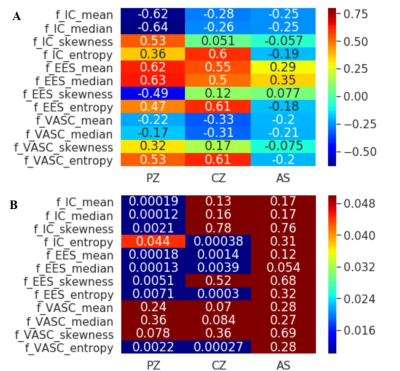 |
Age-Related and Zonal Anatomical Microstructure Features of Normal Prostatic Tissues: A Preliminary Study with VERDICT MRI
Xiangyu Wang1, Fan Lin1, and Yi Lei1
1The First Affiliated Hospital of Shenzhen University, Shenzhen, China 31 male volunteers were examined with VERDICT MRI of the prostate. The derived VERDICT parameters (the fIC, fEES and fVASC) were analyzed with a histogram based on the whole prostate volume and the PZ, CG and AS ROIs. There were statistical differences between histogram parameters and regions of the prostate. Among different anatomical zones, for PZ, Age has a negative correlation with fIC, and positive correlation with fEES. According to CG, there was also positive correlation between age and fEES. |
|
2442.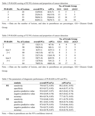 |
Validation of the Diagnostic Accuracy of Multi-parametric MRI with PI-RADS V2.1 for Detecting Clinically Significant Prostate Cancer
Xiangyu Wang1, Fan Lin1, and Yi Lei1
1The First Affiliated Hospital of Shenzhen University, Shenzhen, China 229 PZ and 270 TZ lesions were included in this study. The proportion of overall PCa and clinically important PCa detection (Gleason score≥3+4) for each PI-RADS version 2.1 category was determined. The performance of PI-RADS version 2.1 in cancer detection was evaluated. The diagnostic accuracy of the overall(82.6%) and csPCa TZ lesions(83.0%) is higher than the accuracy of PZ lesions(80.4%, 77.7%). For the csPCa diagnosis, the AUCs of PI-RADS version 2.1 in TZ(0.84) was higher than in PZ(0.77) without significance(P=0.06). Higher PI-RADS version 2.1 scores were associated with increasing likelihood of the presence of clinically important PCa (P<0.01). |
|
2443.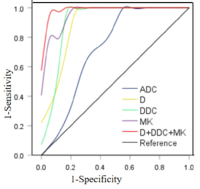 |
Differential diagnosis of Prostatic Hyperplasia with inflammation and Low-grade Transitional Zone Carcinoma: Advanced DWI VS Conventional DWI
Ren Huipeng1, Fan Qing1, Wang Xiaohu1, Wei Xiaocheng2, and Ren Zhuanqin1
1Baoji Central Hospital, Baoji, China, 2GE Healthcare China, Beijing, China
The purpose of this study is to evaluate the efficacy of advanced diffusion weighted imaging (DWI) in the differential diagnosis of benign prostatic hyperplasia(BPH) with inflammation and Low-grade prostate transitional zone (TZ) cancer. By comparing the D, DDC and MK from advanced DWI with ADC value, it is found that the diagnostic efficiency of advanced DWI is better than the conventional DWI, and the combined diagnosis of (D + DDC + MK) has the highest efficiency.
|
|
2444.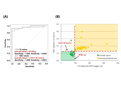 |
Quantitative Approach by the Simultaneous Acquisition of ADC and T2 Values Using Echo-Planar Imaging Sequence for Prostate Cancer Detection
Hirotoshi Maruyama1, Yasuhiro Fujiwara2, and Akira Takahashi1
1Radiology, Kumamoto Saishun Medical Center, Kumamoto, Japan, 2Department of Medical Imaging, Faculty of Life Sciences, Kumamoto University, Kumamoto, Japan
We propose a sequence to simultaneously acquire apparent diffusion coefficient (ADC) and T2 values for detecting prostate cancer. ADC and T2 values of prostate cancer can simultaneously be acquired using not only a diffusion-weighted image, but also multiple echo time (TE) images without motion-probing gradient using echo-planar imaging (EPI). Moreover, the quantitative ADC and T2 maps can be produced in approximately 5 min. Combining ADC and T2 values was effective in differentiating between prostate cancer and the normal peripheral zone. Our rapid quantification imaging technique improves the accuracy of the diagnosis of prostate cancer.
|
|
2445. |
Assessment of aggressiveness of prostate cancer: correlation of MRI ADC map textures with Gleason grade after radical prostatectomy
Liang WANG 1
1Tongji Hospital,Tongji Medical College, Huazhong University of Science & Technology, Wuhan, China Poster Permission Withheld
Prostate cancer is the 2nd leading diagnosed cancer in men worldwide. Radiomics extracts large amounts of quantitative image features from radiologic images and selects stable and clinically relevant radiomics biomarkers for disease assessment. 90 patients underwent mpMRI before radical prostatectomy, with subsequent pathologic evaluation. The texture features were extracted by the python-based pyradiomics package. The AUC of the model was 0.841, with sensitivity 69.6% and specificity 91.2%, which was significantly higher than mean ADC value or single texture feature. MRI ADC map texture evaluation may facilitate noninvasive assessment of aggressiveness of prostate cancer.
|
|
2446.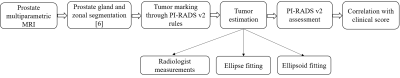 |
Tumor Estimation for PI-RADS v2 Assessment of Prostate Cancer using Multiparametric MRI
Dharmesh Singh1, Saumya Diwan2, Virendra Kumar3, Chandan J Das4, Anup Singh1,5, and Amit Mehndiratta1,5
1Centre for Biomedical Engineering, Indian Institute of Technology (IIT) Delhi, New Delhi, India, 2Delhi Technological University, Delhi, India, 3Department of NMR, All India Institute of Medical Sciences Delhi, New Delhi, India, 4Department of Radiology, All India Institute of Medical Sciences Delhi, New Delhi, India, 5Department of Biomedical Engineering, All India Institute of Medical Sciences Delhi, New Delhi, India
Accurate estimation of tumor size is a challenging task due to the large variation in shape of prostatic tumors. Measurement of diameter and volume of tumor are useful for the staging of prostate cancer(PCa). Therefore, an appropriate method for tumor assessment is essential to assist clinical management of PCa. The goal of this study was to investigate the role of tumor size estimation methods (2D vs. 3D) for the Prostate Imaging Reporting and Data System version-2(PI-RADS v2) assessment of PCa using multiparametric MRI. Automatic tumor size assessment using ellipse-fitting approach showed relatively better performance of PCa assessment compared to radiologist-assessment.
|
|
2447.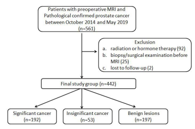 |
Prostate cancer: Inter-reader Agreement and Diagnostic Performance with PI-RADS Version 2.1 at Multiparametric MRI
Li Zhang1, Longchao Li1, and Min Tang1
1Department of MRI, Shaanxi Provincial People’s hospital, Xi’an, China
In 2019, the updated version of PI-RADS Version 2.1 was introduced. This version did not change the entire framework, but made several modifications to address limitations and simplify the scoring system. Between 442 patients, both readers had 96 discordant cases in terms of five-point Version 2.1 scoring. Version 2.1 provides high accuracy for detecting clinically significant PCa with category 4 as the threshold. Both readers have good inter-reader reliability. However, agreement for TZ was lower than for PZ lesions.
|
|
2448.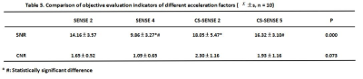 |
Highly accelerated prostate water/fat 3D MRI with compressed sensing and SENSE, compared to the standard of care
Ya nan Wu1, Mei yu Sun1, Ai lian Liu1, Jia zheng Wang2, Yi shi Wang2, Cheng yan Wang1, Li hua Chen1, Qing wei Song1, Ren wang Pu1, and Bing bing Gao1
1The First Affiliated Hospital of Dalian Medical University, Da lian, China, 2Philips Healthcare,China, Bei jing, China
Compressed sensing(CS)technology is currently used less in prostate diseases. This study aims to explore the feasibility of CS and SENSE technology with different acceleration factors for prostate 3D magnetic resonance imaging(MRI). The preliminary exploration concluded that the SNR and CNR with CS 5 in prostate 3D mDIXON imaging are similar to the SENSE 2, and the imaging time is reduced by 28%, which effectively improves the MR scanning efficiency.
|
|
2449.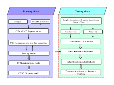 |
Automated diagnosis of prostate cancer from dynamic contrast-enhanced MRI using a Convolution Neural Network–based deep learning approach
Ming Deng1, Haibo Xu2, Xiaoyong Zhang3, and Yingao Zhang4
1Department of Radiology, Zhongnan Hospital of Wuhan University, Wuhan University,, Wuhan, China, 2Department of Radiology, Zhongnan Hospital of Wuhan University, Wuhan University, Wuhan, China, 3MR Collaborations, Siemens Healthcare Ltd, Shenzhen, China, 4Zhongnan Hospital of Wuhan University, Wuhan University, Wuhan, China The aim of this study was to evaluate the diagnostic performance of a convolutional neural network (CNN)-based deep learning technique for the differentiation of prostate cancer (PC) using dynamic contrast agent–enhanced magnetic resonance imaging (DCE-MRI) data. Our patient study demonstrated that the quantitative image features derived from the DCE-MR images based on the self-defined CNN model can be effective in distinguishing PC from the normal, and the automated extraction of Ktrans, TDC, DR, and DY features can significantly promote PC diagnosis. The high performance of the proposed CNN-based deep learning method statistical analysis demonstrated its potential for improving PC diagnosis. |
|
2450.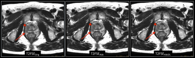 |
Initial experience in abbreviated T2-weighted Prostate MRI using a Deep Learning reconstruction.
Aileen O'Shea1, Arnaud Guidon2, Robert Marc Lebel3, Ersin Bayram4, Theodore Pierce1, Amirkasra Mojtahed1, and Mukesh G Harisinghani1
1Massachusetts General Hospital, Boston, MA, United States, 2Applications and Workflow, GE Healthcare, Boston, MA, United States, 3Applications and Workflow, GE Healthcare, Calgary, AB, Canada, 4Applications and Workflow, GE Healthcare, Houston, TX, United States
Increasing the speed of multiparametric prostate MRI (mpMRI) is highly desirable. However, usual tradeoffs between signal-to-noise (SNR), scan time and lesion conspicuity must be considered. One recently proposed approach consists of using a bi-parametric protocol, whereby only the T2 and diffusion-weighted images are collected, thus highlighting the particular significance of achieving a robust, high-quality T2-weighted acquisition. As such, this work focuses on evaluating a Deep Learning reconstruction technique which shows promises to cut acquisition time of prostate T2-weighted imaging in half and would therefore benefit both bi-parametric as well as mpMRI.
|
|
2451.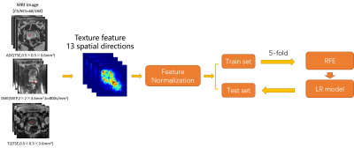 |
Mean or Extremum? Comparison of Two Strategies to Extract Orientation-Dependent Texture Features in Radiomics Studies
Jing Zhang1, Yang Song1, Yu-dong Zhang2, Xu Yan3, Yefeng Yao1, and Guang Yang1
1Shanghai Key Laboratory of Magnetic Resonance, Department of Physics, East China Normal University, shanghai, China, 2Department of Radiology, The First Affiliated Hospital with Nanjing Medical University,, Nanjing, China, 3MR Scientific Marketing, Siemens Healthcare, shanghai, China
Texture features plays an important role in radiomics. To make the texture features rotation-invariant, pyradiomics computes the texture features along all directions and use their mean values. In this study, we demonstrated that maximum and minimum values of these features along different directions, which is also rotation-invariant, may provide added value to radiomics studies. We trained models using mean, maximum and minimum values of texture features along different directions to classify clinically significant (CS) prostate cancer (PCa) and non-CS PCa on PROSTATEx dataset. We found that using extremum instead of mean texture features improved the performance of model.
|
|
2452. |
A Deuterated 13C-Urea Reference to Eliminate 1H Imaging Artifacts in Hyperpolarized 13C MRI Studies
Collin J. Harlan1, Zhan Xu 1, Keith A. Michel1,2, Christopher M. Walker1, Sanjaya D. Lokugama3, Mark D. Pagel2,3, and James A. Bankson1,2
1Department of Imaging Physics, The University of Texas MD Anderson Cancer Center, Houston, TX, United States, 2The University of Texas MD Anderson Cancer Center UT Health Graduate School of Biomedical Sciences, Houston, TX, United States, 3Department of Cancer Systems Imaging, The University of Texas MD Anderson Cancer Center, Houston, TX, United States Multiparametric and hyperpolarized (HP) 13C Magnetic Resonance Imaging (MRI) are attractive multi-modality methods for prostate cancer imaging studies because of their non-invasiveness and characterization of changes in tumor metabolism. A 13C-urea calibration reference is required when conducting HP 13C MRI studies, which must be heavily doped with Gd+ to facilitate rapid calibrations. Care must be taken to ensure that the highly relaxed 1H signal does not compromise DCE-MRI via hyperintense imaging artifacts. Deuteration and lyophilization of 13C-urea can be useful in creating a calibration reference that does not lead to imaging artifacts in 1H Images. |
2453.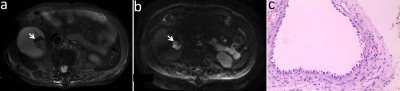 |
Comparison of Diagnostic Performance of Magnetic Resonance Imaging and Computed Tomography for Evaluation of Complex Renal Cysts
Anqin Li1, Zhen Li1, and Daoyu Hu1
1Tongji Hospital, Tongji Medical College, Huazhong University of Science and Technology, Wuhan, China Poster Permission Withheld
To improve understanding of DWI features of complex renal cysts, and compare diagnostic performance of MRI and CT in discrimination of benign and malignant masses. Images of each lesion were analyzed, including size, thickness of wall, number of septum, enhancement of wall/septum, wall nodule, calcification, and cyst content. CT and MRI image characteristics were compared with pathology or follow-up results. The incidences of high signal intensity on DWI were significantly higher in malignant than in benign masses. MRI showed higher AUC than CT for differentiating benign from malignant masses. MRI could be useful in improving diagnosis of complex renal cysts.
|
|
2454.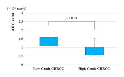 |
Is diffusion-weighted imaging effective in differentiating chromophobe renal cell carcinoma and oncocytoma as per cell grade?
Tatsuya Yamamoto1 and Atsushi Kohno1
1Department of Diagnostic Radiology, The Cancer Institute Hospital of the Japanese Foundation for Cancer Research, Tokyo, Japan Poster Permission Withheld
Although the prognosis of chromophobe renal cell carcinoma (CHRCC) is more favorable than that of other RCC subtypes, it is a malignant tumor with possible metastasis. On the other hand, there is only one confirmed case of renal oncocytoma metastasis. This study assessed the apparent diffusion coefficient (ADC) value derived from diffusion-weighted imaging to differentiate renal oncocytoma from CHRCC according to cell grade. The ADC values were significantly different between low-grade and high-grade CHRCCs and between oncocytoma and high-grade CHRCC. Thus, the ADC value is useful for differentiating oncocytoma from high-grade CHRCC but not for differentiating oncocytoma from low-grade CHRCC.
|
|
2455.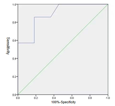 |
Evaluate the efficiency of the ESWAN value for differentiating renal clear cell carcinoma with different pathological grades.
Xinmiao Bu1, Ailian Liu1, Dahua Cui1, Xin Li2, and Qingwei Song1
1The First Affiliated Hospital of Dalian Medical University, dalian, China, 2GE Healthcare, Beijing, China
To investigate the potential of ESWAN in differentiating renal clear cell carcinoma with different pathological grades, to help preoperative non-invasive prediction of renal clear cell carcinoma pathological grading.
|
|
2456. |
Evaluate the efficiency of T1 Mapping and amide proton transfer imaging in differentiating chronic kidney disease and healthy volunteers.
Xinmiao Bu 1, Ailian Liu1, Lihua Chen1, Weiping Yang1, Yunsong Liu1, Yaxin Niu1, Qingwei Song1, Renwang Pu1, Jiazheng Wang2, Zhiwei Shen2, and Zhongping Zhang2
1The First Affiliated Hospital of Dalian Medical University, dalian, China, 2Clinical Science, Philips Healthcare, Beijing, China
To evaluate the joint diagnosis value of T1 Mapping and amide proton transfer imaging in the chronic kidney disease and healthy volunteers.
|
|
2457.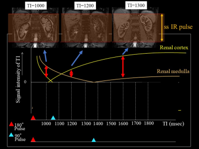 |
Multiparametric MR imaging in diabetic nephropathy: New insights to evaluate early diabetic nephropathy noninvasively
Akira Yamamoto1, Tsutomu Tamada2, Yu Ueda3, Takeshi Fukunaga2, Ayumu Kido2, and Atsushi Higaki2
1Radiology, Kawasaki Medical School, Kurashiki, Japan, 2Kawasaki Medical School, Kurashiki, Japan, 3Philips Japan, Minato-ku, Japan
The purpose of this study was to identify the changes in multiparametric magnetic resonance imaging (MRI) findings in early diabetic nephropathy. Measurements were made of the renal cortex and renal medulla T2 values, T2* values and R2* values, as well as optimal TI, inverted TI value in SSFP with ssIR pulse with multi TI. Also, renal cortical thickness and renal length were measured, representing morphological changes. Significant differences were seen between the healthy and early diabetic nephropathy groups in values of T2 and inverted TI. This study suggests the possibility that MRI using the T2 value and inverted TI of SSFP with an ssIR pulse can be used to evaluate early diabetic nephropathy non-invasively and in a short period of time.
|
|
2458. |
Multiparametric-MRI in the Evaluation of Early diabetic kidney damage
Youzhen Feng1, Zhongyuan Cheng1, Xiaoqing Xiong1, Qiting Lin1, Dingkun SiTu1, Pingkang Chen1, Zhifei Liu2, Long Qian3, and Xiangran Cai1
1Medical Imaging Center, First Affiliated Hospital, Jinan University, Guangzhou, Guangdong, China, Guangzhou, China, 2Medical Imaging Center, First People's Hospital of Kashgar, Xinjiang, china., Xinjiang, China, 3MR Research, GE Healthcare, Beijing, 100176, China., Beijing, China
Multiparametric-MRI (mp-MRI) has shown promising results in the diagnosis of clinical disease. It provides us an approach to perform investigation from multiple dimension and avoids to ignore the worthwhile diagnosis information. Previous studies indicated that the perfusion and dispersion of renal parenchyma have been changed in diabetic mellitus (DM) without biochemical indicators of significant renal damage. However, the convergence and divergence of those diffusion and perfusion related MRI parameters in the evaluation of DM associated kidney damage is still unclear. To assess the renal functional changes in DM patients, three MRI modalities were applied in current study.
|
|
2459.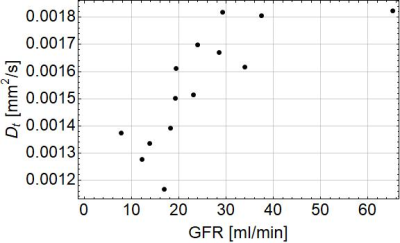 |
Non-invasive assessment of renal function using intravoxel incoherent motion imaging taking account of renal tubular water and relaxation times
Hajime Tamura1, Hideki Ota2, Tatsuo Nagasaka3, and Eikan Mishima4
1Division of Medical Physics, Tohoku University, Graduate school of medicine, Sendai, Japan, 2Department of Advanced MRI Collaboration Research, Tohoku University, Graduate school of medicine, Sendai, Japan, 3Department of Radiology, Tohoku University hospital, Sendai, Japan, 4Division of Nephrology, Endocrinology, and Vascular Medicine, Tohoku University, Graduate school of medicine, Sendai, Japan
Renal IVIM imaging taking account of water in the urinary tubules and relaxation times was performed in patients with renal-artery stenosis. Several estimated parameters were significantly correlated with the functional parameters obtained by renal scintigraphy (water fraction with the time to peak height (Tmax) on the renogram curve of renal scintigraphy, parenchymal diffusion coefficient with the glomerular filtration rate (GFR), and transverse relaxation time of flowing blood with GFR).
|
|
2460. |
Spin-lock MRI Reveals Tubular Dilation and Fibrosis after Unilateral Ureter Obstruction
Feng Wang1,2, Tadashi Otsuka3, Keiko Takahashi3, Daniel C. Colvin1, Hua Li1, Raymond C. Harris3, Takamune Takahashi3, and John C. Gore1,2,4
1Vanderbilt University Institute of Imaging Science, Vanderbilt University Medical Center, Nashville, TN, United States, 2Department of Radiology and Radiological Sciences, Vanderbilt University Medical Center, Nashville, TN, United States, 3Division of Nephrology and Hypertension, Vanderbilt University Medical Center, Nashville, TN, United States, 4Department of Biomedical Engineering, Vanderbilt University, Nashville, TN, United States
Assessing the severity and progression of tubular dilation and fibrosis in kidneys are important clinical challenges. Here we evaluate spin-lock MR imaging with different locking fields for detecting tubular dilation and fibrosis in a mouse model of unilateral ureter obstruction. We fit the dispersion of spin-lock relaxation rates R1ρ at different locking fields (frequencies) to a model of exchanging water pools and assessed the sensitivity of the derived quantities for detecting tubular dilation and fibrosis in kidney. Histological scores for tubular dilation and fibrosis, based on luminal space and positive picrosirius red area in sections, were obtained for comparison.
|
|
2461.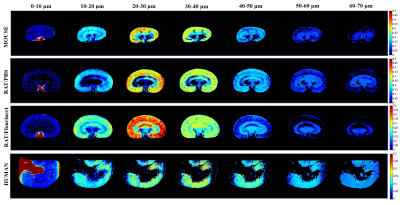 |
Towards non-invasive measurement of renal tubule size by diffusion MRI
Darya Morozov1, Jennifer R. Charlton2, Kimberly A. deRonde2, and Kevin M. Bennett1
1Radiology, Washington University in St. Louis, School of Medicine, Saint Louis, MO, United States, 2Pediatrics, University of Virginia Children's Hospital, Charlottesville, VA, United States
Tubular hypertrophy is an early feature of many renal pathologies. A direct, non-invasive measure of kidney morphology is lacking. Current diagnostics are primarily based on biopsy after suspected renal disease, which is prone to sampling error and is invasive. A robust non-invasive imaging tool to study tubular changes in vivo is greatly needed and would provide early tissue biomarker for initiation and progression of kidney injury. In this work, we used a single diffusion encoding MRI approach to study distribution of restricting tissue compartment sizes in kidney cortex as a potential noninvasive marker of tubule size in the intact kidney.
|
|
2462.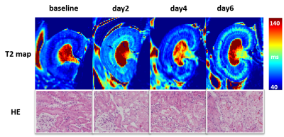 |
Non-invasive assessment of progressive changes in aristolochic acid (AA) induced AKI with T2 mapping
Yingjie Mei1,2, Guixiang Yang3, Yihao Guo1,4, Kaixuan Zhao1,4, Chenggong Yan3, Yikai Xu3, and Yanqiu Feng1,4
1Guangdong Provincial Key Laboratory of Medical Image Processing, Southern Medical University, Guang Zhou, China, 2Philips Healthcare, Guang Zhou, China, 3Department of Medical Imaging Center, Nanfang Hospital, Southern Medical University, Guangzhou, China, 4School of Biomedical Engineering, Southern Medical University, Guang Zhou, China
Cases of Aristolochic acid nephropathy (AAN) have been reported worldwide, yet no serum or urinary biomarkers have been shown to have clinical utility in detection of AAN. In this study, we aim to detect renal changes in the early phase of AAN with T2 mapping. The results suggest that T2 mapping may be a valuable tool in experimental studies and clinical practice of acute phase of AAN.
|
|
2463.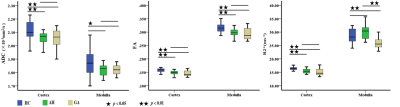 |
Monitoring the microenvironment changes of kidney in patients with hyperuricemia using dual-modal functional MRI
Zhongyuan Cheng1, Qiting Lin1, Xiaoqing Xiong1, Dingkun Situ1, Pingkang Chen1, Zhifei Liu2, Long Qian3, Youzhen Feng1, and Xiangran Cai1
1Medical Imaging Center, First Affiliated Hospital, Jinan University., Guangzhou, China, 2Medical Imaging Center, First People's Hospital of Kashgar, Xinjiang, china., Xinjiang, China, 3MR Research, GE Healthcare., Beijing, China
The aim of this study was to investigate the feasibility of diffusion tensor imaging (DTI) and blood oxygenation level dependent imaging (BOLD) in assessing early renal function changes in hyperuricemia.
|
|
2464.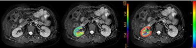 |
Is renal blood supply a predictor of protein synthesis in the kidney? A preliminary study in the healthy population
Lihua Chen1, Ailian Liu1, Jiangzheng Wang2, Zhiwei Shen2, Qingwei Song1, Renwang Pu1, and Nan Wang1
1The First Affiliated Hospital of Dalian Medical University, Dalian, China, 2Philips Healthcare, Beijing, China
3D amide proton transfer weighted (APTw) MR imaging is a novelcontrast-agent-free MRI technique for detection of amide protons in mobile cellular proteins and peptides 1-3.On the other hand, 3D pseudo-continuous arterial spin labeling (3D-pCASL)is amethod to measure renal blood supply and hence potentially a metric for renal function. This study reveals a close association between the vascularity and concentration of mobile cellular proteins in kidney.
|
|
2465.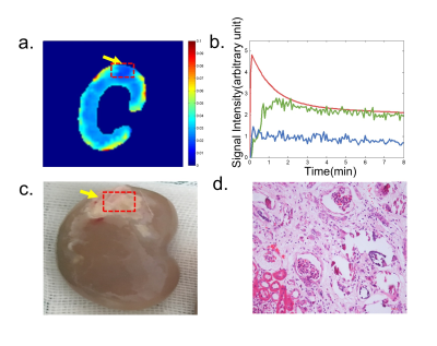 |
Quantitative Renal Function Assessment of Renal embolization
Hanjing Kong1
1Peking University, Beijing, China
Renal embolization is part of a multisystemic disease and has attracted enhanced attention in recent years for its increasing incidence in the elderly.It is a significant cause of renal loss in patients who suffer from valvular cardiopathy, aortic atheromatosis, and hypercoagulable states. In this study, we attempt to investigate the feasibility of compressed sensing (CS) based DCE-MRI in the assessment of AERD in animal models.
|
2466.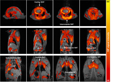 |
In-vivo 19F-isoflurane MR imaging in adipose tissue
Gang Zheng1, Michael de Veer1, Hongxin Wang1, Shenpeng Li1, and Gary Egan1
1Monash Biomedical Imaging, Monash University, Clayton, Australia
The detection of adipose tissue (AT) is of great importance in studying obesity. Isoflurane is a common fluorinated anesthetic with an excellent safety record in preclinical studies, which has a high blood–gas partition coefficient in adipose tissue. This abstract demonstrates that 19F-isoflurane can be used as an MR contrast agent for noninvasive imaging of AT in a mouse body.
|
|
2467. |
Measurement of pancreatic volume: a comparison between the single-layer ROI based and 3D volume Extraction methods.
Wan Dong1, Ailian Liu1, Qinhe Zhang1, Liangjie Lin2, and Jiazheng Wang2
1Department of Radiology, the First Affiliated Hospital of Dalian Medical University, Dalian, China, 2Philips Healthcare, Beijing, China
It has been reported that pancreatic volume is closely related to pancreatic function. The 3D mDixon Quant MRI can achieve accurate water and fat images. Here, two measurement methods, including the single-layer region-of-interest (ROI) based and the 3D volume extraction methods, were compared for determination of pancreatic volume on the 3D mDixon Quant images. Results showed that pancreatic volumes measured by the two methods were with favorable consistency (ICC=0.971) and strong stability (P=0.169).
|
|
2468.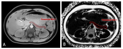 |
The value of 3D mDIXON Quant in quantitative evaluation of factors related to pancreatic volume and fat content in healthy subjects.
Wan Dong1, Ailian Liu1, Qinhe Zhang1, Liangjie Lin2, and Jiazheng Wang2
1Department of Radiology, the First Affiliated Hospital of Dalian Medical University, Dalian, China, 2Philips Healthcare, Beijing, China
Functions of pancreas are associated with its volume and fat fraction. 3D mDixon Quant has been proved to be valuable in measuring fat fraction, which was employed in this study for determination of volume and fat content of pancreas. Then the correlations of pancreas volume and fat content to age, gender and BMI were examined. Results showed a significant negative correlation of age to the pancreas volume, while positive to the fat fraction. Therefore, 3D mDixon Quant is helpful in assessing pancreas volume and fat fraction. As age increases, the pancreas tends to shrink and its fat fraction to increase.
|
|
2469.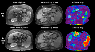 |
Early Effect of 90Y locoregional therapy on tumor and liver parenchyma stiffness measured with MR elastography: initial experience
Paul Kennedy1,2, Sara Lewis3, Octavia Bane1,2, Stefanie Hectors1,2,4, Edward Kim5, and Bachir Taouli1,2
1BioMedical Engineering and Imaging Institute, Icahn School of Medicine at Mount Sinai, New York, NY, United States, 2Department of Diagnostic, Molecular and Interventional Radiology, Icahn School of Medicine at Mount Sinai, New York, NY, United States, 3Radiology, Icahn School of Medicine at Mount Sinai, NY, NY, United States, 4Department of Radiology, Weill Cornell Medicine, New York, NY, United States, 5Diagnostic, Molecular and Interventional Radiology, Icahn School of Medicine at Mount Sinai, NY, NY, United States
In this study we assess early changes following 90Y radioembolization (RE) in hepatocellular carcinoma and liver parenchyma using MR elastography and predict response at 6 weeks/6 months. Tumor stiffness (TS) and liver stiffness (LS) adjacent to the tumor were significantly increased 6w following RE. Baseline TS and LS adjacent to the tumor were significantly lower in patients who achieved complete response. Baseline TS correlated with necrosis at 6w and was a significant predictor of partial response or stable disease based on mRECIST criteria following RE. MR elastography may be a useful tool for predicting necrosis and tumor response to RE.
|
|
2470.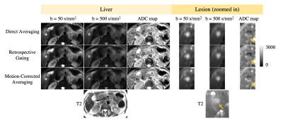 |
Liver Diffusion MRI using Optimized Gradient Waveforms, Free-Breathing Acquisitions, and Motion-Corrected Averaging: Validation in Patients
Ruiqi Geng1,2, Yuxin Zhang1,2, Matthias Robert Muhler1, Jitka Starekova1, and Diego Hernando1,2
1Department of Radiology, University of Wisconsin-Madison, Madison, WI, United States, 2Medical Physics, University of Wisconsin-Madison, Madison, WI, United States
Diffusion weighted imaging (DWI) of the liver has multiple important applications in the assessment of liver disease, including cancer. However, liver DWI faces several major challenges including signal dropout due to cardiovascular pulsation, mis-registration due to respiratory motion, and low SNR. We have developed a DWI technique that addresses these challenges by combining motion-robust diffusion waveforms to address cardiovascular pulsation, high SNR from non-gated free-breathing acquisitions, and motion-corrected averaging for respiratory motion-correction. This technique was evaluated in patients with known or suspected liver metastases. The proposed technique has the potential to enable improved assessment of liver lesions.
|
|
2471.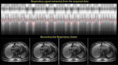 |
Motion-resistant XD-GRASP for free-breathing liver MRI: qualitative comparison with standard breath-hold hepatobiliary phase imaging
Nathanael Kim1, Shannan Dickinson2, Maggie Fun3, Ersin Bayram3, Li Feng4, Ricard Do2, and Ricardo Otazo1,2
Video Permission Withheld
1Medical Physics, Memorial Sloan Kettering Cancer Center, New York, NY, United States, 2Radiology, Memorial Sloan Kettering Cancer Center, New York, NY, United States, 3GE Healthcare, Waukesha, WI, United States, 4Biomedical Engineering and Imaging Institute and Department of Radiology, Icahn School of Medicine at Mount Sinai, New York, NY, United States
Breath-hold post contrast T1-weighted liver MRI is limited by the patient’s ability to cooperate with breathing instructions. Free-breathing radial imaging and compressed sensing-based motion-resolved image reconstruction (XD-GRASP) has been specifically developed to resolve motion artifacts over standard cartesian techniques. In this work, we apply XD-GRASP to enable free-breathing MRI in the hepatobiliary liver phase, a clinically relevant problem. We demonstrate that the image quality of free-breathing XD-GRASP is equal to or superior to the one using standard breath hold acquisitions.
|
|
2472.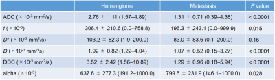 |
Comparison of Mono-, Bi-, and Stretched Exponential Diffusion-weighted MR Imaging in Differentiating Hepatic Hemangiomas and Liver Metastases
Yoshifumi Noda1, Keita Fujimoto1, Nobuyuki Kawai1, Kimihiro Kajta1, Yuta Akamine2, Hiroshi Kawada1, Yukichi Tanahashi1, and Masayuki Matsuo1
1Radiology, Gifu University, Gifu, Japan, 2Philips Japan, Tokyo, Japan Poster Permission Withheld
Intravoxel incoherent motion, a bi-exponential model of diffusion-weighted imaging with multiple b values, can represent pure molecular diffusion and perfusion, and be used in characterizing focal hepatic lesions. Recently, stretched exponential model has been used in several cancers. In this study, we evaluated the feasibility of stretched exponential model for differentiating hepatic hemangiomas and liver metastases. Our results showed that DDC value from a stretched exponential model was the highest diagnostic potential, so it could be a quantitative imaging biomarker for differentiating hepatic hemangiomas and liver metastases.
|
|
2473.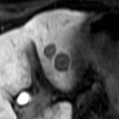 |
Hepatobiliary Contrast Uptake Patterns in Liver Metastases from Pancreatic Ductal Adenocarcinoma: Prediction for Patients’ Overall Survival
Yoshifumi Noda1,2, Vinit Baliyan1, Hamed Kordbacheh1, and Avinash R Kambadakone1
1Radiology, Massachusetts General Hospital, Boston, MA, United States, 2Radiology, Gifu University, Gifu, Japan Poster Permission Withheld
Gadoxetic acid is a liver-specific contrast agent and has been reported to be useful for the detection of liver metastases in patients with pancreatic ductal adenocarcinoma (PDAC). Recently, aberrant OATP expression has been reported in liver metastases from PDAC and is associated with poor prognosis. Our results show that patients with liver metastases demonstrating heterogeneous hypointensity on hepatobiliary phase images are associated with worse overall survival compared to those with homogeneous hypointensity.
|
|
| 2474. | A preliminary study on differential diagnosis of the benign and malignant liver lesions with amide proton transfer-weighted (APTw) imaging
Tao Lin1, Ailian Liu 1, Jiazheng Wang 2, Nan Wang1, Lihua Chen 1, Dahua Cui1, Ying Zhao 1, Qingwei Song 1, Yishi Wang2, Renwang Pu 1, and Bingbing Gao1
1The First Affiliated Hospital of Dalian Medical University, Dalian, China, 2Philips Healthcare, Beijing, China Poster Permission Withheld
Our work aimed to explore the value of amide proton transfer-weighted (APTw) imaging in diagnosing liver lesions. The result showed that novel imaging tool had a high value in differentiating benign liver lesions from malignant liver lesions (AUC: 0.801; sensitivity: 76.9%; specificity: 83.3%).
|
|
2475.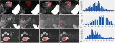 |
The value of texture analysis of multiple high b-values DWI in distinguishing different types of hepatic malignant tumors
Ying Zhao1, Jiazheng Wang2, Zhiwei Shen2, Jianqing Sun2, Nan Wang1, Lihua Chen1, Dahua Cui1, Tao Lin1, Qingwei Song1, Renwang Pu1, Bingbing Gao1, and Ailian Liu1
1The First Affiliated Hospital of Dalian Medical University, Dalian, China, 2Philips Healthcare, Beijing, China
In the current study, texture analysis of multiple high b-values DWI was demonstrated to be capable to identify primary hepatic cancer and hepatic metastases, which provided abundant quantitative information that may facilitate clinical management. The results showed that texture features based on b800 and b2000 DWI images was the optimal strategy to identify different types of hepatic malignant tumors (b800: sensitivity 92.0%, specificity 84.2%; b2000: sensitivity 84.0%, specificity 94.7%).
|
|
2476.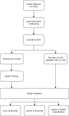 |
Development of a deep learning algorithm for detection of liver cancers on Magnetic Resonance Imaging
Sailong Zhang1, Keith Wan-Hang Chiu1, Siu Hin Mak2, TSOUGENIS Efstratios3, and Peng Cao1
1Diagnostic Radiology, The University of Hong Kong, Hong Kong, Hong Kong, 2The University of Hong Kong, Hong Kong, Hong Kong, 3Imsight Medical Technology Company (Hong Kong), Hong Kong, Hong Kong
Early detection of liver cancer is crucial for improving patient management outcome. However, liver lesions can be differencaited to be identified. In recent years, the fully convolution neural network (FCNN) has shown to be able to achieve commensurate and comparable performance of detecting various pathology on medical imaging. The goal of this study is to show the possibility of applying FCNN deep learning for training the hepatic lesion detection on dynamic contrast-enhanced Magnetic Resonance Imaging.
|
|
2477.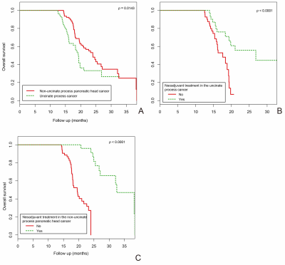 |
Clinical Outcome of Uncinate Process Cancer versus Non-Uncinate Process Pancreatic Head Cancer: A Singe Center Retrospective Study
Yun Bian1, Chao Ma1, Xu Fang1, Jin Li1, Kai Cao1, Li Wang1, Jin Gang2, Jianping Lu1, and Hui Jiang3
1Radiology, Changhai hospital, Shanghai, China, 2Pancreatic Surgery, Changhai hospital, Shanghai, China, 3Pathology, Changhai hospital, Shanghai, China
The uncinate process has unique anatomical characteristics, which may make cancers that develop there radiopathologically different from cancers that develope elsewhere in the pancreas. Previous studies on uncinate and non-uncinate process pancreatic head cancer have yielded conflicting results. Some investigators concluded that location of pancreatic head cancer did not correlate with prognosis. Meanwhile, other investigators demonstrated a significant association between tumor location and prognosis, showing that uncinate process pancreatic head cancer was associated with a higher mortality risk versus non-uncinate process pancreatic head cancer. However, it still remains unelucidated whether tumor location is an independent prognostic factor of overall survival (OS) of pancreatic head cancer patients. In addition, it is unclear whether tumor location could guide prognostic stratification of pancreatic head cancer patients. So, the primary objectives of our study were to investigate the relationship between uncinate and non-uncinate process pancreatic head cancer and overall survival (OS) and further explore prognostic stratification between the two tumor locations and OS in different subgroups of patients.
|