
Digital Poster Session
Body: Abdominopelvic MRI - Benign
Body
2478 -2492 Abdominopelvic MRI - Benign - Diffuse Liver: Fat & Contrast
2493 -2506 Abdominopelvic MRI - Benign - Liver Fibrosis & Cirrhosis
2507 -2520 Abdominopelvic MRI - Benign - Pancreas
2521 -2534 Abdominopelvic MRI - Benign - Female Pelvis, Placenta & Fetal
2535 -2549 Abdominopelvic MRI - Benign - Liver: Iron & Bile
2550 -2561 Abdominopelvic MRI - Benign - Diabetes, Nutrition & Metabolism
2562 -2575 Abdominopelvic MRI - Benign - Female Pelvis & Maternal-Fetal Imaging
2576 -2591 Abdominopelvic MRI - Benign - Gastrointestinal & Abdominal
2592 -2605 Abdominopelvic MRI - Benign - Machine Learning & Radiomics in Body MRI
2606 -2620 Abdominopelvic MRI - Benign - Emerging & Advanced MRI Techniques in Body MRI
2621 -2634 Abdominopelvic MRI - Benign - Pancreas & Hepatobiliary
2635 -2649 Abdominopelvic MRI - Benign - Genitourinary Imaging Innovation, Quality Control & Post-Processing
2478.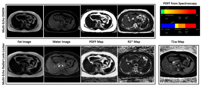 |
Simultaneous T1 and Fat Fraction Quantification using Multi-Echo Radial Look-Locker Imaging
Mahesh Bharath Keerthivasan1, Xiaodong Zhong2, Marcel Dominik Nickel3, David Garcia4, Maria Altbach4, Berthold Kiefer3, and Vibhas Deshpande5
1Siemens Healthcare USA, Tucson, AZ, United States, 2Siemens Healthcare USA, Los Angeles, CA, United States, 3Siemens Healthcare Gmbh, Erlangen, Germany, 4University of Arizona, Tucson, AZ, United States, 5Siemens Healthcare USA, Austin, TX, United States
Abdominal T1 mapping has been used for the quantification of various pathologies including the characterization of focal liver lesions and liver fibrosis. The presence of fat and iron in the liver act as confounding factors resulting in T1 estimation errors. In this work, we present a multi-echo inversion-recovery radial Look-Locker technique with FLASH readouts for simultaneous T1w, T2* and PDFF quantification. We also propose two fitting strategies to generate water-only T1 estimates from the acquired data. Performance of the method is evaluated on phantoms and in vivo results are presented.
|
|
2479.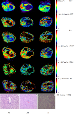 |
Multiparametric MRI evaluation of liposomal prostaglandin E1 intervention on hepatic warm ischemia-reperfusion injury in rabbits
Qian Ji1, JingYao Li1, Jiabing Jiang1, Robert Grimm2, and Jinxia Zhu3
1Tianjin First Central Hospital, Tianjin, China, 2Siemens Healthcare GmbH, Erlangen, Germany, 3MR collaboration, Siemens Healthcare Ltd, Beijing, China
This study evaluated if multiparametric magnetic resonance imaging (MRI) could identify changes in warm ischemia-reperfusion injury (WIRI) after liposomal prostaglandin E1 (Lipo-PGE1) intervention in rabbit livers. We measured blood oxygen level–dependent, diffusion tensor, and intravoxel incoherent motion imaging on functional hepatic parameters in rabbits exposed to 1) warm ischemia at different times, 2) Lipo-PGE1 interventions, and 3) sham operations. The parameters were sensitive to perfusion and oxygenation changes during WIRI. Multiparametric MRI successfully identified changes after Lipo-PGE1 intervention. These results offer new insights to quantitatively evaluate the effect of drugs on hepatic WIRI after liver transplantation and hepatectomy.
|
|
2480.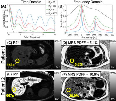 |
Limits of Fat Quantification with Chemical Shift Encoded Magnetic Resonance Imaging in the Presence of Iron Overload
Timothy J Colgan1, Diego Hernando1,2, and Scott B Reeder1,2,3,4,5
1Radiology, University of Wisconsin - Madison, Madison, WI, United States, 2Medical Physics, University of Wisconsin - Madison, Madison, WI, United States, 3Biomedical Engineering, University of Wisconsin - Madison, Madison, WI, United States, 4Medicine, University of Wisconsin - Madison, Madison, WI, United States, 5Emergency Medicine, University of Wisconsin - Madison, Madison, WI, United States
Quantitative confounder-corrected chemical shift encoded MRI estimates of proton density fat fraction (PDFF) are corrected for R2* signal decay. However, at very high levels of iron overload, the observed signal in CSE-MRI decays rapidly (high R2*), complicating the separation of water and fat signals, and therefore the quantification of PDFF. This work characterized the degree of iron overload above which the quantification of PDFF is limited and developed practical clinical and research guidelines for PDFF estimation protocol design for 1.5T and 3.0T.
|
|
2481.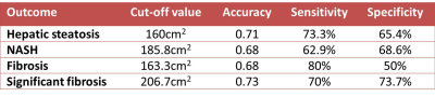 |
Association between visceral adipose tissue and hepatic steatosis and steatohepatitis in obese patients
Ilkay S Idilman1,2, Hsien Min Low3, Tolga Gidener1, Kenneth Philbrick1, Taofic Mounajjed4, Jiahui Li1, Alina Allen5, Meng Yin1, and Sudhakar K Venkatesh1
1Radiology, Mayo Clinic, Rochester, MN, United States, 2Radiology, Hacetep University, Ankara, Turkey, 3Radiology, Tan Tock Seng Hospital, Singapore, Singapore, 4Pathology, Mayo Clinic, Rochester, MN, United States, 5Gastroenterology and Hepatology, Mayo Clinic, Rochester, MN, United States
In this study we measured visceral adipose tissue (VAT), proton density fat fraction (PDFF) of liver, liver volume and correlated with liver biopsy features in obese patients at risk for non-alcoholic fatty liver disease (NAFLD). VAT shows moderately significant association with PDFF and liver volume. VAT also shows weak but significant correlation with hepatic steatosis, non-alcoholic steatohepatitis (NASH) and fibrosis at histology. Liver volume was significantly larger in patients in hepatic steatosis, NASH and fibrosis. VAT has moderate accuracy for detection of hepatic steatosis (0.71), NASH (0.68), fibrosis (0.68) and significant fibrosis (0.73) at histology.
|
|
2482.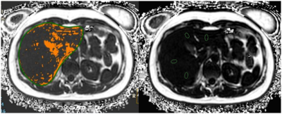 |
Feasibility of Liver Fat Quantification based on the Threshold Extraction on fast 3D mDIXON images
Nan Zhang1, Qingwei Song1, Renwang Pu1, Haonan Zhang1, Yu Song1, Jiazheng Wang2, Liangjie Lin2, and Ailian Liu1
1The First Affiliated Hospital of Dalian Medical University, Dalian, China, 2Philips Healthcare, Beijing, China
The present study aims to explore feasibility of the threshold extraction method for liver fat quantification on the CS-SENSE 3D mDIXON-Quant images. Different from the conventional measurement of liver fat contents based on regions of interest that can be limited when the patients are associated with inhomogeneous fat distribution, liver fat quantification based on the 3D threshold-extraction can be more convenient and reliable.
|
|
2483.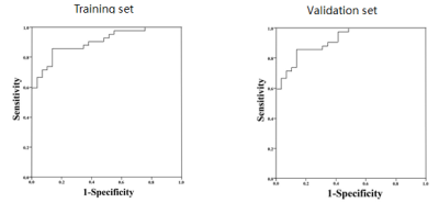 |
Gd-BOPTA-enhanced MRI predicts the severity of esophageal varices (EV) and portal vein pressure in hepatitis B cirrhosis patients
Xinya Zhao1, Xianshun Yuan1, Mengxiao Liu2, Xiang Feng3, Xiangtao Lin1, and Ximing Wang1
1Department of Radiology, Shandong Provincial Hospital, Jinan, China, 2MR Scientific Marketing, Diagnostic Imaging, Siemens Healthcare Ltd, shanghai, China, 3MR Scientific Marketing, Diagnostic Imaging, Siemens Healthcare Ltd, Beijing, China The characteristics of liver cirrhosis are usually hepatic dysfunction and portal hypertension. Portal hypertension further leads to ascites and esophageal varices (EV). To our knowledge, no methods are currently available for the simultaneous evaluation of portal hypertension and EV severity. The results of this study positively illustrate that Gd-BOPTA-enhanced MRI can serve as a novel and efficient tool to simultaneously and accurately evaluate portal hypertension and high-risk EVs. Relative enhancement ratio (RE) is correlated with estimated HVPG. The formula (-6.483+15.612*portal vein width + 2.251 * RE - 0.176 * platelet count) could be used for the prediction of high-risk EVs. |
|
2484.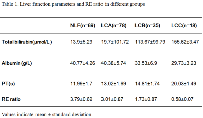 |
Gadolinium-BOPTA Contrast-enhanced MRI in the Hepatobiliary Phase: Evaluating Liver Function based on the visualizing degree of Biliary System
Xinya Zhao1, Xianshun Yuan1, Xiang Feng2, Mengxiao Liu3, Xiangtao Lin1, and Ximing Wang1
1Department of Radiology, Shandong Provincial Hospital, Jinan, China, 2MR Scientific Marketing, Diagnostic Imaging, Siemens Healthcare Ltd, Beijing, China, 3MR Scientific Marketing, Diagnostic Imaging, Siemens Healthcare Ltd, shanghai, China
Gd-BOPTA, as a widely used hepatobiliary-specific MR contrast agent, has advantages in diagnosing focal liver lesions and may be a valuable assessment tool for the estimation of liver function. The aim of our study was to evaluate liver function according to the degree of biliary system visualization using Gd-BOPTA. Our results suggested that the degree of biliary system visualization using Gd-BOPTA-enhanced MRI may be used as a quantifiable metric to estimate liver function and liver function reserve.
|
|
2485.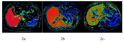 |
Evaluation of liver function using the hepatocyte fraction based on gadoxetic acid–enhanced MR imaging
Mao-Tong LIU1, Xue-Qin ZHANG1, Jian LU1, and Wei-Bo CHEN2
1Third Affiliated Hospital of Nantong University & Nantong Third People's Hospital, Nan Tong, China, 2Philips Healthcare Shanghai, China, Shang Hai, China
The purpose of this study was to evaluate the feasibility of using the hepatocyte fraction based on gadoxetic acid–enhanced MRI for the assessment of liver function. Firstly, T1 mapping imaging was performed before and 20 minutes after Gd-EOB-DTPA administration, The following parameters are then obtained from the images: pre- and postcontrast T1 values of the liver (T1pre and T1post), increase in the T1 relaxation rate (Δ R1), rate of the decrease of the T1 relaxation time (Δ T1), hepatocyte fraction (HeF), and uptake coefficient (K). Our study showed that hepatocyte fraction is an effective method to evaluate liver function in patients with hepatitis B cirrhosis.
|
|
2486.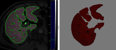 |
Radiomics Analysis on Gd-EOB-DTPA-Enhanced MRI for Prediction of Liver Function and Hepatic Cirrhosis
Xie Yuanliang1, Wang Xiang1, Li Hui1, Liu Xiaoyu1, and Sun Jianqing2
1Radiology, Central Hospital of Wuhan, Tongji Medical College, Huazhong University of Science and Technology, Wuhan, China, 2Clinical Science,Philips Healthcare, Shanghai, China
This retrospective study explored the value of a radiomics-based model on Gd-EOB-DTPA-Enhanced MRI for predicting liver function and cirrhosis in clinic. Multi-class radiomics feature extraction was performed on 2D-view whole liver at portal level on HBP MRI obtained 20 min after Gd-EOB-DTPA-enhanced MRI. A prediction model including 15 radiomics features using a machine learning logistic regression classifier showed the mean AUCs on train dataset and test dataset were 0.91 and 0.87 for diagnosing Child-Pugh A respectively; 0.93 and 0.93 for diagnosing liver cirrhosis, respectively.
|
|
2487.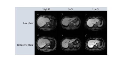 |
A New Possibility of Late Phase Image in Gd-EOB-DTPA-enhanced MRI: Visual Assessment of Hepatic Function and Fibrosis Based on Uptake Rate of EOB
Yasuhiro Inokuchi1, Masahiro Uematsu1, and Tsuneyuki Takashina1
1Radiology, Edogawa Hospital, Tokyo, Japan
We identified visual assessment value of late phase image to assess abnormal hepatic function and fibrosis. We retrospectively selected 41 patients who underwent Gd-EOB-DTPA enhanced MRI and classified them into three groups based on visual assessment of late phase images. Kruskal–Willis t-test was used to assess significant differences in the LSR of hepatobiliary phase, FIB-4, APRI, and platelet count. Intra-class correlation coefficient (ICC) was used for intra-reader visual assessment of late phase image. Significant differences in all parameters were observed in the groups. ICC was 0.85. Hepatic function or fibrosis might be assessed by visual assessment of late phase image.
|
|
2488.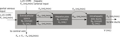 |
A unified model for hepatobiliary transporter function assessment using Gadoxetate DCE-MRI
Sirisha Tadimalla1,2 and Steven Sourbron2,3
1Institute of Medical Physics, University of Sydney, Sydney, Australia, 2Department of Biomedical Imaging Sciences, University of Leeds, Leeds, United Kingdom, 3Department of Infection, Immunity and Cardiovascular Disease, University of Sheffield, Sheffield, United Kingdom
A variety of models for Gadoxetate DCE-MRI in the liver has been proposed, but comparing results of different groups is difficult due to a lack of consistency in the definitions, nomenclature and units. We perform a rigorous classification of existing models and definitions by defining a general unified Gadoxetate DCE-MRI liver model, and identifying the relationship with existing models. Six distinct models were identified in the literature and a translation table was derived to allow direct comparison of measured quantities. The method provides a rational basis for a new standard in this field.
|
|
2489.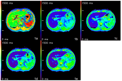 |
Quantitative Assessment of Liver Function by Using T1mapping based on Gadoxetic Acid–enhanced MRI
Xueqin Zhang1, Jian LU1, Jifeng JIANG1, and Weibo CHEN2
1the Third People’s Hospital of Nantong, Nantong, China, 2Philips Healthcare, Shanghai, China
The purpose of this study was to investigate whether liver function as determined by indocyanine green (ICG) clearance can be estimated quantitatively from magnetic resonance T1mapping with Gd-EOB-DTPA. We used Look-Locker sequences to acquire T1 mapping images pre and post-contrast at 5, 10, 15 and 20 minutes after Gd-EOB-DTPA administration, T1 relaxation times of the liver were measured, reduction rates of T1 relaxation times were calculated, our study showed that Gd-EOB-DTPA-enhanced T1mapping MRI is helpful for the quantitative evaluation of liver function, T1 relaxation time post-contrast at 20 minutes was the independent factor to predict ICG R15>20%.
|
|
2490.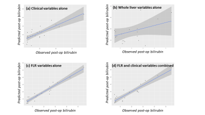 |
Prediction of post-hepatectomy liver function with Dynamic Gadoxetate-Enhanced MRI
David Longbotham1, Daniel Wilson2, Ian Rowe3, Dhakshinamoorthy Vijayanand4, Magdy Attia4, Ashley Guthrie5, Mark Gilthorpe3, Rajendra Prasad4, and Steven Sourbron6
1University of Leeds, Leeds, United Kingdom, 2Department of Medical Physics, Leeds Teaching Hospitals NHS Trust, Leeds, United Kingdom, 3Leeds Institute of Data Analytics, University of Leeds, Leeds, United Kingdom, 4Hepatobiliary and Transplantation Surgery, Leeds Teaching Hospitals NHS Trust, Leeds, United Kingdom, 5Department of Radiology, Leeds Teaching Hospitals NHS Trust, Leeds, United Kingdom, 6Department of Infection, Immunity and Cardiovascular Disease, University of Sheffield, Sheffield, United Kingdom
The aim of this study was to identify Dynamic Gadoxetate-Enhanced MRI (DGE-MRI) biomarkers that can improve predictions of post-hepatectomy liver function. 29 patients requiring resection for colorectal liver metastases were recruited, with post-operative bilirubin as outcome measure. The results suggest that: (a) functional imaging substantially improves outcome predictions over demographical and biochemical tests; (b) it is critical to separately characterise the future liver remnant; (c) volumetry does not offer any added predictive value. We conclude that DGE-MRI may improve patient selection for hepatectomy, potentially reducing the risk of post-hepatectomy liver failure while allowing more patients to be operated.
|
|
2491. |
Comparative study of T1ρ and Gd-EOB-DTPA-enhanced T1mapping with extracellular volume fraction in assessment of liver fibrosis in rabbit model
Qing Wang1, wei xing1, Yanan Du1, Zuhui Zhu1, Yufeng Li1, and Jilei Zhang2
1Third Affiliated Hospital of Soochow University & First People's Hospital of Changzhou., changzhou, China, 2Clinical Science,Philips Healthcare, shanghai, China
This study aimed to compare the diagnostic performance for liver fibrosis staging of T1ρ and Gd-EOB-DTPA-enhanced T1mapping with extracellular volume fraction measurement in CCL4 rabbit model. Result showed that T1ρ performed better diagnostic performance and correlation with LF and fibrosis percentage of postive staining(%) than T1native.
|
|
| 2492. | Diffusion Kurtosis Imaging Quantification of Liver Microscopic Changes in Rats Following Partial Hepatectomy at Different Proportions
Caixin Qiu1, Shuangshuang Xie1, Jinxia Zhu2, Chengwen Liu2, Robert Grimm3, Qing Li1, and Wen Shen1
1Tianjin First Center Hospital, Tianjin, China, 2MR Collaboration, Siemens Healthcare Ltd, Beijing, China, 3Siemens Healthcare GmbH, Erlangen, Germany
In this study, we used DKI to quantify the microscopic changes of residual liver in two groups of rats with different proportions of a partial hepatectomy (PH). All the rats underwent DWI to acquire DKI data at baseline and multiple time points after surgery. The results showed that DKI-derived MD in the 70% PH group were lower than in the 30% PH group at all time points after surgery. MD decreased to a minimum by the fifth day before rising back to baseline. This suggests that DKI is a practical technique for the timely evaluation of the liver regeneration process.
|
 View the Poster
 Watch the Video
2493.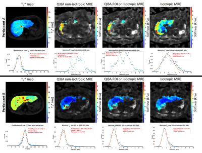 |
Effect of spatial resolution on Gradient Echo Magnetic Resonance Elastography at 3T
Chris R Bradley1,2, Deirdre McGrath1,2, Eleanor F Cox1,2, and Susan T Francis1,2
1Sir Peter Mansfield Imaging Centre, University of Nottingham, Nottingham, United Kingdom, 2NIHR Nottingham Biomedical Research Centre, Nottingham University Hospitals NHS Trust and University of Nottingham, Nottingham, United Kingdom
Iron-mediated T2* effects are more prominent in MRE data acquired at 3T compared to 1.5 T, and have been suggested to lead to failure rates of up to 15% for MRE at 3T. MRE based liver stiffness was measured using the QIBA recommendation with a 2D gradient‐recalled‐echo MRE sequence using a 1.5x4.5x10 mm3 acqusition. For comparison, MRE data was also collected at 4.5mm isotropic spatial resolution. A larger voxel volume in the MRE acquisition provided higher SNR which in turn resulted in a higher proportion of voxels being fit for stiffness with confidence >0.95.
|
|
2494.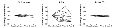 |
Assessing the variation of MR and non-invasive markers in compensated cirrhosis: insights for assessing disease progression
Chris R Bradley1,2, Eleanor F Cox1,2, Naaventhan Palaniyappan2, Guruprasad P Aithal2, I Neil Guha2, and Susan T Francis1,2
1Sir Peter Mansfield Imaging Centre, University of Nottingham, Nottingham, United Kingdom, 2NIHR Nottingham Biomedical Research Centre, Nottingham University Hospitals NHS Trust and University of Nottingham, Nottingham, United Kingdom
Baseline multi-organ MRI measures of structure and haemodynamics in the liver, spleen, heart and kidneys were collected in healthy volunteers, compensated cirrhosis (CC) and decompensated cirrhosis patients to benchmark the change in measures with disease severity. In a stable CC cohort, observed annually for 3 years, we show liver T1, liver perfusion, superior mesenteric artery flow, spleen perfusion, and renal cortex T1 (measures that predict negative liver related outcomes) have sufficient resolution to track disease progression.
|
|
2495.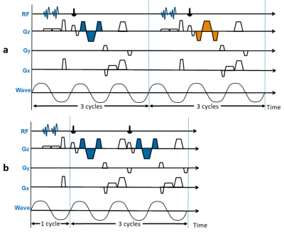 |
A Fast MR Elastography Sequence with Interleaved Inflow Saturation and Compressed SENSE
Hui Wang1,2,3, Amol Pednekar2,3, Jean A. Tkach2,3, Kaley R. Bridgewater2, Andrew T. Trout2,3, Jonathan R. Dillman2,3, and Charles L. Dumoulin2,3
1Philips, Cincinnati, OH, United States, 2Department of Radiology, Cincinnati Children’s Hospital Medical Center, Cincinnati, OH, United States, 3Department of Radiology, University of Cincinnati College of Medicine, Cincinnati, OH, United States
We describe a fast field-echo Magnetic Resonance Elastography pulse sequence to measure liver stiffness in less than half the breath hold time (≈6.3 sec/slice) of the conventional implementation. Key features include: 1) non-alternating motion encoding gradients to allow a shorter TR while maintaining appropriate gradient waveform polarity synchronization with the applied mechanical motion; 2) interleaved flow saturation pre-pulses to suppress flow; and 3) pseudorandom undersampling k-space with Compressed SENSE reconstruction. The technique was validated in two gel phantoms differing in stiffness and used to evaluate liver stiffness in five volunteers.
|
|
2496.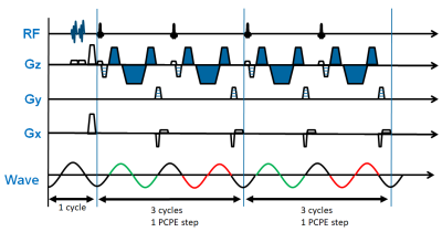 |
A Fast 3D MR Elastography Sequence for Measurement of Liver Stiffness in One Breath Hold
Hui Wang1,2,3, Amol Pednekar2,3, Jean A. Tkach2,3, Charles L. Dumoulin2,3, Kaley R. Bridgewater2, Andrew T. Trout2,3, and Jonathan R. Dillman2,3
1Philips, Cincinnati, OH, United States, 2Department of Radiology, Cincinnati Children’s Hospital Medical Center, Cincinnati, OH, United States, 3Department of Radiology, University of Cincinnati College of Medicine, Cincinnati, OH, United States
We describe a 3D fast field echo (FFE) Magnetic Resonance Elastography (MRE) pulse sequence for measurement of liver stiffness in a single breath hold. The key features of the sequence include: 1) 3D acquisition; 2) mechanical wave magnitude labelling of 1.5 cycles of motion for each RF excitation; 3) flow saturation pre-pulses to suppress vascular flow; and 4) pseudorandom undersampling of k-space with Compressed SENSE reconstruction. The technique was validated in two gel phantoms with different stiffness and used to measure liver stiffness in five volunteers.
|
|
2497.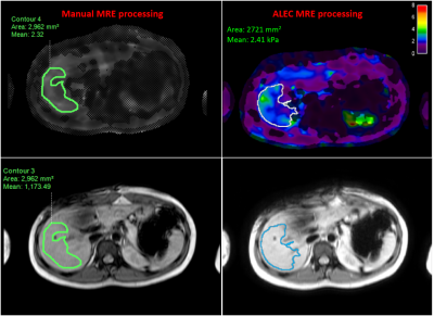 |
Comparison Of Manual And Automatic Liver MR Elastography Processing For Shear Stiffness Estimation In Children And Young Adults
Deep B. Gandhi1, Adebayo B. Braimah1, Jonathan Dudley1, Jean A. Tkach1, Amol Pednekar1, Andrew T. Trout1, Alexander G. Miethke2, Jeremiah A. Heilman3, Bogdan Dzyubak4, David S. Lake4, and Jonathan R. Dillman1
1Imaging Research Center, Department of Radiology, Cincinnati Children's Hospital Medical Center, Cincinnati, OH, United States, 2Division of Hepatology, Gastroenterology and Nutrition, Cincinnati Children's Hospital Medical Center, Cincinnati, OH, United States, 3Resoundant Inc., Rochester, MN, United States, 4Department of Radiology, Mayo Clinic, Rochester, MN, United States
Autoimmune liver diseases lead to fibrosis and is manifested as excessive accumulation of extracellular matrix and collagen that ultimately causes increase in liver stiffness. MR Elastography (MRE) has proven to be an important tool to clinically diagnose liver fibrosis. In this study we performed MRE at 1.5T on 65 subjects with autoimmune liver disease. The data was then manually processed by 2 independent readers and an automated algorithm. Near-perfect correlation and excellent agreement were observed between Reader1 and Reader2 against the automated algorithm (r=0.987 and r=0.981, respectively). Readers had excellent inter-reader agreement(ICC=0.988) and the automated algorithm also demonstrated perfect reproducibility.
|
|
2498.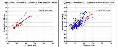 |
Comparison Of GraSE Versus FSE T2 Mapping In The Liver And Correlation With Histologic Fibrosis Stage In Pediatric Autoimmune Liver Disease
Deep B. Gandhi1, Jonathan Dudley1, Ruchi Singh2, Jean A. Tkach1, Divya Sharma3, Amy Taylor2, Alexander G. Miethke2, and Jonathan R. Dillman1
1Imaging Research Center, Department of Radiology, Cincinnati Children's Hospital Medical Center, Cincinnati, OH, United States, 2Division of Hepatology, Gastroenterology and Nutrition, Cincinnati Children's Hospital Medical Center, Cincinnati, OH, United States, 3Department of Pathology and Laboratory Medicine, University of Cincinnati Medical Center, Cincinnati, OH, United States
Autoimmune liver diseases can lead to hepatic fibrosis which, when progressive, can lead to liver failure ultimately requiring transplantation. In this study 31 patients with autoimmune liver diseases underwent conventional and GraSE T2 mapping at 1.5T as well as liver biopsy with histologic fibrosis staging. Significant positive correlations between GraSE and conventional liver T2 measurements were observed for the weighted mean of all slices (r=0.80; p<0.0001) and for individual slices from all subjects (r=0.72; p<0.0001). Conventional T2 measurements were higher, on average, than GraSE T2 measurements. There was no significant correlation observed between liver T2 measurements and histologic fibrosis stage.
|
|
2499.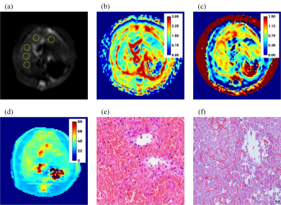 |
Comparison of T1rho and Diffusion Kurtosis Imaging in Evaluation of Hepatic Sinusoidal Obstruction Syndrome in Rats
Jian Lyu1,2, Guixiang Yang3,4, Yingjie Mei5, Li Guo1,2,6, Yihao Guo1,2, Kaixuan Zhao1, Xinyuan Zhang1,2, Yikai Xu3,4, and Yanqiu Feng1,2
1School of Biomedical Engineering, Southern Medical University, Guangzhou, China, 2Guangdong Provincial Key Laboratory of Medical Image Processing, Southern Medical University, Guangzhou, China, 3Department of Medical Imaging Center, Nanfang Hospital, Southern Medical University, Guangzhou, China, 4Key Laboratory of Mental Health of the Ministry of Education, Southern Medical University, Guangzhou, China, 5Philips Healthcare, Guangzhou, China, 6Department of MRI, The First People’s Hospital of Foshan (Affiliated Foshan Hospital of Sun Yat-sen University), Foshan, China
T1rho represents the spin-lattice relaxation time constant in the rotating frame, which sever as a biomarker for liver function associated with alteration in the macromolecular content of tissues. Diffusion kurtosis imaging (DKI) was developed to measure non-Gaussian diffusion, which have increasingly been used to characterize microstructural heterogeneity in vivo. Sinusoidal obstruction syndrome (SOS) is a dynamic process with complex histopathological changes in liver. Our study attempts to investigate the relationship between T1rho and DKI parameters in the light of pathological examinations to help us better understand the contribution of the possible factors to changes in T1rho.
|
|
2500.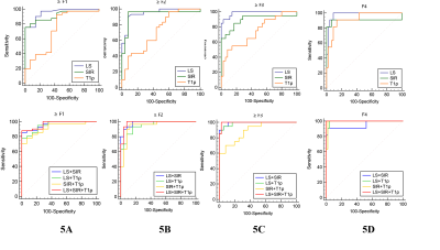 |
Staging liver fibrosis on multi-parametric MRI in a rabbit model: elastography, susceptibility-weighted imaging and T1ρ imaging
Hai-Feng Liu1, Li-Qiu Zou2, Qing Wang1, Yu-Feng Li1, Ya-Nan Du1, and Wei Xing1
1Third Affiliated Hospital of Soochow University, changzhou, China, 2Sixth Affiliated Hospital of Shenzhen University, shenzhen, China
In this prospective experimental study, we evaluated the independent value and diagnostic efficacy of multi-parametric MRI using quantitative measurements of the liver stiffness (LS) on MRE, liver-to-muscle signal intensity ratio (SIR) on SWI, and T1ρ value for staging LF in a rabbit model.
|
|
2501.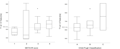 |
Evaluation of liver fibrosis and cirrhosis on the basis of T1 mapping and the impact of inflammation, age and liver volume as confounding factors
Hanns-Christian Breit1, David Winkel2, and Tobias Heye2
1Radiology, University Hospital of Basel, Basel, Switzerland, 2University Hospital of Basel, Basel, Switzerland
Aim of our study was to evaluate confounding factors for the assessment of liver fibrosis. A total of 200 patients were retrospectively included (67 patients with fibrosis or cirrhosis, 40 patients with acute elevation of laboratory parameters, 93 healthy patients). T1 values were significantly lower in healthy patients without known fibrotic changes than in patients with acute liver disease or known fibrosis or cirrhosis. Therefor T1 mapping seems to be a capable predictor for the detection of liver fibrosis and cirrhosis.
|
|
2502.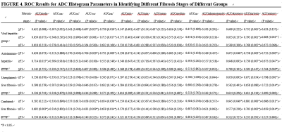 |
Whole-liver apparent diffusion coefficient histogram analysis for the diagnosis and staging of liver fibrosis
Zheng You1,2, Lei Jun-qiang2, and Xu Yong-sheng2
1First Clinical Medical College of LanZhou University, Lanzhou, Gansu, China, 2Radiology, First Hospital of LanZhou University, Lanzhou, Gansu, China
This study aimed to determine whether whole-liver apparent diffusion coefficient (ADC) histogram parameters can contribute to hepatic fibrosis staging. We evaluated quantitative histogram parameters between different pathological fibrosis stages. And the diagnostic performance of ADC histogram parameters in discriminating stage 1 or greater (≥F1), stage 2 or greater (≥F2), and stage 3 or greater (≥F3) liver fibrosis were compared. The results showed that many histogram parameters (kurtosis, skewness, entropy, mode, 75th and 90th percentiles) had statistical significance among the pathologic liver fibrosis stages (P<0.05), and kurtosis yielded the highest area under the curve (0.801).
|
|
2503.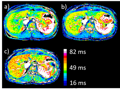 |
Comparison of the degree of R1rho dispersion in liver between healthy volunteers and patients with liver disease
Minori Onoda1, Yu Ueda2, Satoshi Kobayashi1,3,4, Tosiaki Miyati3, Naoki Ohno3, Yudai Shogan1, Tadanori Takata1, Yukihiro Matsuura1, and Toshifumi Gabata4
1Department of Radiological Technology, Kanazawa University hospital, Kanazawa, Japan, 2Philips Japan, Tokyo, Japan, 3Faculty of Health Sciences, Institute of Medical, Pharmaceutical and Health Sciences, Kanazawa University, Kanazawa, Japan, 4Department of Radiology, Kanazawa University Hospital, Kanazawa, Japan
In this study, we compared the degree of R1rho dispersion in liver of healthy volunteers and patients with liver disease to better understand the behavior of T1rho in liver. T1rho in normal liver tissue was correlated with the perfusion-independent diffusion coefficient (D) calculated by IVIM analysis, whereas in patients with liver disease, there was no correlation between T1rho and D. Furthermore, there was no significant difference between the R1rho dispersion values in liver between volunteers and patients.
|
|
2504.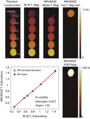 |
Free-Breathing, Confounder-Corrected T1 Mapping in the Liver with 3D Radial Inversion Recovery MRI
Yavuz Muslu1,2, Steven Kecskemeti2,3, Diego Hernando1,2,4, and Scott B. Reeder1,2,4,5,6
1Department of Biomedical Engineering, University of Wisconsin-Madison, Madison, WI, United States, 2Department of Radiology, University of Wisconsin-Madison, Madison, WI, United States, 3Waisman Center, University of Wisconsin-Madison, Madison, WI, United States, 4Department of Medical Physics, University of Wisconsin-Madison, Madison, WI, United States, 5Department of Medicine, University of Wisconsin-Madison, Madison, WI, United States, 6Department of Emergency Medicine, University of Wisconsin-Madison, Madison, WI, United States
Quantitative T1 mapping in the liver is an emerging biomarker of hepatic fibrosis and characterization of liver function. Existing T1 mapping methods in abdomen are generally sensitive to tissue fat and B1 inhomogeneities , both of which confound estimates of T1. Further, Cartesian methods may suffer from motion related ghosting artifacts. In this work, we propose to combine 3D-radial inversion recovery with chemical shift encoded imaging to jointly estimate T1 of water, T1 of fat, proton density fat fraction (PDFF), and B0 and B1 inhomogeneities. The feasibility and performance of the proposed method are evaluated with simulations, and phantom experiments.
|
|
2505.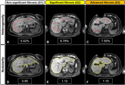 |
Assessment of fibrosis grades in chronic liver disease using liver heterogeneity and nodularity quantification program
Tae-Hoon Kim1, Ji Eon Kim1, Jong-Hyun Ryu1, SeungJin Kim2, Min-Gi Pak2, Chang-Won Jeong1, and Kwon-Ha Yoon1,3
1Medical Convergence Research Center, Wonkwang University, Iksan, Korea, Republic of, 2Medical Science, Wonkwang University, Iksan, Korea, Republic of, 3Radiology, Wonkwang University, Iksan, Korea, Republic of Poster Permission Withheld
Liver fibrosis is a hallmark of chronic liver disease (CLD) characterized by the excessive accumulation of extracellular matrix proteins. To diagnose and grade the liver fibrosis, liver biopsy is the reference standard, however the method has some limitations, including potential pain, sampling variability, and low patient acceptance. Therefore, there have been efforts to develop noninvasive imaging techniques and quantification softwares for diagnosis, staging, and monitoring of liver fibrosis. This study developed a MRI-suitable quantification program for assessing heterogeneity and nodularity in the liver and compared the difference between fibrosis grades in CLD.
|
|
2506.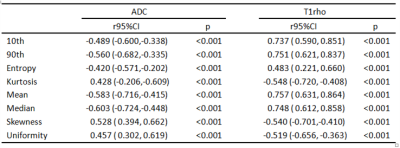 |
Histogram Analysis of T1 relaxation time in the rotating frame and Apparent Diffusion Coefficient for Diagnosis of Liver Fibrosis
Jing Li1, Xinming Li1, Xianyue Quan1, Genwen Hu2, Yingjie Mei3, Da Shi1, Shisi Li4, Zhendong Qi1, and Xiao Zhang5
1Zhujiang Hospital, Southern Medical University, Guangzhou, China, 2Shenzhen People’s Hospital, Clinical Medical College of Jinan University, Shenzhen, China, 3Phlips Healthcare, Guangzhou, China, 4The Third Affiliated Hospital of Southern Medical University, Guangzhou, China, 5Guangdong Provincial Key Laboratory of Medical Image Processing, School of Biomedical Engineering, Southern Medical University, Guangzhou, China
To explore the potential of histogram analysis to evaluate the liver fibrosis stages, histogram of T1rho and ADC were acquired from liver fibrosis model built in seventy-five rats by injecting 50% carbon tetrachloride and olive oil. In this study, we found that the parameters of histogram analysis showed strong correlations with liver fibrosis stages, as well as inflammatory activity,while T1rho is regarded to be better than ADC.
|
2507.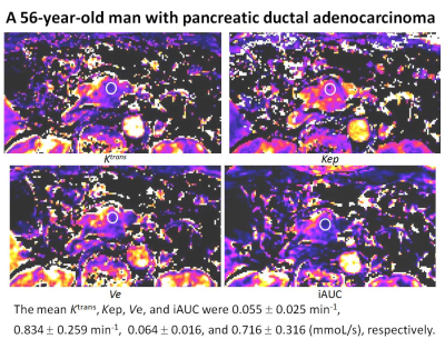 |
DCE-MRI with a free-breathing compressed sensing VIBE for pancreatic ductal adenocarcinoma: correlation with ECV fraction
Yoshihiko Fukukura1, Yuichi Kumagae1, Hiroaki Nagano1, Koji Takumi1, Hiroshi Imai2, Marcel Dominik Nickel3, and Takashi Yoshiura1
1Kagoshima University Graduate School of Medical and Dental Sciences, Kagoshima, Japan, 2Siemens Healthcare K.K., Tokyo, Japan, 3Siemens Healthcare, Erlangen, Germany
This study focused on the feasibility of dynamic contrast-enhanced MRI (DCE-MRI) with compressed sensing T1-weighted volumetric interpolated breath-hold examination (csVIBE) for pancreatic ductal adenocarcinoma (PDAC) and correlation with extracellular volume fraction (ECV). Our results indicated that DCE-MRI obtained with csVIBE is feasible for the assessment of PDACs and the ECV fraction can be used in place of DCE-MRI parameters for predicting treatment response or survival in patients with PDAC.
|
|
2508. |
Acute pancreatic fat change in probiotics and intermittent fasting trial
Jun Lu1, Dech Dokpuang1, Rinki Murphy2, Lindsay Plank2, John Zhiyong Yang1, Reza Nemati3, and Kevin Haokun He4
1School of Science, Auckland University of Technology, Auckland, New Zealand, 2School of Medicine, University of Auckland, Auckland, New Zealand, 3Canterbury Health Laboratories, Canterbury District Health Board, Christchurch, New Zealand, 4Saint Kentigern College, Auckland, New Zealand
Pancreatic fat has been reported to be closely related to type 2 diabetes risk, hence is the subject of our investigation in a clinical trial. Pancreatic fat changes before and after a 12-week intermittent fasting programme with or without daily probiotic were determined using magnetic resonance imaging (MRI). Two-point Dixon protocol was used to scan patients and manual image-processing method was used to quantify the fat. A significant reduction in pancreatic fat was observed after intermittent fasting, while addition of probiotic did not increase pancreatic fat reduction.
|
|
| 2509. | Visualization of pancreas and reproducibility of metrics with diffusion kurtosis imaging in rats at 11.7T MRI
Tingting Zhang1, Yimei Lu1, He Wang2, and Dengbin Wang1
1Radiology, Xinhua Hospital, Shanghai Jiao Tong University School of Medicine, Shanghai, China, 2Institute of Science and Technology for Brain-Inspired Intelligence, Shanghai, China
This study aimed to visualize the pancreas of rats with uncontrast MRI and validate the contour of pancreas by infusing gadolinium solution into the biliopancreatic duct for post-mortem imaging at 11.7 T MRI. Also, the reproducibility of metrics with diffusion kurtosis imaging (DKI) were evaluated in the pancreas of rats. We found that MRI at 11.7T could facilitate preclinical experiments in rodent pancreas and DKI appeared to be a useful noninvasive imaging tool to research pancreatic diseases with satisfactory reproducibility.
|
|
2510. |
Quantitative study on DWI, T1 mapping, T2 mapping, APT, IVIM and 3D mDIXON-quant of pancreas in fatty liver patients
Yuhui Liu1,2, Ailian Liu3, Queenie Chan4, Qinhe Zhang3, and Wan Dong3
1Department of Radiology, the First Affiliated Hospital of Dalian Medical University, Dalian, Dalian, China, 2Dalian medical university, Dalian, China, 3Department of Radiology, the First Affiliated Hospital of Dalian Medical University, Dalian, China, 4Philips Healthcare, Beijing, China
At present study, ADC, T1, T2, APT, sADC, D*,D, f, FF and R2* of pancreas in patients with fatty liver and control subjects were measured. This study viewed that there was a significant difference in sADC between two groups (P=0.01). This may illustrate that fatty liver has an impact on the function of pancreas.
|
|
2511.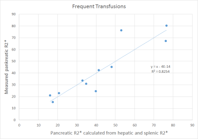 |
MRI Relaxometry: Pancreatic R2* Values in Relation to Hepatic and Splenic R2* with Respect to Transfusion Frequency
Arthur Peter Wunderlich1,2, Stephan Kannengießer3, Lena Kneller1, Berthold Kiefer3, Holger Cario4, Meinrad Beer1, and Stefan Andreas Schmidt1
1Dept. for Diagnostic and Interventional Radiology, Ulm University, Medical Center, Ulm, Germany, 2Section for Experimental Radiology, Ulm University, Medical Center, Ulm, Germany, 3Siemens Healthcare GmbH, Erlangen, Germany, 4Dept. for Pediatric and Adolescent Medicine, Ulm University, Medical Center, Ulm, Germany
To study pancreatic iron accumulation in liver overloaded patients in relation to hepatic and splenic iron content, 90 patients were investigated at 1.5 T MRI with a prototype breath hold volumetric 3D GRE sequence with in-line R2* calculation. Mean R2* values were determined in liver, spleen and pancreas by manually drawn ROIs. Pancreatic R2* values were related to hepatic and splenic R2* by multiple linear regression, yielding significant correlation in two subgroups: a) frequently transfused patients, and b) regular transfused patients. Correlation, and therefore, ability to predict pancreatic R2*, improved by including splenic R2* compared to solely hepatic R2*.
|
|
2512.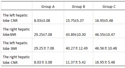 |
Effect of Compressed SENSE on 3D mDixon Sequences for Liver Imaging : A Comparative Study with 3D Vane Sequences
Qiang Wei1, Ailian Liu1, Dongna Yi1, Queenie Chan2, Qingwei Song1, Renwang Pu1, Lihua Chen1, Yu Zhang1, and Jiazheng Wang2
1The First Affiliated Hospital of Dalian Medical University, Dalian, China, 2Philips Healthcare, Beijing, China
The challenging problem of MR living examination is how to obtain images with diagnostic image quality in a shorter scan time. The purpose of this study is to explore the value of 3D mDixon sequence using compressed SENSE (CS) technology in liver examination. Compared with free-breathing mDixon with 3D Vane sequence, breath-hold (BH) 3D mDixon SENSE factor 2 and BH 3D mDixon CS factor 2 sequence can effectively improve the signal-to-noise ratio, contrast-to-noise ratio of the image and the image quality, and significantly shorten the scanning time.
|
|
2513.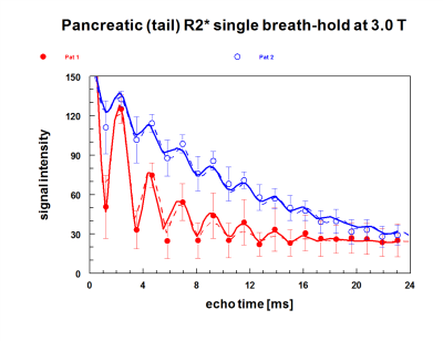 |
PANCREATIC IRON IN PATIENTS WITH HEMOCHROMATOSIS: NOT AS RARE AS CARDIAC IRON
Jin Yamamura1, Peter Nielsen2, Sarah Keller3, Regine Grosse4, Björn Schönnagel1, and Roland Fischer2,5
1Diagnostic and Interventional Radiology, University Medical Center Hamburg-Eppendorf, Hamburg, Germany, 2Biochemistry, University Medical Center Hamburg-Eppendorf, Hamburg, Germany, 3Diagnsotic and Interventional Radiology, Universitätsmedizin Charité, Berlin, Germany, 4Hemato-Oncology, University Medical Center Hamburg-Eppendorf, Hamburg, Germany, 5Diagnostic Radiology, UCSF-Benioff Children's Hospital Oakland, Oakland, CA, United States
HFE-associated hereditary Hemochromatosis (HFE-HH) is the most frequent monogenic genetic disorder in the Caucasian population. The excessive iron storage in organs are common, affecting the liver mostly (> 90 %) resulting in a potentially severe liver damage (fibrosis, cirrhosis). In recent years, hepatic and cardiac iron deposition has been studied in detail. Due to its involvement in the development of diabetes - a frequent co-morbidity in iron overload diseases - pancreatic iron should become a field of interest. This study aims to determine the pancreatic iron and fat content in patients with HFE-HH.
|
|
2514.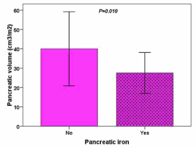 |
Quantification of pancreatic volume and clinical correlations in patients with hemoglobinopathies
Antonella Meloni1, Massimiliano Missere2, Gennaro Restaino2, Laura Pistoia1, Vincenzo Positano1, Emanuele Grassedonio3, Nicolò Schicchi4, Giuseppe Peritore5, Francesco Massei6, Nicola Dello Iacono7, Angelica Barone8, and Alessia Pepe1
1MRI Unit, Fondazione G. Monasterio CNR-Regione Toscana, Pisa, Italy, 2Fondazione di Ricerca e Cura "Giovanni Paolo II", Campobasso, Italy, 3Policlinico "Paolo Giaccone", Palermo, Italy, 4Azienda Ospedaliero-Universitaria Ospedali Riuniti "Umberto I-Lancisi-Salesi", Ancona, Italy, 5"ARNAS" Civico, Di Cristina Benfratelli, Palermo, Italy, 6Azienda Ospedaliero Universitaria Pisana – Stabilimento S.Chiara, Pisa, Italy, 7Ospedale Casa Sollievo della Sofferenza IRCCSOspedale Casa Sollievo della Sofferenza IRCCS, San Giovanni Rotondo, Italy, 8Azienda Ospedaliero-Universitaria di Parma, Parma, Italy
In patients with hemoglobinopathies fat infiltration is a common finding and pancreatic volume is significantly lower than in normal subjects but it is not associated to age or gender. Pancreatic iron overload is present in the 74% of the patients and it is associated with reduced pancreatic volume. Patients with diabetes have a lower pancreatic volume.
|
|
| 2515. | Quantitative assessment of the pancreas in healthy subjects using DWI, IVIM, and 3D mDixon-quant: correlation with age, gender and BMI
Qinhe Zhang1, Wan Dong1, Jiazheng Wang2, Yishi Wang2, and Ailian Liu1
1The first affiliated hospital of Dalian Medical University, Dalian, China, 2Philips Healthcare, Beijing, China
This study measured the quantitative imaging metrics of the pancreas using DWI, IVIM, and 3D mdixon-quant sequences in healthy subjects and correlated these quantitative metrics with age, gender and BMI. This study showed that there were noticeable differences in sADC, f between genders (p<0.05). It shows that pancreatic cells of male are closely arranged, cell gap is reduced, and water diffusion is limited and pancreatic perfusion reduce,which may be because pancreatic fat content of male overweigh that of female(5.16% vs.3.68%).
|
|
2516.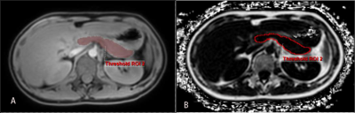 |
Explore the relationship between liver and pancreatic fat contents through 3D mDIXON Quant
Yaru You1, Qinhe Zhang1, Ailian Liu1, Jiazheng Wang2, and Liangjie Lin2
1The First Affiliated Hospital of Dalian Medical University, Dalian, China, 2Philips Healthcare, Beijing, China
In recent years, population of non-alcoholic fatty liver diseases (NAFLD) have shown a trend of increasing. Besides, studies have reported that NAFLD was associated with pancreatic fat infiltration. 3D mDixon Quant has been widely used for evaluation of fat fraction in various organs/tissues. In the present study, the 3D mDixon Quant sequence was employed to assess the relationship between liver and pancreatic fat contents. Results suggested that there was a significant positive correlation between liver and pancreatic fat contents (r=0.624, P<0.05), which may be helpful for clinical diagnoses.
|
|
2517.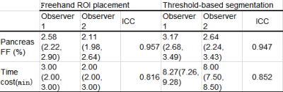 |
Quantification of pancreatic fat content: A Comparison of freehand regions of interest placement and threshold-based segmentation
Yaru You1, Qinhe Zhang1, Ailian Liu1, and Jiazheng Wang2
1The First Affiliated Hospital of Dalian Medical University, Dalian, China, 2Philips Healthcare, Beijing, China
The diagnosis pancreatic fat infiltration is commonly performed with consideration of histopathology in combination with CT /MRI images. This study evaluated the performance of two fat content measurement methods using 3D mDIXON Quant images, based on interest-based hand-drawn area (ROI) placement and threshold-based segmentation. Results by the two methods show good agreement, indicating that both of the two methods can be reliable for evaluation of the pancreas fat content. While, the method based threshold segmentation is recommended in this study, since it can provide a full evaluation of fat contents across the pancreas and can be more convenient in implementation.
|
|
2518.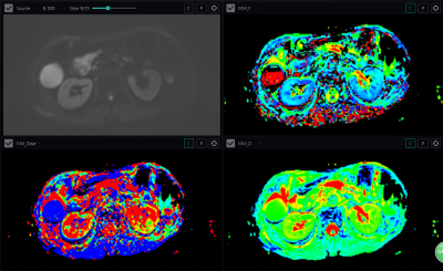 |
Predicting the resectability and pathological grading of pancreatic cancer by intra voxel incoherent motion diffusion-weighted imaging
Qi liu1, Jing Gang Zhang1, Jie Chen1, Wei Xing1, and Jilei Zhang2
1Changzhou First People's Hospital, Changzhou, China, 2Clinical Science, Philips Healthcare, Shanghai, China
The resectability and pathological grade of pancreatic cancer are important for prognosis of patients. We try to predict the pathological grade and the resectability of pancreatic cancer by quantitative IVIM-DWI. Comparing the IVIM parameters of resectable and unresectable pancreatic cancer, poorly differentiated and highly-moderately differentiated pancreatic cancer, we found that poorly differentiated pancreatic cancer had lower f value than highly-moderately differentiated pancreatic cancer, and resectable pancreatic cancer had higher d, f values than unresectable pancreatic cancer. The quantitative IVIM parameters can predict the resectability and pathological stage of pancreatic cancer, which can be helpful for assessment of resectability.
|
|
2519.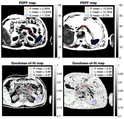 |
Why you shouldn’t report pancreas MRI-PDFF ‘for free’ from a liver scan
Alexandre Triay Bagur1, Ged Ridgway2, Michael Brady2, and Daniel Bulte1
1Department of Engineering Science, University of Oxford, Oxford, United Kingdom, 2Perspectum Diagnostics, Oxford, United Kingdom
The proximal locations of the liver and pancreas in the abdomen make it tempting to report the PDFF of both in a single scan. However, published methods for liver fat measurement need revision before they accurately measure pancreatic fat. UK Biobank scans were used to quantify the quality of fits of a method developed previously for liver PDFF when applied to the pancreas. Pancreas fits were an order of magnitude lower than fits in the liver or the spleen. This could be due to a suboptimal acquisition or because the liver fat model does not approximate well to the pancreas.
|
|
2520.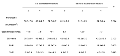 |
Effect of different Compressed-SENSE acceleration factors on pancreas volume using 3D mDIXON Quant
Jie Yang1, Qinhe Zhang1, Ailian Liu1, Jiazheng Wang2, and Zhongping Zhang2
1The first affiliated hospital of Dalian Medical University, Dalian, China, 2Philips Healthcare, Beijing, China
The pancreatic volume can reflect the function of the pancreas to a certain extent. The 3D mDixon Quant can be used to assess the volume of the tissue structure, but some patients cannot tolerate the long-term breath test. This study was designed to ensure pancreas volume using different CS acceleration factors on the premise of ensuring image quality. The results show that CS-SENSE 6 guarantees image quality and reduces scan time.
|
2521.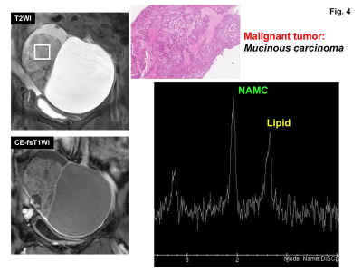 |
Clinical value of N-acetyl mucinous compounds and lipid peaks in differentiating benign and malignant ovarian mucinous tumors by MR spectroscopy
Mayumi Takeuchi1, Kenji Matsuzaki2, and Masafumi Harada1
1Department of Radiology, Tokushima University, Tokushima, Japan, 2Department of Radiological Technology, Tokushima Bunri University, Sanuki-city, Japan
MR spectroscopy of pathologically proven 26 ovarian mucinous tumors (9 benign and 17 malignant) was retrospectively evaluated. N-acetyl mucinous compounds (NAMC) peak at 2 ppm was observed in all 26 lesions. Lipid peak was observed in 1 of 9 benign tumors (11%) and 12 of 17 malignant tumors (71%). The presence of lipid peak for the diagnosis of malignancy had a sensitivity of 71%, specificity of 89%, PPV of 92%, and NPV of 62%. We conclude that the bimodal peaks of NAMC and necrosis-associated lipid are suggestive of malignant mucinous tumors.
|
|
2522.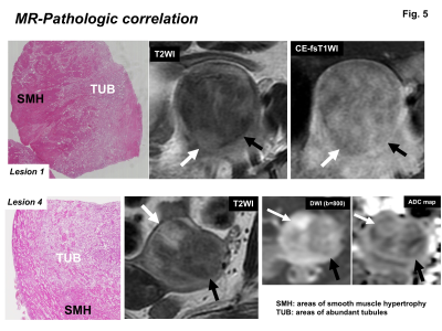 |
MRI findings of uterine adenomatoid tumors including diffusion-weighted imaging with pathologic correlation
Mayumi Takeuchi1, Kenji Matsuzaki2, and Masafumi Harada1
1Department of Radiology, Tokushima University, Tokushima, Japan, 2Department of Radiological Technology, Tokushima Bunri University, Sanuki-city, Japan
Uterine adenomatoid tumor (AT) is a rare benign neoplasm of mesothelial origin. MRI findings of surgically proven 10 ATs were retrospectively evaluated. Most ATs (9/10) appeared as a heterogeneous intensity mass on T2WI with admixture of well-defined myoma-like low intensity area reflecting the areas of smooth muscle hypertrophy, and ill-defined high intensity areas reflecting the areas of abundant tubules. AT may contain high intensity areas on DWI, occasionally appearing as ring-like high intensity area (3/9), with relatively high ADC (6/9) due to T2 shine-through effect. Intra-tumoral hemorrhage was observed on SWAN (1/6) but not on T1WI (0/10).
|
|
2523.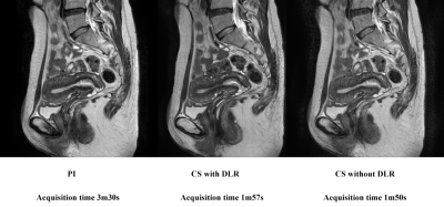 |
Compressed Sensing with and without Deep Learning Reconstruction: Comparison of the Utility for Women’s Pelvic MRI with Parallel Imaging
Takahiro Ueda1, Yoshiharu Ohno1, Kaori Yamamoto2, Akiyoshi Iwase3, Takashi Fukuba3, Yuki Obama1, Kazuhiro Murayama4, and Hiroshi Toyama1
1Radiology, Fujita Health University School of Medicine, Toyoake, Japan, 2Canon Medical Systems Corporation, Otawara, Japan, 3Radiology, Fujita Health University Hospital, Toyoake, Japan, 4Joint Research Laboratory of Advanced Medical Imaging, Fujita Health University School of Medicine, Toyoake, Japan
There have been no major reports for assessing the utility of compressed sensing (CS) and deep learning reconstruction (DLR) on women’s pelvic MRI as compared with routinely applied parallel imaging (PI). We hypothesized that CS with DLR was able to improve image quality and shorten examination time on women’s pelvic MRI, when compared with PI. The purpose of this study was to directly compare the utility of CS and DLR with PI at women’s pelvic MRI examination in patients with different women’s pelvic diseases.
|
|
2524. |
Can T2 mapping be used to differentiate endometrial cancer from benign lesion?
han xu1, jie zhang2, ximing wang3, qingwei liu3, and xiang feng4
1radiology, shandong provincial hospital, JINAN, China, 2shandong provincial hospital, JINAN, China, 3shandong provincial hospital, jinan, China, 4MR Scientific Marketing,Diagnostic Imaging, Siemens Healthcare Ltd, beijing, China
This study aimed to investigate the feasibility of T2 mapping in differentiating endometrial cancer (ECA) from benign endometrial lesions (BEL), as well as to evaluate the histopathological stages, grades and types of ECA.We measured the mean T2 values of 51 endometrial cancer,12 benign endometrial lesions and 23 normal endometrium of volunteers.We found the mean T2 values were signifcantly different among ECA, BEL and normal volunteers, and could be used to distinguish different types of ECA, but could not distinguish different stages or grades of ECA.
|
|
2525.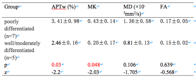 |
Application value of DKI combined with APT in differentiating pathological grades of squamous cell carcinoma of cervix
Yaxin Niu1, Shifeng Tian1, Wan Dong1, Xing Meng1, Lihua Chen1, Qingwei Song1, Jiazheng Wang2, Zhiwei Shen2, Ailian Liu1, and Queenie Chan2
1The First Affiliated Hospital of Dalian Medical University, Dalian Medical University, Da Lian, China, 2Philips Healthcare, Beijing, China, Bei jing, China
We aimed to explore the value of amide proton transfer-weighted (APTw) imaging combined with diffusion kurtosis imaging (DKI) in differentiating pathological grades of squamous cell carcinoma (SCC) of cervix. The result showed that the highest diagnostic efficacy (AUC: 0.971) was acquired using APTw combined with mean kurtosis (MK).
|
|
2526.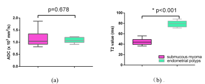 |
Quantitative analysis with T2 mapping in the differentiation between uterine submucous myoma and endometrial polyps
Liuhong Zhu1, Puyeh Wu2, and Jianjun Zhou1
1Xiamen Branch, Zhongshan Hospital, Fudan University, Xiamen, China, 2MR Research China, GE Healthcare, Beijing, China
It is challenging to differentiate between uterine submucous myoma and endometrial polyps, due to a high similarity of their manifestations in conventional MRI. Meanwhile, T2 mapping is an objective and stable technique which has been applied to diagnosis of many diseases. Here we evaluate the value of quantitative measurements derived from T2 mapping and DWI in differentiating between uterine submucous myoma and polyps. We found a descending order of T2 values from healthy endometrium, endometrial polyp to submucous myoma group. We concluded that T2 mapping can be used as a quantitative tool in the differentiation between submucous myoma and polyps.
|
|
2527. |
The value of ESWAN in diagnosis and differential diagnosis of endometrial carcinoma and endometrial polyp
Xing Meng1, Ailian Liu 1, Shifeng Tian1, Ye Ju1, and Qingwei Song1
1Department of Radiology, the First Affiliated Hospital of Dalian Medical University, Dalian, China
Enhanced T2* weighted angiography (ESWAN) has been applied in the diagnosis of female pelvic tumor, but no study on the differentiation of endometrial cancer and related benign lesions by ESWAN has been found. We investigated the value of ESWAN multiple quantitative parameters in the identification of endometrial carcinoma and endometrial polyps.
|
|
2528. |
Application of amide proton transfer weighted imaging in differential diagnosis of endometrial carcinoma and benign endometrial lesions
Ye Ju1, Xing Meng1, Shifeng Tian1, Liangjie Lin2, Jiazheng Wang2, Zhiwei Shen2, Yishi Wang2, Yaxin Niu1, Wan Dong1, and Ailian Liu1
1First affiliated hospital of dalian medical university, Dalian, China, 2Philips Healthcare, Beijing, China
APT imaging has been preliminarily explored for diagnoses of cervical diseases. In this study, we investigated the potential of APTw-MRI in differential diagnosis of endometrial carcinoma and benign endometrial lesions.
|
|
2529. |
Use of amide proton transfer-weighted imaging to distinguish ovarian cysts due to endometriosis from cystadenoma and cystadenocarcinoma
Ye Li1, Yaxin Niu1, Xuedong Wang1, Lihua Chen1, Yanling Wu2, and Ailian Liu1
1The First Affiliated Hospital of Dalian Medical University, Dalian, China, 2Philips Healthcare, Beijing, China Poster Permission Withheld
The intensity of the APT signal depends on the pH and protein content within the tissue. The later the bleeding stage is, the lower the APT value is. The ectopic endometrium repeatedly bleeds in the ovary. The blood components deposited in the cyst fluid decomposition, so the APT value of the cyst fluid is reduced. The metabolic level of tumor tissue is increased, and more protein is synthesized. Therefore, the protein content of ovarian cystadenoma and cystadenocarcinoma fluid is higher, so the APT value is increased.
|
|
2530.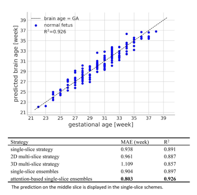 |
Fetal brain age estimation and anomaly detection using attention-based deep ensembles with uncertainty
Wen Shi1,2, Guohui Yan3, Yamin Li2,4, Haotian Li1, Tintin Liu1, Yi Zhang1, Yu Zou3, and Dan Wu1
1Key Laboratory for Biomedical Engineering of Ministry of Education, Department of Biomedical Engineering, College of Biomedical Engineering & Instrument Science, Zhejiang University, Hangzhou, China, 2Key Laboratory of Biomedical Information Engineering of Ministry of Education, Department of Biomedical Engineering, School of Life Science and Technology, Xi'an Jiaotong University, Xi'an, China, 3Department of Radiology, Women's Hospital, School of Medicine, Zhejiang University, Hangzhou, China, 4School of Biomedical Engineering, Shanghai Jiaotong University, Shanghai, China
Accurate estimation of the brain age is important for the evaluation of brain development, especially in the fetal stage when little diagnostic tools are available. This study designed attention-based deep ensembles to estimate brain age in the normal developing fetus, based on axial T2-weighted in-utero MRI images from routine clinical scans. Mean absolute error of 0.803 week was achieved, and the attention maps highlighted the regions of interest associated with the estimation. Predictive uncertainty was simultaneously quantified, and together with the proposed prediction confidence, we were able to detect several types of anomalies, including small head circumference, malformations, and ventriculomegaly.
|
|
2531. |
Development of fetal MRI in a tertiary referral center: how it impacts prenatal diagnosis and alters clinical outcome
Jonan Chun Yin Lee1, Renata Kiri Mak1, and Janice Wong Li Yu1
1Radiology, Queen Elizabeth Hospital, Hong Kong, Hong Kong
Fetal MRI is a useful investigation to visualize congenital anomalies and allow for appropriate counselling and timely intervention. Since early 2017, our hospital has begun to perform fetal MRI from obstetric referrals. 18 pregnant patients underwent fetal MRI for suspected fetal anomalies. The neurological system was the most common region of concern during fetal MRI (67%). When compared to antenatal ultrasonography, fetal MRI provided significant additional and/or change in diagnostic information in 44% of patients , and results in change in postnatal outcome and/or facilitation of postnatal management in 44% of patients, including termination of pregnancy and prompt surgical intervention.
|
|
2532.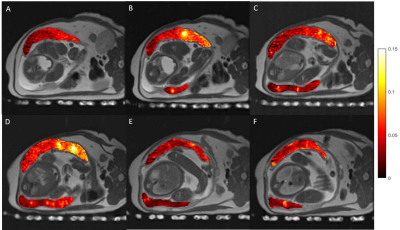 |
Placental Perfusion Imaging on Zika-Infected Rhesus Macaques using Velocity-Selective ASL MRI
Ruiming Chen1, Sydney Nguyen2,3,4, Kai D. Ludwig1, Daniel Seiter1, Megan E. Murphy2,3,4, Kathleen M. Anthony2,3,4, Terry K. Morgan5, Ante Zhu6,7, Dahan Kim1, Sean B. Fain1,6,7, Oliver Wieben1,6, Thaddeus G. Golos2,3,4, and Kevin M. Johnson1,6
1Medical Physics, University of Wisconsin - Madison, Madison, WI, United States, 2Wisconsin National Primate Research Center, University of Wisconsin - Madison, Madison, WI, United States, 3Comparative Biosciences, University of Wisconsin - Madison, Madison, WI, United States, 4Obstetrics & Gynecology, University of Wisconsin - Madison, Madison, WI, United States, 5Pathology, Oregon Health & Science University, Portland, OR, United States, 6Radiology, University of Wisconsin - Madison, Madison, WI, United States, 7Biomedical Engineering, University of Wisconsin - Madison, Madison, WI, United States
Effective non-invasive assessment of placental health, particularly in early pregnancy, is of clinical interest but currently lacking. Arterial spin labeling (ASL) MRI can safely provide local functional assessment of placental perfusion, however, placental perfusion imaging is challenging and current approaches have shortcomings. Here we investigate a multi-slice velocity-selective (VS ASL) sequence with volumetric placental perfusion assessment and report on local and global perfusion across multiple gestational stages for zika-infected rhesus macaques and healthy controls.
|
|
| 2533. | Quantification of diffusion and perfusion of the placenta using whole-placenta volumetric IVIM analysis
Tao Lu1
1Sichuan academy of medical Medical Sciences & Sichuan Provincial People’s Hospital, Chengdu, China
Placental morphological and physiological characteristics are related to health of the newborn and the adult. IVIM offers a quantitative and objective technique to measure maternal placental function without use of the contrast agent. In our study, Whole-placenta volumetric IVIM analysis is used to evaluate the parameters from IVIM of the entire placenta and avoids the subjectivity of ROI placement to ensure calculation accuracy and repeatability.
|
|
2534.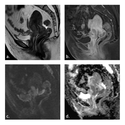 |
Diagnostic accuracy of bladder invasion in cervical cancer: Comparison of T2-weighted, diffusion-weighted, and contrast-enhanced MR imaging.
Yen-Ling Huang1,2, Yu-Ting Huang1,3, Kueian Chen1,2,4, and Gigin Lin1,2,4
1Department of Medical Imaging and Intervention, Chang Gung Memorial Hospital at Linkou, Taoyuan, Taiwan, 2Imaging Core Laboratory, Institute for Radiological Research, Chang Gung Memorial Hospital at Linkou and Chang Gung University, Taoyuan, Taiwan, 3Department of Diagnostic Radiology, Chang Gung Memorial Hospital at Keelung, Keelung, Taiwan, 4Clinical Metabolomics Core Laboratory, Chang Gung Memorial Hospital at Linkou, Taoyuan, Taiwan
Cervical cancer with bladder invasion is rare and carries a poor prognosis. MR imaging is useful in the detection of bladder mucosal involvement from cervical cancer, with reported high negative predictive value, and therefore can serve to justify the necessities of invasive cystoscopy. Diffusion-weighted imaging is superior to T2-weighted and contrast-enhanced MR study in accurately diagnosing bladder invasion.
|
2535.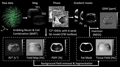 |
Free-Breathing QSM and R2* Mapping of Hepatic Iron Overload Using a 3D Multi-Echo Cones Trajectory
Youngwook Kee1, Christopher M. Sandino2, Joseph Y. Cheng1, Timothy Colgan3, Diego Hernando3, Ann Shimakawa4, and Shreyas S. Vasanawala1
1Radiology, Stanford University, Stanford, CA, United States, 2Electrical Engineering, Stanford University, Stanford, CA, United States, 3Radiology, University of Wisconsin-Madison, Madison, WI, United States, 4GE Healthcare, Redwood City, CA, United States
In this study, free-breathing QSM and R2* mapping of liver iron overload was enabled using a 3D multi-echo cones trajectory. The proposed method was compared with a chemical-shift-encoded MRI technique that requires breath-holding. QSM and R2* exhibit similar image quality in axial, sagittal, and coronal views as well as good agreement in ROI-based quantitative value based on Bland-Altman and linear correlation plots. The imaging time for free-breathing liver iron quantification takes ~4 min, which can be further accelerated by increasing readout duration or using compressed sensing.
|
|
2536.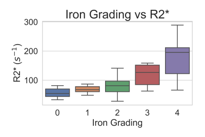 |
Histological Validation of Multipeak Fat-Corrected Complex R2* Mapping for Quantification of Liver Iron
Collin J Buelo1,2, Jing Zhou3, Ante Zhu2,4, Kritisha Rajlawot5, Bingjun He5, Diego Hernando1,2, Jin Wang5, and Scott B. Reeder1,2,4,6,7
1Medical Physics, University of Wisconsin-Madison, Madison, WI, United States, 2Radiology, University of Wisconsin-Madison, Madison, WI, United States, 3Pathology, The third Affiliated Hospital, Sun Yat-Sen University, Guangzhou, China, 4Biomedical Engineering, University of Wisconsin-Madison, Madison, WI, United States, 5Radiology, The third Affiliated Hospital, Sun Yat-Sen University, Guangzhou, China, 6Medicine, University of Wisconsin-Madison, Madison, WI, United States, 7Emergency Medicine, University of Wisconsin-Madison, Madison, WI, United States
Histological grading of iron deposition in the liver has been shown to correlate with conventional magnitude-based R2* mapping methods. However, magnitude-based R2* mapping methods are known to exhibit bias in the presence of fat and when signal-to-noise ratio (SNR) is low. Multipeak fat-corrected complex R2* mapping enables accurate R2* measurements by correcting for fat-related bias and the bias due to low SNR, but has not been compared to histological iron grading. In this work, multipeak fat-corrected R2* mapping is validated by comparing with histological iron grading as the reference standard.
|
|
2537.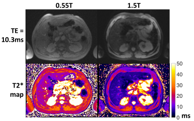 |
Evaluation of severe hepatic iron overload using a high-performance 0.55T MRI system
Adrienne E Campbell-Washburn1, Christine Mancini1, Anna Conrey1, Lanelle Edwards1, Hui Xue1, Peter Kellman1, W. Patricia Bandettini1, and Swee Lay Thein1
1National Heart, Lung, and Blood Institute, National Institutes of Health, Bethesda, MD, United States
Iron overload in the liver can be assessed with MRI by measurements of T2* and R2*. In this study, we assessed the dynamic range of hepatic T2* values using a high-performance 0.55T, compared to 1.5T, in patients with iron overload. Patients with iron overload had increased dynamic range of T2* values at 0.55T (T2* = 11.8 ± 9.4ms) compared with 1.5T (T2* = 5.7 ± 6.0ms), and the measurements were closely correlated between 0.55T and 1.5T (r = 0.96). The improved field homogeneity at 0.55T may provide value for stratification and monitoring of patients with severe iron overload by T2*.
|
|
2538.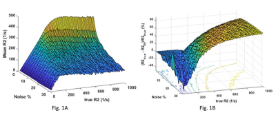 |
Effect of noise on R2 based iron quantitation: Numerical simulation and phantom verification
Zili David Chu1,2 and Raja Muthupillai3
1Radiology, Baylor College of Medicine, MISSOURI CITY, TX, United States, 2Pediatric Radiology, Texas Children's Hospital, Houston, TX, United States, 3Radiology, Baylor St. Luke's Medical Center, Houston, TX, United States
T2/T2* based relaxometry is increasingly used to non-invasively quantify tissue iron content in lieu of biopsy. However, studies have shown that the observed linear relationship between R2 (1/T2) and tissue iron concentrations does not hold true above an iron concentration threshold (dubbed as the ‘saturation threshold’) [1,2]. Our numerical simulations and phantom experiments show that the choice of interval between the echo times used to sample T2 decay, and noise levels play a crucial role in determining the saturation threshold, and that the linear relationship between iron concentration and R2 can be extended by judiciously varying echo spacing.
|
|
2539.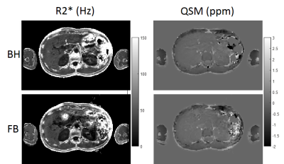 |
Feasibility of Free Breathing Quantitative Susceptibility Mapping of the Liver: Comparison with a Breath-Hold Acquisition
Ramin Jafari1,2, Yan Wen1,2, Pascal Spincemaille2, Thanh D. Nguyen2, Martin R. Prince2, Xianfu Luo3, Daniel Margolis2, and Yi Wang1,2
1Cornell University, Ithaca, NY, United States, 2Weill Cornell Medicine, New York, NY, United States, 3Northern Jiangsu People's Hospital, Yangzhou, China
Quantitative Susceptibility Mapping (QSM) enables accurate non-invasive monitoring of liver iron to guide iron-chelation therapy. Feasibility of a breath-hold sequence has been demonstrated in healthy subjects and in patients. In this work we investigate feasibility of a free-breathing navigator sequence in healthy volunteers to generate QSM maps and compare its correlation with the breath-hold sequence.
|
|
2540.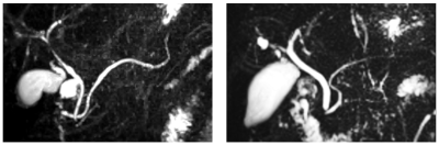 |
Breath-hold MRCP using Accelerated 3D-Spiral Turbo Spin-Echo Imaging
John P. Mugler1, Elisabeth Weiland2, Thomas Benkert2, Craig H. Meyer1, Josef Pfeuffer2, and Berthold Kiefer2
1University of Virginia, Charlottesville, VA, United States, 2Siemens Healthcare GmbH, Erlangen, Germany
Current free-breathing, respiratory-triggered, heavily T2-weighted 3D fast/turbo spin-echo acquisitions work well in many patients for MRCP, but non-diagnostic results are obtained in some patients, particularly those with irregular breathing patterns during the several-minute acquisition. Thus, there has been renewed interest in 3D MRCP techniques that can be completed within a single breath-hold period as an alternative for patients in whom free-breathing techniques are inadequate. This work demonstrates that breath-hold 3D MRCP based on a 3D stack-of-spirals turbo spin-echo acquisition, which uses in-plane acceleration to achieve a reasonable breath-hold time, can provide good image quality for evaluation of major ductal structures.
|
|
2541.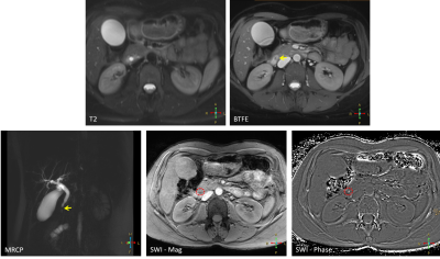 |
Relative contribution of SWI, compared to conventional MRI, in the detection of common bile-duct (CBD) calculi
Vishal Singh1, Jaladhar Neelavalli2, Suhail P Parvaze2, Mamta Gupta1, A K Seth1, and Rakesh Kumar Gupta1
1Radiology, Fortis Memorial Research Institute, Gurgaon, India, 2Clinical Science, Philips Innovation Campus, Philips India Limited, Bengaluru, Karnataka, India
Confident detection of small common bile duct stones is clinically challenging. Ultrasound (US) has highly variable sensitivity and the sensitivity of MRCP, while better than that of US, reduces significantly for stones<5mm. In this work, we have evaluated the relative contribution of SWI in the detection of CBD stones, relying on their susceptibility property.
|
|
2542. |
Is Liver Function Affected by Resective Surgery? – Hepatocyte Uptake and Efflux to Bile in Liver Cancer Determined Using Gadoxetate Enhanced MRI
Christian Simonsson1,2, Markus Karlsson1,3, Bengt Norén1, Gunnar Cedersund2, Per Sandström4, Anna Lindhoff Larsson4, Nils Dahlström1,3, and Peter Lundberg1,3
1Departments of Radiation Physics, Radiology, Department of Medical and Health Sciences, Linköping University, Linköping, Sweden, 2Department of Medical Engineering, Linköping University,, Linköping Univeristy, Linköping, Sweden, 3Center for Medical Image Science and Visualization (CMIV), Linköping Univeristy, Linköping, Sweden, 4Department of Surgery, Department of Clinical and Experimental Medicin, Linköping University, Linköping, Sweden
Just after surgery there is a period were the remnant tissue needs to match the requirement of normal liver function while recovering. This could lead to fatal consequences after too extensive surgical procedures, or due to insufficient liver function. In contrast, it might also be the case that the surgical procedure is too conservative because the predicted liver function is underestimated. This would not have been the case if it was possible to use more precise measures of liver function. We investigated the capabilities of gadoxetate enhanced MRI for determining liver function pre- and post-surgery using two separate approaches.
|
|
2543. |
Improved visualization of common bile duct stone with compressed sensing MRCP
Mengke Wang1, Yan Bai1, Wei Wei1, Jing An2, Yuyu Wang2, Muhammed Labeeb2, Xianchang Zhang3, and Meiyun Wang1
1Department of Medical Imaging, Henan Provincial People’s Hospital & Zhengzhou University People’s Hospital, Zhengzhou, China, 2Siemens Shenzhen Magnetic Resonance Ltd., Shenzhen, China, 3MR Collaboration, Siemens Healthcare Ltd., Beijing, China
Patients with common bile duct (CBD) stone disease suffer from the relatively long scan time of magnetic resonance cholangiopancreatography (MRCP). Compressed sensing (CS) is a technique that can accelerate the speed of MRI and has been applied to MRCP. This study investigated the utility of CS-MRCP in diagnosing CBD stone disease in comparison with conventional MRCP on the 1.5T scanner. The results showed that CS-MRCP could provide comparable image quality with conventional MRCP for the diagnosis of CBD stone disease but with a substantially shorter acquisition time (1:35 vs. 4:08 mins).
|
|
2544.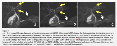 |
Clinical feasibility of compressed SENSE accelerated MRCP with Vital Eye in pancreaticobiliary disorders: a preliminary study.
Ming He1, Xiaoqi Wang2, Jiazheng Wang2, Zhengyu Jin1, and Huadan Xue1
1Department of radiology, Peking Union Medical College Hospital, Beijing, China, 2Philips Healthcare, Beijing, China
The purposes of this study were to prospectively evaluate the clinical feasibility of a MRCP protocol using both Vital Eye and compressed SENSE(CS-VE-MRCP) and to compare its performance with original navigator-triggered (NT) CS-NT-MRCP and NT-MRCP. The results show that the imaging quality and diagnostic performance of CS-VE-MRCP was comparable to that of NT-MRCP and slightly superior than that of CS-NT-MRCP. Besides, the scan time of CS-VE-MRCP was significantly decreased compared to that of NT-MRCP. The combination of compressed SENSE and Vital Eye in MRCP was feasible, suggesting the potential of imaging time reduction without pampering the diagnostic capability.
|
|
2545.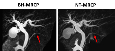 |
Pancreaticobiliary System Evaluation via BH-MRCP using SPACE: A Comparison with Conventional NT-MRCP at 3 Tesla
Qiuxia Luo1, Xiaoyong Zhang2, Qianwei Xie1, Guijin Li3, Bingjun He1, and Jin Wang1
1The Third Affiliated Hospital,Sun Yat-sen University, Guangzhou, China, 2MR Collaborations, Siemens Healthcare Ltd, Shenzhen, China, 3Siemens Healthcare Ltd, Guangzhou, China
Conventional three-dimensional navigator-triggered magnetic resonance cholangiopancreatography (NT-MRCP) is widely used in clinics for the evaluation of the anatomy and abnormalities of the pancreaticobiliary system. However, the method has limitations because of its extensive time to acquire images. This study explored the efficiency and the clinical utility of 3D breath-hold MRCP (BH-MRCP) as an alternative to conventional NT-MRCP at 3 Tesla. Based on these, methods used on a sample population of 25 patients, the results showed that the BH-MRCP technique can be a viable alternative to enhance the clinical workflow of pancreatobiliary MRI.
|
|
2546.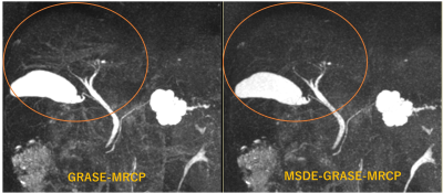 |
Better Background suppression with GRASE-MRCP using MSDE
Mamoru Takahashi1, Yasuo Takehara2, Takuya Matsumoto3, Yoshikazu Nagura3, Tomoyasu Amano3, Norihiro Tooyama1, Katsutoshi Ichijo1, Yasutomo Katsumata4, and Satoshi Goshima5
1Radiology, Seirei Mikatahara General Hospital, Hamamatsu, Japan, 2Nagoya University, Graduate School of Medicine, Nagoya, Japan, 3Seirei Mikatahara General Hospital, Hamamatsu, Japan, 4Philips Electronics Japan, Ltd., Tokyo, Japan, 5Hamamatsu University School of Medicine, Hamamatsu, Japan
MRCP accelerated with GRASE (GRASE-MRCP) allowed single breath-holding 3D MRCP and better depiction of the cystic duct because of short TE. On the other side, background signal such as blood vessels sometimes remained. Incorporating MSDE into GRASE-MRCP made it possible to suppress the background signals without extending the image time and reducing the image quality.
|
|
2547. |
Comparison of breath-hold and respiratory-triggered 3D-SPACE MRCP sequences in the diagnosis of choledocholithiasis
Xin Li1, Yue Qin1, Yifan Qian1, Juan Tian1, Shaoyu Wang2, Yinhu Zhu1, Liyao Liu1, Yanqiang Qiao1, and Boyuan Jiang1
1XI’AN DAXING HOSPITAL, ShaanXi, Xi’an, China, 2Siemens Healthcare Ltd, ShaanXi, Xi’an, China
Magnetic resonance cholangiopancreatography (MRCP) is an effective imaging modality for the evaluation of anatomy and abnormalities of biliary and pancreatic system. The main drawback of conventional navigator-triggered (NT) MRCP is the long acquisition time resulting in a greater variability in the depth of respiration, which may create image blurring and motion artifacts. In this study, we performed MRCP scanning using a single breath hold (BH) 3D SPACE and NT SPACE protocols to evaluated the image quality and acquisition time in patients with choledocholithiasis.
|
|
2548.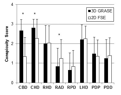 |
MR cholangiopancreatography in a single breath-hold: comparative effectiveness between 3D GRASE and 2D thick-slab SSFSE
Cheng-Ping Chien1,2, Feng-Mao Chiu3, and Hsiao-Wen Chung1
1Graduate Institute of Biomedical Electronics and Bioinformatics, National Taiwan University, Taipei, Taiwan, 2Taipei Beitou Health Management Hospital, Taipei, Taiwan, 3Philips Healthcare, Taipei, Taiwan
3D MR cholangiopancreatography (MRCP) based on gradient- and spin-echo (GRASE) and 2D thick-slab MRCP using fast spin-echo (FSE), both acquired within single breath-hold, were compared using a 4-point score at 3T on 95 healthy subjects (age range = 25-75) in eight different segments of hepatic and pancreatic ducts. 3D GRASE outperformed 2D thick-slab FSE in the common bile duct and common hepatic duct, but compared inferiorly in right anterior hepatic duct (p < 0.001), with insignificant difference (p > 0.05) for the other five ducts. It is concluded that 2D thick-slab FSE MRCP complements 3D GRASE MRCP if performed additionally.
|
|
2549.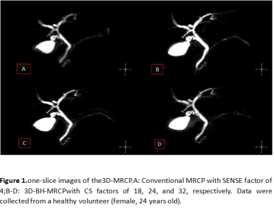 |
Explore performance of 3D MRCP with different compressed-sensing acceleration factors
Renwang Pu1, Jiazheng Wang2, Liangjie Lin2, Jingjun Wu1, Qingwei Song1, Nan Zhang1, and Ailian Liu1
1Department of Radiology, the First Affiliated Hospital of Dalian Medical University, Dalian, dalian, China, 2Philips Healthcare, Beijing, China, beijing, China
The present study aims to explore feasibility of three-dimensional(3D)breath-hold(BH)magnetic resonance cholangiopancreatography (MRCP) through acceleration of the combination of compressed sensing and sensitivity encoding(CS-SENSE),and choose an optimal acceleration factor. 3D-MRCP scans on healthy volunteers were carried out with different acceleration factors (conventional SENSE factor of 4, and CS-SENSE factors of18, 24, and 32).The CS-SENSE acceleration factor of 24 was recommended because of the favorable image quality and reasonable scan duration (15s).
|
2550.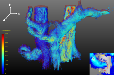 |
Hemodynamics Comparison Between Type2 Diabetes Mellitus Patients and Healthy Controls Using 4D Flow MRI
Jiachen Ji1, Di Wu2, Yunduo Li1, Shenghong Ju2, and Rui Li1
1Center for Biomedical Imaging Research, Department of Biomedical Engineering, Tsinghua University, Beijing, China, 2Zhong Da Hospital Southeast University, Nanjing, China
Type2 Diabetes Mellitus (T2DM) is a metabolic disease with high morbidity. 4D Flow MRI is an advanced technique which could provide visualization and quantification of blood flow. In the study, we identified the reproducibility of the processing and measuring procedure of abdominal 4D Flow data and discovered the significant hemodynamic differences in the affected vessels between T2DM patients and controls using 4D Flow MRI. The differences indicated the systematic hemodynamic changes caused by the disease and hinted 4D Flow MRI could offer more help in the evaluation and better understanding of the disease.
|
|
2551.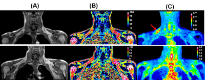 |
Investigation of Supraclavicular Brown Adipose Tissue in Hyperthyroid Patients using Simultaneous PET-MR Imaging
Sanjay K Verma1, Lijuan Sun2, Suresh Anand Sadananthan3, Navin Michael3, Hui Jen Goh2, Govindharajulu Priya2, Melvin Khee-Shing Leow2,4, and S Sendhil Velan1
Video Permission Withheld
1Singapore Bioimaging Consortium, Agency for Science Technology and Research (A*STAR), Singapore, Singapore, 2Clinical Nutrition Research Centre, Agency for Science, Technology and Research (A*STAR), Singapore, Singapore, 3Singapore Institute of Clinical Sciences, Agency for Science, Technology and Research (A*STAR), Singapore, Singapore, 4Department of Endocrinology, Tan Tock Seng Hospital, Singapore, Singapore
BAT dissipates heat energy during adaptive thermogenesis regulated by several complex neuronal mechanisms including thyroid hormones. Twelve hyperthyroid subjects, treated with an anti-thyroid drug intervention were investigated using simultaneous PET-MR imaging. A significant increase in body weight and decrease in resting metabolic rate after treatment were observed. MR fat fraction significantly increased after treatment, whereas there is no significant change in PET-SUV. MR is more sensitive to the thyroid hormone induced changes, and can be utilized to study the BAT activity.
|
|
2552.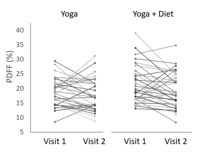 |
Evaluating a Community-Based Diet and Lifestyle Intervention For Improved Metabolic Health in India: A Pilot Randomized Controlled Trial (RCT)
Stephen Bawden1,2, K Manoj3, Elizabeth Simpson1, K Leena4, Moira Taylor1, Sally Hibbert5, Amrita Vijay1, Laura Miller1, Jane Grove1, Ana Valdes1, Penny Gowland2, Thrivikrama Shenoy4, and Guruprasad P Aithal1
1NIHR Nottingham Biomedical Research Centre, University of Nottingham, Nottingham University Hospitals NHS Trust and the University of Nottingham, Nottingham, United Kingdom, 2Sir Peter Mansfield Imaging Centre, University of Nottingham, Nottingham, United Kingdom, 3Metro Scan and Laboratory, Trivandrum, India, 4Population Health and Research Institute, Trivandrum, India, 5Centre for Research in the Behavioural Sciences, Nottingham University Business School, Nottingham, United Kingdom
The Indian population provides important cohorts for research into metabolic disorders due to the greater prevalence of NAFLD and diabetes, and the relatively low complexity of dietary intake. In this study 71 male participants with NAFLD were recruited from a cohort in India and randomized into two groups - a 16 week low glycaemic index dietary intervention arm and a control arm. 1H MRS was used to assess liver fat fractions, alongside other metabolic markers. Results show a trend towards reduced fat fractions in the diet arm only.
|
|
2553.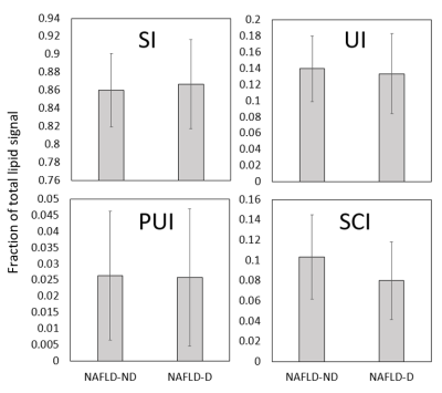 |
Using 1H MRS to Investigate Hepatic Fat Fractions and Lipid Composition in NAFLD Patients with Type 2 Diabetes
Stephen Bawden1,2, Scott Willis3, Aron Sherry3, Penny Gowland2, James King3, and Guruprasad P Aithal1
1NIHR Nottingham Biomedical Research Centre, University of Nottingham, Nottingham University Hospitals NHS Trust and the University of Nottingham, Nottingham, United Kingdom, 2Sir Peter Mansfield Imaging Centre, University of Nottingham, Nottingham, United Kingdom, 3School of Sport, Exercise and Health Sciences, Loughborough University, Loughborough, United Kingdom 1H MRS offers unique insights into hepatic lipid composition. In this study, NAFLD patients with and without Type 2 Diabetes were scanned and hepatic lipid composition indices measured. Diabetic patients then underwent a randomized controlled trial to investigate the effectiveness of 6 weeks moderate exercise vs control. Lipid composition indices were similar to previous studies in obesity, and show no differences in diabetic v non-diabetic groups. The intervention also shows no change in hepatic fat fraction or lipid composition. |
|
2554.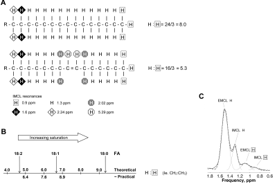 |
Accumulation of saturated IMCL is associated with insulin resistance in both males and females
Laura Watson1, Katie Carr1, Claire Adams2, Jieniean Worsley1, Krishna K Chatterjee1,2, Leanne Hodson3, Chris Boesch4, Graham J Kemp5, David B Savage2, and Alison Sleigh1,2,6
1National Institute for Health Research Cambridge Clinical Research Facility, Cambridge University Hospitals NHS Foundation Trust, Cambridge, United Kingdom, 2Metabolic Research Laboratories, Wellcome Trust-MRC Institute of Metabolic Science, University of Cambridge, Cambridge, United Kingdom, 3Oxford Centre for Diabetes, Endocrinology and Metabolism (OCDEM), University of Oxford, Oxford, United Kingdom, 4Department of Clinical Research and Radiology, AMSM, University Bern, Bern, Switzerland, 5Department of Musculoskeletal Biology, and MRC-Arthritis Research UK Centre for Integrated research into Musculoskeletal Ageing (CIMA), University of Liverpool, Liverpool, United Kingdom, 6Wolfson Brain Imaging Centre, University of Cambridge, Cambridge, United Kingdom In females the accumulation of saturated IMCL is more strongly associated with whole-body insulin resistance than IMCL concentration alone. Using a recently-validated 1H MRS approach we studied both the IMCL composition and concentration independent of composition in 30 control males, 41 age- and BMI-matched female controls and 16 female insulin resistant lipodystrophic subjects. We demonstrate that in both males and females markers reflecting the accumulation of saturated IMCL are more strongly associated with whole-body insulin sensitivity than IMCL concentration alone.
|
|
2555.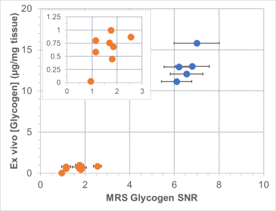 |
Natural abundance 13C MRS based glycogen detection in Pompe disease: Test/retest characterization in a Gaa KO mouse model
Deanne Lister1, Prodromos Parasoglou2, Andrew Baik2, Katherine Cygnar2, Aris Economides2, Brendan Cook1, Sven Macholl1, and Patrick McConville1
1Molecular Imaging Center, Invicro, A Konica Minolta Company, La Jolla, CA, United States, 2Regeneron Pharmaceuticals, Tarrytown, NY, United States
Pompe disease is a debilitating condition that results in abnormally high tissue glycogen concentrations and is the subject of significant therapeutic focus by the pharma industry, including promising gene therapies. Non-invasive biomarkers that are specific and sensitive are sought after to address major gaps in early phase clinical trials. We characterized 13C MRS measurements of muscle glycogen concentration as potential biomarkers in Pompe disease, including test/re-test measurements and correlation with tissue biochemical measurements. Strong specificity and reasonable reproducibility was found, suggesting potential for 13C MRS to be used in Pompe disease patients.
|
|
2556.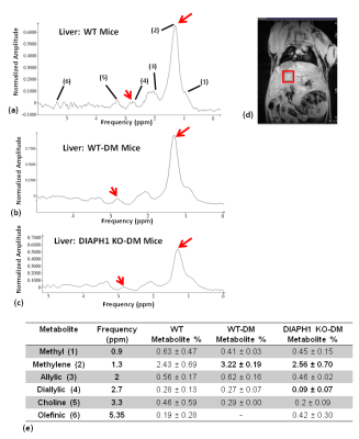 |
Assessment of cardiac and liver metabolite changes in diabetic and DIAPH1 knockout mice using 1H-MRS and chemical-shift encoded MRI
Rajiv G Menon1, Dimitri Martel1, Nosirudeen Quadri2, Charlotte Detremmerie2, Laura Frye2, Ann Marie Schmidt2, Ravichandran Ramasamy2, and Ravinder R Regatte1
1Center for Biomedical Imaging, Department of Radiology, New York University Langone Health, New York, NY, United States, 2Department of Biochemistry and Molecular Pharmacology, Department of Medicine, New York University Langone Health, New York, NY, United States
Diabetes Mellitus causes systemic changes in a number of metabolites in multiple organs. Previous studies have shown that RAGE and its cytoplasmic domain partner Diaph1 are key mediators of metabolic and functional changes in diabetic mice. The goal of this study was to use 1H-MRS and CSE-MRI to investigate the metabolite and water-fat fraction changes in the liver and heart in wild type (WT), WT-diabetic (WT-DM) mice, and DIAPH1 knockout diabetic (DIAPH1 KO-DM) mice. The metabolite levels and water-fat distribution in the three cohorts suggest that DIAPH1 KO-DM mice, despite being diabetic, experience a protective effect owing to DIAPH1 deletion.
|
|
2557.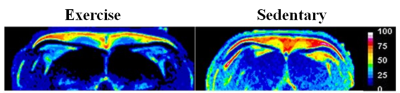 |
Metabolic imaging of brown adipose tissue with intense exercise intervention
Venkatesh Gopalan1, Kavita Kaur1, Jadegoud Yaligar1, Sanjay Kumar Verma1, Rengaraj Anantharaj1, Le Thi Thu Giang1, and S Sendhil Velan1
1Laboratory of Molecular Imaging, Singapore BioImaging Consortium,A*STAR Research Entities, Singapore, Singapore
There is a large interest in development of non-invasive methods for activation of brown adipose tissue (BAT) due to its potential to combat obesity. Exercise is a non-pharmacological strategy to combat obesity by increasing EE. In this study we have investigated exercise induced BAT activation, heat and RER in sedentary and exercise groups. Our results show that exercise activates the brown fat with reduction in fat fraction, increased heat and RER. UCP1 expression shows that reduction in “white” like adipocytes in exercise group.
|
|
2558.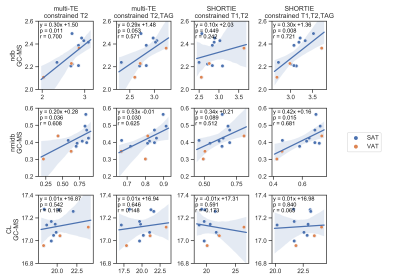 |
Estimation of the triglyceride fatty acid composition in human adipose tissue using multi-TE and SHORTIE STEAM: in vitro validation using GC-MS
Stefan Ruschke1, Dominik Weidlich1, Julius Honecker 2, Claudine Seeliger2, Josef Ecker3, Olga Prokopchuk4, Hans Hauner2, and Dimitrios C Karampinos1
1Department of Diagnostic and Interventional Radiology, School of Medicine, Technical University of Munich, Munich, Germany, 2Else Kröner Fresenius Center for Nutritional Medicine, Technical University of Munich, Freising, Germany, 3ZIEL Institute for Food & Health, Research Group Lipid Metabolism, Technical University of Munich, Freising, Germany, 4Department of Surgery, Klinikum rechts der Isar, School of Medicine, Technical University of Munich, Munich, Germany
There is a growing interest in utilizing single-voxel $$$^1$$$H MR spectroscopy (MRS) for probing the triglyceride fatty acid composition by means of the number of double bounds (ndb) and number of methylene interrupted double bounds (nmidb) per triglyceride as well as the mean fatty acid carbon chain length (CL). The present study investigates the in vitro measurement agreement of fatty acid composition parameters between single-voxel multi-TE and short-TR multi-TI multi-TE (SHORTIE) STEAM spectroscopy and the gold standard gas chromatography-mass spectrometry (GC-MS) in human subcutaneous and visceral adipose tissue samples.
|
|
2559.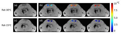 |
Studying the effect of ambient temperatures on heat production of activated BAT in rats using magnetic resonance thermometry
Chuanli Cheng1, Qian Wan1, Yangzi Qiao1, Min Pan2, Changjun Tie1, Hairong Zheng1, Xin Liu1, and Chao Zou1
1Shenzhen Institutes of Advanced Technology,Chinese Academy of Sciences, Shenzhen, China, 2Shenzhen Hospital of Guangzhou University of Chinese Medicine, Shenzhen, China
Cold exposure is the commonly used method in evaluating the brown adipose tissue (BAT) activation. In this study, the heat production capacity of BAT in rats housed at different ambient temperatures were evaluated using magnetic resonance thermometry after injecting norepinephrine (NE). The results show that the rats housed at lower ambient temperature (15°C) for 2 weeks have surprisingly lower temperature rise after NE injection, implying that long-term cold exposed rats have lower heat production capacity after drug activation.
|
|
2560.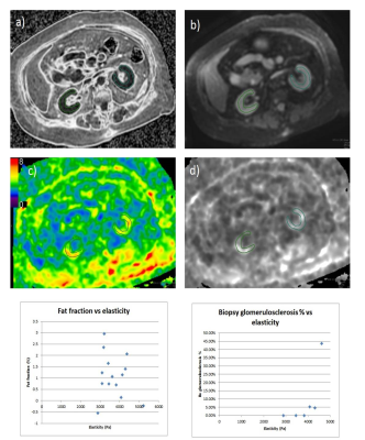 |
MR Elastography and PDFF as Imaging Biomarkers for Diabetic Kidney Disease: Correlating MR Measurements with Histopathology
Anshuman Panda1, Justin Yu1, Michael Schwartz1, Helen Looker2, Robert Nelson2, Kelly Smith1, Aidan McGirr1, Sophia Fasani1, Sukhdeep Singh1, Kristina Flicek1, and Alvin Silva1
1Mayo Clinic Arizona, Scottsdale, AZ, United States, 2National Institutes of Health, Bethesda, MD, United States
The purpose of this pilot study is to determine technical feasibility and correlate MR Elastography (MRE) and Proton Density Fat Fraction (PDFF) with gold standard assessments of glomerular function and histopathology markers.
|
|
2561.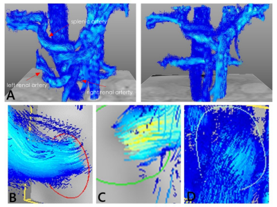 |
Assessments of Blood Flow Curve Characteristics of Type2 Diabetes Using 4D Flow MRI
Di Wu1, Jiachen Ji2, Shenghong Ju1, and Rui Li2
1Department of Radiology, Zhong Da Hospital,Southeast Univ., Nanjing, China, 2Department of Biomedical Engineering, Center for Biomedical Imaging Research,Tsinghua Univ., Beijing, China
The aim of this study is to identify the abdominal blood flow curve characteristics of patients with type2 diabetes using advanced 4D Flow MRI, compared with the healthy subjects. Left and right renal arteries as well as splenic artery were visualized and quantified by dedicated software. The characteristics of flow curves showed significant differences in the skewness and time to peak, which may indicate the variation in renal progression rates and become promising non-invasive biomarkers of the T2DM function assessment.
|
2562.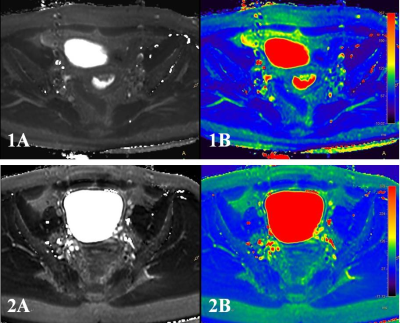 |
Quantitative identification of endometrial carcinoma and endometrial polyp by T2 mapping
Shifeng Tian1,2, Ailian Liu2, Jiazheng Wang3, and Liangjie Lin3
1The First Affiliated Hospital of Dalian Medical University, Dalian, China, 2the First Affiliated Hospital of Dalian Medical University, Dalian, China, 3Philips Healthcare, Beijing, China Poster Permission Withheld
T2 mapping is a novel MRI sequence that enables quantitative assessment of various diseases. However, its potential for diagnosis of endometrium diseases has not been explored,andit may also help differential diagnosisof endometrial carcinoma and endometrial polyp.In this study, the T2mapping was used for quantitative analyses of endometrial carcinoma and endometrial polyp. Results shown that endometrial carcinoma was associated with significant higher T2 values than endometrial polyp, therefore, it is feasible to use T2 mapping for discrimination of endometrial carcinoma and endometrial polyp.
|
|
2563.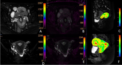 |
The combination use of APT and R2* enhances the differentiation of endometrial carcinoma and endometrial benign lesions
Xing Meng1, Ailian Liu 1, Shifeng Tian1, Zhiwei Shen2, Yaxin Niu1, Wan Dong1, Zhongping Zhang2, and Yishi Wang2
1Department of Radiology, the First Affiliated Hospital of Dalian Medical University, Dalian, China, 2Philips Healthcare, Beijing, China
Amide proton transfer (APT) and mDixon - Quant techniques have been applied in clinical work, but few studies have been conducted in female pelvic system. We discussed the value of APT combined with R2* in differentiating endometrial cancer from benign endometrial lesions. The AUC, sensitivity and specificity of APT combined R2* were 0.925, 88% and 100%, respectively.
|
|
2564.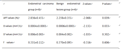 |
APT combined with IVIM in differential diagnosis of endometrial carcinoma and endometrial benign lesions
Xing Meng1, Ailian Liu 1, Shifeng Tian1, Zhiwei Shen2, Tianjing Zhang2, Yishi Wang2, Yaxin Niu1, and Wan Dong1
1Department of Radiology, the First Affiliated Hospital of Dalian Medical University, Dalian, China, 2Philips Healthcare, Beijing, China
Amide proton transfer (APT) combined with intravoxel incoherent motion (IVIM) has been preliminarily applied in the diagnosis and differentiation of cervical squamous cell carcinoma, but has not been in the identification of endometrial lesions. We discussed the value of APT combined with IVIM in differentiating endometrial carcinoma from endometrial benign lesions. The AUC, sensitivity and specificity of APT combined IVIM were 0.979, 87.5% and 100%, respectively.
|
|
2565.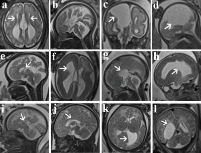 |
Comparison of ultrasound and magnetic resonance imaging for the detection of fetal corpus callosum abnormalities
Cong Sun1, Yufan Chen1, Jinxia Zhu2, and Guangbin Wang1
1Shandong Medical Imaging Research Institute, Shandong University, Jinan, China, 2MR Collaboration, Healthcare Siemens Ltd., Beijing, China, Beijing, China
We compared ultrasound and magnetic resonance imaging (MRI) for the detection of fetal corpus callosum (CC) dysplasia in 375 fetuses at our Institute. The results showed that MRI was significantly better than ultrasound in being able to diagnose normal and abnormal fetal CC. Also, fetal MRI can be helpful in assessing associated abnormalities and enhancing prognostic consultations.
|
|
2566.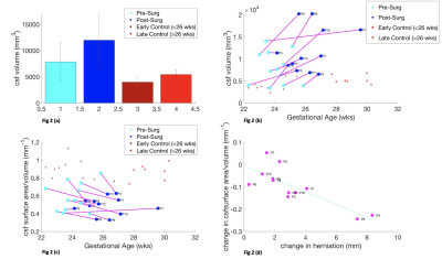 |
Shape and Volume Analysis of the Fetal Brain Pre and Post Fetal Spina Bifida Surgery
Nada Mufti1,2,3, Michael Ebner2,3, Lucas Fidon2, Dimitra Flouri2,3, Riine Heinsalu2, Tom Vercauteren2,4, Sebastien Ourselin2, Luc De Catte4, Jan Deprest1,4, Anna L David1,4, Philippe Demaerel5, Andrew Melbourne2,3, and Michael Aertsen5
1Institute for Women's Health and Department of Medical Physics and Biomedical Engineering, University College London (UCL), London, United Kingdom, 2School of Biomedical Engineering and Imaging Sciences (BMEIS), King's College London, London, United Kingdom, 3Department of Medical Physics and Biomedical Engineering, University College London (UCL), London, United Kingdom, 4Department of Obstetrics and Gynaecology, University Hospitals Katholieke Universiteit (KU) Leuven, Leuven, Belgium, 5Department of Radiology, University Hospitals Katholieke Universiteit (KU) Leuven, Leuven, Belgium
Thorough assessment of the fetal central nervous system is required to select the most suitable candidates for fetal spina bifida surgery, and for post-operative monitoring to predict outcome. Children with myelomeningocele exhibit difficulties in cognitive performance, and motor skills1. This is related to the Chari II malformation and ventriculomegaly2,3. Our aim is to determine if MRI technology can quantify volume, surface area, and shape of cerebral structures before and after fetal spina bifida surgery. We explore if these parameters can be used as potential biomarkers for efficacy of fetal surgery by correlating with herniation level.
|
|
2567.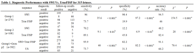 |
Imaging the Fetal Spine using MRI: A Preliminary Exploration on Scanning Strategy
Xianyun Cai1, Guangbin Wang2, and Jinxia Zhu3
1radiology, Shandong Medical Imaging Research Institute, Shandong University, jinan, China, 2Shandong Medical Imaging Research Institute, Shandong University, jinan, China, 3MR Collaboration, Siemens Healthcare Ltd., beijing, China
This study explored the scanning strategy of fetal spine imaging using Magnetic Resonance Imaging (MRI) on 315 volunteer pregnant subjects. Whole-spine MRI was performed on fetuses using Susceptibility-Weighted Imaging (SWI) and True Fast Imaging with Steady-state Precession (TrueFISP) sequences. Images from both methods were acquired, and the diagnostic efficacy was compared. The SWI showed superior performance in visualizing osseous spinal anomalies, while TrueFISP better presented spinal canal contents lesions. Additionally, the MRI was preferred for the diagnosis of fetal spinal diseases to US. These mixed results suggest that a combination of both techniques is appropriate for fetal spine imaging.
|
|
2568.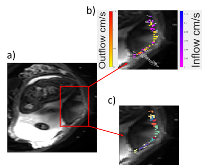 |
First in vivo demonstration of venous return in the human placenta
George Hutchinson1, Neele Dellschaft1, Simon Shah2, Nia Jones3, Lopa Leach4, and Penny Gowland1
1SPMIC, The University of Nottingham, Nottingham, United Kingdom, 2Kings College London, London, United Kingdom, 3Queens Medical Centre, The University of Nottingham, Nottingham, United Kingdom, 4School of Life Sciences, The University of Nottingham, Nottingham, United Kingdom
Little is known about maternal venous return from the placenta, despite the fact this good venous drainage is critical for adequate flow into, and percolation through the placenta. In this abstract we aim to identify and characterise venous return by identifying regions of blood flowing across the placental wall, and we find similar velocities of blood flow entering and leaving the placenta.
|
|
2569.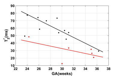 |
Relationship between Placenta T2* and Gestational Age at 3.0 T
Feifei Qu1, Edgar Hernandez-Andrade2, Julio Marin-Concha3, Swati Mody4, Pavan Jella1, Roberto Romero3,5,6, and E. Mark Haacke1,7,8
1Department of Radiology, Wayne State University School of Medicine, Detroit, MI, United States, 2Department of Obstetrics and Gynecology, Wayne State University School of Medicine, Detroit, MI, United States, 3Perinatology Research Branch, NICHD/NIH/DHHS, Detroit, MI, United States, 4Department of Radiology, Children Hospital of Michgan, Detroit, MI, United States, 5Department of Obstetrics and Gynecology, University of Michigan, Ann Arbor, MI, United States, 6Department of Epidemiology and Biostatistics, Michigan State University, East Lansing, MI, United States, 7Department of Biomedical Engineering, College of Engineering, Wayne State University, Detroit, MI, United States, 8The MRI Institute for Biomedical Research, Bingham Farms, MI, United States
Healthy growth and function of the placenta are important to support normal fetal development throughout the second and third trimester. T2* values affected by the tissue oxygenation could be a valuable tool to detect the placenta function. In this work, we collected T2* map for both health pregnancy and the pregnancy complicated by IUGR at different GA at 3.0 T and also investigated the relationship between them.
|
|
2570.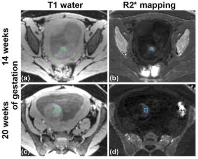 |
Quantification of T2* relaxation times of human liver in the midtrimester fetus at 1.5T
Matthias R Muehler1, Ante Zhu1,2, Oliver Wieben1,2,3, Diego Hernando1,2,3,4, Dinesh Shah5, and Scott B. Reeder1,2,3,6,7
1Radiology, University of Wisconsin-Madison, Madison, WI, United States, 2Biomedical Engineering, University of Wisconsin-Madison, Madison, WI, United States, 3Medical Physics, University of Wisconsin-Madison, Madison, WI, United States, 4Electrical and Computer Engineering, University of Wisconsin-Madison, Madison, WI, United States, 5Obstetrics and Gynecology, University of Wisconsin-Madison, Madison, WI, United States, 6Emergency Medicine, University of Wisconsin-Madison, Madison, WI, United States, 7Medicine, University of Wisconsin-Madison, Madison, WI, United States
In preeclampsia, the erythropoetin level in the fetus has been reported to be moderately elevated and the number of erythroid cells in the fetal liver reduced. Assessment of fetal liver health and function over gestation, including hepatic hematopoiesis, is of great interest. T2* relaxometry is an emerging quantitative biomarker of liver iron content. In this study, we assessed fetal liver T2* in the midtrimester pregnancy. A fetal liver T2* of 22ms and 25ms at 14 and 20 weeks of gestation, respectively, was observed at 1.5T. T2* values are significantly correlated with gestational age but not with obesity or mothers’ age.
|
|
2571.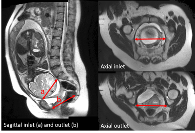 |
Upright pelvimetry using MRI for the prediction of birth associated cephalo-pelvic disproportion following induction of labour.
Hannah Grace Williams1, Andrew Cooper1, Lesley Hodgen2, Catriona Hussain2, Nia Jones3, and Penny Gowland1
1Sir Peter Mansfield Imaging Centre, University of Nottingham, Nottingham, United Kingdom, 2Queens Medical Centre, NUH NHS Trust, Nottingham, United Kingdom, 3Faculty of Medicine and Health, University of Nottingham, Nottingham, United Kingdom
Dystocia, difficult birth due to either a big baby, small maternal pelvis, or malposition of the presenting part is an important obstetric problem and a major cause of emergency caesarean section and birth injury. The risk of caesarean section is 30% for first time mothers who have had their labour induced. We have developed an MRI protocol to assess fetal size and maternal pelvic measurements in supine and upright positions to determine whether they can be used as a predictor of a woman's risk of emergency C-section after the induction of labour.
|
|
2572.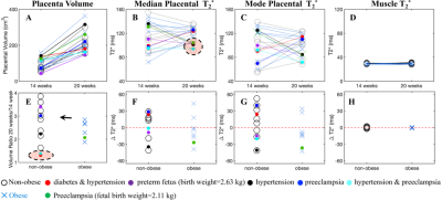 |
Longitudinal Study of T2*-based BOLD Placental MRI in Obese Pregnant Human Subjects
Ante Zhu1,2, Jitka Starekova2, Tom Batan3, Kevin Johnson2,4, Scott B. Reeder1,2,4,5,6, Oliver Wieben1,2,4, Dinesh Shah7, and Diego Hernando1,2,4,8
1Biomedical Engineering, University of Wisconsin-Madison, Madison, WI, United States, 2Radiology, University of Wisconsin-Madison, Madison, WI, United States, 3Integrative Biology, University of Wisconsin-Madison, Madison, WI, United States, 4Medical Physics, University of Wisconsin-Madison, Madison, WI, United States, 5Emergency Medicine, University of Wisconsin-Madison, Madison, WI, United States, 6Medicine, University of Wisconsin-Madison, Madison, WI, United States, 7Obstetrics and Gynecology, University of Wisconsin-Madison, Madison, WI, United States, 8Electrical and Computer Engineering, University of Wisconsin-Madison, Madison, WI, United States
Maternal obesity is associated with increased risk of gestational hypertension, preeclampsia and fetal growth restriction, complications related to reduced uteroplacental blood flow and placental vascularity malformation. T2* measurements of blood oxygen level-dependent effects have promise for assessing placental function. Here, we measured placental T2* in non-obese and obese pregnant women at 14 and 20 weeks of gestation. Our study showed that the range of placental T2* is similar between the two populations at both gestation ages. Trends of decreased and increased placental T2* with gestation were observed in different subjects, suggesting longitudinal changes in placental T2* may
vary among subjects.
|
|
2573.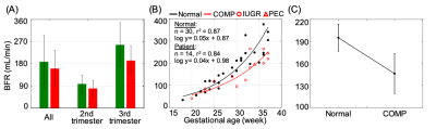 |
MR Assessment of Blood Flow Rate in Umbilical Vein and Its Correlation with Ultrasound
Pei-Hsin Wu1, Michael C. Langham1, Ana E. Rodríguez-Soto1, Eileen Hwuang2, Felix W. Wehrli1, and Nadav Schwartz3
1Department of Radiology, University of Pennsylvania, Philadelphia, PA, United States, 2Department of Bioengineering, University of Pennsylvania, Philadelphia, PA, United States, 3Maternal Child Health Research Center, Perelman School of Medicine, Philadelphia, PA, United States
Intrauterine growth restriction (IUGR) and preeclampsia (PEC) have been associated with increased perinatal morbidity and mortality. Umbilical vein (UV) blood flow reflects the efficiency of fetoplacental circulation. The hypothesis that UV blood flow rate (BFR) is lower in abnormal pregnancies (e.g. IUGR, PEC) was investigated. The results show 1) a high correlation in UV BFR between MR and ultrasound confirms the utility of PC-MRI, and 2) a lower MR-based UV BFR, found in the adverse composite birth outcome group as compared to normal subjects, suggests that UV flow may be a significant clinical indicator of adverse pregnancy outcomes.
|
|
2574.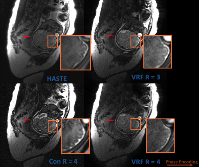 |
Single-shot T2-weighted Fetal MRI with variable flip angles, full k-space sampling, and nonlinear inversion: towards improved SAR and sharpness
Yamin Arefeen1, Borjan Gagoski2,3, Esra Turk2,3, Ellen Grant2,3, Jacob White1, Kawin Setsompop3,4,5, and Elfar Adalsteinsson1,5,6
1Electrical Engineering and Computer Science, Massachusetts Institute of Technology, Cambridge, MA, United States, 2Fetal-Neonatal Neuroimaging and Developmental Science Center, Boston Children’s Hospital, Boston, MA, United States, 3Harvard Medical School, Boston, MA, United States, 4Athinoula A. Martinos Center for Biomedical Imaging, Boston, MA, United States, 5Harvard-MIT Health Sciences and Technology, Cambridge, MA, United States, 6Institute for Medical Engineering and Science, Cambridge, MA, United States
Fetal MRI utilizes Half-Fourier-acquisition-single-shot-turbo-spin-echo (HASTE) for rapid T2-weighted imaging to mitigate motion. However, T2-decay and partial fourier induce blurring, while refocusing trains extend total acquisition time due to specific absorption rate (SAR) constraints. Variable flip angles (VFA) with full Fourier acquisitions have been shown to improve image quality, maintain contrast, and accelerate acquisition through SAR reduction. We combine nonlinear reconstruction, VFA, and full Fourier undersampling to improve sharpness and reduce SAR while maintaining similar contrast to HASTE. Simulation and phantom experiments illustrate improved sharpness. Preliminary, in-vivo fetal results demonstrate improved sharpness and SAR with slight tradeoffs in contrast and SNR.
|
|
2575.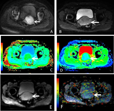 |
Comparative analysis of the value of APT and different diffusion models in evaluating the histological features of cervical squamous carcinoma
Mengyan Hou1, Dongming Han1, and Kaiyu Wang2
1The First Affiliated Hospital,Xinxiang Medical, Xinxiang, China, 2GE Healthcare, MR Research China, Beijing, China
This study aims to compare the performances of APT, DWI, and DKI in estimating histological grading of cervical squamous carcinoma. We find that the parameter MTRasym of APT and the parameter MK and MD of DKI are helpful for the differentiation diagnosis of cervical squamous cell carcinoma. Compared with DWI and DKI, APT is more effective in identifying the histological grades of cervical squamous carcinoma, and the combination of APT and DWI parameters can greatly improve diagnostic effect.
|
2576.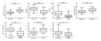 |
Quantitative evaluation of ileal crohn’s disease with intravoxel incoherent motion diffusion-weighted imaging (IVIM-DWI)
Peiwen Sun1, Junheng Li1, Meiying Zeng1, Shuai Wang1, Diru Zhu1, Jilei Zhang2, and Binghui Zhao*1
1Department of Radiology, Shanghai Tenth People’s Hospital of Tongji University, School of Medicine, Shanghai, China, 2Clinical Science, Philips Healthcare, Shanghai, China
Crohn’s disease (CD) is a chronic inflammatory bowel disorder that can occur in any section of the gastrointestinal tract, commonly affecting the small intestine, especially the ileum. Colonoscopy used as the gold standard for assessing disease activity, but its invasiveness limits the use. We aimed to assess inflammatory activity of CD patients by using Intravoxel incoherent motion diffusion-weighted imaging (IVIM-DWI), and found IVIM-DWI may serve as a promising method for noninvasive detecting and monitoring the activity of CD.
|
|
2577.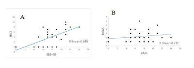 |
MRE for the detection of inflammatory diseases of the juvenile Crohn’s disease: quantifying the inflammatory activity.
zheng xian ying1 and Zhang Zhong shuai2
1First affiliated Hospital of Fujian Medical University, fuzhou, China, 2MR , Siemens Healthcare, Shanghai, China
This study aimed to demonstrate that Magnetic resonance enterography global score (MEGS) shows equally values in pediatric crohn's patients and had comparable diagnostic efficacy comparing with other MRE scores. 52 juvenile Crohn patients were included, and MEGS, SES-CD and eAIS of the terminal ileum were calculated. The results show that for both endoscopically defined and histologically defined active inflammation and ulcerative lesions MEGS have high diagnostic accuracy, are consisting with other studies. The results provided in the current study suggest that MR may be considered as an alternative to endoscopy.
|
|
2578.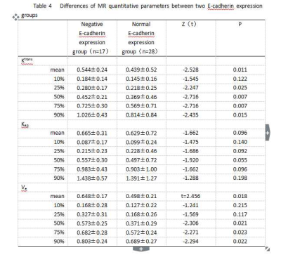 |
Correlation between MR DCE quantitative parameters and E-cadherin expression in gastric cancer
Liangliang Yan1 and Jinrong Qu1
1Radiology, Affiliated Cancer Hospital of Zhengzhou University, Zhengzhou, China
Multiple MR quantitative parameters are associated with E-cadherin expression, and Ktrans 75% is the optimal parameter to evaluate the invasion of gastric cancer. In addition, our study also determine the specific boundary range of Ktrans 75%, Ktrans mean, and Ve mean to predict whether E-cadherin expression has decreased or absented, which provides a new method to early evaluate gastric cancer invasion.
|
|
2579.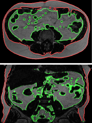 |
MRI Changes in Visceral Fat in Crohn’s Disease
Iyad Naim1,2, Gordon Moran2, Penny Gowland1,2, Caroline Hoad1,2, E Simpson 2,3, and Jordan McGing2
1Sir Peter Mansfield Imaging Centre, School of Physics, University of Nottingham, Nottingham, United Kingdom, 2Nottingham Digestive Disease Biomedical Research Centre, University of Nottingham, Nottingham, United Kingdom, 3Metabolic and Molecular Physiology, School of Life Sciences, University of Nottingham, Queen's Medical Centre Campus, Nottingham, United Kingdom
Crohn’s disease (CD) is an inflammatory bowel disease which effects 150k people in the UK alone. Visceral fat hypertrophy is a characteristic radiological finding in CD.To date radiologists just describe it but are not able to provide more objective data regarding visceral fat volume. In this retrospective study we used 2-point mDixon fat Images along with image processing techniques to study visceral fat properties in CD patients compared with healthy volunteers. The data demonstrated a tendency of CD patients to have a higher ratio of visceral fat and more volume of intestines fully surrounded by fat compared with healthy volunteers.
|
|
2580.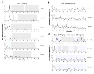 |
Hyoscine butylbromide for bowel movement reduction in mouse abdominal MRI
Carlos Bilreiro1, Francisca F. Fernandes1, Rui V. Simões1, Celso Matos1, and Noam Shemesh1
1Champalimaud Research, Champalimaud Centre for the Unknown, Lisbon, Portugal
Bowel movement is a source of motion artifacts in mouse abdominal MRI. This work aimed to evaluate fasting and hyoscine butilbromide (Buscopan®) for its reduction. Thirteen mice were imaged in a 9.4T MRI scanner with a FLASH sequence for 90 minutes (~19s temporal resolution): 6 were injected with a Buscopan® bolus, 4 with a bolus and constant infusion, and 3 were food deprived. A single bolus of Buscopan® was effective for bowel movement reduction in mice, for up to 1h, whereas other methods were not effective. These data favor the use of Buscopan® for abdominal MRI in the mouse.
|
|
2581.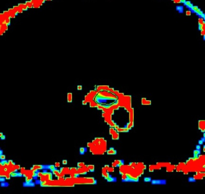 |
Application of intravoxel incoherent motion in preoperative assessment of clinicopathological features of Esophageal Squamous Cell Carcinoma
TAO SONG1
1radiology, Henan Cancer Hospital, Zhengzhou, China
Preoperative assessment of intravoxel incoherent motion in clinicopathologic features of esophageal squamous cell carcinoma. the uploaded file includes objective,method,results,conclusion,references, acknowledgements and figure.
|
|
2582.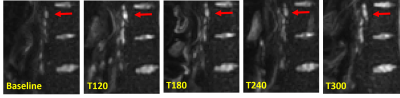 |
Imaging of the intestinal lymphatics following oral consumption of lipids using MRI.
Hannah Grace Williams1, Adelaide Jewell2, Caroline Hoad1,3, Luca Marciani3,4, Snow Stolnik-Trenkic2, Pavel Gershkovich2, and Penny Gowland1
1Sir Peter Mansfield Imaging Centre, University of Nottingham, Nottingham, United Kingdom, 2School of Pharmacy, University of Nottingham, Nottingham, United Kingdom, 3NIHR Nottingham Biomedical Research Centre, Nottingham University Hospital and University of Nottingham, Nottingham, United Kingdom, 4Nottingham Digestive Disease Centre, School of Medicine, University of Nottingham, Nottingham, United Kingdom
It has been shown previously that lipophilic (lipid soluble) drugs administered in lipid-based formulations (or using lipophilic prodrugs) can be delivered in very high concentrations to the intestinal lymphatics. In order for the intestinal lymphatic targeting to be clinically relevant, the response of intestinal lymph following oral administration of lipids needs to be investigated in humans. This is the first study we are aware of using MRI to study changes in lymph nodes following the consumption of a fatty meal. With optimisation this method could provide a novel marker of which nodes could be target for treatment using lipophilic drugs.
|
|
2583. |
Diffusion Weighted whole body Imaging with Background body signal suppression with three b value for tumorlike lesion detection in abdomen
bingbing gao1, yanwei miao1, ailian liu1, jiazheng wang2, zhiwei shen2, jianqing sun2, qingwei song1, and nan zhang1
1first Affiliated hospital of Dalian medical university, Dalian, China, 2Philips Healthcare, Beijing, China, Beijing, China
The purpose of this study was to evaluate image quality of diffusion weighted whole body Imaging with background body signal suppression (DWIBS) sequence using different b values (b=0,800 and b=0,800,1500) for predicting tumor or tumorlike lesions in abdomen. The results showed index ADC image with b=0,800 depicted more lesions than b=0,1500, including false positive lesions. The ADC value of b=0,800, 1500 is less than that of b=0, 800, indicating tumor’s precise ADC value is lower than we thought before.
|
|
2584.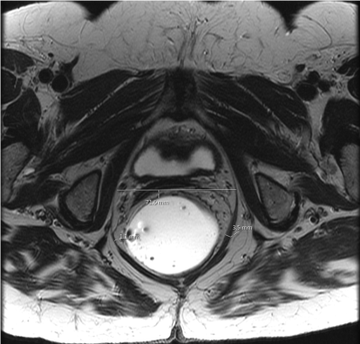 |
Levator hiatus width measurement and its correlation with pelvic floor dysfunction
Anugayathri Jawahar1, Perla Subbaiah 2, and Vipul R Sheth 1
1Body MRI, Stanford University, PALO ALTO, CA, United States, 2Mathematics, Oakland University, Rochester, MI, United States
Pelvic floor dysfunction affects about half of women over 50 years of age, causing significant morbidity. MR defecography compliments the clinical examination in the evaluation of pelvic floor dysfunction. We investigated if the measurement of levator hiatus width is a useful and reliable supplement or alternative measurement in predicting pelvic floor dysfunction. We retrospectively reviewed MR defecography examinations and recalculated the levator hiatus width and the anorectal angle with 2 blinded readers. We found that the levator hiatus width had a statistically significant correlation with pelvic organ prolapse and can add to the confidence in reporting H-line and M-line abnormalities.
|
|
2585.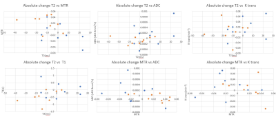 |
Multi-parameter assessment of Perianal Crohn’s disease using quantitative MRI: A Multi-centre study.
Ali Alyami1,2,3, Caroline Hoad 2,4, Ross Little5, Konstantinos Argyriou4, Uday Bannur6, Khalid Latief6, Christopher Clarke6, Phillip Lung7, Michael Berks5, Susan Cheung5, James PB O'Connor5,8, Geoff JM Parker9,10, Penny Gowland2,
and Gordon W. Moran1,4
1Nottingham Digestive Diseases Centre, University of Nottingham, Nottingham, United Kingdom, 2Sir Peter Mansfield Imaging Centre, University of Nottingham, Nottingham, United Kingdom, 3Faculty of Applied Medical Sciences, Diagnostic Radiology, Jazan University, Jazan, Saudi Arabia, 4NIHR Nottingham Biomedical Research Centre at Nottingham University Hospitals NHS Trust and University of Nottingham, University of Nottingham, Nottingham, United Kingdom, 5Quantitative Biomedical Imaging Laboratory, Division of Cancer Sciences, University of Manchester, Manchester, United Kingdom, 6Nottingham University Hospitals NHS Trust, Radiology, Nottingham, United Kingdom, 7St Mark's Hospital and London North West Healthcare NHS Trust, Radiology, London, United Kingdom, 8Radiotherapy Related Research, Division of Cancer Sciences, University of Manchester, Manchester, United Kingdom, 9Bioxydyn Limited, Manchester, United Kingdom, 10Centre for Medical Image Computing, University College London, London, United Kingdom
Perianal Crohn’s disease (pCD) is a potential complication of CD patients. Disease monitoring is imprecise due to unreliable clinical scores and subjective radiological reporting. Quantitative MRI sequences, e.g. diffusion-weighted image (DWI), dynamic contrast enhancement (DCE), magnetization transfer (MT) and T2 relaxometry offer opportunities to improve diagnostic capability. This study aimed to attain pilot data regarding the diagnostic utility of these sequences before and after 12 weeks of biological treatment in active pCD. Significant negative correlation was found between MTR and T2, MTR and DCE parameter ktrans and MTR with apparent diffusion coefficient, reflecting competing disease effects of inflammation and fibrosis.
|
|
| 2586. | Relaxometry and magnetisation transfer to quantify the small bowel wall in healthy volunteers
Ali Saleh Alyami1,2,3, Hannah Grace Williams2, Konstantinos Argyriou4, Penny Gowland2,4, Gordon W. Moran1,4, and Caroline Hoad 2,4
1Nottingham Digestive Diseases Centre, University of Nottingham, Nottingham, United Kingdom, 2Sir Peter Mansfield Imaging Centre, University of Nottingham, Nottingham, United Kingdom, 3Faculty of Applied Medical Sciences, Diagnostic Radiology, Jazan University, Jazan, Saudi Arabia, 4NIHR Nottingham Biomedical Research Centre at Nottingham University Hospitals NHS Trust and University of Nottingham, University of Nottingham, Nottingham, United Kingdom
Quantitative MRI datasets based on relaxometry and Magnetisation Transfer (MT) sequences could be used as potential biomarkers of disease activity in Inflammatory Bowel Disease. The aim of the study was to measure the repeatability of T2, T1 and MT small bowel measurements following bowel preparation and administration of an anti-spasmodic agent. Inter-observer variations in these measurements were also assessed. The quantitative MRI methods examined showed excellent repeatability for T2 and good repeatability for T1 parameters for both repeatability between visits and reproducibility of results between observers from data of a small group of healthy subjects.
|
|
2587.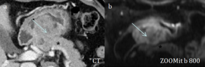 |
The application value of ZOOMit DWI in the diagnosis of surgical plan for patients with gastric sinus cancer with obstructive symptoms
Yubo Liu1, Ximing Wang1, Xianshun Yuan1, Haiyan Jing2, Xiankui Cheng2, Liang Shang3, Jinglei Liu3, Mengxiao Liu4, and Xiang Feng5
1Department of Radiology, Shandong Provincial Hospital, Jinan, China, 2Department of Pathology, Shandong Provincial Hospital, Jinan, China, 3Department of gastrointestinal surgery, Shandong Provincial Hospital, Jinan, China, 4MR Scientific Marketing, Diagnostic Imaging, Siemens Healthcare Ltd, shanghai, China, 5MR Scientific Marketing, Diagnostic Imaging, Siemens Healthcare Ltd, Beijing, China
For gastric antrum cancer with obstructive symptoms, CT has been a common method for preoperative diagnosis. But because it shows poor tumor boundaries, it does not give clear guidance on treatment options. ZOOMit DWI has higher image quality and smaller deformations make tumor boundaries better, so it can provide better preoperative guidance.
|
|
2588.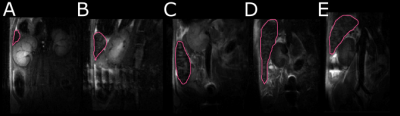 |
Serial Imaging of Splenomegaly in the Sleeping Sickness Infected Mouse
Samantha Paterson1, William Holmes1, Linda Carberry1, and Jean Rodgers1
1University of Glasgow, Glasgow, United Kingdom
Splenomegaly is a symptom of the African parasitic disease, sleeping sickness. This study is the first research to demonstrate splenomegaly non-invasively in a murine model of sleeping sickness. This researched showed an increase in the size of the spleen of over 20 times the healthy volume during the late stage of the disease whilst also demonstrating a change in the rate of increase during different stages of the disease. Overall, new information about splenomegaly in sleeping sickness has been demonstrated for the first time.
|
|
2589.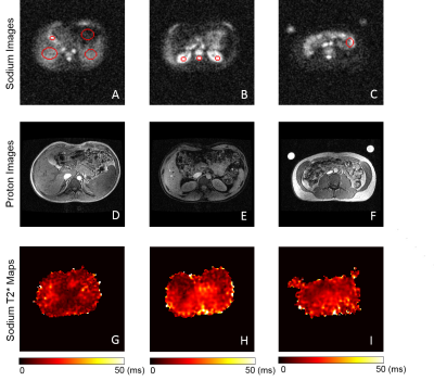 |
Quantitative sodium imaging of the abdomen
Arnold Julian Vinoj Benjamin1, Mary A McClean1, Joshua D Kaggie1, Lucian Beer1, Frank Riemer2, Rolf F Schulte3, Titus Lanz4, Martin J Graves5, and Ferdia A Gallagher1
1Department of Radiology, University of Cambridge, Cambridge, United Kingdom, 2Department of Radiology, Haukeland University Hospital, Bergen, Norway, 3GE Global Research, Munich, Germany, 4RAPID Biomedical GmbH, Rimpar, Germany, 5Department of Radiology, Cambridge University Hospitals NHS Foundation Trust, Cambridge, United Kingdom
In this study, we report sodium T2* values and total sodium concentration (TSC) values in healthy volunteers in the abdomen using a large field-of-view (FOV) birdcage sodium body coil that provides a uniform excitation over the whole abdomen. We explore whether sodium will be more specific to the BOLD effect, as sodium is a major component of blood and oxygenation of this blood will affect the sodium T2* values directly.
|
|
2590.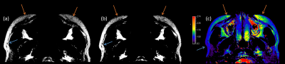 |
MRI Application for Autologous Fat Transplantation Assessment
Xueyin Liao1, Yu Chen2, Xiaoqi Wang3, Mingyong Yang1, and Huadan Xue2
1Plastic Surgery Hospital, Chinese Academy of Medical Sciences, Peking Union Medical Collage, Beijing, China, 2Department of Radiology, Peking Union Medical College Hospital, Beijing, China, 3Philips HealthCare, Beijing, China
We have investigated the clinical feasibility of applying MRI to predict the fat survival rate of autologous fat transplantation on faces 7 days after surgery. High resolution T1W and T2W fat-only images, T2 mapping, IVIM diffusion, and spectroscopy methods were implemented. The morphological images provided extradentary value in visualizing transplanted fat in vivo, the biomarker T2 mapping identified edema, IVIM measured perfusion and localized rebuilding vascularization, and MRS quantified lactate content for predicting post-op fat survival rate. The non-contrast MRI technologies proved values in imaging for autologous fat transplantation surgery.
|
|
2591.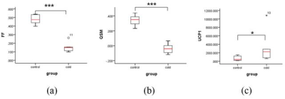 |
Detection of Active Brown Adipose Tissue Using Quantitative Susceptibility Mapping (QSM)
Cuiling Zhu1, Yihao Guo2, Wenbin Si2, Yingjie Mei3, Qiaoling Zhong1, Lijie Zhong1, Yanqiu Feng2, and Xiaodong Zhang1
1Department of Radiology, The Third Affiliated Hospital of Southern Medical University, Guangzhou, China, 2School of Biomedical Engineering, Southern Medical University, Guangzhou, China, 3Philips Healthcare, Guangzhou, China
Activated and inactivated brown adipose tissue showed some differences in function, protein expression and intracellular components, which remained non-invasive MRI testing possible. Despite its strong relevance with iron contents, QSM detect BAT in large lipid depots remains a major challenge. This work proposes a method that use values of QSM and (fat fraction) FF to quantitatively distinguish the two groups in brown adipose tissue depot in vivo, histological validation is then performed. Preliminary results are broadly in line with expectation.
|
2592.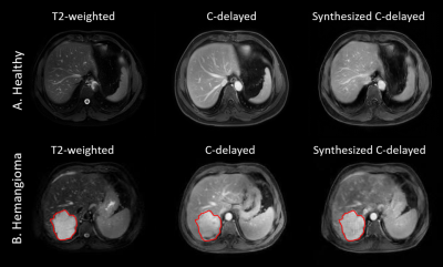 |
Prediction of contrast-enhanced liver MRI with CycleGAN
Yajing Zhang1 and Hanlin Fu1
1Philips Healthcare, Suzhou, China
Contrast-enhanced MR scans have been commonly used in the diagnosis of liver lesions. However, it has limited use to the patients allergic to the contrast agent. Recently, there are increasing concerns about the safety of the use of Gadolinium-based contrast agents. In this study, we employed a cycle generative adversarial network strategy to test the feasibility of predicting the contrast enhancement images in liver MRI.
|
|
2593.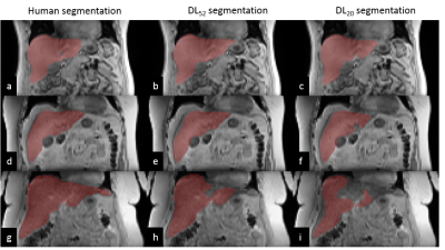 |
Deep Learning for Liver Segmentation and Quantification of Obese Patients
Philipp Madörin1, Xeni Deligianni1,2, Francesco Santini1,2, Simon Andermatt2, Philippe Claude Cattin2, Anne Christin Meyer-Gerspach3,4, Bettina Karin Wölnerhanssen3,4, Oliver Bieri1,2, and Orso Pusterla1,2,5
1Department of Radiology, Division of Radiological Physics, University Hospital Basel, Basel, Switzerland, 2Department of Biomedical Engineering, University of Basel, Basel, Switzerland, 3St. Clara Research Ltd, St. Clara Hospital, Basel, Switzerland, 4University of Basel, Basel, Switzerland, 5Institute for Biomedical Engineering, University and ETH Zurich, Zurich, Switzerland
Obesity is one of the greatest health risks and strongly related to fatty liver disease. Magnetic resonance imaging enables non-invasive measurement of fat-water distribution in tissue. To provide an automated evaluation of the liver volume and fat percentage, we trained a Multi-Dimensional Gated Recurrent Units network to segment multi-contrast data. The neural network was trained with a limited number of data comprising 52, 20, 10 datasets and was evaluated for liver volume and fat percentage quantification.
|
|
2594.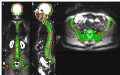 |
Automatic Bone Segmentation on Whole body Diffusion-Weighted MRI using Deep Learning
Asha K Kumaraswamy1,2, Punith B Venkategowda1, Chandrashekar M. Patil2, and Robert Grimm3
1Siemens Healthcare Private Limited, Bengaluru, India, 2Vidyavardhaka College of Engineering, Mysuru, India, 3MR Application Predevelopment, Siemens Healthcare, Erlangen, Germany
Whole body diffusion-weighted MR imaging is a promising technique for the evaluation of bone metastases e.g. in prostate and breast cancer. Segmenting and quantifying tumor burden and treatment response based on DWI and corresponding ADC has been proposed previously. However, treatment effects may influence the actual segmentation and signal intensities. Here, a more reproducible approach would be preferred for segmenting and analyzing consistent regions of interest also in follow-up examinations. We present a deep learning method for automatic bone segmentation, based on T1-weighted imaging or DWI. Feasibility of the new approach is shown, resulting in significantly reduction in computation time.
|
|
2595.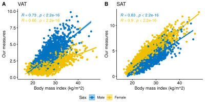 |
Unsupervised assessment of abdominal adipose tissue from whole body Dixon MRI
Ying Xia1, Pierrick Bourgeat1, Samantha Burnham1, and Jurgen Fripp1
1The Australian eHealth Research Centre, CSIRO Health and Biosecurity, Brisbane, Australia
Quantitative assessment of visceral adipose tissue (VAT) is important in studies of obesity and metabolic abnormalities. The study developed and evaluated an unsupervised method for abdominal adipose tissue quantification in a large sample from the general population. The results revealed strong correlations with reference measures using dual energy X-ray and MR based methods with human intervention. We also demonstrate the relationship between body mass index (BMI) and adipose tissue compartments. As expected, BMI did not fully reflect fat accumulation in VAT. With increasing BMI, males were more likely to have higher VAT accumulation than females.
|
|
2596.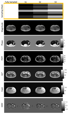 |
Quantitative Susceptibility Mapping from Deep-Learning Based Reconstruction of Undersampled Gradient-Recalled Echo Data
Ramin Jafari1,2, Pascal Spincemaille2, Thanh D. Nguyen2, Junghun Cho1,2, Martin R. Prince2, and Yi Wang1,2
1Cornell University, Ithaca, NY, United States, 2Weill Cornell Medicine, New York, NY, United States A single breath-hold 3D GRE acquisition with tens of seconds is used to acquire data for water/fat separation and QSM generation in liver. However, in elderly and paediatric patients long breath-holds are not feasible. Compressed sensing along with Deep Learning is an alternative to shorten the scan time and perform reconstruction on undersampled data. In this work we compare how undersampling GRE data at different rates and use of Deep Learning for reconstruction will affect the water/fat separation and QSM results. |
|
2597.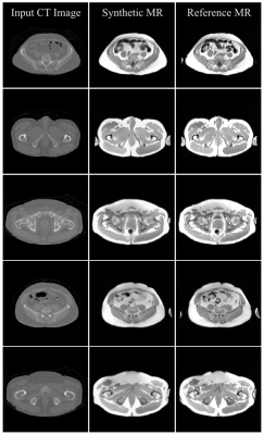 |
CT-based Synthetic pelvic T1-weighted MR image generation using a deep convolutional neural network (CNN)
Reza Kalantar1, Jessica M Winfield1,2, Christina Messiou1,3, Dow-Mu Koh1,3, and Matthew D Blackledge1
1Division of Radiotherapy and Imaging, The Institute of Cancer Research, London, United Kingdom, 2The Royal Marsden Hospital, London, United Kingdom, 3Department of Radiology, The Royal Marsden Hospital, London, United Kingdom
The MR-Linac exploits excellent soft-tissue contrast of MRI images with the option of daily re-planning for irradiation of pelvic tumours. However, this requires significant clinician interaction as contours need manual redefining for each radiotherapy fraction. Recently, machine learning-based segmentation approaches are being developed to automate this process. One major limitation of this approach is the lack of available fully-annotated MRI data for training. In this study, a deep learning model was developed and trained on CT/MR registered images of 21 patient datasets, generating synthetic T1-weighted MR images from input CT data, which could be useful towards disease segmentation on MRI.
|
|
2598.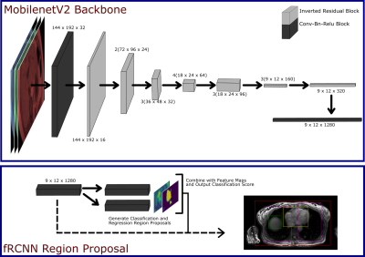 |
Deep learning based multi-organ detection: a step closer to liver T1 map quality assurance
Charles E Hill1,2, Ferenc Emil Mozes2, Liam AJ Young2, Luca Biasiolli2, Matthew D Robson3, and Vicente Grau1
1Engineering Science, University of Oxford, Oxford, United Kingdom, 2Oxford Centre for Magnetic Resonance Research (OCMR), University of Oxford, Oxford, United Kingdom, 3Perspectum Diagnostics LTD., Oxford, United Kingdom
Quantitative Liver T1 mapping techniques are becoming more prevalent in the clinic, for the diagnosis and prognosis of various liver diseases. However, time pressure and technique complexity can often lead to incorrect acquisition due to sub-optimal slice location. We implement a faster Region Proposal CNN for the detection of multiple organs in the body, and report back the findings promptly via a local user interface, to inform the radiographers if they need to re-acquire. We show that this network has a high localisation accuracy and high speed, such that it will be applicable in the clinical environment.
|
|
2599.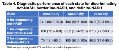 |
Characterization of NAFLD disease evolution with multiparametric MRI/MRE and multi-state Hidden Markov Model
Jingbiao Chen1,2, Yunyi Kang3, Feng Ju3, Jiahui Li1, Jie Chen1, Xin Lu1, Richard L Ehman1, Vijay H Shah2, and Meng Yin1
1Radiology, Mayo Clinic, Rochester, MN, United States, 2Gastroenterology and Hepatology, Mayo Clinic, Rochester, MN, United States, 3School of Computing, Informatics, and Decision Systems Engineering, Arizona State University, Tempe, AZ, United States
Non-invasively diagnosing non-alcoholic fatty liver disease (NAFLD) and predicting disease prognosis in individual patients are two main unmet clinical needs. There are very few longitudinal studies that evaluate imaging, biochemical, and histopathological variables that predict disease progression of NAFLD. This study established a multi-state Hidden Markov model (HMM) of NAFLD evolution in an animal model with three imaging biomarkers: MRI derived proton density fat fraction (PDFF) and MR elastography (MRE) assessed liver stiffness (LS) and loss modulus (LM). Results have shown that a 3-state HMM can well characterize the natural history of NAFLD, and predict disease progression or regression.
|
|
2600.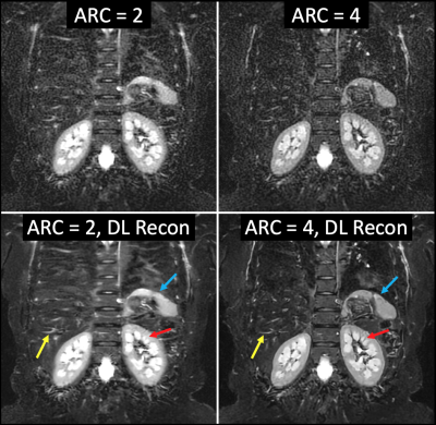 |
Accelerated Whole-body Imaging with Uniform Fat-Suppression and Deep-Learning Reconstruction
Alan B McMillan1, Lloyd Estowski2, Ty A Cashen2, R. Marc Lebel3, Xinzeng Wang4, Ersin Bayram4, and Ali Pirasteh1
1Radiology, University of Wisconsin Madison, Madison, WI, United States, 2Global MR Applications & Workflow, GE Healthcare, Waukesha, WI, United States, 3Global MR Applications & Workflow, GE Healthcare, Calgary, AB, Canada, 4Global MR Applications & Workflow, GE Healthcare, Houston, TX, United States
We present a single-shot fast-spin-echo pulse-sequence (SSFSE) with short-tau inversion recovery (STIR) and deep-learning (DL) image reconstruction that provides whole-body images in less than 3 minutes, achieving uniform fat suppression, detailed delineation of the anatomy, and favorable signal-to-noise (SNR). This was achieved through the reduction of echo-train-length by increasing the acceleration to a factor of 4x, hence substantially improving image quality through blurring reduction in the phase encoding direction, and ameliorating the subsequent SNR reductions by utilization of a DL reconstruction algorithm. This whole-body MRI technique can be used in the setting of various pathologies for both pediatrics and adults.
|
|
2601.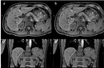 |
Deep convolution neural network exploration for super resolution of abdominal 3D mDixon scans
Johannes M Peeters1 and Marcel Breeuwer1,2
1MR Clinical Science, Philips, Best, Netherlands, 2Biomedical Engineering – Medical Image Analysis, Eindhoven University of Technology, Eindhoven, Netherlands
Breath holding is often applied for abdominal imaging to avoid motion artifacts. However, breath holding limits the acquisition time and thus the resolution of the images. We propose to use 3D super resolution deep convolutional neural networks (CNN) to enhance the sharpness of 3D mDixon MRI. We found that sharpness increases with increasing number of network layers, but levels off already at 6 layers.
|
|
2602.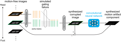 |
Retrospective Motion Artifact Reduction with CNN (MARC) combined with model-based artifact simulation for T2WI of the liver
Motohide Kawamura1, Daiki Tamada1, Tetsuya Wakayama2, Hiroshi Onishi1, and Utaroh Motosugi1
1Department of Radiology, University of Yamanashi, Yamanashi, Japan, 2MR Collaboration and Development, GE Healthcare, Tokyo, Japan
Motion artifact is a problem in abdominal imaging. Respiratory-triggering techniques are commonly used to suppress the motion artifact. However, it is not always perfect in clinical practice. Deep learning-based motion correction is an attractive solution. However, it requires pairs of images with and without motion artifacts, which is difficult in body MRI. Here, we propose a deep learning-based method to remove motion artifact using a simulation of artifacts based on a simple model for respiratory gating failure. Preliminary results showed that the proposed method can remove motion artifacts in respiratory-triggered FSE-T2WI of the liver, which were corrupted by irregular breathing.
|
|
2603.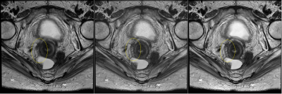 |
Deep learning based image reconstruction for T2-weighted rectal cancer imaging
Ken-Pin Hwang1, Xinzeng Wang2, Randy D Ernst3, Sarah M Palmquist3, Marc Lebel2, Gaiane M Rauch3, George J Chang4, Craig Messick4, Melissa W Taggart5, Ersin Bayram2, Jingfei Ma1, and Harmeet Kaur3
1Department of Imaging Physics, The University of Texas M.D. Anderson Cancer Center, Houston, TX, United States, 2MR Applications and Workflow, GE Healthcare, Waukesha, WI, United States, 3Department of Radiology, The University of Texas M.D. Anderson Cancer Center, Houston, TX, United States, 4Department of Surgical Oncology, The University of Texas M.D. Anderson Cancer Center, Houston, TX, United States, 5Department of Pathology, The University of Texas M.D. Anderson Cancer Center, Houston, TX, United States
Proper evaluation of rectal cancer with MR imaging requires high resolution imaging of the rectal wall. The image quality demands are difficult to achieve due to the increasing risk of peristaltic motion with longer scan times. In this work, we apply a novel deep learning based reconstruction (DL recon) method to an accelerated sequence using reduced averages and increased acceleration. Radiologist scores indicate that the combined method provides superior SNR and definition with less motion degradation when compared to the routine sequence with conventional reconstruction. Thus improved motion robustness can be gained from applying DL Recon to an accelerated sequence.
|
|
2604.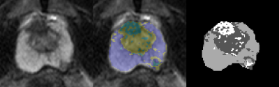 |
The feasibility of model-free machine learning with variable features of MRI images on the prostate cancer application
Feng-Mao Chiu1,2, Ting Chun Lin1, Queenie Chan3, Cheng-Chun Li4, Jen-I Hwang4, and You Yin Chen1
1Department of Biomedical Engineering, National Yang-Ming University, Taipei, Taiwan, 2Health system, Philips, Taipei, Taiwan, 3Healthcare, Philips, Hong Kong, China, 4Department of Medical Imaging, Tungs’ Taichung Metroharbor Hospital, Taichung, Taiwan
Several advanced post-processing methods were used to assess prostate cancer, including diffusion model of IVIM, T2 mapping with multi-echo turbo spin echo , and permeability analysis with Tofts model. We found that it is not easy to overcome the fitting error, and it is not reasonable to directly integrate IVIM, T2 mapping and permeability together as well. The aim of this study is to evaluate the feasibility of the prostate cancer screening with model-free machine leaning , and to test different combinations of input data, including multiple b-value diffusion weighted imaging (DWI), multi-echo T2w TSE, and dynamic contrast enhanced (DCE) images.
|
|
2605.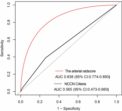 |
Correlation between CT based Radiomics Features and Mesenteric Vein Resection Margin in Patients with the Pancreatic Head Cancer
Yun Bian1, Chao Ma1, Xu Fang1, Jin Li1, Kai Cao1, Li Wang1, Jin Gang2, Jianping Lu1, and Xiangxue Wang3
1Radiology, Changhai hospital, Shanghai, China, 2Pancreatic Surgery, Changhai hospital, Shanghai, China, 3Shanghai Institute for Advanced Communication and Data Science, School of Computer Engineering and Science, Shanghai University, Shanghai, China
A striking number of patients are diagnosed with resectable pancreatic cancer (PC) or borderline resectable PC by computed tomography (CT) but end up with positive (R1) resection at surgery according to the National Comprehensive Cancer Network (NCCN) criteria. If the pathological resection margin can be accurately and noninvasively predicted prior to surgery, an appropriate treatment plan can be developed and patients with PC can avoid futile surgery. Hence, we sought to accurately identify the relationship between the arterial radiomics score (rad-score) and pathologic superior mesenteric vein (SMV) resection margin in patients with pancreatic head cancer and to evaluate the diagnostic performance of the rad-score in differentiating between negative (R0) and R1 resection.
|
2606.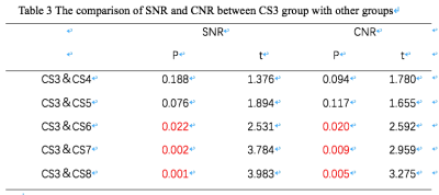 |
Compressed Sensing and Parallel Imaging on 3D Uterus Imaging
Yaxin Niu1, Ailian Liu1, Shifeng Tian1, Lihua Chen1, Qingwei Song1, Jiazheng Wang2, and Zhiwei Shen2
1The First Affiliated Hospital of Dalian Medical University, Dalian Medical University, Da Lian, China, 2Philips Healthcare, Beijing, China, Beijing, China
Acquisition time is one of the main limiting factors in MRI. Compressed sensing is an emerging technology proven to effectively shorten scan time in several clinical scenarios. Here we propose to apply a combination of compressed sensing and sensitivity encoding in 3D uterus imaging that yields 1x1x2 mm3 resolution and uncompromised image quality with 5-fold acquisition acceleration.
|
|
2607.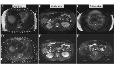 |
Effect of Continuous table movement (CTM) on fat saturation quality in T2 weighted imaging
Hina Arif1, Stephane Chartier 2, Tanmay Tiwari3, Bobby Kalb2, and Manoj Saranathan2
1Medical Imaging, University of Arizona, Tucson, AZ, United States, 2University of Arizona, Tucson, AZ, United States, 3University of Arziona, TUCSON, AZ, United States
Uniform fat suppression is critical for optimal MR assessment of lesions in the abdomen and pelvis. Recent advances using SPAIR sequencing have improved fat suppression and increased the signal-to-noise ratio versus older techniques. However, fat suppression still remains challenging in anatomical regions away from the magnetic isocenter due to field inhomogeneity. Here we demonstrate that continuous table movement (CTM) is superior to fixed table (FT) technology for SPAIR-mediated fat suppression across the entire image stack. As field inhomogeneity away from isocenter is a common challenge, CTM may provide a solution to many frequently encountered problems.
|
|
2608.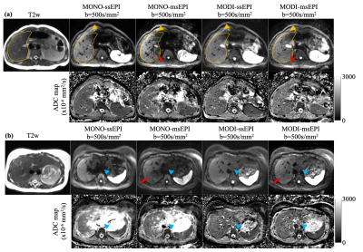 |
Motion-Robust, Reduced-Distortion Diffusion MRI of the Liver with Optimized Motion Compensated Waveforms and Multi-shot EPI
Yuxin Zhang1,2, Matthias Muhler1, Ruiqi Geng1,2, Arnaud Guidon3, and Diego Hernando1,2
1Radiology, University of Wisconsin Madison, Madison, WI, United States, 2Medical Physics, University of Wisconsin Madison, Madison, WI, United States, 3Applications and Workflow, GE Healthcare, Boston, MA, United States
Conventional liver diffusion MRI acquisitions suffer from several challenges including low spatial resolution, B0-induced distortions, and elastic motion-induced signal voids. In this work, motion-robust and distortion-corrected liver diffusion-weighted imaging (DWI) was enabled by combining optimized motion compensated diffusion waveforms with multi-shot EPI acquisitions. Quantitative validation of distortions and ADC measurements was performed in healthy volunteer to assess the robustness and reproducibility of the combined techniques, as well as the synergistic effect of motion-robust gradient waveform and multi-shot EPI acquisitions. Preliminary patient example demonstrated the feasibility in patients with metastatic liver lesions.
|
|
2609.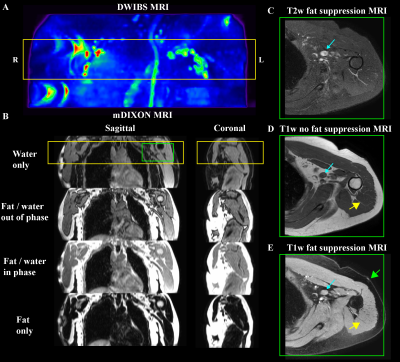 |
Quantitative magnetic resonance relaxometry for identifying lymphatic insufficiency in patients with secondary lymphedema
Paula Donahue1, Rachelle Crescenzi2, Chelsea Lee3, Niral J Patel3, Maria Garza2, Kalen Petersen2, and Manus Joseph Donahue2
1Physical Medicine and Rehabilitation, Vanderbilt Medical Center, Nashville, TN, United States, 2Radiology and Radiological Sciences, Vanderbilt Medical Center, Nashville, TN, United States, 3Pediatrics, Vanderbilt Medical Center, Nashville, TN, United States
MRI methods capable of evaluating tissue changes in response to lymphatic dysfunction have not been incorporated into lymphedema diagnosis and management owing to limited clinical evaluation of these technologies. We performed quantitative relaxometry in the skin and deep tissue of patients with lymphedema and matched controls, in sequence with standard biophysical bedside measures used for lymphedema assessment. In both deep tissue and skin, T2 was elevated in patients relative to control volunteers, consistent with edema, and relaxometry values were more discriminatory for category (i.e., control versus lymphedema) compared to common bedside tools of arm volume asymmetry and tissue dielectric constant.
|
|
2610.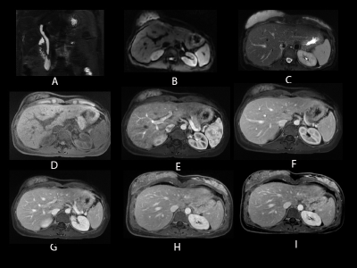 |
Integration of novel respiratory navigation techniques into routine abdominal MR examinations
Melany Atkins1 and Ersin Bayram2
1Fairfax Radiological Consultants, Arlington, VA, United States, 2General Electric, Houston, TX, United States
Breath-held acquisitions have long been a staple of body MRI to cope with respiratory motion. While this strategy works for many patients, it is challenging for some. Change in breath-hold capacity from one patient to the next requires frequent parameter tweaking, thus operator skillset becomes a key factor in image quality and consistency. Recent advances in novel motion robust acquisition methods and improved hardware such as Air coils have opened the possibility to perform complete free breathing exams. In this study, we report the feasibility results of free breathing integration into our routine abdominal MRI practice.
|
|
2611.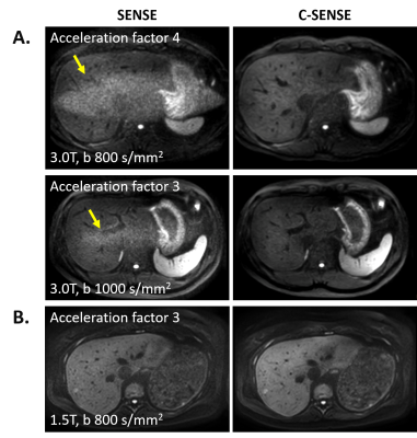 |
Body Diffusion MR Imaging with Compressed SENSE Based on Single-Shot EPI at 3T and 1.5T: Technical Feasibility and Initial Clinical Experience
Paul Sprenger1,2, Kosuke Morita3, Takeshi Nakaura4, Masami Yoneyama5, Shuo Zhang2,6, Maike Bode2, Christiane K. Kuhl2, and Nils A. Kraemer2
1Faculty of Medicine, RWTH Aachen University, Aachen, Germany, 2Diagnostic and Interventional Radiology, University Hospital RWTH Aachen, Aachen, Germany, 3Kumamoto University Hospital, Kumamoto, Japan, 4Graduate School of Medical Sciences, Kumamoto University, Kumamoto, Japan, 5Philips Japan, Tokyo, Japan, 6Philips GmbH DACH, Hamburg, Germany
Compressed sensing technologies have recently become commercially available that serve the purpose to shorten the image acquisition time for morphological imaging. Here we explore the reconstruction algorithm that allows the combination of wavelet transformation of compressed sensing with coil information of sensitivity encoding (SENSE) in body diffusion MRI based on 2D single-shot EPI and uniform undersampling, and report our results in both healthy volunteers and patients on both 3T and 1.5T, in comparison to the conventional SENSE parallel imaging technique.
|
|
2612.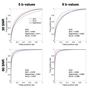 |
Variability from complexity: assessing IVIM acquisition schemes through parameter estimation uncertainty
Sean C Epstein1, Timothy J. P. Bray2, Margaret A. Hall-Craggs2, and Hui Zhang1
1Department of Computer Science & Centre for Medical Image Computing, University College London, London, United Kingdom, 2Centre for Medical Imaging, University College London, London, United Kingdom
This work uses the quantification of parameter-estimation uncertainty to assess the clinical suitability of IVIM acquisition schemes. We apply this analysis to two bone marrow classification tasks. We simulate IVIM data and show that fitting a simplified, biased, diffusion model (ADC), can, under certain clinically relevant conditions, outperform ground-truth IVIM fitting. We further show that, within the same disease, the opposite can also be true: IVIM outperforms ADC. Such results can play an important role in guiding clinical DWI practice and we show that they can be predicted by explicitly quantifying the uncertainty in parameter estimation.
|
|
2613.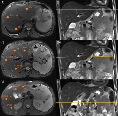 |
Comparison and correlation study between DKI and IVIM in assessing liver parenchyma
Guifang Lin1, Lili Wang1, Zhongshuai Zhang2, Bin Sun1, Weiwen lin1, Ruolan Lin1, Jingming Chen1, Jiangao Xie1, Yuanfeng Liu1, Qing Duan1, and Yunjing Xue1
1Department of Radiology, Fujian Medical University Union Hospital, Fuzhou, China, 2SIEMENS Healthcare, Shanghai, China
The aim of this study was to assess the liver parenchyma by evaluating the regional variability, the measurement stability and the correlations of parameters calculated from IVIM and DKI models using different ROIs. The results demonstrated that S8, S5/8, S5 and S3 delegating different level of liver tissue can be chosen as the representative area in assessing the IVIM-DKI imaging of the liver. ADC, MD, MK and D with lower deviation were suitable for the quantitative evaluation of liver tissue and can be used to reflect different sides of the microstructure of the hepatic parenchyma.
|
|
2614.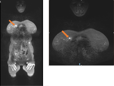 |
Whole body MRI evaluation for adult patients with hereditary cancer syndromes
Vishnupriya Kuchana1, Nobuhiro Fujita1, Elina Shrestha1, Amro Saad Aldine1, Khalid Balawi1, and Jinha M Park1
1Radiology, University of Iowa Hospitals and Clinics, Iowa City, IA, United States
Patients diagnosed with hereditary cancer syndromes (HCS) mutations are at increased risk of developing tumors. We are trying to evaluate the utility and cost-effectiveness of WB MRI for screening and monitoring adult HCS patients for new lesions (malignant or pre malignant). We retrospectively collected the data from the HCS patients who had whole-body MRI scans and are aged 18 -72 years old. Overall sensitivity and specificity of WB MRI is 100% and 87.5% and mean per patient cost responsibility per scan is $358.83 which proves WB MRI can be an effective tool for screening and surveillance of HCS patients.
|
|
2615.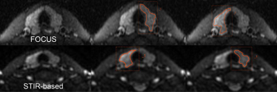 |
Optimizing diffusion-weighted imaging of thyroid gland using dedicated surface coil
Yunfei Wang1, Chaofan Zhu1, Dehong Luo1, Long Qian2, Yayuan Geng3, Wenming Deng1, and Zhou Liu1
1Radiology, National Cancer Center/National Clinical Research Center for Cancer/Cancer Hospital & Shenzhen Hospi, Shenzhen, China, 2MR Research, GE Healthcare, Beijing, China, 3Huiying Medical Technology Co., Ltd., Beijing, China
It is challenging to perform diffusion-weighted imaging (DWI) on thyroid gland with high quality due to susceptibility artifacts caused by complex structure compositions, and physiological motion artifacts and low signal-to-noise ratio (SNR) using conventional head and neck joint coil. We used a histogram-based approach to evaluate the images of the small field-of-view (FOV) DWI (Focus, GE) and DWI with short-tau inversion-recovery (STIR) fat suppression using a state-of-the-art surface-coil specifically designed for thyroid gland. We found significantly different level and considerably inhomogeneity in signal intensity between right and left lobe of thyroid gland and between two DWIs.
|
|
2616.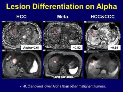 |
Diffusion-Weighted Imaging of the Liver with Stretched-Exponential Model
Takeshi Yoshikawa1, Yoshiharu Ohno2, Masao Yui3, Yoshimori Kassai3, Tatsuya Ohkubo3, Shinichiro Seki4, Katsusuke Kyotani5, and Yuji Kishida6
1Kobe University Graduate School of Medicine, Kobe, Japan, 2Fujita Health University School of Medicine, Toyoake, Japan, 3Canon Medical Systems Corporation, Otawara, Japan, 4Hyogo Prefectural Tamba Medical Center, Tamba, Japan, 5Kobe University Hospital, Kobe, Japan, 6Konan Medical Center, Kobe, Japan
Stretched-exponential model enables diffusion analysis considering diffusion varieties in each voxel on abdominal DWI. Our study showed stretched-exponential model has a potential to increase diagnostic performance of liver DWI. Alpha has a potential to be used for malignant lesion differentiation.
|
|
2617. |
Diffusion-Weighted Imaging with Stretched-Exponential Model for Pancreatic Tumors
Takeshi Yoshikawa1, Yoshiharu Ohno2, Masao Yui3, Yoshimori Kassai3, Tatsuya Ohkubo3, Shinichiro Seki4, Katsusuke Kyotani5, and Yuji Kishida6
1Radiology, Kobe University Graduate School of Medicine, Kobe, Japan, 2Fujita Health University School of Medicine, Toyoake, Japan, 3Canon Medical Systems Corporation, Otawara, Japan, 4Hyogo Prefectural Tamba Medical Center, Tamba, Japan, 5Kobe University Hospital, Kobe, Japan, 6Konan Medical Center, Kobe, Japan
Stretched-exponential model enables diffusion analysis considering diffusion varieties in each voxel on abdominal DWI. Our study showed stretched-exponential model has a potential to increase diagnostic performance of pancreatic DWI. Alpha has a potential to be used for malignant lesion differentiation.
|
|
2618.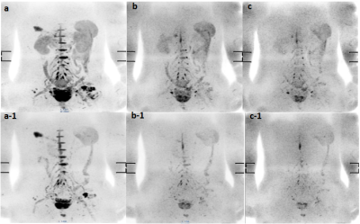 |
The Value of Oral Paramagnetic Contrast Agent in Eliminating Gastrointestinal Interference with DWIBS
Nan Zhang1, Qingwei Song1, Renwang Pu1, Jiazheng Wang2, Liangjie Lin2, Ailian Liu1, and Haonan Zhang1
1The First Affiliated Hospital of Dalian Medical University, Dalian, China, 2Philips Healthcare, Beijing, China
The present study aims to explore the effect of oral paramagnetic contrast agent on image quality of Diffusion weighted Whole body Imaging with Background body signal Suppression(DWIBS).This study confirmed that the oral paramagnetic contrast agent could eliminate gastrointestinal interference without affecting the diagnostic efficacy of DWIBS images.
|
|
2619.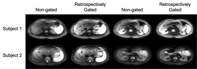 |
Retrospective respiratory and cardiac gating in abdominal diffusion-weighted imaging
Sean McTavish1, Christoph Zoellner1, Johannes M. Peeters2, Omar Kamal1, Rickmer F. Braren1, and Dimitrios C. Karampinos1
1Department of Diagnostic and Interventional Radiology, Technical University of Munich, Munich, Germany, 2Philips Healthcare, Best, Netherlands
Diffusion-weighted imaging (DWI) remains a valuable tool in abdominal lesion detection. However, respiratory and cardiac motion remain major effects confounding the robustness of abdominal DWI. Respiratory triggering and prospective gating techniques are typically employed to reduce the effect of respiratory motion on abdominal DWI. However, these techniques can become either inefficient or result in artifacts in patients with irregular breathing patterns. In addition, cardiac triggering further reduces the SNR efficiency of a respiratory triggered acquisition. The purpose of the present work is to develop an algorithm for combined retrospective gating of abdominal DWI based on recorded respiratory and cardiac signals.
|
|
2620.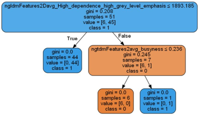 |
Texture analysis based on T2-weigted images show high differentiation between IVDs with and without annular fissuring
Stefanie Eriksson1, Hanna Hebelka2,3, Leif Torén2,3, Christian Waldenberg2, Helena Brisby2, and Kerstin Lagerstrand1,4
1Medical Physics and Biomedical Engineering, Sahlgrenska University Hospital, Gothenburg, Sweden, 2Institute of Clinical Sciences, Gothenburg University, Gothenburg, Sweden, 3Department of Radiology, Sahlgrenska University Hospital, Gothenburg, Sweden, 4Institute of Clinical Scences, Gothenburg University, Gothenburg, Sweden
Texture analysis provides quantitative image analysis based on mathematically calculated features. A high number of features calculated with texture analysis showed to be significantly different between the intervertebral discs with and without annular fissuring that had been classified from CT discograms. When the features based on T2-weighted images were used for classification by "Random Forest" a very high accuracy in differentiating between discs with and without annular fissuring was achieved.
|
2621. |
Flow quantification with navigator-gated 4D flow MRI in portal hypertension
Octavia Bane1,2, Daniel Stocker1,2, Paul Kennedy1,2, Stefanie Hectors1,2,3, Susanne Schnell4, Scott Friedman5, Thomas Schiano5, Isabel M Fiel6, Swan Thung6, Aaron Fischman2, Michael Markl4,7, and Bachir Taouli1,2
1BioMedical Engineering and Imaging Institute, Icahn School of Medicine at Mount Sinai, New York, NY, United States, 2Department of Diagnostic, Molecular and Interventional Radiology, Icahn School of Medicine at Mount Sinai, New York, NY, United States, 3Department of Radiology, Weill Cornell Medicine, New York, NY, United States, 4Department of Radiology, Northwestern University, Chicago, IL, United States, 5Department of Medicine, Icahn School of Medicine at Mount Sinai, New York, NY, United States, 6Department of Pathology, Icahn School of Medicine at Mount Sinai, New York, NY, United States, 7Biomedical Engineering, Northwestern University McCormick School of Engineering, Evanston, IL, United States
The purpose of our prospective study was to correlate the hepatic vasculature hemodynamic parameters measured with navigator-gated 4D flow MRI with the hepatic venous pressure gradient (HVPG) in patients with chronic liver disease and suspicion of portal hypertension (PH). Peak velocity in the celiac trunk (CT) was moderately correlated with HVPG, and was significantly elevated in patients with PH. CT flow was significantly elevated in patients with clinically significant PH (CSPH), while portal vein peak velocity was significantly reduced in patients with cirrhosis. Our results suggest that 4D flow MRI may be sensitive to hemodynamic changes in patients with PH.
|
|
2622.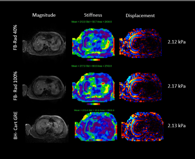 |
An Inline Implementation of Free Breathing 2D Radial MRE with Radial Self Gating
Bradley Bolster, Jr.1, Stephan Kannengiesser2, and Vibhas Deshpande3
1Siemens Medical Solutions USA, Inc., Salt Lake City, UT, United States, 2Siemens Healthcare GmbH, Erlangen, Germany, 3Siemens Medical Solutions USA, Inc., Austin, TX, United States
This study demonstrates in inline implementation of a radial free breathing MRE sequence with integrating radial self gating. Measured stiffness values are consistent across self-gating threshold values and results are in good agreement with standard breath-hold MRE techniques.
|
|
2623.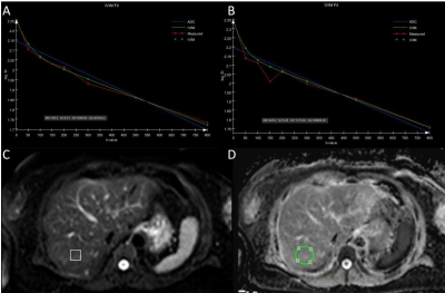 |
Post-treatment recovery of perfusion-related diffusion in the fatty liver of type 2 diabetes mellitus: an IVIM-DWI study
Wei Wei1, Yan Bai1, Xianchang Zhang2, Robert Grimm3, and Meiyun Wang1
1Department of Radiology, Henan Provincial People’s Hospital, Zhengzhou, China, 2MR Collaboration, Siemens Healthcare Ltd, Beijing, China, 3MR Application Predevelopment, Siemens Healthcare, Erlangen, Germany
This study aimed to investigate the utility of intra-voxel incoherent motion diffusion-weighted imaging (IVIM-DWI) for monitoring the liver function change during type 2 diabetes mellitus (T2DM) treatment. The results showed that liver fat fractions were significantly decreased and perfusion-related diffusion (D*) was significantly increased, but pure molecular diffusion (D) values showed no significant change in T2DM patients after treatment for three months. Our findings indicated that improvement of hepatic parenchymal perfusion may occur earlier than water molecule diffusion during T2DM treatment. D* may be a more sensitive biomarker than D to characterize the liver function change during T2DM treatment.
|
|
2624.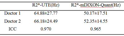 |
The relativity (R2*) between ultra-short echo time MRI and mDIXON-Quant sequence in liver cirrhosis
Nan Wang1, Ailian Liu1, Qingwei Song1, Renwang Pu1, Mingli Gao1, Lihua Chen1, Jiazheng Wang2, Zhiwei Shen2, and Yishi Wang2
1The First Affiliated Hospital of Dalian Medical University, Dalian, China, 2Philips Healthcare, Beijing, China
The detection and quantification of iron depositon in cirrhotic liver are important for the diagnosis and treatment. The MR-UTE sequence has advantages in quantifying severe liver iron overload, and partially solve the problem that severe iron overload cannot be accurately quantified. mDIXON-Quant sequence could quantify liver R2* value.
|
|
2625.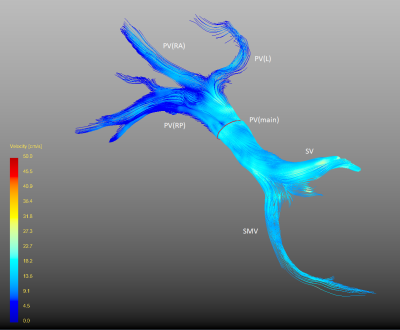 |
Feasibility of 4D-Flow MRI of the Liver using Pencil Beam Navigator for Respiratory Gating and Compressed Sensing
Markus Karlsson1, Jens Tellman1, Fredrik Testud2, Ning Jin3, Nils Dahlström1, and Peter Lundberg1
1Department of Medical and Health Sciences, and Center for Medical Image Science and Visualization, Linköping University, Linköping, Sweden, 2Siemens Healthcare AB, Malmö, Sweden, 3Siemens Medical Solutions USA, Inc, Cleveland, OH, United States
Four-dimensional flow magnetic resonance imaging of the liver can be used to characterize the blood flow in the portal vein. This could be a useful tool to evaluate portal hypertension. However, four-dimensional flow magnetic resonance imaging typically requires a long acquisition time, which limits clinical usefulness. We therefore investigated if compressed sensing can be used to shorten the acquisition time. We found that the acquisition time could be reduced to 8-10 minutes, without impacting the flow measurements.
|
|
2626.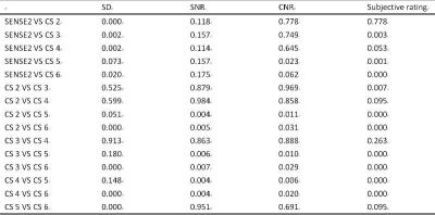 |
The effect of Compressed SENSE acceleration factor on 3D mDIXON sequence for liver imaging
Shiyu Wang1, Jiazheng Wang2, Qingwei Song1, Zhongping Zhang3, Bingbing Gao1, Lihua Chen1, and Ailian Liu4
1The First Affiliated Hospital of Dalian Medical University, Dalian, China, 2Philips Healthcare, Dalian, China, 3Philips Healthcare, Beijing, China, 4The First Affiliated Hospital of Dalian Medical University, Dlian, China
Image acquisition of an mDixon sequence typically requires a breath hold to minimize respiratory motion artifact. Minimization the acquisition time of mDixon could be very useful in clinical daily routine. The purpose of the study is to explore the effect of the acceleration factor on the image quality of 3D mDIXON sequence for liver imaging using Compressed SENSE . And CS acceleration factor 4 can reduce the scanning time to 10s on the premise of ensuring the image quality, which is the best scheme for clinical 3D mDixon scanning of liver.
|
|
2627. |
Quantitative assessment of the pancreas in subjects using DWI, IVIM, and 3D mDxion-quant: correlation with BMI, SAT, VAT and TAT
Qinhe Zhang1, Wan Dong1, Yishi Wang2, Jiazheng Wang2, and Ailian Liu1
1The first affiliated hospital of Dalian Medical University, Dalian, China, 2Philips Healthcare, Beijing, China
This study measured the quantitative imaging metrics of the pancreas and correlated ADC, sADC, D*,D, f, FF and R2* of pancreas with BMI, TAT, SAT and VAT. This study viewed that there was significant relationship between R2* and VAT, TAT (r=0.495, 0.533, respectively). And, ADC was related to SAT (r=0.460). These may illustrate the impact of different fat distribution on the function of pancreas.
|
|
2628.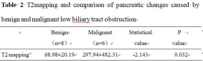 |
T2 mapping quantitative imaging to evaluate pancreatic changes caused by benign and malignant low biliary tract obstruction
Xuedong Wang1, Ailian Liu1, Jiazheng Wang2, Ke Jiang2, and Anliang Chen1
1The first affiliated hospital of dalian medical university, DaLian, China, 2Philips Healthcare, China, Beijing, China
T2 mapping is an effective sequence of MRI that enables quantitative assessment of diseases. However, currently there are few trials to confirm its feasibility in distinguishing pancreatic changes due to benign and malignant low biliary obstruction. In this experiment, we used T2 mapping to quantify these changes, and found that T2 mapping is feasible for evaluating the changes of pancreatic parenchyma due to benign and malignant low biliary obstruction. And the value of T2 mapping in malignant group is higher than that in benign group.
|
|
2629.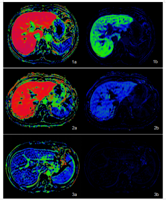 |
Quantitative Assessment of Liver Function by using Gd-EOB-DTPA-Enhanced MRI: Hepatocyte Fraction
Xueqin ZHANG1, Jian LU1, Jifeng JIANG1, and Weibo CHEN2
1the Third People’s Hospital of Nantong, Nantong, China, 2Philips Healthcare, Shanghai, China
The purpose of this study was to assess the ability of hepatocyte fraction on Gd-EOB-DTPA-enhanced MRI for the quantitative evaluation of liver function. 69 patients with cirrhosis and 22 healthy volunteers with normal liver function who underwent Gd-EOB-DTPA enhanced MRI were collected in this study. T1 mapping images were obtained by using the Look-Locker sequences before and 20 minutes after Gd-EOB-DTPA administration, hepatocyte fraction (HeF) and KHep were measured. Our study showed that hepatocyte fraction (HeF and KHep vlaues) based on Gd-EOB-DTPA- enhanced T1 mapping MRI is an efficient diagnostic tool for the quantitative evaluation of liver function.
|
|
2630.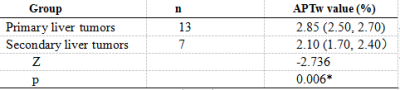 |
A preliminary study on the value of amide proton transfer-weighted imaging in differentiating primary from secondary malignant liver tumors
Tao Lin1, Ailian Liu 1, Jiazheng Wang 2, Nan Wang1, Lihua Chen 1, Dahua Cui1, Ying Zhao 1, Qingwei Song 1, Renwang Pu 1, Bingbing Gao1, and Yanling Wu2
1The First Affiliated Hospital of Dalian Medical University, Dalian, China, 2Philips Healthcare, Beijing, China
Our work aimed to explore the value of amide proton transfer-weighted (APTw) imaging in diagnosing primary and secondary malignant liver tumors. The result manifested that the novel imaging tool had a valuable utility in differentiating primary from secondary malignant liver tumors (AUC: 0.879; sensitivity: 76.9%; specificity: 100%).
|
|
2631.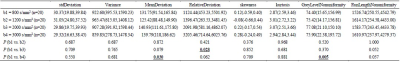 |
The value of texture analysis based on multiple high b-values DWI for evaluation of tumor heterogeneity in HCC patients
Ying Zhao1, Jiazheng Wang2, Zhiwei Shen2, Zhongping Zhang2, Nan Wang1, Lihua Chen1, Dahua Cui1, Tao Lin1, Qingwei Song1, Renwang Pu1, Bingbing Gao1, and Ailian Liu1
1The First Affiliated Hospital of Dalian Medical University, Dalian, China, 2Philips Healthcare, Beijing, China
Previous studies have reported preliminary but promising results that high b‐value DWI has a remarkably high sensitivity in neuro or genitourinary malignancies. However, the value of high b-value DWI in liver remains controversial. In the current study, multiple high b-values DWI - based texture analysis was applied to evaluate tumor heterogeneity in hepatocellular carcinoma (HCC) patients. The results demonstrated that ultra-high b-value (b = 2000 and 3000 s/mm2) DWI images could offer more information in tumor heterogeneity evaluation compared to the conventional b-value (b = 800 s/mm2) DWI images based on texture analysis in HCC patients.
|
|
2632.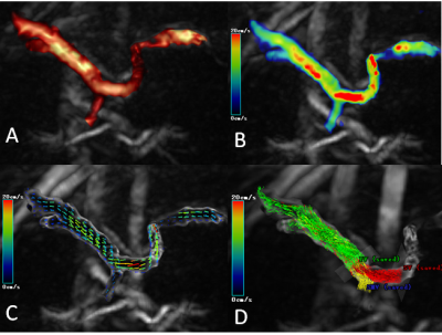 |
Evaluation of Portal System Flow in Response to a Meal Challenge with 4D-Flow MRI
Lihua Chen1, Ailian Liu2, Jiazheng Wang3, Yishi Wang3, Yaxin Niu2, and Qingwei Song2
1The First Affiliated Hospital of Dalian Medical University, DaLian, China, 2The First Affiliated Hospital of Dalian Medical University, Dalian, China, 3Philips Healthcare, Beijing, China
The quantitative flow measurements at 4D - flow MRI in the portal system and flow change in response to a meal challenge. Significant increased blood flow velocity and volume were observed in PV and SMV and significantly decreased blood flow was observed in SV after a meal.
|
|
2633.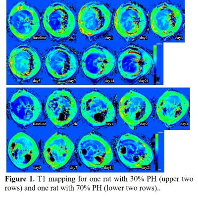 |
Assessing Liver Changes after 30% and 70% Partial Hepatectomy Using T1 and T2 mapping
Qing Li1, Shuangshuang Xie1, Caixin Qiu1, Jinxia Zhu2, Chengwen Liu2, and Wen Shen1
1Tianjin First Central Hospital, Tianjin, China, 2MR Collaboration, Siemens Healthcare, Ltd, Beijing, China
T1 and T2 mapping of rats with 30% and 70% partial hepatectomy (PH) were compared, and trends were similar with some differences, indicating liver changes after PH in the 70% PH group was more active. The results suggested that T1 and T2 mapping are potential to assess liver regeneration after PH.
|
|
2634.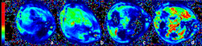 |
T2* Relaxation Time as an Imaging Biomarker for Monitoring Liver Arterial Blood Supply After Partial Hepatectomy
shuangshuang xie1, caixin qiu1, yajie sun1, qing li1, jinxia zhu2, and wen shen1
1Tianjin First Center Hospital, Tianjin, China, 2MR Collaboration, Siemens Healthcare, Beijing, China Poster Permission Withheld
The value of effective T2 (T2*) relaxation time in monitoring changes in liver arterial blood supply after partial hepatectomy was investigated using a rat model. Ten rats received a 30% hepatectomy, and the other ten received a 70% hepatectomy. Before and after the surgery, all animals underwent T2* mapping at multiple time points. Liver T2* relaxation times were measured and compared between different groups at each time point. The results indicated that T2* relaxation time can be used to monitor changes in liver arterial blood supply after partial hepatectomy.
|
| 2635. | Consensus-based technical recommendations for clinical translation of renal MRI
Fabio Nery1, Iosif Mendichovszky2, Pim Pullens3, Ilona Dekkers4, Octavia Bane5, Andreas Pohlmann6, Anneloes de Boer7, Alexandra Ljimani8, Aghogho Odudu9, Charlotte Buchanan10, Kanishka Sharma11, Christoffer Laustsen12, Anita Harteveld7,
Xavier Golay13, Ivan Pedrosa14, David Alsop15, Sean Fain16, Anna Caroli17, Pottumarthi Prasad18, Susan Francis10, Eric Sigmund19, Maria Fernández‐Seara20, and Steven Sourbron21
1Developmental Imaging and Biophysics Section, UCL Great Ormond Street Institute of Child Health, London, UK, London, United Kingdom, 2Department of Radiology, Addenbrooke’s Hospital, Cambridge University Hospitals NHS Foundation Trust, Cambridge, United Kingdom, 3Department of Radiology, Ghent University Hospital and Ghent Institute for Functional and Metabolic Imaging, Ghent University, Ghent, Belgium, 4Department of Radiology, Leiden University Medical Center, Leiden, Netherlands, 5Translational and Molecular Imaging Institute and Department of Radiology, Icahn School of Medicine at Mount Sinai, New York, NY, United States, 6Berlin Ultrahigh Field Facility, Max Delbrueck Center for Molecular Medicine in the Helmholtz Association, Berlin, Germany, 7Department of Radiology, University Medical Center Utrecht, Utrecht University, Utrecht, Netherlands, 8Department of Diagnostic and Interventional Radiology, Medical Faculty, University Dusseldorf, 40225, Dusseldorf, Germany, 9Division of Cardiovascular Sciences, School of Medical Sciences, Faculty of Biology, Medicine and Health, University of Manchester, Manchester, United Kingdom, 10Sir Peter Mansfield Imaging Centre, University of Nottingham, University Park, Nottingham, United Kingdom, 11Imaging Biomarkers Group, Department of Biomedical, Imaging Sciences, University of Leeds, Leeds, United Kingdom, 12Department of Clinical Medicine, MR Research Centre, Aarhus University, Aarhus, Denmark, 13Brain Repair and Rehabilitation, Institute of Neurology, University College London, London, United Kingdom, 14Department of Radiology, Advanced Imaging Research Center, University of Texas Southwestern Medical Center, Dallas, TX, United States, 15Department of Radiology, Beth Israel Deaconess Medical Center, Harvard Medical School, Boston, MA, United States, 16Departments of Biomedical Engineering, Radiology, and Medical Physics, University of Wisconsin, Madison, WI, United States, 17Department of Biomedical Engineering, Istituto di Ricerche Farmacologiche Mario Negri IRCCS, Bergamo, Italy, 18Department of Radiology, Center for Advanced MR Research, NorthShore University Health System, Evanston, IL, United States, 19Department of Radiology, Center for Biomedical Imaging (CBI), Center for Advanced Imaging Innovation and Research (CAI2R), NYU Langone Health, New York, NY, United States, 20Department of Radiology, Clínica Universidad de Navarra, Pamplona, Spain, 21Department of Infection, Immunity and Cardiovascular Disease, University of Sheffield, Sheffield, United Kingdom Poster Permission Withheld
The field of renal MRI has undergone significant developments over the last decade. However, the lack of standardisation of acquisition and analysis methods remains an important barrier to clinical translation. We present a consensus project initiated through the Cooperation in Science and Technology (COST) Action PARENCHIMA whereby a group of renal MRI experts employed a Delphi-based approach to develop technical recommendations for renal T1 and T2 mapping, arterial spin labelling, diffusion-weighted imaging and blood oxygenation level-dependent MRI. These will promote standardisation of renal MRI protocols and thus facilitate clinical translation and comparison of data across sites.
|
|
2636.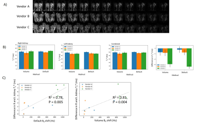 |
Travelling kidneys: Multicentre multivendor variability of renal BOLD and T1 mapping – preliminary results
Charlotte E Buchanan1, Fabio Nery2, Andrew Priest3, Eleanor F Cox1, Joao Sousa4, Michael Nation5, Iosif Mendichovszky3, Steven Sourbron6, David Thomas2,7, and Susan T Francis1
1Sir Peter Mansfield Imaging Centre, University of Nottingham, Nottingham, United Kingdom, 2Developmental Imaging and Biophysics Section, University College London, London, United Kingdom, 3Cambridge University Hospitals NHS Foundation Trust, Cambridge, United Kingdom, 4Imaging Biomarkers Group, Department of Biomedical Imaging Sciences, University of Leeds, Leeds, United Kingdom, 5Kidney Research UK, Peterborough, United Kingdom, 6Department of Infection, Immunity and Cardiovascular Disease, University of Sheffield, Sheffield, United Kingdom, 7Queen Square Institute of Neurology, University College London, London, United Kingdom
We have performed an intra-individual, inter-vendor comparison of renal T1 and BOLD T2* measures using predominantly product sequences. Renal T2* is sensitive to dephasing due to background magnetic field gradients, the degree of which differs across the MR vendors. A strong B0 L-R gradient, especially prominent for one vendor, leads to an asymmetry in T2* values between kidneys. B1 maps vary across subjects with no vendor-specific bias. Renal cortex T1 values computed using a 2-parameter fit were shown to systematically vary across vendors; a 3-parameter fit reduced this bias but differences remain. This highlights areas of future development for standardisation.
|
|
2637.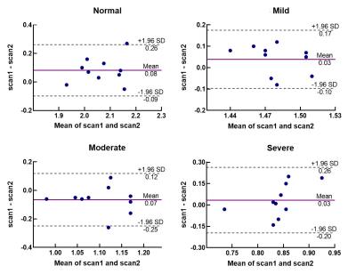 |
The reproducibility of DWI and BOLD in Early Diagnosis of Acute Kidney Injury in Animal Models
Hanjing Kong1, Weizheng Gao1, Fei Gao1, Chengyan Wang2, Xiaodong Zhang1, Min Yang1, Xiaoying Wang3, and Jue Zhang1
1Peking University, Beijing, China, 2Fudan University, Shanghai, China, 3Peking University First Hospital, Beijing, China
Early diagnosis of acute kidney injury (AKI) has clinical importance. Blood oxygen level-dependent (BOLD) imaging and diffusion-weighted imaging (DWI) showed its priority in diagnosis of AKI in the early phase. However, the reproducibility of these methods remains unknown. In this study, we present repeated measurements of the DWI and BOLD renal MR imaging and further compared the quantitative results in terms of reproducibility.
|
|
2638.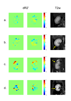 |
A reproducibility study on the changes of R2’ in quantitative evaluation of acute renal injury
Weizheng Gao1, Hanjing Kong1, Chengyan Wang2, Xiaodong Zhang1, Min Yang1, Xiaoying Wang3,4, and Jue Zhang1
1Peking University, Beijing, China, 2Fudan University, Shanghai, China, 3Department of Radiology, Peking University First Hospital, Beijing, China, 4Academy for Advanced Interdisciplinary Studies, Peking University, Beijing, China
Recent studies have demonstrated that a novel MRI imaging sequence termed psMASE, together with hemodynamic response imaging, is able to effectively distinguish different severities of acute renal injury. In this study, we performed repeated scanning to verify the repeatability of the psMASE strategy.
|
|
2639.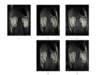 |
Improved Diffusion-Weighted Imaging of Rat Kidney Using Interleaved Multishot EPI with 2D Navigators
Qiang Liu1, Zhongbiao Xu2, Xinyuan Zhang1, Zhifeng Chen1, Rongli Zhang3, Yaohui Wang4, Kaixuan Zhao1, Ed X. Wu5,6, and Yanqiu Feng1
1School of Biomedical Engineering, Guangdong Provincial Key Laboratory of Medical Image Processing, Southern Medical University, Guangzhou, China, 2Department of Radiotherapy, Cancer Center, Guangdong Provincial People's Hospital & Guangdong Academy of Medical Science, Guangzhou, China, 3School of Medicine, Guangdong Provincial People's Hospital , South China University of Technology, Guangzhou, China, 4Institute of Electrical Engineering, Chinese Academy of Sciences, Beijing, China, 5Laboratory of Biomedical Imaging and Signal Processing, The University of Hong Kong, Hong Kong SAR, China, 6Department of Electrical and Electronic Engineering, The University of Hong Kong, Hong Kong SAR, China
This work aimed to improve rat kidney diffusion-weighted imaging (DWI) by using interleaved multishot echo planar imaging (EPI) with 2D navigators. The data of shots were combined to reconstruct the final images by using the IRIS processing pipeline. The in vivo rat imaging results show that the multishot EPI approach obtained renal diffusion-weighted images with higher resolution and less geometric distortion, compared single-shot EPI. The DWI of rat kidney can be significantly improved by 2D navigated multishot EPI.
|
|
2640. |
Second-order Motion Compensated Intravoxel Incoherent Motion Analysis of the Kidney
Yuki Koshino1, Naoki Ohno2, Tosiaki Miyati2, Tetsuo Ogino3, Yu Ueda3, Naoki Hori1, Yukihiro Matsuura1, Toshifumi Gabata4, and Satoshi Kobayashi1,2,4
1Radiology Division, Kanazawa University Hospital, Ishikawa, Japan, 2Faculty of Health Sciences, Institute of Medical, Pharmaceutical and Health Sciences, Kanazawa University, Ishikawa, Japan, 3Philips Japan, Tokyo, Japan, 4Department of Radiology, Kanazawa University Graduate School of Medical Sciences, Ishikawa, Japan Poster Permission Withheld
To investigate the potential benefit of second-order motion compensated (2nd-MC) diffusion encoding scheme on intravoxel incoherent motion (IVIM) analysis of the kidney, we compared the IVIM diffusion parameters and repeatability between the 2nd-MC and conventional diffusion gradients. Our results showed that the 2nd-MC diffusion gradients show better fitting accuracy and repeatability of IVIM diffusion parameters of the kidney compared with the conventional diffusion gradients. Therefore, the 2nd-MC diffusion encoding scheme could enable us to reduce the bulk motion effect mainly caused by cardiac and respiratory motion during diffusion sensitization.
|
|
2641.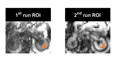 |
Reliability of Water-Lipid Separation in the Kidney Measured by Accelerated In Vivo Magnetic Resonance Spectroscopic Imaging at 3 T
Ahmad Alhulail1,2, Mahsa Servati1,3, Nathan Ooms1,3, Oguz Akin4, Alp Dincer5,6, M. Albert Thomas7, Ulrike Dydak1,3, and Uzay E Emir1,8
1School of Health Sciences, Purdue University, West Lafayette, IN, United States, 2Department of Radiology and Medical Imaging, Prince Sattam bin Abdulaziz University, Al Kharj, Saudi Arabia, 3Department of Radiology and Imaging Sciences, Indiana University School of Medicine, Indianapolis, IN, United States, 4Department of Radiology, Memorial Sloan Kettering Cancer Center, New York, NY, United States, 5Department of Radiology, School of Medicine, Acıbadem Mehmet Ali Aydinlar University, Istanbul, Turkey, 6Center for Neuroradiological Applications and Research, Acıbadem Mehmet Ali Aydinlar University, Istanbul, Turkey, 7Department of Radiology, University of California Los Angeles, Los Angeles, Los Angeles, CA, United States, 8Weldon School of Biomedical Engineering, Purdue University, West Lafayette, IN, United States
MRS could be useful for characterization of the renal cell carcinoma by detecting lipid content. However, single-voxel MRS cannot show the distribution of lipids for heterogeneous tumors. We propose a fast MRSI sequence to map the distribution of lipid metabolites within a clinically acceptable time frame at 3T. The proposed MRSI technique provided high-quality data with reproducible quantitative outputs. The coefficient of variance for the quantified fat fraction was less than 1%.
|
|
2642.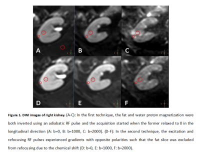 |
Fat suppression efficiency in kidney diffusion imaging with different techniques
Jingjun Wu1, Ailian Liu1, Jiazheng Wang2, Zhongping Zhang2, Dahua Cui1, Lihua Chen1, Qingwei Song1, Renwang Pu1, and Bingbing Gao1
1Department of Radiology, the First Affiliated Hospital of Dalian Medical University, Dalian, China, 2Philips Healthcare, Beijing, China
The reverse gradient technique showed equal fat suppression efficiency and higher SNR and CNR when compared to the RF inversion method in liver DWI, while the measured ADC values were consistent among the two. Chemical shift artifacts remain problematic in abdominal diffusion imaging due to the challenges in B0 field shimming, and the adoption of the optimal fat suppression technique for different application scenarios is important to balance between fat suppression, SNR, and ADC accuracy.
|
|
2643.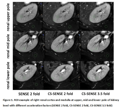 |
Feasibility of highly accelerated high resolution two-dimensional T2-weighted MRIusing compressed sensing and parallel imaging in healthy kidney
Jingjun Wu1, Ailian Liu1, Jiazheng Wang2, Xiaofang Xu2, Dahua Cui1, Lihua Chen1, Qingwei Song1, Renwang Pu1, and Bingbing Gao1
1Department of Radiology, the First Affiliated Hospital of Dalian Medical University, Dalian, China, 2Philips Healthcare, Beijing, China
When compared to SENSE 2, a clinical routine kidney T2WI protocol and hence the golden standard in this study, CS-SENSE with a 75% higher acceleration factor achieved comparable image quality, both qualitatively and quantitatively, while approximately halved the imaging time. CS-SENSE with acceleration factor 3.5 has the potential to replace the 2 fold SENSE acceleration as a clinical routine to increase patient comfort and throughput, which is especially beneficial for ultra-high resolution acquisitions which normally demands extended imaging time.
|
|
2644.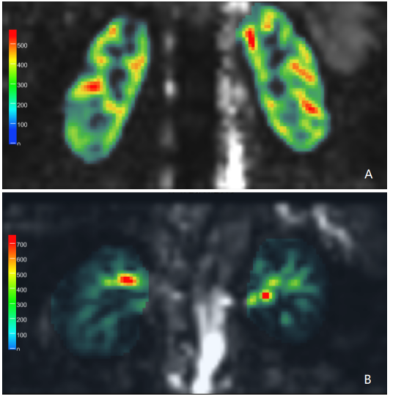 |
Application of the Free-Breathing 3D Turbo Gradient Spin Echo pCASL Sequence to Assess Renal Perfusion in Patients with Diabetic Nephropathy
Xiao-dong Zheng1, Shi-hong Li1, Guang-wu Lin1, Cai-xia Fu2, and Bernd Kühn3
1Department of Radiology, Hua Dong Hospital, Fudan University, Shanghai, China, 2MR Application Development, Siemens Shenzhen Magnetic Resonance Ltd., Shenzhen, China, 3MR Application Predevelopment, Siemens Healthcare, Erlangen, Germany
In this study, we aimed to compare the renal blood flow (RBF) values obtained with the 3D Turbo Gradient Spin Echo (TGSE) pCASL sequence with glomerular filtration rates (GFRs) measured using single-photon emission computed tomography (SPECT) for nuclide nephro-dynamic imaging to assess renal perfusion in patients with diabetic nephropathy. Our study demonstrated that the RBF values were well correlated with GFR, indicating that the 3D-TGSE pCASL sequence could be used as an alternative - method for the prediction of early diabetic nephropathy.
|
|
2645. |
Explore the performance of compressed-SENSE accelerated 3D mDIXON on kidney of healthy subjects
Lijuan Sun1, Qingwei Song1, Meiyu Sun1, Dianxiu Ning1, Nan Zhang1, Nan Wang1, Jiazheng Wang2, and Liangjie Lin2
1The First Affiliated Hospital of Dalian Medical University, Dalian, China, 2Philips Healthcare, Beijing, China
The compressed sensing (CS) technique, which implements k-space sparses ampling and iterative reconstruction algorithm, has now been widely used in acceleration of MR examinations of various anatomies and anatomical contrasts. This study aims to explore the performance of 3D water-fact imaging accelerated using a combination of compressed-sensing and sensitivity encoding on kidney of healthy subjects. Several acceleration factors of the proposed technique were compared against the traditional sensitivity-encoding technique and an acceleration factor of 2 was recommended for a balance between imaging time and image quality.
|
|
2646.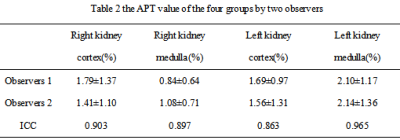 |
3D amide proton transfer MR imaging of kidney: a feasibility study
Nan Wang1, Ailian Liu1, Jiazheng Wang2, Zhiwei Shen2, Qingwei Song1, Renwang Pu1, Bingbing Gao1, and Lihua Chen1
1The First Affiliated Hospital of Dalian Medical University, Dalian, China, 2Philips Healthcare, Beijing, China
MRI imaging is a radiographic imaging technology widely used in human body. The application of MRI at the molecular level had became a hot research topic in the near future. 3D amide proton transfer weighted(APTw) MR imaging is a new contrast-agent-free MRI technique that addresses the need for a more confident diagnosis in oncology[1,2]. The APTw was widely used in the nervous system. Recently, The APTw was gradually applied to the abdomen[3,4]. At present, there is no related research reported on renal APT imaging.
|
|
2647.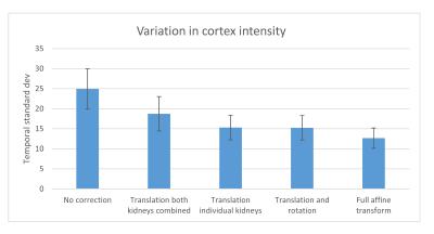 |
Retrospective affine motion correction of arterial spin labeling kidney perfusion measurements
Steffen Ringgaard1 and Anne Dorte Blankholm2
1MR Research Centre, Aarhus University, Aarhus, Denmark, 2Department of Radiology, Aarhus University Hospital, Aarhus, Denmark
When doing arterial spin labeling perfusion measurements in the kidneys, respiratory motion is an important issue. We compared different methods for doing retrospective motion correction of single-shot ASL images. We evaluated the temporal signal variation in the cortex after correction by translation, translation+rotation and affine transformation and found that the variation was lowest when using full affine correction. Besides, we also compared different similarity measures as used during the motion correction.
|
|
2648.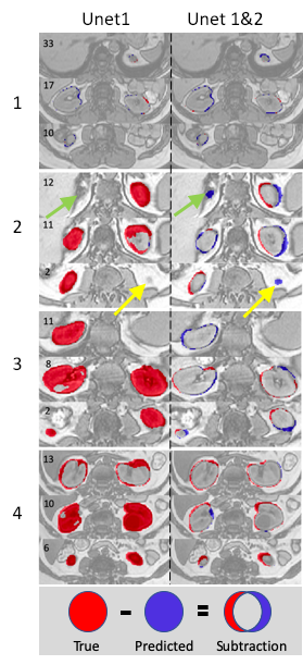 |
Kidney segmentation using dual neural network and one-shot deep learning
Junyu Guo1 and Ivan Pedrosa1
1Radiology, UT southwestern medical center, Dallas, TX, United States
Image segmentation is important for quantitative analysis in many medical applications. Although deep learning methods generate segmentation with high accuracy, the model has to be trained for different organs and image modalities. These training processes are very time-consuming and labor-intensive because of the requirements of drawing masks manually. In this project, we present a dual neural network method trained using one set of 3D MRI data from a single subject. We demonstrate the feasibility of using a single 3D dataset to train a dual neural network for kidney segmentation, which greatly reduce the burden of drawing masks for large datasets.
|
|
2649.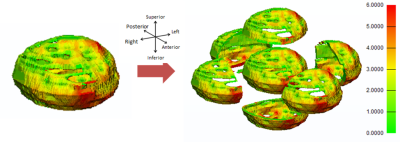 |
Segmentation and 3D Anatomical Renderings of Pelvic MRI Allow Quantitative Analysis of Lower Urinary Tract Anatomy
Lucille E Anzia1, Cody J Johnson1, Diego Hernando1, Wade A Bushman2, Shane A Wells1, and Alejandro Roldán-Alzate3
1Radiology, University of Wisconsin Madison, Madison, WI, United States, 2Urology, University of Wisconsin Madison, Madison, WI, United States, 3Mechanical Engineering, University of Wisconsin Madison, Madison, WI, United States
Previous anatomic studies of the lower urinary tract predominantly utilize ultrasound and computed tomography. We present a novel method for measuring bladder wall volume (BWV), bladder wall thickness (BWT) and prostate volume (PV) using 30 (14 male) patients aged 30-39. Through MRI segmentation of the bladder and prostate in Mimics software, 3D models were quantified for BWV and PV. 3-matic was used to measure BWT. There were no significant differences in BWV or BWT between males and females (p=0.52,0.25). Mean PV was 28.7cm3 (16.7-47.8cm3). Non-significant variations in BWT were observed regionally. Future directions will use this methodology across age groups.
|