
Digital Poster Session
Musculoskeletal: Bone, Cartilage, and Muscle
Musculoskeletal
2696 -2710 Bone, Cartilage, and Muscle - Muscle 1
2711 -2725 Bone, Cartilage, and Muscle - Muscle 2
2726 -2741 Bone, Cartilage, and Muscle - Cartilage 1
2742 -2757 Bone, Cartilage, and Muscle - Cartilage 2
2758 -2772 Bone, Cartilage, and Muscle - Bone 1
2773 -2785 Bone, Cartilage, and Muscle - Bone 2
2696.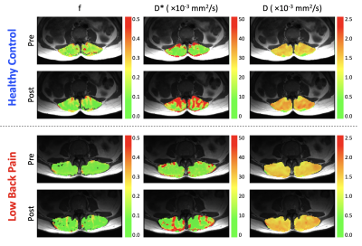 |
IVIM MRI evaluation of blood flow response to exercise in patients with low back pain compared to healthy, pain-free controls
Erin K Englund1, David B Berry2, John J Behun1, Lawrence R Frank3, Samuel R Ward1,3,4, and Bahar Shahidi1
1Orthopaedic Surgery, University of California San Diego, La Jolla, CA, United States, 2Nanoengineering, University of California San Diego, La Jolla, CA, United States, 3Radiology, University of California San Diego, La Jolla, CA, United States, 4Bioengineering, University of California San Diego, La Jolla, CA, United States
IVIM MRI was used to evaluate changes in muscle blood flow before and after lumbar extension exercise in patients with low back pain and healthy controls. Results showed that IVIM was sensitive to blood flow changes from exercise in both groups. Pain free controls had a higher mean diffusion coefficient (D) and the change in D from exercise was also greater in this cohort. A subset of patients with low back pain who had a limited response to exercise per the pseudo-diffusion coefficient (D*) were also found to have no improvement in disability following a 12-week physical therapy regimen.
|
|
2697.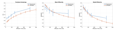 |
Comparison of simulated and experimental stimulated echo diffusion at varying diffusion times in skeletal muscle
Erin K Englund1, David B Berry2, Vitaly Galinsky3, Lawrence R Frank3, and Samuel R Ward1,3,4
1Orthopaedic Surgery, University of California San Diego, La Jolla, CA, United States, 2Nanoengineering, University of California San Diego, La Jolla, CA, United States, 3Radiology, University of California San Diego, La Jolla, CA, United States, 4Bioengineering, University of California San Diego, La Jolla, CA, United States
Diffusion tensor imaging provides insight into the underlying tissue microstructure. Here, we compare simulated DTI data to experimentally acquired data in skeletal muscle using stimulated echo DTI at multiple diffusion encoding times. After adjusting for the difference in simulated versus measured apparent diffusion coefficient, there was relative agreement of mean diffusivity and radial diffusivity between the measured and simulated data. Fractional anisotropy was overestimated experimentally, likely due to limited image SNR.
|
|
2698.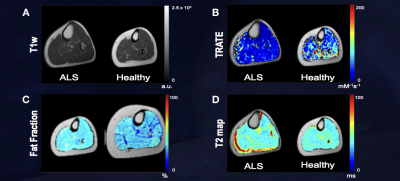 |
Evaluating Longitudinal Muscle Degeneration in ALS Patients using MR Cytography
Sudarshan Ragunathan1, Laura C Bell1, Ashley M Stokes1, Natenael Semmineh1, Jessie Duncan2, Nicole Turcotte2, Shafeeq Ladha2, and Chad C Quarles1
1Neuroimaging Research, Barrow Neurological Institute, Phoenix, AZ, United States, 2Gregory W. Fulton ALS and Neuromuscular Center, Barrow Neurological Institute, Phoenix, AZ, United States
Amyotrophic lateral sclerosis (ALS) is a neurodegenerative disease characterized by loss of upper and lower motor neurons (UMN and LMN), resulting in muscle atrophy and eventual death. Clinical indicators such as ALSFRS-R scores can be confounded by inter-rater variability and low sensitivity. MR Cytography based TRATE has been shown to identify changes to myofiber microstructure. This work aims to demonstrate that TRATE is a more sensitive and consistent metric to evaluate longitudinal muscle degeneration in ALS patients than existing clinical indicators such as ALSFRS-R scores.
|
|
2699.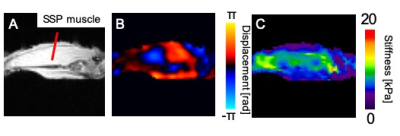 |
Stiffness change of the rotator cuff muscle before and after the tendon tear with magnetic resonance elastography and ultrasound elastography.
Akihisa Koga1, Yoshiaki Itoigawa1, Mikio Suga2, Yuri Suganuma3, Tomoki Wada1, Daichi Morikawa1, Yuichiro Maruyama1, and Kazuo Kaneko4
1Orthopaedic surgery, Juntendo Urayasu Hospital, Chiba, Japan, 2Center for Frontier Medical Engineering, Chiba University, Chiba, Japan, 3Graduate School of Science and Engineering, Chiba University, Chiba, Japan, 4Orthopaedic surgery, Juntendo University, Tokyo, Japan
The purpose of this study was to explore the feasibility of Magnetic Resonance Elastography (MRE) measurement for quantification of the stiffness change of the shoulder rotator cuff muscle compared with Shear Wave ultrasound Elastography (SWE). Six porcine shoulders were used in this study. MRE and SWE measurement of the rotator cuff muscle was performed before and after the rotator cuff tendon detachment. Stiffness values were significantly lower after the tendon detachment in both MRE and SWE measurements (p<0.05). This result suggests that MRE could be a feasible method for quantification of the rotator cuff muscle stiffness as well as SWE.
|
|
2700.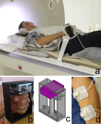 |
Compressed sensing three-directional 3D velocity imaging of the skeletal muscle during evoked isometric contraction of the arm
Xeni Deligianni1,2, Francesco Santini1,2, Anna Hirschmann3, Ning Jin4, Nicolas Place5, Oliver Bieri1,2, and Claudia Weidensteiner1,2
1Radiology/Division of Radiological Physics, University Hospital of Basel, Basel, Switzerland, 2Biomedical Engineering, University of Basel, Basel, Switzerland, 3Radiology, University Hospital of Basel, Basel, Switzerland, 4Siemens Medical Solutions, Cleveland, Cleveland, OH, United States, 5Institute of Sport Sciences, University of Lausanne, Lausanne, Switzerland
MRI of the upper extremities is important for the follow-up of muscle dystrophies. The goal of this study was to investigate the feasibility of 4D-velocity imaging during neuromuscular electrical stimulation(NMES) of the arm, standardized with the force output. Two healthy volunteers were scanned at 3T during isometric contraction of the biceps brachii for different elbow angles. While the force output was different in intensity, the waveforms of force and strain were similar. In conclusion, it was shown that it is possible to acquire 3D-dynamic velocity data synchronized with NMES of the arm and simultaneously record the evoked force.
|
|
2701.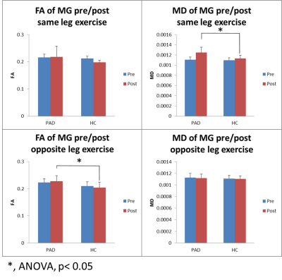 |
Diffusion tensor imaging of calf muscle damage in peripheral arterial disease
Jianli Wang1, Jonathan Stavres2, Christopher T Sica1, Cheryl Blaha2, Michael Herr2, Samuel Pai2, Aimee Cauffman2, Jeffrey Vesek3, Qing X Yang1,4, and Lawrence I Sinoway2
1Radiology, Penn State College of Medicine, Hershey, PA, United States, 2Heart & Vascular Institute, Penn State College of Medicine, Hershey, PA, United States, 3Molecular Biology, Penn State College of Medicine, Hershey, PA, United States, 4Neurosurgery, Penn State College of Medicine, Hershey, PA, United States
Peripheral arterial disease (PAD) is a systemic atherosclerotic vascular disease characterized by impaired skeletal muscle perfusion and mitochondrial respiration. Over time, persistent exposure to muscle ischemia eventually leads to muscle atrophy, myopathy, and mitochondrial dysfunction in PAD patients. In this study we investigated the utilization of diffusion tensor imaging to characterize the pathophysiological changes of calf muscles before and after graded plantar flexion exercise. At rest, the clinically worse leg of the PAD subjects had higher diffusivity in the calf muscles. Significant greater increases of diffusivity in the post-exercise muscle of PADs than the HCs suggest acute exercise-related muscle damage.
|
|
2702. |
Comparison of measurements of intramuscular fat from T1-weighted and mDixon MRI scans in people with and without spinal cord injury
Bart Bolsterlee1,2, Elizabeth A Bye3,4, Junya Eguchi1,5, Joanne Glinsky3, Jeanette Thom5, and Robert D Herbert1,5
1Neuroscience Research Australia, Randwick, Australia, 2Graduate School of Biomedical Engineering, University of New South Wales, Randwick, Australia, 3John Walsh Centre for Rehabilitation Research, Sydney University, Sydney, Australia, 4Spinal Injury Unit, Prince of Wales Hospital, Randwick, Australia, 5School of Medical Sciences, University of New South Wales, Randwick, Australia
Accurate quantification of fat content of human muscles could help assess disease status and test effectiveness of interventions in people with neurological conditions, whose muscles are frequently infiltrated with fat. We compared two commonly used MRI methods based on T1-weighted and mDixon scans to quantify intramuscular fat in 112 muscles from people with and without spinal cord injury. Fat fraction measurements agreed well in muscles with high proportions of fat, but the T1-weighted method could not be used in muscles with small proportions of fat. We recommend against the use of T1-weighted methods to quantify intramuscular fat.
|
|
 |
2703.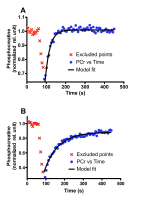 |
The Molecular Basis for 31P MRS-Based Phosphocreatine Muscle Resynthesis Rate Measurements in Healthy Adults
Maninder Singh1, Moses M Darpolor2, Jeffrey Covington1, Sebastian Hanet1, Eric Ravussin1, and Owen T Carmichael1
1Pennington Biomedical Research Center, Baton Rouge, LA, United States, 2Stillman College, Tuscaloosa, AL, United States
Dynamic phosphorus magnetic resonance spectroscopy (31P-MRS) is a method used for in vivo studies of skeletal muscle function including measurements of phosphocreatine (PCr) synthesis rate during recovery from submaximal exercise. However, the molecular events underlying the PCr resynthesis rate are still under debate. Therefore, our goal was to assess the PCr resynthesis rate from 31P-MRS spectra collected from the skeletal muscle (vastus lateralis) of healthy adults and investigate associations between PCr resynthesis and levels of mitochondria-related transcripts and proteins in the same muscle (NAMPT, NQO1, PGC-1α, and SIRT1).
|
2704.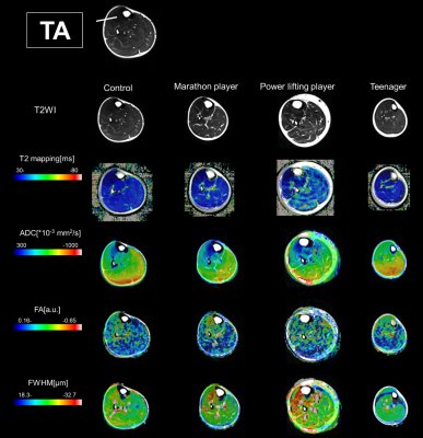 |
The visualization technique of the distribution of muscle quality by sport characteristics / changes due to growth using q-space imaging
Daisuke Nakashima1, Junichi Hata2, Yasushi Sera1, Hirotaka James Okano2, Kazuki Sato1, Takeo Nagura1, Hideyuki Okano1, Morio Matsumoto1, and Masaya Nakamura1
1Keio University School of Medicine, Tokyo, Japan, 2Jikei University School of Medicine, Tokyo, Japan
We evaluated the muscle quality of lower limbs among four groups: controls, marathon players, powerlifters, and teenagers using q-space imaging. Initially, we confirmed that fast muscle fiber has a larger cell size than slow muscle fiber in cadaveric immunohistology study of the tibialis anterior muscle (TA). In addition, there were many fast muscle fiber in the TA. In q-space imaging study, the cell diameter increased in the process of growing up from teenage to adulthood, which indicates an increase in fast muscle cells according to growth. Furthermore, the powerlifting players had more enlarged fast muscle cells than other groups.
|
|
2705. |
31P MRS assessments of mitochondrial dysfunction in patients with peripheral arterial disease undergoing revascularization.
Jabrane Karkouri1,2,3, Jill Slade4, Helene Ratiney1, Sylvain Grange1, Anne Tonson4, Pierre Croisille1, and Magalie Viallon1
1Université de Lyon, INSA-Lyon, Université Claude Bernard Lyon 1, UJM-Saint Etienne, CNRS, Inserm, CREATIS UMR 5220, U1206, Lyon, France, Lyon, France, 2SIemens Healthcare SAS, Saint-Denis, France, 3Wolfson Brain Imaging Center, University of Cambridge, Cambridge, United Kingdom, 4Radiology, Michigan state university, East Lansing, MI, United States
Obliterative arterial disease of the lower limbs is a disease that obstructs lower extremities arteries, resulting in reduced lower limb perfusion and possibly mitochondrial dysfunction. Mitochondrial function of the calf assessed via 31P MRS at moderate and low exercise intensities before and after revascularization and phase contrast angiography of the posterior tibial artery enabled the assessment of vascular and mitochondrial contributions of the patients.
|
|
2706.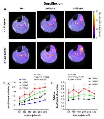 |
Detecting motor unit activity during volitional muscle contraction – Motor Unit MRI
Linda Heskamp1, Matthew G. Birkbeck1,2,3, Ian S. Schofield4, Roger G. Whittaker4, and Andrew M. Blamire1
1Newcastle Magnetic Resonance Center, Institute of Cellular Medicine, Newcastle University, Newcastle upon Tyne, United Kingdom, 2Northern Medical Physics and Clinical Engineering, Freeman Hospital, Newcastle upon Tyne NHS Foundation Trust, Newcastle upon Tyne, United Kingdom, 3Newcastle Biomedical Research Centre (BRC) NIHR, Newcastle University, Newcastle upon Tyne, United Kingdom, 4Institute of Neuroscience, Newcastle University, Newcastle upon Tyne, United Kingdom
Motor units (MUs) play a fundamental role in muscle physiology and disease. They can be imaged using a diffusion weighted imaging technique, motor unit MRI (MUMRI). Previous work activated MUs using electrical stimulation, limiting MUMRI to muscles innervated from superficial nerves. Here, we demonstrated the feasibility of MUMRI during volitional muscle activation. We confirmed that the MU activity detected with MUMRI during muscle contraction increased with b-value, force level and was dependent on the diffusion-sensitisation direction. This work allows us to image MUs in situations reflecting real muscle physiology making it a promising tool for studying motor neuron diseases.
|
|
2707. |
Variability of Diffusion Tensor MRI, 2-Point Dixon fat fraction and T2 relaxation in ROIs of thigh muscle in rheumatoid arthritis patients
Dominic Bertham1,2, Matthew Farrow3, Ai Lyn Tan3, Steven Tanner1, Paul Emery3, and John Biglands1
1Leeds Biomedical Research Centre, Leeds, United Kingdom, 2Department of Health Sciences, University of York, York, United Kingdom, 3Leeds institute of Rheumatic and Musculoskeletal Medicine, University of Leeds, Leeds, United Kingdom
Muscular weakness is associated with fatty infiltration and changes to muscle fibre structure. Quantitative magnetic resonance imaging (MRI) may be able to detect subtle muscle changes in rheumatoid arthritis (RA) patients. Before this method is used, variability of MRI measurements involving regions of interest (ROI) must first be measured. Fat fraction, T2 measurement and diffusion tensor imaging was performed on 19 participants with RA. Intra-rater and inter-rater variability of MRI measurements for thigh muscle ROIs were assessed. Inter-rater and intra-rater variability scores were high, suggesting that these measurements are sufficiently precise to allow the study of subtle changes to muscle.
|
|
2708.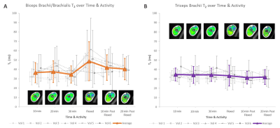 |
Detecting Muscle Activity using a 0.5T Upright Open MRI – A Pilot Study Using a Double Echo Steady State Sequence to Measure T2 in Biceps and Triceps
Noor Shaikh1,2,3, Andrew Yung4, Honglin Zhang5, John Street2,6, Cornelia Laule2,7,8,9, Thomas Oxland2,3,6, and David Wilson2,5,6
1Biomedical Engineering, University of British Columbia, Vancouver, BC, Canada, 2ICORD, University of British Columbia, Vancouver, BC, Canada, 3Mechanical Engineering, University of British Columbia, Vancouver, BC, Canada, 4UBC MRI Research Center, University of British Columbia, Vancouver, BC, Canada, 5Centre for Hip Health and Mobility, University of British Columbia, Vancouver, BC, Canada, 6Orthopaedics, University of British Columbia, Vancouver, BC, Canada, 7Radiology, University of British Columbia, Vancouver, BC, Canada, 8Physics & Astronomy, University of British Columbia, Vancouver, BC, Canada, 9Pathology & Laboratory Medicine, University of British Columbia, Vancouver, BC, Canada
We used a 0.5T upright open MRI to investigate T2 as a marker for muscle activity. Six volunteers’ arms were imaged with a Double Echo Steady State (DESS) sequence while relaxing (30min), following weighted bicep curls, and again after relaxing (20min). Directly after bicep curls T2 increased (average 41%) in the biceps but not the triceps, then subsequently decreased (average 14%) with relaxing. Intra-rater repeatability was promising. T2 standard deviation was relatively high, which is likely due to tissue heterogeneity. This preliminary study supports the potential of using DESS in upright open MRI to assess muscle injury or dysfunction.
|
|
2709. |
Assessment of peripheral muscle deconditioning using 31P-MRS during high intensity ischaemic plantar flexion exercise
Jordan McGing1, Rosemary Nicholas1, Sebastien Serres2, Paul Greenhaff2, Gordon Moran3, and Susan Francis1
1Sir Peter Mansfield Imaging Centre, Nottingham, United Kingdom, 2University of Nottingham, Nottingham, United Kingdom, 3Nottingham Digestive Diseases Centre, Nottingham, United Kingdom
Non-invasive assessment of muscle quality is of relevance in chronic disease, where muscle deconditioning is prevalent. To quantify PCr kinetics, localised and non-localised 31P acquisitions were performed during ischemic plantar flexion exercise and recovery. Application of non-localised 31P acquisition resolved motion induced phasing issues and low temporal resolution apparent in 31P ISIS data acquisitions. This allowed quantitation of PCr kinetics in response to high intensity ischemic exercise, successfully providing a surrogate marker of muscle quality in fatigued CD patients. Application of this within-bore exercise protocol to fatigued CD patients and healthy control volunteers will provide insight into pathological fatigue mechanisms.
|
|
2710.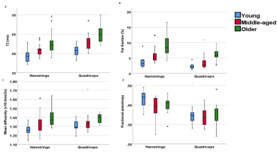 |
The effect of ageing on skeletal muscle as assessed by quantitative MR imaging: an association with frailty and muscle strength
Matt Farrow1, John Biglands1, Steven F Tanner1, Elizabeth Hensor1, Philip O'Connor1, Paul Emery1, and Ai Lyn Tan1
1University of Leeds, Leeds, United Kingdom The proportion of older people in the population is increasing. It is known that muscle health deteriorates with age, resulting in a loss of muscle mass and strength, known as sarcopenia. This study demonstrates that ageing is associated with a significant increase in fat fraction and T2 and a significant decrease in muscle volume, grip strength and muscle power. Quantitative MRI parameters correlated with frailty index, grip strength and muscle power. Therefore, quantitative MRI measurements have the potential to be useful markers of age and muscle health and can be used in the management and treatment of sarcopenia and frailty. |
 Watch the Video
 View the Poster
2711.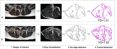 |
Is fractal dimension value of the lumbar multifidus muscle associated with low back pain?
Junchao Ma1, Nan Yu1, Ruifeng Wang1, and Shaoyu Wang2
1The Affiliated Hospital of Shaanxi University of Chinese Medicine, Xian Yang, China, 2MR Scientific Marketing Specialist Diagnostic Imaging Healthcare Greater China, Xi'an, China
Magnetic resonance imaging (MRI) is considered the best imaging method to evaluate the multifidus degeneration. The existing evaluation methods based on fat infiltration fraction (FSF) cannot fully reflect the muscle integrity and functional status. In order to obtain a tool for objective and continuous grading of multifidus degeneration. Fractal method was used to analyze the signal heterogeneity of multifidus. The fractal dimension of lumbar multifidus muscle(LMM) is closely related to LBP. The fractal dimension may be a reliable imaging marker for monitoring the degree of multifidus degeneration in symptomatic patients.
|
|
2712.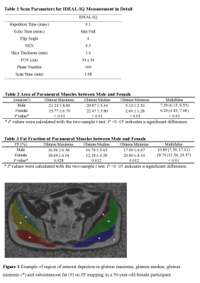 |
Quantification of Fat Fraction in Muscles Adjacent to Lumbosacral Plexus within Healthy People Using IDEAL-IQ Sequence
Mengyue Wang1, Yin Shi1, Weiqiang Dou2, and Yuefen Zou1
1Radiology, The First Affiliated Hospital of Nanjing Medical University, Nanjing, China, 2GE Healthcare, MR Research China, Beijing, China
Many diseases can result in increased fatty infiltrations within muscles adjacent to lumbosacral plexus. However, the normal level of fat content in paraneural muscles is not clear within healthy people. In this study, we applied iterative decomposition of water and fat with echo asymmetry and least-squares estimation intelligent quantification (IDEAL-IQ) to measure fat fraction (FF) of paraneural muscles within healthy participants. We found that FF values of paraneural muscles in healthy people depend on gender and age. Therefore, both factors of gender and age need to be taken into account, when assessing the FF levels of muscles in diseases.
|
|
2713.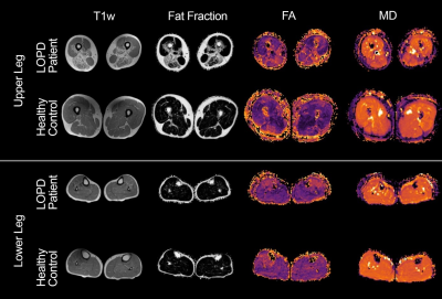 |
Muscle Diffusion tensor imaging shows changes in non-fat infiltrated muscles in late-onset Pompe disease (LOPD)
Lara Schlaffke1, Martijn Froeling2, Marlena Rohm1, Johannes Forsting1, Martin Tegenthoff1, Matthias Vorgerd1, and Robert Rehmann1
1Neurology, BG UK Bergmannsheil, Bochum, Germany, 2Radiology, UMC Utrecht, Utrecht, Netherlands
Quantitative MRI-markers are essential for monitoring disease progression in late-onset pompe disease (LOPD). Using muscle diffusion tensor imaging (mDTI) and mDixon we evaluated differences in diffusion parameters in six thigh and seven calf muscles - with <10% and >10% fat-fraction - of 18 LOPD and 29 healthy controls (HC). Upper leg muscles with <10% fat-fraction showed significant differences in MD, RD, λ1-3 and MD positively correlated with 6-MWT (p=0.003). mDTI reveals an increased diffusion restriction in muscles of LOPD-patients with and without fat-infiltration and could reflect structural changes prior to fatty degeneration.
|
|
2714.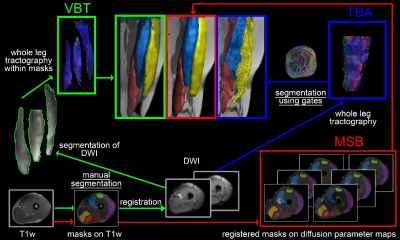 |
Evaluation of Interrater comparability of manual and tractography based thigh muscle segmentation of diffusion tensor images
Lara Schlaffke1, Robert Rehmann1, Martijn Froeling2, and Johannes Forsting1
1Neurology, BG UK Bergmannsheil, Bochum, Germany, 2Radiology, UMC Utrecht, Utrecht, Netherlands
Muscle diffusion tensor imaging is a quantitative magnetic resonance image (MRI) technique, which can provide information about muscular microstructure and integrity. Due to high intermuscular variability, the manual separation of the muscles is essential. Here, we have compared three methods, which allow assessing of diffusion properties of the thigh muscles. In each of the three investigated methods, two independent raters performed the muscle segmentation. We could show, that volume-based tractography proved to be the most reliable and robust method (ICC = 0.923-0.985) and has the advantage to achieve additional information about muscle architecture.
|
|
2715.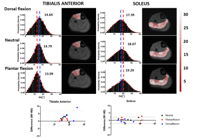 |
A method for quantification of changes in leg muscle fiber orientations
Laura Secondulfo1, Joep J M Suskens2, Ozgur Kilic2, Valentina Mazzoli3, Mario Maas4, Hans J.L Tol2, Aart Nederveen4, Melissa Hooijmans1, and Gustav J Strijkers1
1Biomedical Engineering and Physics, UMC Amsterdam, Location AMC, Amsterdam, Netherlands, 2Orthopedic Surgery, UMC Amsterdam, Location AMC, Amsterdam, Netherlands, 3Radiology, Lucas Center for Imaging, Stanford University, Stanford, CA, United States, 4Radiology and Nuclear Medicine, UMC, Amsterdam, Location AMC, Amsterdam, Netherlands
Hamstring injuries have high recurrence rates in elite athletes, which motivates the investigations in novel diagnostic methods for muscle injury and follow-up. Diffusion tensor imaging facilitates direct and indirect monitoring of the muscle condition and architecture. Fiber orientations and changes therein due to injury or training are considered a key parameter; however, the assessment over the full volume of an individual muscle is still difficult. Therefore, we developed a method to generate reproducible quantitative fiber-angle color maps of the whole volume of leg muscles, which proved sensitive to changes due to muscle stretch and a training intervention.
|
|
2716.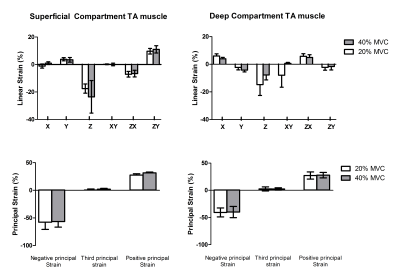 |
A novel approach for 3D whole muscle strain measurements during isometric muscle contractions
Melissa Tamara Hooijmans1,2, Crystal L. Coolbaugh1, Hannah L. Kilpatrick1, Mark K George1, and Bruce M Damon1,3
1Vanderbilt University Institute of Imaging Science, Nashville, TN, United States, 2Department of Biomedical Engineering & Physics, Amsterdam University Medical Centers, Amsterdam, Netherlands, 3Department of Radiology and Radiological Sciences, Biomedical Engineering, and Molecular Physiology and Biophysics, Vanderbilt University Medical Center, Nashville, TN, United States
In this study we explore the feasibility of using displacement fields to quantify whole muscle 3D strain patterns during submaximal voluntary contractions of the dorsiflexor muscles. Our results showed a consistent pattern, of large negative (shortening) and a large positive (lengthening) principal strains. Both the magnitude and pattern of strain agree with other studies performed during submaximal contractions which indicates the feasibility of this approach to quantify 3D strain.
|
|
2717.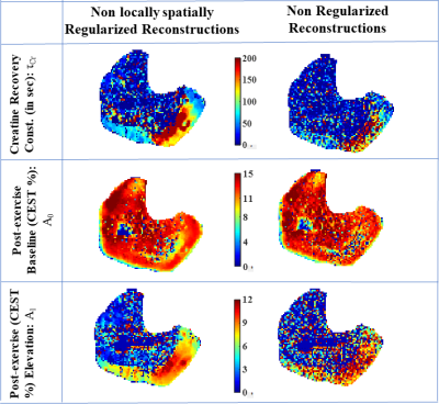 |
Parametric Maps of Creatine Recovery Constants in Exercised Muscle
Dushyant Kumar1, Deepa Thakuri1, Blake Benyard1, Hari Hariharan1, Ravi Prakash Reddy Nanga1, and Ravinder Reddy1
1Radiology, University of Pennsylvania, Philadelphia, PA, United States
Though oldest noninvasive imaging biomarker for creatine kinase (CK) reaction in exercised muscle, phosphorous magnetic resonance spectroscopy (31PMRS) suffers from the poor resolution. The 2D creatine Chemical Exchange Saturation Transfer allows for the assessment of creatine recovery with excellent in plane spatial resolution, it was not possible to construct voxel wise parametric maps for recovery time constant due to low signal to noise ratio (SNR). Recently developed 3D implementation of CrCEST allowed for higher SNR and increased volumetric coverage and we exploit these two advantages in our novel non-local spatially regularized approach to reconstruct parametric map for recovery time constant.
|
|
2718.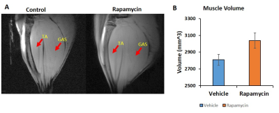 |
Metabolic Imaging of Skeletal Muscle in Aging Rats: Potential for improving muscle metabolism with Rapamycin
Rengaraj Anantharaj1, Jadegoud Yaligar1, Giang Thi Thu Le1, Venkatesh Gopalan1, Sanjay Kumar Verma1, Kavita Kaur1, Kasthuri Thirumurugan2, Johan G Eriksson2,3, Brian Kennedy4, and S Sendhil Velan1,2
1Laboratory of Molecular Imaging, Singapore Bioimaging Consortium, A*STAR, Singapore, Singapore, 2Singapore Institute for Clinical Sciences, A*STAR, Singapore, Singapore, 3Department of Obstetrics & Gynecology, National University of Singapore, Singapore, Singapore, 4Center for Healthy Aging, National University of Singapore, Singapore, Singapore
Aging associated loss of muscle mass leads to metabolic diseases and compromised quality of life. Increase in intramyocellular lipid (IMCL) and reduced skeletal muscle mass are associated with insulin resistance and diabetes. Rapamycin increases muscle mass by inhibiting the mTOR signalling pathway. In this study, we investigated IMCL metabolism and muscle mass in response to rapamycin intervention in an aging rodent model. We observed significant reduction in IMCL along with increase in muscle mass indicating improved muscle metabolism with rapamycin intervention. Muscle differentiation gene, MyoD was upregulated and myostatin which is negative regulator of muscle growth factor was down regulated.
|
|
2719. |
Measuring Spontaneous Muscular Activities in Neuromuscular Disease: Preliminary Results
Martin Schwartz1,2, Petros Martirosian1, Günter Steidle1, Thomas Küstner1,2,3, Bin Yang2, Alto Stemmer4, Thorsten Feiweier4, Ludger Schöls5,6, Matthis Synofzik5,6, and Fritz Schick1
1Section on Experimental Radiology, University Hospital of Tuebingen, Tuebingen, Germany, 2Institute of Signal Processing and System Theory, University of Stuttgart, Stuttgart, Germany, 3School of Biomedical Engineering & Imaging Sciences, King's College London, St. Thomas' Hospital, London, United Kingdom, 4Siemens Healthcare GmbH, Erlangen, Germany, 5Department Neurodegenerative Diseases, Hertie Institute for Clinical Brain Research, Tübingen & Center for Neurology, Tuebingen, Germany, 6German Center for Neurodegenerative Diseases (DZNE), Tuebingen, Germany
Quantification of spontaneous mechanical activities in musculature like fibrillations or fasciculations is of high interest for the assessment of neuro-muscular function in normal and impaired subjects. The diagnostic assessment of neuromuscular disease focuses at specific muscular regions, and the measurement protocol was optimized in order to robustly quantify spontaneous activities in these areas. This work shows preliminary results regarding activity patterns of healthy and diseased subjects.
|
|
2720.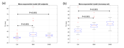 |
Differences between inflammatory and dystrophic myopathies revealed by the multi-exponential behavior of water-T2 decays in skeletal muscle
Ericky Caldas de Almeida Araujo1,2, Harmen Reyngoudt1,2, and Pierre G. Carlier1,2
1NMR Laboratory, Neuromuscular Investigation Center, Institute of Myology, Paris, France, 2NMR Laboratory, CEA/DRF/IBFJ/MIRCen, Paris, France
Although sensitive to disease activity, the mono-exponential muscle water T2 is non-specific to the underlying dominant pathophysiological processes taking place in the different neuromuscular disorders. In this work we performed multi-exponential analysis of muscle water T2-relaxation data acquired in healthy subjects, Duchenne Muscular Dystrophy and Inclusion Body Myositis patients. T2-decay curves were obtained using 1H NMR spectra acquired at different echo times. The results put in evidence a distinct water T2-relaxation behavior between inflammatory and dystrophic myopathies, supporting the hypothesis that a multi-exponential analysis of T2-relaxation data can reveal specific pathophysiological information that is missing from mono-exponential analysis.
|
|
2721.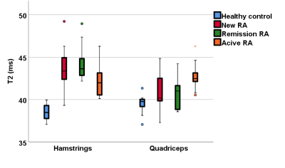 |
Quantitative MRI of muscles is different in rheumatoid arthritis patients compared to healthy controls
Matt Farrow1, John Biglands1, Steven F Tanner1, Elizabeth Hensor1, Paul Emery1, and Ai Lyn Tan1
1University of Leeds, Leeds, United Kingdom Rheumatoid arthritis can present with the loss of muscle mass and a decrease in strength and functional capability. This study demonstrates that Quantitative MRI can detect differences in the muscles of RA patients, whether they are newly diagnosed, in remission or with persistently active disease. Differences in T2, FF and muscle volume were apparent at diagnosis, suggesting muscle changes in RA occur early. Furthermore, despite RA treatment, patients in remission show worse MRI parameters and strength compared to healthy individuals, suggesting the muscles have pathology. This warrants attention in improving the muscle health throughout the spectrum of the RA continuum. |
|
2722.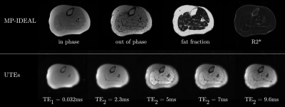 |
Short T2 Fraction Mapping of Skeletal Muscle Integrating Ultralow TE (UTE) and MP-IDEAL multiecho data
Usha Sinha1, Vadim Malis2, Edward Smitaman3, and Shantanu Sinha3
1Physics, San Diego State University, San Diego, CA, United States, 2Physics, UC San Diego, La Jolla, CA, United States, 3Radiology, UC San Diego, La Jolla, CA, United States
Ultralow TE imaging of skeletal muscle enables the visualization of low T2* species. In skeletal muscle, the signal from the small fraction of short-T2 (T2S) components are masked by the larger component long-T2 (T2L) species. One method is to extract the short T2 components from subtracted images of a dual echo UTE sequence. However, the subtracted images invariably have residual signal from lipids due to the chemically shifted frequencies of signals produced by lipids. We integrate the fat fraction and T2L extracted from MP-IDEAL sequence to the subtracted UTE images to extract the fibrosis fraction.
|
|
2723.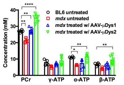 |
Monitoring micro-dystrophin treatment effects in mdx4cv mice using magnetic resonance imaging and spectroscopy
Ravneet Vohra1, Guy Odom2, Jeffrey S Chamberlain2,3,4, and Donghoon Lee1
1Radiology, University of Washington, Seattle, WA, United States, 2Neurology, University of Washington, Seattle, WA, United States, 3Medicine, University of Washington, Seattle, WA, United States, 4Senator Paul D. Wellstone Muscular Dystrophy Specialized Research Center, University of Washington, Seattle, WA, United States
Mutations in the X-linked dystrophin gene disrupts protein expression resulting in Duchenne muscular dystrophy (DMD), a neuromuscular disorder characterized by body-wide muscle cell degeneration. The mdx mouse model is one of the most commonly used animal models for DMD. Recombinant adeno-associated viral (rAAV) vector-mediated gene transfer represents a promising approach for DMD. Magnetic resonance has emerged as a noninvasive method in monitoring disease progression and treatment response for muscular dystrophy. The aim of this study was to elucidate the functional impact of micro-dystrophin on skeletal muscles using magnetic resonance imaging and spectroscopy.
|
|
2724.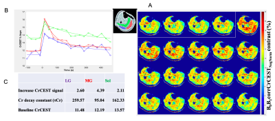 |
CrCEST effects in Knee Osteoarthritis Patients following Plantar Flexion Exercise of lower leg skeletal muscle at 7.0T
Deepa Thakuri1, Dushyant Kumar1, Abigail Cember1, Ravi Prakash Reddy Nanga1, Lizbeth Novelo2, Hari Hariharan1, Joshua Baker2, and Ravinder Reddy1
1Radiology, University of Pennsylvania, Philadelphia, PA, United States, 2Rheumatology & Epidemiology, University of Pennsylvania, Philadelphia, PA, United States
Osteoarthritis (OA) is the leading cause of disability in adult and aging populations. Knee OA increases difficulty in the functional use of various upper and lower leg muscles. In the recent years, CrCEST technique has emerged as a potential imaging biomarker to study muscle metabolism and its application was shown in different systemic diseases like mitochondrial disorders. Here, we are particularly interested in exploring and investigating the correlation of knee OA with post exercise CrCEST increase.
|
|
2725. |
Association between Abdominal Fat and Skeletal Muscle Tissues Fat Deposition in Type 2 Diabetes Mellitus: A Pilot Investigation
Manoj Kumar Sarma1, Andres Saucedo1, Daniel Kohanghadosh1, Kavya Umachandran1, Ely R. Felker1, Christine H. Darwin2, and M. Albert Thomas1
1Radiological Sciences, David Geffen School of Medicine at UCLA, Los Angeles, CA, United States, 2Medicine, David Geffen School of Medicine at UCLA, Los Angeles, CA, United States
Increased intramyocellular and extramyocellular lipids (IMCL and EMCL), abdominal lipids and decreased lipid unsaturation ratios, are of prime interest in determining relationships of these changes to insulin sensitivity and progression to type 2 diabetes (T2DM). We acquired 6-point Dixon based MRI of abdomen and accelerated MR Spectroscopic Imaging in calf muscle. In the T2DM group, BMI correlated positively with visceral fat, total fat and hepatic fat fractions. In contrast, BMI correlated negatively with pancreatic fat fractions (body/tail) in age-matched healthy volunteers. Mostly association between IMCL unsaturation index and abdominal fat content was observed. Also, we found an excellent correlation between abdominal lipid accretion and calf muscle lipid infiltration in T2DM and healthy controls predominantly inside muscle fibers (IMCL).
|
2726.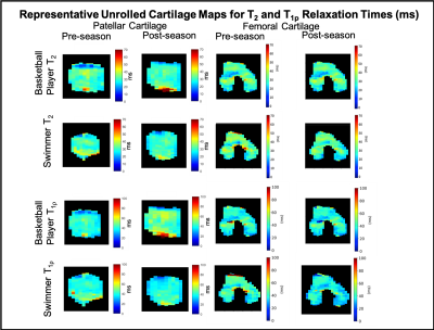 |
Longitudinal Analysis of Knee Articular Cartilage in Collegiate Basketball Players and Swimmers: Preliminary Results
Elka B Rubin1, Valentina Mazzoli1, Marianne Black1, Arjun D Desai1, Kate Young1, Feliks Kogan1, Ashwin Sreedhar1, Dominic J Vincentini1, Katelin A Knox1, Tomoo Yamada1, Andrew McCabe2, Marc Safran1, Sharmila Majumdar3, Hollis
G Potter4, and Garry E Gold1
1Stanford University, Stanford, CA, United States, 2Santa Clara University, Santa Clara, CA, United States, 3University of California San Francisco, San Francisco, CA, United States, 4Hospital for Special Surgery, New York, NY, United States
Basketball players place high loads on their knee joints that can lead to chronic knee injuries. In this study, we used advanced MRI methods and a cluster analysis to longitudinally study cartilage structure and potential early degenerative changes in Division 1 (D1) basketball players and swimmers. Pre-season and post-season quantitative results indicate an increase in T2 and T1p relaxation times in the central compartment of the femoral cartilage in the basketball players compared to the swimmers. The results of this study suggest that microstructural changes in knee cartilage can occur in one season of D1 college play.
|
|
2727.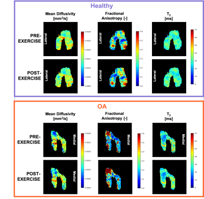 |
Diffusion tensor imaging of articular cartilage at 3T: repeatability and sensitivity to degenerative changes
Valentina Mazzoli1, Lauren Elizabeth Watkins1, Elka Brooke Rubin1, Brian Hargreaves1, Feliks Kogan1, and Garry Evan Gold1
1Department of Radiology, Stanford University, Stanford, CA, United States
Mean Diffusivity (MD) and Fractional Anisotropy (FA) derived from DTI are a very promising biomarker of cartilage health. Many current implementations of DTI require high field scanners, and its feasibility utility at 3T is unclear. The aim of this study was to develop an optimized DTI acquisition and postprocessing protocol for the evaluation of femoral articular cartilage at 3T in a clinically feasible scan time, and to assess its repeatability and sensitivity to transient changes induced by exercise. Our results show that MD and FA measurements at 3T are repeatable and robust to changes induced by exercise.
|
|
2728.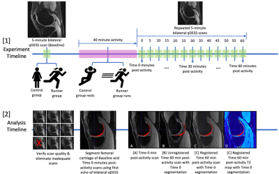 |
Short Term Effects of Running on Knee Cartilage: Global and Regional T2 Relaxation Times in Femoral Cartilage of Female Recreational Runners
Hollis Crowder1, Valentina Mazzoli2, Marianne Black2, Lauren Watkins3, Feliks Kogan2, Brian Hargreaves4, Marc Levenston4, and Garry Gold4
1Mechanical Engineering, Stanford University, Palo Alto, CA, United States, 2Radiology, Stanford University, Palo Alto, CA, United States, 3Bioengineering, Stanford University, Palo Alto, CA, United States, 4Stanford University, Palo Alto, CA, United States
To identify potential changes in cartilage hydration or microstructure resulting from exercise we compared global and regional T2 relaxation times of eleven female recreational runners and five controls at baseline, time 0, and time 60 minutes post-exercise. No significant difference in mean T2 relaxation times between the runner group and the control group were found at any time point in either global or regional analysis. Suspected changes in cartilage hydration or microstructure resulting from biomechanical running forces may not manifest in such a way that is detectable by global or regional T2 relaxation analysis compared to controls.
|
|
2729.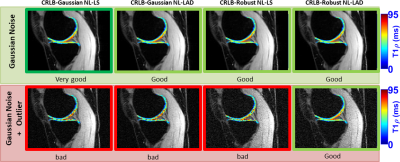 |
Robust Statistics for T1ρ-Mapping of Knee Cartilage on Mono- and Bi-Exponential Models: Optimal Spin-Lock Times and Fitting Methods
Marcelo Victor Wust Zibetti1, Azadeh Sharafi1, and Ravinder Regatte1
1Radiology, NYU, New York, NY, United States
T1ρ-mapping using mono- or bi-exponential models usually require multiple spin-lock times (#TSLs). Choosing the optimal #TSLs for improved SNR, i.e. minimizing the Cramer-Rao lower bound (CRLB), is important. However, Gaussian statistics with CRLB usually lead to the repetition of longer TSLs and the minimum number of shorter TSLs. This choice is non-robust to large data acquisition errors caused by subject motion or other scan related problems that strongly affect quantitative parameters. To alleviate this problem, we propose a robust T1ρ-protocol based on optimized #TSLs using CRLB with robust statistics, and outlier-robust fitting method.
|
|
2730.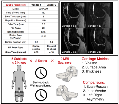 |
Scan-Rescan Variability and Left-Right Knee Asymmetry of Cartilage Morphometry Assessed with Rapid MRI in a Harmonized Multi-Vendor Study
Akshay Chaudhari1, Quin Lu2, Anna Wisser3,4, Wolfgang Wirth3,4, Garry E Gold1, Brian A Hargreaves1, and Felix Eckstein3,4
1Stanford University, Stanford, CA, United States, 2Philips, San Francisco, CA, United States, 3Paracelsus Medical University, Salzburg, Austria, 4Chondrometrics GmbH, Ainring, Germany
Changes in cartilage morphology have shown to predict and monitor osteoarthritis progression with great sensitivity. However, the lack of a rapid and inexpensive MRI technique for imaging cartilage that can be implemented across vendors is a challenge in large multi-site clinical studies. In this work, we evaluate multi-vendor and scan-rescan reliability and explore left-right knee asymmetries of cartilage morphology measured using a rapid, 4-minute quantitative double-echo steady-state (qDESS) sequence. We demonstrate qDESS harmonization across vendors, which can produce high scan-rescan repeatability and high repeatability of left-right knee asymmetry of cartilage volume, surface area, and thickness metrics.
|
|
2731.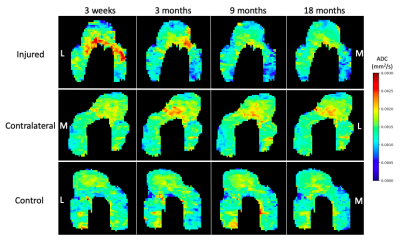 |
Detecting Early Changes in ACL-Reconstructed Knee Cartilage Using Diffusion-Weighted MRI
Halston J.C. Sandford1, Marianne Black2,3, Akshay S. Chaudhari2, Arjun Desai2,4, Feliks Kogan1, Brian A. Hargreaves2,4,5, Garry E. Gold2,5,6, and Valentina Mazzoli2
1Stanford University, Stanford, CA, United States, 2Radiology, Stanford University, Stanford, CA, United States, 3Mechanical Engineering, Stanford University, Stanford, CA, United States, 4Electrical Engineering, Stanford University, Stanford, CA, United States, 5Bioengineering, Stanford University, Stanford, CA, United States, 6Orthopedic Surgery, Stanford University, Stanford, CA, United States
ACL-injured individuals, despite reconstructive surgery, have an increased risk of developing osteoarthritis and methods to identify early changes in cartilage are needed to allow implementation of treatment. Diffusion-weighted MRI (DWI) has been shown to provide information on femoral cartilage health. This study aims to assess the potential of DWI to detect early degenerative changes in articular cartilage following ACL reconstruction. We found that DWI shows elevated ADC values in both ACL-injured and contralateral knees compared with controls at baseline. DWI did not show significant changes in cartilage over time within 18 months of post-ACL-reconstructive surgery.
|
|
2732. |
Functional Gradient Indices for the Early Detection and Prediction of Osteoarthritis: Data from Human Explants and the OAI Database
Robert L. Wilson1, Nancy C. Emery2, David M. Pierce3,4, and Corey P. Neu1
1Mechanical Engineering, University of Colorado Boulder, Boulder, CO, United States, 2Ecology and Evolutionary Biology, University of Colorado Boulder, Boulder, CO, United States, 3Mechanical Engineering, University of Connecticut, Storrs, CT, United States, 4Biomedical Engineering, University of Connecticut, Storrs, CT, United States
Spatial gradients in articular cartilage structure trend toward homogeneity with onset and progression of osteoarthritis (OA). Exploiting MRI, we compared heterogeneous variations in tissue structure, and the corresponding function, to OA severity through the use of spatial first derivatives, herein termed functional gradient indices (FGIs), providing new biomarkers indicating tissue state. We evaluated FGI maps of MRI data visualizing osteochondral human explants and OAI subjects for relationships to OA severity. FGI data correlated significantly with tissue health, while traditional metrics did not. The sensitivity and versatility of FGI analysis suggests a novel biomarker with improved detection of early disease pathogenesis.
|
|
2733.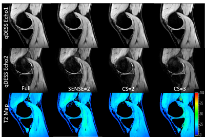 |
Application of Compressed Sensing to Quantitative Double Echo steady state (qDESS) for Rapid T2 Relaxometry in Knees
Quin Lu1, Brian A Hargreaves2, and Akshay S Chaudhari2
1Clinical Science, Philips Healthcare NA, San Francisco, CA, United States, 2Radiology, Stanford University, Stanford, CA, United States
Quantitative Double Echo Steady State (qDESS) is a fast 3D MRI sequence capable of simultaneous knee morphometry and T2 relaxometry. Compressed-sensing based acceleration methods are more and more readily available from major MRI vendors, and have the potential to further speed up qDESS and to provide greater clinical value. In this study, we investigate the feasibility of applying Philips Compressed SENSE (CS) to qDESS. We show that CS does not bias the T2 accuracy of qDESS. Our findings support the clinical application of CS accelerated qDESS for fast and quantitative MR knee assessment.
|
|
2734.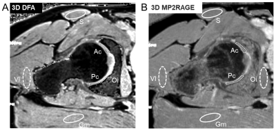 |
MP2RAGE improves robustness of 3D T1-Mapping of Hip Cartilage compared to Conventional Dual-Flip-Angle Acquisition
Bernd A Jung1, Onur Afacan2, Till Lerch1, Young-Jo Kim3, Tobias Kober4,5,6, Michael Ith1, Markus Klarhoefer7, and Florian Schmaranzer1
1Diagnostic, Interventional and Pediatric Radiology, University Hospital Bern, Bern, Switzerland, 2Dept of Radiology, Boston Children`s Hospital, Boston, MA, United States, 3Dept of Orthopaedic Surgery, Boston Children`s Hospital, Boston, MA, United States, 4Advanced Clinical Imaging Technology, Siemens Healthcare, Lausanne, Switzerland, 5Dept of Radiology, Lausanne University Hospital and University of Lausanne, Lausanne, Switzerland, 6LTS5, École Polytechnique Fédérale de Lausanne (EPFL), Lausanne, Switzerland, 7Siemens Healthcare, Zuerich, Switzerland
Although commonly used for T1 mapping of hip cartilage, 3D dual flip angle (DFA) techniques are sensitive to flip angle variations related to B1 inhomogeneities. Therefore, the IR-based MP2RAGE technique was compared with a standard 3D DFA acquisition including B1 field mapping for T1 mapping of hip cartilage in a phantom study and in healthy volunteers at 3T. For the phantom study, a 2D IR-based sequence was additionally acquired to provide reference T1 maps. MP2RAGE resulted in more accurate T1 values with less regional variations compared to DFA method and therefore enables a more robust T1 mapping of hip cartilage.
|
|
2735.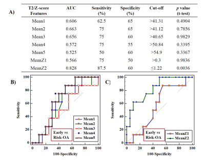 |
To Evaluate the Effect of Normalization on Femur Cartilage T2 Values in Diagnosis of Osteoarthritis
Rafeek Thaha1, Sandeep Panwar Jogi1,2, Sriram Rajan3, Amit Mehndiratta1,4, Anup Singh1,4, and Dharmesh Singh1
1Centre for Biomedical Engineering, Indian Institute of Technology, New Delhi, India, 2Biomedical Engineering, ASET, Amity University Haryana, Gurgaon, India, 3Mahajan Imaging Centre, New Delhi, India, 4Department of Biomedical Engineering, All India Institute of Medical Sciences, New Delhi, India
Quantitative MR parameters(such as T2-relaxation time) are sensitive to biochemical changes in the cartilage. The objective of the study was to evaluate the performance of different statistics parameters from T2 values of femur cartilage and normalized T2 map (Z-score T2 map) values for differentiating healthy vs OA patients as well as Early-OA (Grade-I and Grade-II), vs Risk-OA (Grade-III) patients. Multiple Statistic parameter from T2 map provided high accuracy in differentiation between healthy vs OA cartilage, while one of the static parameter of Z-score T2 map provided high accuracy in differentiation between early-OA vs Risk-OA.
|
|
2736.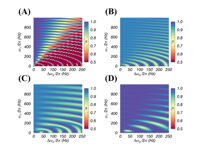 |
A self-compensated spin-locking scheme for quantitative R1ρ dispersion in articular cartilage
YUXI PANG1
1Department of Radiology, University of Michigan, Ann Arbor, MI, United States
A self-compensated spin-locking (SL) scheme for quantitative R1ρ dispersion in cartilage has been developed. The performance of this new method was evaluated by Bloch simulations and R1ρ dispersion (with 6 SL RF strengths ranging from 50 to 1000 Hz) studies on agarose (1-4%, w/v) phantom and on one healthy human knee in vivo at 3T, with respect to three reported SL approaches. The simulated and experimental results indicate that the proposed SL method was less susceptible to B0 and B1 field artifacts for a wide range of SL strengths, and thus more suitable for quantitative R1ρ dispersion in ordered tissue.
|
|
2737.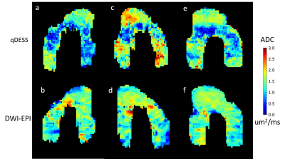 |
qDESS ADC as a Biomarker for Early Degeneration in Femoral Cartilage of Post-Reconstruction ACL Tear Patients and Correlation with DWI-EPI ADC
Mary Elizabeth Hall1,2, Valentina Mazzoli2, Marianne Black1,2, Halston Sandford2, Katherine Young2, Daehyun Yoon2, Bragi Sveinsson3, Akshay Chaudhari2, Emily McWalter4, Feliks Kogan2, Marc Levenston1,2,5, Brian Hargreaves2,5,6, and Garry Gold2
1Mechanical Engineering, Stanford University, Stanford, CA, United States, 2Radiology, Stanford University, Stanford, CA, United States, 3Massachusetts General Hospital, Boston, MA, United States, 4Mechanical Engineering, University of Saskatchewan, Saskatoon, SK, Canada, 5Bioengineering, Stanford University, Stanford, CA, United States, 6Electrical Engineering, Stanford University, Stanford, CA, United States
This study evaluates apparent diffusion coefficient (ADC) as measured by a quantitative double echo steady state (qDESS) sequence as a biomarker for early osteoarthritis detection in articular cartilage in the femur and its correlation with ADC from a diffusion weighted echo planar (DWI-EPI) scan. 9 injured knees and contralateral knees of patients undergoing reconstruction surgery following anterior cruciate ligament tears were scanned with qDESS and DWI-EPI sequences up to 18 months post surgery. There were no consistent patterns of qDESS ADC change on a global or regional basis in the femoral cartilage. qDESS ADC did not correlate with DWI-EPI ADC.
|
|
2738.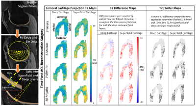 |
Detecting Early Changes in ACL-Reconstructed Knees: Cluster Analysis of T2 Relaxation Times from 3 Months to 18 Months Post-Surgery
Marianne S Black1,2, Katherine A Young1, Akshay S Chaudhari1, Feliks Kogan1, Bragi Sveinsson3, Emily J McWalter4, Garry E Gold1,5, Marc E Levenston1,2, and Brian A Hargreaves1,5,6
1Radiology, Stanford University, Stanford, CA, United States, 2Mechanical Engineering, Stanford University, Stanford, CA, United States, 3Massachusetts General Hospital, Boston, MA, United States, 4Mechanical Engineering, University of Saskatchewan, Saskatoon, SK, Canada, 5Bioengineering, Stanford University, Stanford, CA, United States, 6Electrical Engineering, Stanford University, Stanford, CA, United States
There is a need to detect and quantify early osteoarthritic changes for the development of treatments for osteoarthritis progression. ACL-injured subjects are at an increased risk of developing osteoarthritis, and T2 is sensitive to the structure and composition of cartilage, including osteoarthritic change. This study used a quantitative DESS acquisition to obtain T2 maps in 10 subjects 3-weeks, 3-months, 9-months, and 18-months after ACL reconstruction surgery and 10 controls at matched times. Our results show that T2 cluster analysis was able to detect changes to the ACL-reconstructed cartilage as early as 3-months post-surgery and these differences persisted at 18-months.
|
|
2739.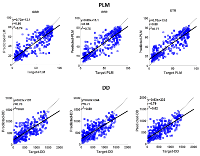 |
Multiparametric MRI Predicts Articular Cartilage Proteoglycan Content and Collagen Fiber Orientation
Abdul Wahed Kajabi1,2, Seyed Amir Mirmojarabian1,2, Juuso Ketola1, Timo Liimatainen1,2, Miika T. Nieminen1,2,3, Mikko J. Nissi4, and Victor Casula1,2
1Research Unit of Medical Imaging, Physics and Technology, University of Oulu, Oulu, Finland, 2Medical Research Center, University of Oulu and Oulu University Hospital, Oulu, Finland, 3Department of Diagnostic Radiology, Oulu University Hospital, Oulu, Finland, 4Department of Applied Physics, University of Eastern Finland, Kuopio, Finland
In this study, multiparametric MRI was used to predict cartilage proteoglycan content and collagen fiber orientation, as measured by quantitative microscopy. Twenty osteochondral samples were obtained from stifle joints of ten Shetland ponies. Measurements of 14 different MRI parameters, including T1, T2, continuous wave T1rho, adiabatic T1rho and T2rho, and TRAFF were performed at 9.4 T. Three ensemble-based regression models (Gradient Boosting, Random Forest and Extra Trees) were used and the highest coefficients of determination (r2) were 0.77 for collagen orientation and 0.62 for proteoglycan content. These findings show that multiparametric MRI can be used to non-invasively estimate cartilage histology.
|
|
2740.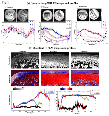 |
Site-Specific Quantitative µMRI and Polarized Light Microscopy (PLM) Study of Young Rabbit Femur Cartilage
Yang Xia1, Syeda Batool1, and Mohammad Hammimi1
1Physics, Oakland University, Rochester, MI, United States Poster Permission Withheld
This work aims to characterize the site-specific and depth dependent molecular and morphological structures in young rabbit femur cartilage, using quantitative µMRI (T2 relaxation) and Polarized light microscopy (PLM).
|
|
2741. |
Assessment of short-term and mid-term knee cartilage changes before and after marathon running using T2* mapping images
Xiaoshuai Chen1, Ranxu Zhang1, Xiaoyue Zhou2, Esther Raithel3, and Jian Zhao1
1The third Hospital of Hebei Medical University, Shijiazhuang, China, 2Siemens Healthineers Ltd, Shanghai, China, 3Siemens Healthcare, Erlangen, Germany
Once articular cartilage is damaged it cannot be readily repaired. However, to what extent marathon running causes cartilage damage to the knees is unclear. We quantitatively assessed the morphologic and T2* value changes in the knee cartilage of marathon runners using an automatic cartilage segmentation method. The cartilage volume, thickness, and T2* values of 21 sub-regions were quantitatively assessed. The results showed that the T2* value of knee cartilage increased right after running and recovered two months later, suggesting that the knee joint cartilage showed a degree of reversible change after marathon running.
|
| 2742. | Quantification of knee cartilage using automated cartilage segmentation prototype software : Clinical validation on volunteers
Ping Zhang1, Xiao Yue Zhou2, Esther Raithel3, Xu Ran Zhang4, Xiao Shuai Chen4, Jian Ling Cui4, and Jian Zhao4
1The third Hospital of Hebei Medical University, Shijiazhuang, China, 2MR Collaboration, Siemens Healthineers Ltd,Shanghai,China, ShangHai, China, 3Siemens Healthcare, Erlangen, Germany., Erlangen, Germany, 4radiology, The third Hospital of Hebei Medical University, Shijiazhuang, China
Manual cartilage segmentation is a time-consuming post-processing procedure, especially for clinical doctors. Automatic cartilage segmentation frees doctors from tedious computer work. Although there have been various automatic segmentation algorithms, accurate automatic segmentation is still a challenge. In this study, we validated the automatic cartilage segmentation results of the knee joint using a 3D high-resolution DESS sequence. We found that the location of cartilage subregions, and the hydrarthrosis and cartilage degeneration may influence the accuracy of the segmentation. In order to derive more accurate results, a manual finetuning of the automatic segmentation was done. Automatic segmentation still saved considerable time.
|
|
2743. |
Distinguishing Exercise-Induced Compositional Changes in Knee Cartilage with Quantitative MR Relaxation Time Mapping
Dimitri A Kessler1, Joshua D Kaggie1, James W MacKay1,2, Scott McDonald3, Andrew Grainger1, Alexandra R Roberts4, Robert L Janiczek5, Martin J Graves1, and Fiona J Gilbert1
1Department of Radiology, University of Cambridge, Cambridge, United Kingdom, 2Norwich Medical School, University of East Anglia, Norwich, United Kingdom, 3Cambridge University Hospitals NHS Foundation Trust, Addenbrooke’s Hospital, Cambridge, United Kingdom, 4Independent Clinical Imaging Consultant, Munich, Germany, 5GlaxoSmithKline, Clinical Imaging, Philadelphia, PA, United States
We introduce a method to reliably determine changes in healthy knee cartilage composition after joint loading. Ten healthy participants were imaged before and after participant repositioning to determine the test-retest repeatability of T1ρ and T2 relaxation time mapping. Additionally, nine healthy participants were imaged before and after a mild, dynamic stepping exercise. Three-dimensional surface analysis of patellar, femoral, and lateral and medial tibial cartilage was performed. The exercise surface data was thresholded with the determined measurement errors from the T1ρ and T2 repeatability data to highlight cartilage regions experiencing reliable exercise-induced compositional changes.
|
|
2744.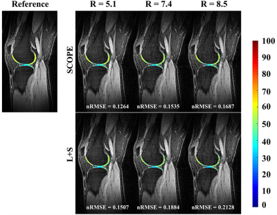 |
Accelerating 3D-T1ρ cartilage imaging using signal compensated low-rank plus sparse matrix decomposition
Yuanyuan Liu1,2, Weitian Chen3, Xin Liu1, Hairong Zheng1, Dong Liang1,2, and Yanjie Zhu1
1Paul C. Lauterbur Research Center for Biomedical Imaging, Shenzhen Institutes of Advanced Technology, Chinese Academy of Sciences, Shenzhen, China, 2Research center for Medical AI, Shenzhen Institutes of Advanced Technology, Chinese Academy of Sciences, Shenzhen, China, 3Department of Imaging and Interventional Radiology, The Chinese University of Hong Kong, Hong Kong, China
The quantitative 3D-T1ρ mapping requires multiple T1ρ-weighted images with different spin lock times (TSLs) to obtain the T1ρ map, which makes the acquisition time very long. In this work, a signal compensation strategy with low-rank plus sparse model (SCOPE) was used to reconstruct T1ρ-weighted images from highly undersampled data. We provide the reconstructed images and the estimated T1ρ maps at an acceleration factor up to 8.5 in fast 3D-T1ρ cartilage imaging.
|
|
2745.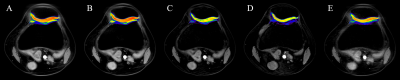 |
Accelerated T1ρ Acquisition for Knee Cartilage Using Compressed Sensing:Quantitative evaluation
Ye Li1, Guangbin Wang1, Weibo Chen2, and Aocai Yang1
1Shandong Medical Imaging Research Institute,Shandong University, Jinan, China, 2Philips Healthcare, Shanghai, China
The current work evaluates the influence of compressed sensing accelerated imaging on the T1ρ mapping. Quantitative evaluation is done using quantitative T1ρ mapping for knee cartilage quantification. T1ρ quantification after CS-accelerated acquisition was compared with non-CS-accelerated acquisition for central patella cartilage.
|
|
2746.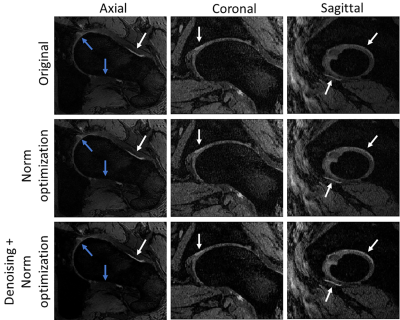 |
Improving DESS contrast in 7T hip images using L1-norm denoising and optimal echo combination
Aurelien Destruel1, Mingyan Li 1, Craig Engstrom2, Ewald Weber1, Jin Jin1,3,4, Rahel Heule5, Oliver Bieri6, Feng Liu1, and Stuart Crozier1
1School of Information Technology and Electrical Engineering, The University of Queensland, Brisbane, Australia, 2School of Human Movement and Nutrition Sciences, The University of Queensland, Brisbane, Australia, 3University of Southern California, Los Angeles, CA, United States, 4Siemens Healthcare Pty Ltd, Brisbane, Australia, 5High Field Magnetic Resonance, Max Planck Institute for Biological Cybernetics, Tübingen, Germany, 6Division of Radiological Physics, Department of Radiology, University Hospital Basel, Basel, Switzerland
The double-echo steady-state (DESS) sequence has been used successfully in 3T MRI imaging of the musculoskeletal system for segmentation of joint. However, in 3D-DESS images acquired at 7T MRI, a reduction in the contrast between tissues due to an increased diffusion sensitivity may complicate cartilage segmentation. Typically, the signals acquired with DESS are averaged without any pre-processing. However, these signals give different contrasts and have different noise behaviours. In this work, we improve the contrast-to-noise ratio (CNR) whilst preserving anatomical detail in high-resolution 7T DESS images through a new approach combining L1-norm denoising and p-norm combination of the echo signals.
|
|
2747.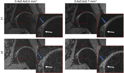 |
Comparison between high-resolution DESS images of the hip joint at 3T and 7T MRI
Aurelien Destruel1, Mingyan Li 1, Ewald Weber1, Jin Jin1,2,3, Shekhar S Chandra1, Jurgen Fripp4, Feng Liu1, Stuart Crozier1, and Craig Engstrom5
1School of Information Technology and Electrical Engineering, The University of Queensland, Brisbane, Australia, 2Mark and Mary Stevens Neuroimaging and Informatics Institute, University of Southern California, Los Angeles, CA, United States, 3ARC Training Centre for Innovation in Biomedical Imaging Technology, The University of Queensland, Brisbane, Australia, 4CSIRO Health and Biosecurity, Herston, Australia, 5School of Human Movement and Nutrition Sciences, The University of Queensland, Brisbane, Australia
Advances in automatic segmentation of cartilage in hip joints has shown the need for high resolution and contrast in 3D sequences. In this work, double-echo steady-state (DESS) hip images from a healthy volunteer were compared at 3T and 7T. 3D-DESS with a resolution up to 0.4x0.4x0.6mm3 was acquired, and showed that the 7T images had superior anatomical detail, signal-to-noise ratio (SNR), contrast ratio (CR) and overall image quality. The improvements observed in 7T imaging of the hip have the potential to boost the performance of automated cartilage segmentation algorithms by reducing segmentation errors.
|
|
2748. |
T2 Relaxation Time Reveals Improvement of Articular Cartilage Quality After Bariatric Surgery at 12-month Follow-up
Sami Lehtovirta1,2, Ahti Kemppainen1,2, Marianne Haapea2,3, Jaro Karppinen2,4,5, Eveliina Lammentausta2,3, Vesa Koivukangas6, Eero Kyllönen7, Mika Nevalainen1,2,3, Anna-Maija Kauppila7, Victor Casula1,2, and Miika T. Nieminen1,2,3
1Research Unit of Medical Imaging, Physics and Technology, University of Oulu, Oulu, Finland, 2Medical Research Center, University of Oulu and Oulu University Hospital, Oulu, Finland, 3Department of Diagnostic Radiology, Oulu University Hospital, Oulu, Finland, 4Center for Life Course Health Research, University of Oulu, Oulu, Finland, 5Finnish Institute of Occupational Health, Oulu, Finland, 6Department of Surgery, Oulu University Hospital, Oulu, Finland, 7Department of Physical Medicine and Rehabilitation, Oulu University Hospital, Oulu, Finland
Obesity has become a worldwide phenomenon with nearly tripling since 1975: 13% of adults over 18 are obese. We set out to study articular cartilage of knee joint in obese individuals using T2 relaxation time. Our study population underwent Roux-en-Y gastric bypass operation and were compared with a control group of obese individuals in a 12-month follow-up. Our results suggest improved quality of cartilage in the lateral compartment of femoral cartilage after bariatric surgery.
|
|
2749.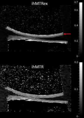 |
Inhomogenous Magnetization Transfer Imaging in Cartilage – Initial Demonstration and Protocol Optimization at 7 Tesla
Andrew C. Yung1, Valentin H. Prevost1, Jane Desrochers2, David Wilson2, and Piotr Kozlowski1
1UBC MRI Research Centre, University of British Columbia, Vancouver, BC, Canada, 2Centre for Hip Health and Mobility, University of British Columbia, Vancouver, BC, Canada
This work presents one of the first examples of ihMT imaging in cartilage, using ex vivo bovine knee specimens at 7 Tesla. Protocol optimization experiments were performed to determine the optimal set of parameters (B1rms, TR, frequency offset, centre frequency) to maximize the ihMTRex signal. Cartilage-specific signal was observed, which may be related to residual dipolar order established by the highly ordered nature of collagen.
|
|
2750.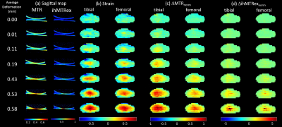 |
Magnetization Transfer Techniques (MTR and ihMTRex) for Detecting Cartilage Strain in Loaded ex vivo Joints at 7 Tesla
Andrew C Yung1, Valentin H. Prevost1, Jane Desrochers2, Emily Sullivan2, Piotr Kozlowski1, and David Wilson2
1UBC MRI Research Centre, University of British Columbia, Vancouver, BC, Canada, 2Centre for Hip Health and Mobility, University of British Columbia, Vancouver, BC, Canada
We investigated the use of magnetization transfer ratio MTR and ihMTRex as a potential surrogate of cartilage strain, using ex vivo cow knee specimens in a uniaxial compression rig. On MTR and ihMTRex image volumes for several load levels, the cartilage strain and average MTR or ihMTRex for each column of pixels within the cartilage were calculated. Normalized change in MTR showed a linear relation with strain throughout the tested range of deformation, whereas ihMTRex only showed response at the highest load levels. These techniques may be an attractive alternative to T2 or T1ρ techniques to map cartilage strain.
|
|
2751. |
Tibial Rotation and Knee Flexion Moment Correlate to Patellofemoral Deep Cartilage UTE-T2* 2 Years After ACL Reconstruction
Ashley A Williams1,2, Jennifer Erhart-Hledik1,2, Jessica L. Asay2,3, Gordhan Mahtani1,2, and Constance R. Chu1,2
1Orthopaedic Surgery, Stanford University, Stanford, CA, United States, 2Veterans Affairs Palo Alto Health Care System, Palo Alto, CA, United States, 3Mechanical Engineering, Stanford University, Stanford, CA, United States
Patellofemoral joint osteoarthritis (PFOA) following anterior cruciate ligament reconstruction (ACLR) is thought to arise, in part, due to increased external rotation of the tibia and decreased quadriceps strength that alter the tracking of the patella in the trochlear groove. In this study of 59 subjects 2 years after ACLR, higher cartilage UTE-T2* values were detected in ACLR knees with greater external tibial rotations and greater knee flexion moments assessed by gait analysis. This study provides evidence that UTE-T2* is sensitive to patellofemoral cartilage degeneration likely due to altered patellar tracking and quadriceps strength in knees at risk of PFOA.
|
|
2752.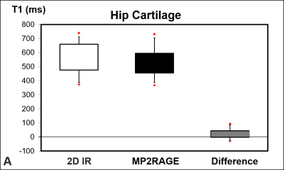 |
Prospective clinical validation of 3D MP2RAGE for T1 mapping of hip cartilage: Preliminary results in patients with hip pain
Florian Schmaranzer1,2, Eduardo Novais2, Young-Jo Kim2, Tobias Kober3,4,5, Bernd Jung1, Sarah Bixby6, and Onur Afacan6
1Department of Diagnostic, Interventional and Pediatric Radiology, University of Bern, Inselspital Bern, Bern, Switzerland, 2Department of Orthopaedic Surgery, Harvard Medical School, Boston Children`s Hospital, Boston, MA, United States, 3Advanced Clinical Imaging Technology, Siemens Healthcare, Lausanne, Switzerland, 4Department of Radiology, Lausanne University Hospital and University of Lausanne, Lausanne, Switzerland, 5École Polytechnique Fédérale de Lausanne (EPFL), Lausanne, Switzerland, 6Department of Radiology, Harvard Medical School, Boston Children`s Hospital, Boston, MA, United States Poster Permission Withheld
Current standard techniques for T1 mapping of hip cartilage are affected by flip angle variations due to B1-field inhomogeneities at 3T. The aim was to prospectively compare a 3D MP2RAGE for T1 mapping of cartilage to a 2D-IR based reference standard in the patients with hip pain undergoing indirect MR arthrography. 3D MP2RAGE correlated well with the 2D-IR reference standard and showed a minor systematic bias. This supports the continued use of 3D MP2RAGE in patients undergoing joint preserving hip surgery with the goal to establish prognostic predictors to predict the success or failure of these interventions.
|
|
2753.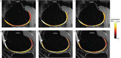 |
Investigation into the effect of spin-lock frequency on the angular dependence of T1rho in in-vivo measurements of femoral knee cartilage at 3T
Laurel Hales1 and Feliks Kogan2
1Electrical Engineering, Stanford University, Stanford, CA, United States, 2Radiology, Stanford University, Stanford, CA, United States
Given the use of T1rho as a measure of cartilage health and an early indicator for osteoarthritis, it is important to understand the T1rho relaxation mechanism including its somewhat unexplained angular dependence. In the hope of finding a range of spin-lock frequencies (FSLs) with a decreased angular dependence, we generated T1rho maps of in-vivo femoral knee cartilage at various FSLs from 100Hz to 1kHz. We found a visible angular dependence in T1rho measurements for all FSLs including 1kHz. If there exists a range of FSL with little or no angular dependence it is higher than 1kHz.
|
|
2754.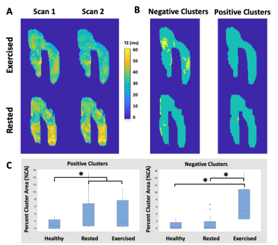 |
Cluster analysis of T2 changes is related to acute exercise in individuals with knee osteoarthritis
Lauren Watkins1, Valentina Mazzoli2, Marianne Black3, Scott Uhlrich3, Brian Hargreaves1,2,4, Garry Gold2, and Feliks Kogan2
1Bioengineering, Stanford University, Stanford, CA, United States, 2Radiology, Stanford University, Stanford, CA, United States, 3Mechanical Engineering, Stanford University, Stanford, CA, United States, 4Electrical Engineering, Stanford University, Stanford, CA, United States
Degradation of articular cartilage related to osteoarthritis is associated with changes in cartilage T2 relaxation times that may not be uniform across the cartilage surface. Analysis of changes in T2 times longitudinally or in response to mechanical loading can assist in detection of regions of cartilage damage. Here we examine the ability of cluster analysis to reflect transient changes in cartilage T2 times in response to acute loading. Osteoarthritic subjects who performed a squat exercise had a greater percent of the cartilage area with negative changes in T2 times compared to healthy and osteoarthritic subjects who did not exercise.
|
|
2755. |
Bias and correction method of UTE trajectory errors in the depiction and relaxometry of deep articular cartilage
Sophia Kronthaler1, Jürgen Rahmer2, Peter Börnert2, Alexandra S. Gersing1, Benedikt J. Schwaiger1, Roland Krug3, and Dimitrios C. Karampinos1
1Department of Diagnostic and Interventional Radiology, Technical University of Munich, School of Medicine, Munich, Germany, 2Philips Research Laboratory, Hamburg, Germany, 3Department of Radiology and Biomedical Imaging, University of California, San Francisco, CA, United States
Imaging of the morphological and biochemical composition of the deep articular cartilage has important implications in the assessment of osteoarthritis. The deep cartilage layer has intrinsically short T2* characteristics and ultra-short echo time (UTE) imaging techniques have been recently employed in the depiction of the deep cartilage layer morphology. UTE imaging is known to be affected by k-space trajectory errors. The purpose of the present work is to investigate the effect of trajectory errors in UTE imaging used for the depiction and relaxometry of deep articular knee cartilage. A GIRF-correction is applied to correct eddy-currents induced k-space trajectory errors.
|
|
2756.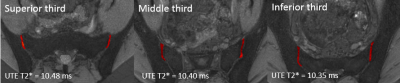 |
Feasibility of UTE T2* cartilage mapping in the sacroiliac joints
Tony T Wong1, Patrick Quarterman1, Phuong Duong1, Diego Jaramillo1, Michael Liu1, and Sachin R Jambawalikar1
1Columbia University Medical Center, New York, NY, United States
Ultrashort echo time (UTE) T2* images of 20 sacroiliac joints were acquired in 10 asymptomatic volunteers. Each sacroiliac (SI) joint was segmented manually and divided into thirds (superior, middle, and inferior). Differences in mean UTE T2* values were assessed. Results demonstrated a mean UTE T2* value of 10.38 ± 0.62 ms in the SI joints. There was no statistical significance between right and left sides (p = 0.89), superior, middle, and inferior third regions (p = 0.86), or males and females (p = 0.47). Intra-observer variability with ICC ranged from 0.89-0.98.
|
|
2757. |
Analysis of T2 and T2* relaxation time reproducibility within hip joint cartilage
Jessica Bugeja1,2, Ales Neubert1,2, Shekhar Chandra1, Jurgen Fripp2, Craig Engstrom1, and Stuart Crozier1
1University of Queensland, Brisbane, Australia, 2CSIRO, Brisbane, Australia
QMRI sequences including T2 and T2* mapping analyse biochemical changes of cartilage including the deterioration of the ECM and changes in water content for early diagnosis of hip OA prior to serious cartilage degradation. Longitudinal studies and clinical assessments with T2 and T2* mapping rely on a high reproducibility of the tissue relaxation times for accurate diagnosis. We present a statistical analysis of the reproducibility of T2 and T2* mapping of hip MR images. An automated cartilage segmentation method is used for the segmentation of FISP MR hip images and biochemical information is obtained from the T2 and T2* images.
|
2758. |
Evaluation of bone marrow proton density fat fraction in β-thalassaemia patients and healthy subjects using 1H-MR spectroscopy.
Umi Nabilah Ismail1, Che Ahmad Azlan1, Shasha Khairullah2, Kuan Jin Lee3, Chai Hong Yeong4, Nur Farhayu Omar5, Mohammad Nazri M Shah1, Raja Rizal Azman Raja Aman1, Nicholas Jackson6, and Kwan Hoong Ng1
1Biomedical Imaging Department, University of Malaya, Kuala Lumpur, Malaysia, 2Department of Medicine, University of Malaya, Kuala Lumpur, Malaysia, 3Singapore Bioimaging Consortium, A*STAR Research, Singapore, Singapore, 4School of Medicine, Faculty of Health and Medical Sciences, Taylor's University Lakeside Campus, Subang Jaya, Malaysia, 5Department of Imaging, Universiti Putra Malaysia, Serdang, Malaysia, 6Department of Pathology, University of Malaya, Kuala Lumpur, Malaysia
Fat in bone marrow is stated to be regulated by the body haematopoietic needs. We explored this by measuring bone marrow fat fraction(FF) of healthy subjects and β-thalassaemia patients who required treatment to suppress their body haematopoietic needs. The results obtained from 1H-MR Spectroscopy suggested that information about marrow adipocytes may be useful in evaluating patients' treatment response.
|
|
2759. |
Variable flip angle T1 Relaxometry of cortical bone free water; correlation with the mechanical compression test
Mahsa Talebi1,2, Shahrokh Abbasi-Rad3, Malakeh Malekzadeh1,4, Mohamad Shahgholi5, Kimia Foudeh5, and Hamidreza Saligheh Rad1,2
1Quantitative MR Imaging and Spectroscopy Group, Research Center for Cellular and Molecular Imaging, Tehran University of Medical Sciences, Tehran, Iran, Tehran, Iran (Islamic Republic of), 2Medical Physics and Biomedical Engineering Department, Tehran University of Medical Sciences, Tehran, Iran, Tehran, Iran (Islamic Republic of), 3Quantitative MR Imaging and Spectroscopy Group, Research Center for Cellular and Molecular Imaging, Tehran University of Medical Sciences, Tehran, Iran, Brisbane, Australia, 43Medical Physics Department, School of Medicine, Iran University of Medical Sciences, Tehran, Iran, Tehran, Iran (Islamic Republic of), 5Department of Mechanical Engineering, Najafabad Branch, Islamic Azad University, Najafabad, Iran, Najaf Abad, Iran (Islamic Republic of)
Cortical bone porosity contributes to bone quality but is under the limit of the current clinical imaging modalities’ resolution. T1 value of water molecules residing in cortical bone pores is linked with their mobility. Since the changes in surface-to-volume ratio of the pores affect cortical bone mechanical properties, we assumed that free water T1 (T1,free) would model the mechanical properties of cortical bone. Variable flip angle, variable TR, and inversion recovery methods were used to quantify T1,free and their correlation with bone toughness was assessed. The results showed VFA T1,free could predict the cortical bone toughness (r = -0.63, p<0.01).
|
|
2760. |
Detection of femoral head ischemia using DCE-MRI in Children with Slipped Capital Femoral Epiphysis (SCFE)
Kojiro Ono1,2, Yasuhiro Oikawa3, Shinya Obara1, and Hideaki Haneishi2
1Radiology, Chiba Children's Hospital, Chiba, Japan, 2Center for Frontier Medical Engineering, Chiba University, Chiba, Japan, 3Orthopedics, Chiba Children's Hospital, Chiba, Japan
In this study, we reveal a scheme to evaluate the femoral head ischemia using optimized DCE-MRI for preoperative diagnosis in children with unstable SCFE. The consistency of the evaluation of DCE-MRI is verified through compare the cases which were classified as ischemia or normal after surgery. As a result, optimized DCE-MRI is a useful tool to detected femoral head ischemia in children with unstable SCFE, which allows doctors to plan a treatment strategy ahead of time. PEI and MTE using DCE-MRI contribute to an objective evaluation of the perfusion status.
|
|
2761. |
Vertebral Marrow Microenvironment Indexes in Middle-aged and Elderly People with Varying Bone Mineral Density: Functional MRI Evaluation
Jingqi Zhu1, Xueli Zhang1, Yun Tu1, Rui Tang1, Rui Ji1, Jilei Zhang2, Ting Hua1, and Guangyu Tang*1
1Department of Radiology, Shanghai Tenth People’s Hospital, Tongji University School of Medicine, Shanghai, China, 2Clinical Science, Philips Healthcare, Shanghai, China
Our study found that the differences of the parameters of mDIXON-Quant and IVIM-DWI [T2*, fat fraction (FF) and f values] among three groups with varying bone mineral density (normal, osteopenic and osteoporotic) were statistically significant. The correlations of the T2* and FF values with T-score were negative and the correlations of the D* and f values with T-score were positive. Our study implies that mDIXON-Quant and IVIM-DWI techniques were helpful to reflect the changes of vertebral marrow microenvironment of osteoporosis, which may provide valuable information for the assessment of the bone quality.
|
|
2762. |
Angular dependence of UTE signal of mouse femur– a pilot study of assessing structure of collagen matrix using MRI
Minghui Tang1, Ken Masuyama2, Takayoshi Nakano3, and Toru Yamamoto1
1Hokkaido University, Sapporo, Japan, 2Teine Keijinkai Hospital, Sapporo, Japan, 3Osaka University, Osaka, Japan
Collagen fibers in the healty cortical bone tends to align to the bone axis, and the degree of this orientation is one of dominant factors to determine bone strength. Because microscopic R2* around a collagen fiber is anisotropic, we investigated how the UTE signal from mice femurs (normal and osteopetrotic) varies by changing the angle between the bone axis and B0 for future MRI evaluation of bone strength. The angular dependence of UTE signal of a knockout osteopetrotic mouse decreased 1/5 of a normal mouse. This decrease reflects deterioration of collagen fiber orientation that is a pathological evidence of osteopetrosis.
|
|
2763. |
Gender-related variation of IVIM diffusion weighted imaging observed in lumbar vertebral bone marrow: a healthy volunteer study
Fan Qing1, Ren Huipeng1, Wang Xiaohu1, Shen Tianbo1, Wei Xiaocheng2, and Ren Zhuanqin1
1Baoji Central Hospital, Shaanxi Province, Baoji, China, 2GE Healthcare, MR Research China, Beijing, China
Despite the fact that diffusion MRI is well established in bone marrow as a valuable imaging modality, only a limited number of studies have investigated the intravoxel incoherent motion (IVIM) approach. In this study, bi-exponential IVIM was used to measure diffusion and perfusion fraction in lumbar vertebral bone marrow in 99 healthy subjects. The results demonstrated a gender-related variation in D and f while D* was an independent factor. Our findings may help to better understand the variation of water diffusion and perfusion imaging in lumbar vertebral bone marrow in-vivo with healthy and pathological condition.
|
|
2764.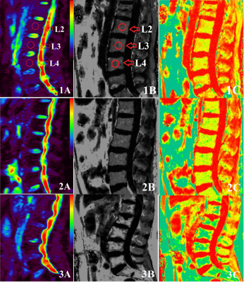 |
Application of Multi-echo Dixon Technique and IVIM-DWI in Patients with Primary Osteoporosis : A Preliminary Study with 3.0 T MRI
Fan Qiuju1, Tan Hui1, Yang Zhen1, Yu Nan1, Yu Yong1, Wang Shaoyu2, Xue Yu3, and Li Yue3
1Affiliated Hospital of Shaanxi University of Chinese Medicine, Shaanxi, China, 2MR senior scientific marketing specialist, Shaanxi, China, 3Shaanxi University of Chinese Medicine, Shaanxi, China
This study aimed to investigate the role of multi-echo Dixon technique and IVIM-DWI in assessing vertebral marrow changes among subjects with osteoporosis, osteopenia and normals. The multi-echo Dixon technique and IVIM-DWI were used to evaluate 26 healthy volunteers, 26 osteopenia patients and 24 osteoporosis patients. The FF, D and D* among osteoporosis, osteopenia and normals were significantly different.Furthermore, we found that there was a statistically significant positive correlation between D value and T-score,while the D* value and FF were negatively correlated to the T-score.
|
|
2765.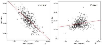 |
The potential clinical value of a quantitative Dixon sequence in predicting bone marrow fat on the lumbar spine
Ying Li1, Cuiping Ren1, Jingliang Cheng1, Jinxia Zhu2, and Stephan Kannengiesser3
1MRI, The first affiliated hospital of Zhengzhou university, Zhengzhou, China, 2MR Collaboration, Siemens Healthcare Ltd., Beijing, China, 3Siemens Healthcare GmbH, Erlangen, Germany
Osteoporosis is a systemic disease characterized by low bone mass with increased fracture risk. This work investigated and evaluated the role of the quantitative Dixon sequence in predicting the bone marrow fat on the lumbar spine, which might provide quantitative imaging parameters for accurately predicting fracture risk in patients with osteoporosis.
|
|
2766.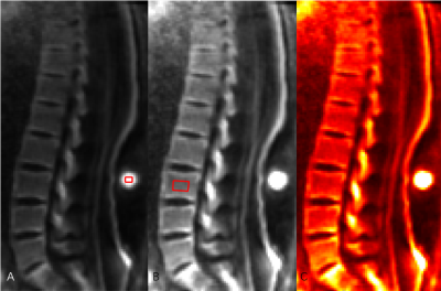 |
The feasibility study of 3D adiabatic inversion recovery prepared ultrashort TE Cones sequence in the evaluation of osteoporosis
Jin Liu1, Yajun Ma2, Xiaojun Chen1, Jianwei Liao1, Wei Li1, Yinghua Zhao3, Yijie Fang1, Long Qian4, Jiang Du2, and Shaolin Li1
1Department of Radiology, The Fifth Affiliated Hospital of Sun Yat-Sen University, Guangdong, China, 2University of California, San Diego, California, San Diego, CA, United States, 3The Tird Affiliated Hospital of Southern Medical University, Guangdong, China, 4MR Research, GE Healthcare, Beijing, China
The current study aims to assess the feasibility of proton density (PD) in the lumbar using 3D adiabatic inversion recovery prepared ultrashort TE cones (3D IR‐UTE‐Cones) sequence. In addition, the latent associations between the PD and BMD, age, gender, the oswestry disability index (ODI) were evaluated, respectively. It was concluded that proton density of trabecular bone can be applied in lumbar. Moreover, the proton density of lumbar were observed to correlate with BMD, age and ODI, which need further study to clarify this topic.
|
|
2767.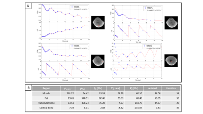 |
Simultaneous imaging of trabecular and cortical bone by analyzing the multi-echo signal decay at UTEs and conventional TEs
Sophia Kronthaler1, Christof Böhm1, Dominik Weidlich1, Maximilian N. Diefenbach1,2, and Dimitrios C. Karampinos1
1Department of Diagnostic and Interventional Radiology, Technical University of Munich, Munich, Germany, 2Division of Infectious Diseases and Tropical Medicine, Munich, Germany
Osteoporosis is characterized by a loss in bone mass and structural decrease of bone tissue which is leading to morbidity and an increased fracture risk. Fractures occur predominantly in areas with trabecular bone and the assessment of the bone volume and microstructural changes in the trabecular bone are therefore highly relevant in fracture prediction. The present study characterizes the decay of trabecularized bone marrow signal in multi-echo imaging sampling both UTEs and conventional TEs. A novel methodology is proposed for simultaneous cortical and trabecular bone imaging in the presence of bone marrow.
|
|
2768.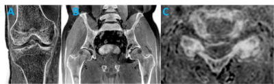 |
FRACTURE: FFE Resembling A CT Using Restricted Echo-spacing: A Novel MRI Technique for Bone Evaluation
Brian Johnson1, Ivan E. Dimitrov1, Sandeep Ganji1, and Molly Dempsey 2
1Philips Healthcare, Gainesville, FL, United States, 2Texas Scottish Rite Hospital for Children, Dallas, TX, United States
Computed tomography (CT) is the current modality of choice for imaging bone yet it suffers from poor soft tissue contrast and utilizes ionizing radiation. Unlike CT, MRI provides excellent soft-tissue contrast, but is limited in its ability to image bone. Here, we describe FRACTURE (FFE Resembling A CT Using Restricted Echo-spacing), a novel 3D gradient-echo approach that offers MRI-based bone contrast to yield clinically relevant information for patient management.
|
|
2769. |
3T CSE-MRI of multiple adipose tissue in osteoporosis patient with varying fracture risk
Dimitri Martel1, Stephen Honig2, and Gregory Chang1
1Radiology, NYU Langone Health, New York, NY, United States, 2Osteoporosis Center, NYU Langone Health, New York, NY, United States Osteoporosis (OP) is a disease of weak bone associated with an increased fracture risk. Recent studies using Chemical Shift Encoded MRI has been previously used to show differences in term of fat amount related to osteoporosis condition in spine and femur. The aim of our study is to use CSE-MRI in muscle, bone marrow and subcutaneous fat of the pelvic region in osteoporosis patient with varying degree of fracture risk. |
|
2770.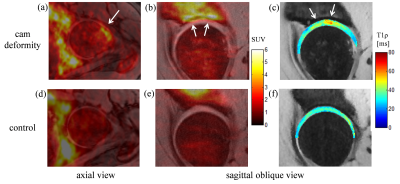 |
Simultaneous evaluation of bone and cartilage status in asymptomatic FAI subjects and healthy controls using PET-MRI
Gerd Melkus1,2, Kawan Rakhra1,2, Reggie Taylor3, Katie Dinelle3, Stephen Dinning4,5, and Paul Beaulé5,6
1Medical Imaging, The Ottawa Hospital, Ottawa, ON, Canada, 2Radiology, University of Ottawa, Ottawa, ON, Canada, 3Brain Imaging Centre, The Royal Hospital, Ottawa, ON, Canada, 4Nuclear Medicine, The Ottawa Hospital, Ottawa, ON, Canada, 5Medicine, University of Ottawa, Ottawa, ON, Canada, 6Orthopaedic Surgery, The Ottawa Hospital, Ottawa, ON, Canada
In this study subjects with asymptomatic FAI and healthy controls underwent PET-MRI to investigate hip bone activity (by [18F]-Sodium Fluoride (NaF) PET) and hip cartilage proteoglycan content (by MRI T1ρ mapping). The FAI subjects showed an increased [18F]-NaF uptake in the femoral head neck junction and in lateral area of the acetabulum, indicating increased bone remodelling. Hip cartilage T1ρ values were significantly higher in the anterior-lateral region for the FAI group, revealing a loss of proteoglycan with a possible connection to an early osteoarthritis stage. PET-MRI has potential to be a useful non-invasive tool to research FAI in vivo.
|
|
2771.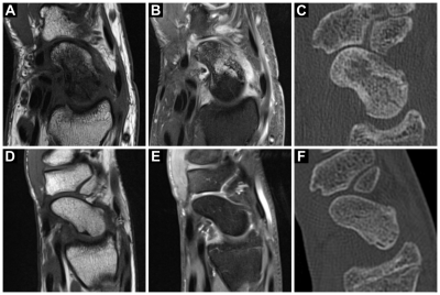 |
Idiopathic osteonecrosis of the scaphoid (Preiser’s disease): Viability patterns in contrast-enhanced MRI
Jan-Peter Grunz1,2, Carsten Herbert Gietzen1,2, Andreas Steven Kunz1, Thorsten Alexander Bley1, and Rainer Schmitt1,2
1Institute of Diagnostic and Interventional Radiology, University Hospital Würzburg, Würzburg, Germany, 2Department of Diagnostic and Interventional Radiology, Rhön-Klinikum Campus Bad Neustadt, Bad Neustadt a. d. Saale, Germany
With the ability to non-invasively display different viability patterns in the scaphoid bone, contrast-enhanced MRI is a powerful tool for detection and staging of Preiser’s disease. To our best knowledge, this study includes the largest patient sample undergoing MRI for idiopathic osteonecrosis of the scaphoid to date, thus expanding the knowledge on this rare disease entity. Our results suggest that the size of scaphoid necrosis is considerably overestimated in unenhanced MRI because necrotic bone cannot be reliably differentiated from vascularized repair tissue. Influencing decision on therapy, the extent of osteonecrosis should consequently not be assessed without administration of contrast agent.
|
|
2772.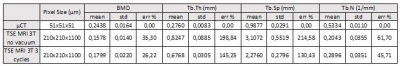 |
MRI assessment of bone microarchitecture in Human Bone samples: The issue of air bubbles artefacts
Enrico Soldati1, Martine Pithioux2, David Bendahan3, and Jerome Vicente1
1IUSTI, AixMarseille, Marseille, France, 2ISM, AixMarseille, Marseille, France, 3CRMBM, AixMarseille, Marseille, France
With the aim of assessing bone microarchitecture, several studies have intended to use MRI but the issue of air bubbles artefacts has been very scarcely reported. In the present study, we assessed air bubbles-related artefacts in MR images of human bone samples and intended to design a protocol to eliminate them. The method was validated using TSE MRI at 3T and high-resolution X-ray micro tomography. Morphological parameters computed from MRI recorded with and without the air bubbles artefacts were compared to those obtained from X-ray micro tomography.
|
2773.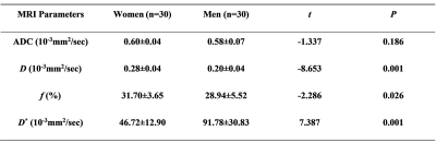 |
The Vertebral Marrow Microstructure in Healthy Young Adults with Intravoxel Incoherent Motion Diffusion-weighted MRI
Jinliang Niu1, Kaiyu Wang2, Wenqi Wu1, Tong Gong3, Jianting Li4, and Rong Zheng4
1Shanxi Medical University Second Affiliated Hospital, Taiyuan, China, 2GE Healthcare, MR Research China, Beijing, Beijing, China, 3Sichuan Provincial People's Hospital, Chengdu, China, 4Shanxi Medical University, Taiyuan, China
Intravoxel incoherent motion diffusion-weighted MRI (IVIM) plays an important role in detecting and monitoring microstructure. It offers diffusion and perfusion information at the same time without contrast agents. In this work, we demonstrated IVIM parameters can discriminate the microstructure of marrow in younger women from men compared with the apparent diffusion coefficient (ADC) values. It may provide noninvasive methods for evaluating cellularity, vascular volume and blood velocity of bone marrow.
|
|
2774.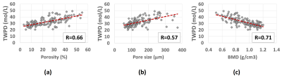 |
Predicting cortical bone microstructural properties by water proton density obtained from ultrashort echo time (UTE) MRI
Saeed Jerban1, Yajun Ma1, Hyungseok Jang1, behnam namiranian1, Nicole Le2, Hoda Shirazian1, Mark Murphy3, Jiang Du1, and Eric Y Chang2
1Radiology, University of California, San Diego, San Diego, CA, United States, 2Radiology Service, VA San Diego Healthcare System, San Diego, CA, United States, 3Orthopaedic Service, VA San Diego Healthcare System, San Diego, CA, United States
Cortical bone porous microstructure can be potentially predicted by the total water density in bone. Comparing bone signal from a relatively fast ultrashort echo time MRI (UTE-MRI) scan against the signal of a known external reference (rubber eraser) enabled us to measure total water proton density (TWPD) in 135 cortical bone specimens from 37 donors. We observed significant correlations between bone TWPD and micro computed tomography (μCT) measures (porosity, pore size, and bone mineral density, (BMD)). This relatively fast MRI technique may aid diagnosing and monitoring bone diseases.
|
|
2775.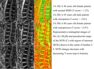 |
Quantitative Magnetization Transfer Imaging of the Lumbar in Primary Osteoporosis at 3T MRI: a Feasibility Study
Fan Qiuju1, Tan Hui1, Yang Zhen1, Yu Nan1, Yu Yong1, Wang Shaoyu2, Xue Yu3, and Li Yue3
1Affiliated Hospital of Shaanxi University of Chinese Medicine, Shaanxi, China, 2MR senior scientific marketing specialist, Shaanxi, China, 3Shaanxi University of Chinese Medicine, Shaanxi, China
This study aimed to assess the association between the magnetization transfer ratio (MTR) value and bone mineral density (BMD) in lumbar vertebral among normal, osteopenia and osteoporosis. The MT technique was used to evaluate 26 healthy volunteers, 26 osteopenia patients and 24 osteoporosis patients. The MTR in the osteoporotic, osteopenic, and normal BMD groups were statistically significant differences. Therefore, MT technique provide quantitative information about the contribution of macromolecules., which can be used as a non-invasive quantitative monitoring MRI tool for osteoporosis progression.
|
|
2776.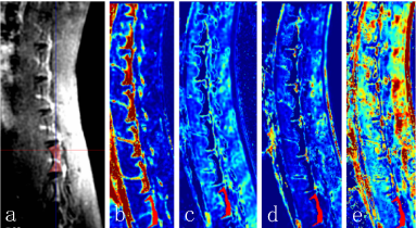 |
Quantitative MRI Study of Diabetic Bone Marrow Microvascular Disease
Pianpian Chen1, Weiyin Vivian Liu2, and Yunfei Zha3
1Hospital of Stomatology Wuhan University, Wuhan, China, 2MR Research, GE Healthcare, Beijing, China, 3Renmin Hospital of Wuhan University, Wuhan, China We quantitatively evaluate the microvascular permeability of lumbar marrow changes in alloxan-induced diabetic rabbits using dynamic contrast enhanced magnetic resonance imaging (DCE-MRI) . And to investigate whether texture analysis based on permeability parameter Ktrans map of DCE-MRI can be used to assess the fine alterations of lumbar marrow structures in early-stage diabetic rabbits. Our results confirm that texture analysis parameters based on DCE-MRI quantitative parameter Ktrans map are feasible to identify the early-stage fine changes of lumbar marrow structure in diabetic rabbits. |
|
2777.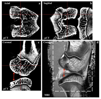 |
Correlation study of rabbit knee joint contour using high-resolution CT and MRI.
Yang Xia1, Farid Badar1, and Sarah Salem1
1Physics, Oakland University, Rochester, MI, United States
Correlated application of high-resolution MRI T2 of intact rabbit knee joints with the aid of bone density properties measured by isotropic high-resolution µCT. The combined multi-model study will aid in the understanding of the relationship between underlying bone-density changes and osteoarthritic cartilage in joint degeneration.
|
|
2778. |
MRI models of the Age-Related Bone Loss: Insights from Multi-modal Imaging Study
Malakeh Malekzadeh1, Shahrokh Abbasi-Rad 2, Mohammad Bagher Shiran1, Mojgan Asadi3, Mehdi Shamsi4, Navid Tofighi Rad4, and Hamidreza Saligheh Rad5
1Iran University of Medical Science, Tehran, Iran (Islamic Republic of), 2Quantitative Medical Imaging Systems Group, Tehran, Iran (Islamic Republic of), 3Tehran University of Medical Science, Tehran, Iran (Islamic Republic of), 4Laleh Hospital, Tehran, Iran (Islamic Republic of), 5Tehran University of medical science, Tehran, Iran (Islamic Republic of)
This study deals with assessing the ability of current clinical measures of bone to model the systematic age-related alteration of bone during aging and where does MRI stand as a new modality. Fifty Healthy volunteers with an average age of 44.53 ±7.95 were enrolled based on oral interviews and blood tests. Quantitative measurements were performed by various modalities for lumbar spine, forearm (DXA), Calcaneus (QUS), and Tiba (QCT and MRI). Pearson correlation coefficients was calculated between the bone parameters and age. The highest correlation coefficient between bone parameters and age was related to T1 (r = 0.766, p <0.01).
|
|
2779.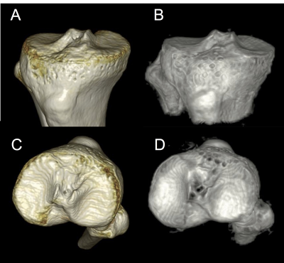 |
Zero Echo Time Imaging of the knee
Saya Horiuchi1, Taiki Nozaki1, Shigekazu Funada1, Atsushi Nozaki2, Hiroyuki Kabasawa2, Yasuyuki Kurihara1, and Nobuto Kitamura3
1Radiology Department, St. Luke's International Hospital, Tokyo, Japan, 2GE Healthcare, Japan, Tokyo, Japan, 3Orthopedic Department, St. Luke's International Hospital, Tokyo, Japan Poster Permission Withheld
Zero Echo Time (ZTE) MR imaging provides “CT-like” contrast for cortical bone of the knee. We compared sagittal images of CT and ZTE of 15 patients, and demonstrated inter-modality agreement between morphology of the cortical bone of the tibia from CT and ZTE images. Metallic screws were blurred on ZTE in patients who underwent Anterior cruciate ligament (ACL) reconstruction. ZTE may be used clinically instead of CT in some cases who can undergo ACL reconstruction.
|
|
2780.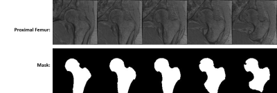 |
3T MRI Textural Analysis of the Proximal Femur in Subjects with Compared to Those without Prior Fragility Fractures
Anmol Monga1, Dimitri Martel1, Stephen Honig2, and Gregory Chang1
1Radiology, NYU Langone Health, New York, NY, United States, 2Osteoporosis Center, NYU Langone Health, New York, NY, United States
In recent years, there is greater emphasis on detecting microstructural information in bones that may lead to improved diagnosis of osteoporosis in patients. Radiomics provides a tool to quantify the textural information of medical images and may serve as a marker of bone microstructure. We aim to analyze if the distribution of the textural features computed in the proximal femur is significantly in subjects with previous fragility fractures compared to those without fracture.
|
|
2781.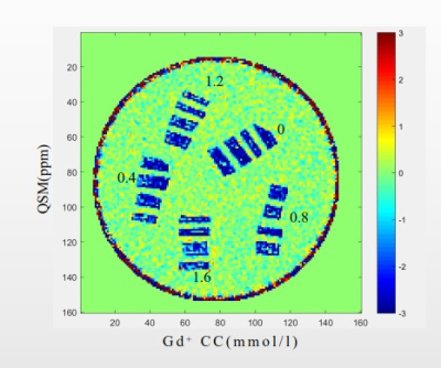 |
Evaluation of Gadolinium Deposition in Cortical Bone Using 3D-UTE-QSM in vitro: Preliminary Study
Xiaodong Zhang1,2, Hyungseok Jang2, Yanjun Chen1,2, Zhenyu Cai2, Ya-Jun Ma2, Wei Zhao2, Yanqiu Feng3, and Jiang Du2
1Department of Radiology, Third Affiliated Hospital of Southern Medical University, Guangzhou, China, 2Department of Radiology,University of California San Diego, San Diego, CA, United States, 3School of Biomedical Engineering, Southern Medical University, Guangzhou, China
After injection of gadolinium-based contrast agents (GBCAs) for enhanced magnetic resonance imaging (MRI), levels of gadolinium (Gd) in the brain are much lower than that measured in bone. The 3D ultrashort echo time Cones Quantitative susceptibility mapping (3D UTE-Cones-QSM) technique has been used to assess calcium in cortical bone. In this study, we investigated the feasibility of evaluating gadolinium deposition in cortical bone in vitro using 3D UTE-Cones-QSM on a 3T clinical scanner. Higher QSM values were observed with higher Gd concentrations, indicating that 3D UTE-Cones-QSM could be a new, non-invasive technique for evaluation of Gd deposition in bone.
|
|
2782.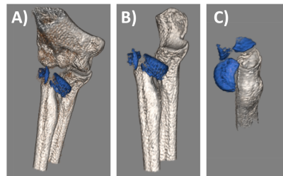 |
Bone Imaging in Musculoskeletal Trauma with Zero Echo-Time MRI
Ryan E Breighner1, Matthew F Koff1, and Hollis G Potter1
1Radiology and Imaging, Hospital for Special Surgery, New York, NY, United States
Zero Echo Time (ZTE) MRI yields positive contrast for bone visualization using MRI. The present study highlights cases in which ZTE provides additional diagnostic information in the context of orthopedic trauma. The presented cases are ones in which MRI was deemed necessary for the evaluation of ligament, tendon, or nerve integrity and ZTE was added to aid in the assessment of fractures. In conjunction with our prior work, this study demonstrates that ZTE MRI provides additional diagnostic information in musculoskeletal MRI, specifically in the context of joint trauma.
|
|
2783.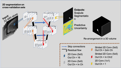 |
Do We Need CT for Producing a Fully-Automatic 3D Scapular Model? MRI meets Deep Learning for Scapular Bone Extraction
Francesco Caliva1, Victoria Wong1, Roland Krug1, Drew Lansdown1, and Valentina Pedoia1
1University of California, San Francisco, San Francisco, CA, United States
We present a fully-automatic, 2D deep learning based strategy for extracting scapular shape from a high-resolution MRI scan and we quantify network’s segmentation uncertainty. Our method has the potential to greatly improve the diagnostic process for patients with shoulder instability, rotator cuff tears, and osteoarthritis by decreasing the need for multiple imaging scans and ionizing radiation while still providing clinically-useful information to clinicians.
|
|
2784.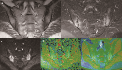 |
Comparative analysis of the diagnostic values of T2 mapping and DWI for sacroiliitis in ankylosing spondylitis
Dongdong Wang1, Huijia Yin1, Wenling Liu1, Zhong Li1, Wenguang Dou1, Dongming Han1, and Kaiyu Wang2
1The First Affiliated Hospital of Xinxiang Medical University, Weihui, China, 2GE Healthcare, MR Research China, Weihui, China
In this study, we compared and analyzed the active, inactive, and healthy control groups using T2 mapping and assessed the activity of AS using the changes in T2 values. The purposes of this study are to use T2 mapping to assess AS activity, to compare the quantitative assessment values of T2 mapping and DWI for subchondral bone marrow edema of the sacroiliac joint, and to provide potential imaging technical support for the clinical diagnosis of AS and efficacy evaluations of AS treatments.
|
|
2785.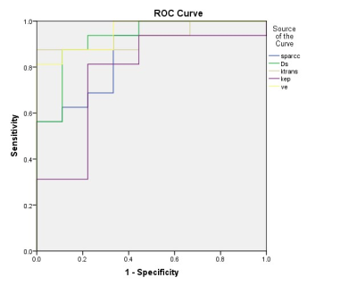 |
Assessment of Ankylosing Spondylitis Activity with Intravoxel Incoherent Motion Diffusion Weighted and Dynamic Contrast-Enhanced MR Imaging
Mengyue Wang1, Wentao Wang2, Lei Xu1, Liang Qi1, and Yuefen Zou1
1Radiology, The First Affiliated Hospital of Nanjing Medical University, Nanjing, China, 2The Affiliated Hospital of Xuzhou Medical University, Xuzhou, China
Effective diagnosis of ankylosing spondylitis (AS) activity plays an important role for clinical treatment and prognosis. In our study, we used intravoxel incoherent motion (IVIM) and dynamic contrast-enhanced (DCE) magnetic resonance imaging (MRI) to differentiate the active and chronic stage of AS. As a result, we found that Dslow, Ktrans and Ve had a high diagnostic sensitivity and specificity.
|