
Digital Poster Session
Musculoskeletal:
Musculoskeletal
2786 -2797 Clinical Applications in MSK and Others - Musculoskeletal Clinical Applications/Translation
2798 -2811 Clinical Applications in MSK and Others - Meniscus/Tendon
2812 -2826 Clinical Applications in MSK and Others - Musculoskeletal Miscellaneous 2
2827 -2842 Clinical Applications in MSK and Others - Musculoskeletal Miscellaneous 1
2786.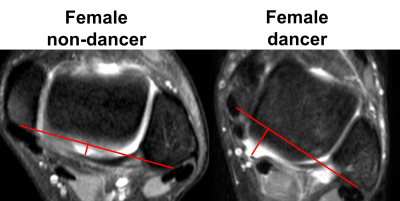 |
Quantification of joint capsule laxity of the tibiotalar and subtalar joints in ballet dancers at 3T MRI
Toshimi Tando1, Saya Horiuchi1, Hon J. Yu1, Alex Luk1, Jeffrey A. Russell2, Kelli Sharp3, and Hiroshi Yoshioka1
1Department of Radiological Sciences, University of California, Irvine, Orange, CA, United States, 2Science and Health in Artistic Performance, Ohio University, Athens, OH, United States, 3Department of Dance, The Claire Trevor School of the Arts, University of California, Irvine, Irvine, CA, United States
Joint capsule laxity/thickening is one of predisposing factors of posterior ankle impingement syndrome. The purpose of this study is to quantitatively evaluate size of the tibiotalar and subtalar joint recesses in MR images in ballet dancers, compared to non-dancers. The results showed that posterior subtalar joint recess volume, distal tibiofibular joint area, medial posterior tibiotalar recess area, and medial posterior capsule distance were larger in female dancers than female non-dancers. These findings indicate that joint capsule laxity can be measured quantitatively and used for quantitative evaluation of posterior ankle impingement.
|
|
2787. |
Advanced Knee Imaging Study in NCAA Division 1 Basketball Update: Study Design and Considerations for Multi-Site Longitudinal Study
Katherine A Young1, Elka Rubin1, Feliks Kogan1, Marianne S Black1, Madeleine Gao2, John M Sabol3, Marc Safran4, Matthew F Koff5, Hollis Potter5, Sharmila Majumdar6, and Garry Gold7
1Radiology, Stanford University, Stanford, CA, United States, 2Radiology, Hospital for Special Surgery, New York City, NY, United States, 3GE Healthcare, Waukesha, WI, United States, 4Stanford University, Redwood City, CA, United States, 5Hospital for Special Surgery, NYC, NY, United States, 6University of California San Francisco, San Francisco, CA, United States, 7Stanford University, Stanford, CA, United States
Knee injuries are especially common in jumping athletes, and, in particular, elite basketball players. We present an update two years into a multi-site study analyzing the results of a common phantom in assessing biases across and within MRI scanners, as well as to characterize the effectiveness of recruitment, assessment, and study design strategies. It was shown that more timepoints of quantitative values potentially corrects for quantitative biases longitudinally within one scanner. As attrition rates are high for athletes in general, consistent and effective methods of recruitment, early identification of key athletes, and standardized methods of assessment are all necessary.
|
|
2788.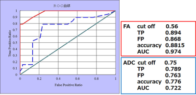 |
Value of fractional anisotropy and apparent diffusion coefficient of diffusion tensor imaging in diagnosing early carpal tunnel syndrome
Tsutomu Inaoka1, Ryosuke Sakai1, Hisanori Tomobe1, Masahiro Sogawa1, Akinori Yamamoto1, Takamitsu Uchi1, Sayuri Kato1, Rumiko Ishikawa1, Tomoya Nakatsuka1, Noriko Kitamura1, Shusuke Kasuya1, and Hitoshi Terada1
1Radiology, Toho University Sakura Medical Center, Sakura, Japan Poster Permission Withheld
There are decreased FA and increased ADC significantly with age in healthy subjects. There are significant differences in FA and ADC between early-stage CTS subjects and age-matched healthy subjects. 0.56 was found to be cutoff value for FA with 89% sensitivity and 88% accuracy and 0.75x10⁻³mm2/s was found to be cutoff value for ADC with 79% sensitivity and 78% accuracy. Although FA and ADC well correlate with clinical examinations and NCS results, the value of FA is much stronger than that of ADC for diagnosing CTS earlier. DTI can be used for the early diagnosis of CTS.
|
|
2789. |
The Feasibility of the Simultaneous Multi-slice BLADE Technique in Shoulder Imaging
Yincong Dou1, Jing An2, Kun Zhou2, Xianchang Zhang3, Fengshan Yan1, Shewei Dou1, and Meiyun Wang1
1Department of Radiology, Henan Provincial People’s Hospital, Zhengzhou, China, 2Siemens Shenzhen Magnetic Resonance Ltd.,, Shenzhen, China, 3MR Collaboration, Siemens Healthcare Ltd.,, Shenzhen, China
The purpose of this study was to evaluate the use of Simultaneous Multi-slice BLADE technique for artifact reduction and overall image quality improvement of the shoulder. All the patients underwent the conventional TSE and SMS BLADE sequences. The images were graded by two radiologists for image artifacts, overall image quality, and delineation of anatomic structure. The results showed that the new technique can significantly reduce artifacts, shorten scanning time, improve overall image quality, display more detailed anatomical structures and provide better clinical diagnostic images than conventional shoulder joint sequence.
|
|
2790. |
Evaluating the preoperative diagnose of lumbosacral radiculopathy using diffusion tensor imaging and tractography
Zhao Feng1, Shi Yin1, Dou Weiqiang2, Shi Haibin1, and Ren Yongxin1
1The First Affiliated Hospital of Nanjing Medical University, Nanjing, China, 2GE Healthcare, MR Research, Beijing, P.R. China, Beijing, China
The aim of this study is mainly to explore the pre-diagnosis value of DTI technology applied to lumbosacral radiculopathy (LSR). By measuring 80 patients with unilateral disc related lumbosacral nerve root compression, we found that DTI technology combined with tractography not only could identify the type of disc herniation, assess the degree of nerve root damage, but also confirm the responsible nerve root. Therefore, we can demonstrate that DTI combined with tractography can improve the diagnosis rate of LSR and reduce iatrogenic injury.
|
|
2791. |
Quantitative evaluation of pubic symphysis in late pregnancy using T2* mapping
Tao Gong1, Jinxia Zhu2, and Guangbin Wang1
1Shandong Medical Imaging Research Institute, Jinan, China, 2Siemens Healthcare, MR Collaborations NE Asia, Beijing, China
Degenerative joint disease of pubic symphysis is the primary cause of groin pain during late pregnancy. T2* mapping is a reliable tool in articular cartilage imaging, has been widely used to evaluate the degeneration of knee joint and intervertebral discs, while no studies was found in the pubic symphysis during pregnancy. Our results indicated the T2* values of cartilage between pubic bones increased significantly during late pregnancy, which were mainly driven by the posterior sub-region of cartilage. This study suggests that T2* mapping is a sensitive quantitative method capable of detecting cartilage changes of pubic symphysis during pregnancy.
|
|
2792. |
Diagnostic value of MAVRIC-SL sequence in patients with suspected infection after orthopedic metal implantation
Jingyi Zhu1, Lizhi Xie2, and Songbai Li1
1the Department of Radiology, First Affiliated Hospital of China Medical University, Shenyang,Liaoning, China, 2GE Healthcare, MR Research, Beijing, China
Multiacquisition with variable resonance image combination (MAVRIC-SL) sequence is a novel metal artifact reduction technique that can effectively reduce metal artifacts in patients who undergo joint replacement or fracture internal fixation. The purpose of this study was to investigate the diagnostic advantages of the MAVRIC-SL sequence MAVRIC-SL compared with conventional FSE sequence for patients with orthopedic metal implants who are suspected of having the infection.
|
|
2793. |
Brachial plexus imaging using DANTE-SPACE: comparison with conventional T2-SPACE at 3T
Xiangchuang Kong1, Jie Meng1, Xiaoyong Zhang2, Huiting Zhang3, Xiaoming Liu1, and Dingxi Liu1
1radiology, Union Hospital, Tongji Medical College, Huazhong University of Science and Technology, Wuhan, China, 2MR Collaborations, Siemens Healthcare, Shenzhen, China, Shenzhen, China, 3MR Scientific Marketing, Siemens Healthcare, Wuhan, China
Three-dimensional high-resolution visualization of brachial plexus using T2-weighted SPACE (T2-SPACE) sequence has very high clinical value for the evaluation of brachial plexopathy. However, conventional T2-SPACE is limited by the lack of relative contrast between nerves and their surrounding tissues. In contrast to T2-SPACE, DANTE-SPACE has been proposed with superior blood flow suppression and might be a potential alternative to address its shortcoming. The objective of this study was to evaluate the T2-weighted DANTE-SPACE for its capability to diagnose brachial plexus due to its superior blood flow suppression.
|
|
2794.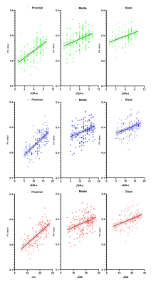 |
Quantitative evaluation of compressed nerve roots treated by PETD in lumbosacral radiculopathy using diffusion tensor imaging
Shi Yin1, Dou Weiqiang2, Shi Haibin1, and Ren Yongxin1
1The First Affiliated Hospital of Nanjing Medical University, Nanjing, China, 2GE Healthcare, MR Research, Beijing, P.R. China, Beijing, China
In this study, we aim to investigate if diffusion tensor imaging can clinically evaluate lumbosacral radiculopathy before and after percutaneous transforaminal endoscopic discectomy. By measuring 66 patients pre- and post-operatively, we found the FA values at the proximal sub-regions (subarticular zone) are more effective and had a significant correlation with clinical symptoms. Besides, both located in the proximal sub-regions, the abnormal parts of DTT visualized nerves coincided with the changes of the FA values in compressed nerves before and after surgery. Therefore, in lumbosacral radiculopathy, DTI is a potential tool for clinical evaluation before and after PETD surgery.
|
|
2795.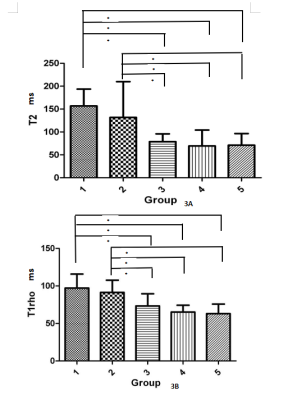 |
Multi-parametric MRI in the evaluation of lumbar intervertebral disc and cartilage endplate degeneration at clinical 3 T
Qianqian Hu1, Weiqiang Dou2, Yong Shen3, Yuan Yin1, Qingqing Zhou1, Yusheng Yu1, and Hong Zhang1
1The affiliated Jiangning hospital of Nanjing medical university, Nanjing, China, 2GE Healthcare, MR Research China, Beijing, China, 3GE Healthcare, MR Research China, Nanjing, China
multi-parametric MRI was applied to qualitatively and quantitatively investigate the degenerations of lumbar intervertebral disc and cartilage endplate. After measuring twenty-nine patients with low back pain, we found that, for intervertebral discs, its relaxation properties of T2 and T1rho, the image SNR as well as the fat fraction revealed strong correlations with its degeneration degrees. Moreover, zero echo time (ZTE) imaging also showed promise in the visualization of cartilage endplate. Therefore, multi-parametric MRI can be considered an effective tool in the evaluation of spinal degeneration, especially at the early stage.
|
|
2796.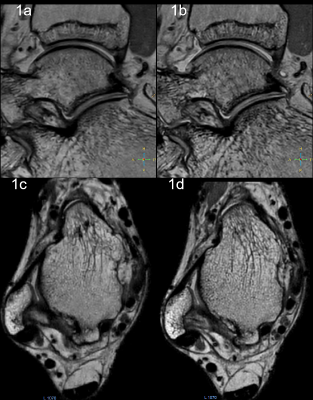 |
Evaluation of the 3D VISTA sequence with compressed sensing acceleration for imaging of the ankle: a Preliminary study
Liu Xiaoming1, Dai Meng1, Liu Xi1, Kong Xiangchuang1, and Wang Jiazheng2
1Departments of Radiology, Union Hospital, Tongji Medical College, Huazhong University of Science and Technology, Wuhan, China, 2Philips Healthcare, Beijing, China
The three-dimensional (3D) high resolution volume isotropic FSE sequence of ankle magnetic resonance imaging has the capabilities of acquiring thin-sliced sections and performing MPR, at the cost of long scan time. Several recent studies showed that the compressed sensing(CS) helps to reduce scan time, which is especially beneficial in 3D imaging. The aim of this study was to investigate the feasibility of 3D VISTA imaging with CS in ankle joint imaging, in comparison to 3D-VISTA with parallel imaging (PI ). The results showed that the acquisition time of 3D-VISTA MRI was reduced with CS while the image quality was retained.
|
|
2797.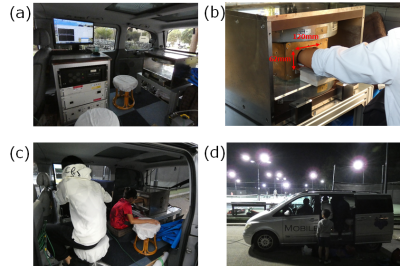 |
Clinical feasibility of mobile medical screening for wrist injuries in tennis players using a small car-mounted MRI
Tomoki Miyasaka1, Michiru Kajiwara1, Akito Kawasaki2, Yoshikazu Okamoto3, and Yasuhiko Terada1
Video Permission Withheld
1Institute of Applied Physics, University of Tsukuba, Tsukuba, Japan, 2Graduate School of Human Sciences, University of Tsukuba, Tsukuba, Japan, 3Institute of Clinical Medicine, University of Tsukuba, Tsukuba, Japan
We have developed a portable MRI that can provide opportunities for mobile operation in many environments including screening and primary care suites. Here we showed clinical feasibility of mobile medical screening using the portable MRI. We transported the scanner to a tennis club and imaged the wrists of 21 junior tennis players. The image quality was high enough to detect the TFCC injuries in most cases. Our results indicate that the portable system could be applicable for mass screening and early diagnosis of wrist injuries.
|
 View the Poster
 Watch the Video
2798.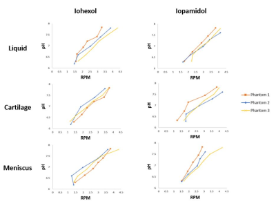 |
AcidoCEST-UTE MRI is a Reproducible Method to Measure Cartilage and Meniscus pH
Rachel A High1,2, Yang Ji1, Ya-Jun Ma1, Qingbo Tang2, Mark E Murphy3, Jiang Du1, and Eric Y Chang1,2
1Radiology, University of California San Diego, San Diego, CA, United States, 2Radiology Service, VA San Diego Healthcare System, San Diego, CA, United States, 3Orthopedic Surgery Service, VA San Diego Healthcare System, San Diego, CA, United States
The acidification of musculoskeletal tissues is being investigated as a way to localize pain in osteoarthritis. AcidoCEST-UTE MRI can measure pH in tissues such as cartilage and meniscus, though in vivo validation is difficult. Thus, it is imperative that the acidoCEST-UTE sequence be reproducible and verifiable ex vivo, where pH measurements can be confirmed with a pH electrode. This study tested the reproducibility of the acidoCEST-UTE sequence in three ex vivo phantoms, confirming the pH sensitivity of the technique and demonstrating the consistency of the method and results.
|
|
2799.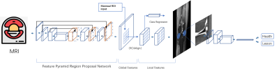 |
A two-stage Convolutional neural network for meniscus segmentation and tear classification
Xing Lu1,2, Chang Guo1, Kai Zheng1, Dashan Gao2, Yunqiang Chen2, Haimei Chen1, and Yinghua Zhao1
1Department of Radiology, The Third Affiliated Hospital of Southern Medical University, Guangzhou, China, 212Sigma Technologies, San Diego, CA, United States Poster Permission Withheld
Artificial intelligence for interpreting MRI meniscal tear could prioritize high risk patients and assist clinicians in making diagnoses. In this study, a two-stage end-to-end convolutional neural network, with Mask rcnn as backbone for object detecting and Resnet for classification, is proposed for automatically detecting torn in the meniscus on MRI exams. With training dataset of 507 MR images and validation dataset of 69 MR images, the meniscus detection achieves a recall of 0.95 when 1 false positive of 1 image, and the ROC for classification of torn meniscus get a AUC of 0.99.
|
|
2800.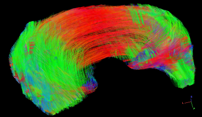 |
High Angular Resolution Diffusion Imaging (HARDI) in Meniscus
Nian Wang1, Gary Cofer1, Louis E. DeFrate2, Abigail Holt2, Amy L. McNulty2, Yi Qi1, Charles E. Spritzer1, and G. Allan Johnson1
1Department of Radiology, Duke University, Durham, NC, United States, 2Department of Orthopaedic Surgery, Duke University, Durham, NC, United States
Application of diffusion magnetic resonance imaging (dMRI) to map the complex collagen fibril structures of meniscus is still challenging, due to the short T2/T2* values, low fractional anisotropy (FA) values, and relatively low signal-to-noise (SNR). In this study, we imaged the porcine menisci in a preclinical 7 T system with relatively short echo time (TE ~ 11 ms). A 3D diffusion-weighted spin-echo pulse sequence was used for whole meniscus tractography at 125 µm isotropic resolution.
|
|
2801.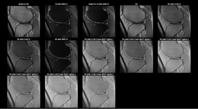 |
Subtle Intensity Graduating Homomorphic Transform with a Small-percentage of Fat-saturation to Improve Conspicuity of Meniscal Tears at 7T
Venkata Veerendranadh Chebrolu1,2, Peter Kollasch1,2, Eric G Stinson1,2, Andrew J Fagan2, Joel P Felmlee2, Benjamin M Howe2, Matthew A Frick2, and Kimberly K Amrami2
1Siemens Medical Solutions USA, Inc., Rochester, MN, United States, 2Department of Radiology, Mayo Clinic, Rochester, MN, United States
In this work, we propose the use of Subtle Intensity Graduating Homomorphic Transform (SIGHT) along with application of a small percentage of fat-saturation (fat-sat) to improve the conspicuity of meniscal tears at 7T. 2 musculoskeletal radiologists compared turbo-spin-echo (TSE) proton-density (PD)-weighted knee MRI (without fat-sat) with 12 different MRI options (TE, TR, fat-sat, field-of-view, and image-processing variations) for conspicuity of meniscal tears and rated 1st, 2nd and 3rd preferred options for each case. SIGHT image-processing applied to PD-weighted MRI along with a small percentage of fat-sat provided better conspicuity of meniscal tears than the regular PD- and T2-weighted MRI images.
|
|
2802.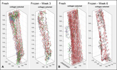 |
Magic Angle Directional Imaging (MADI) visualises changes to collagen fibre orientation in patellar tendons after freezing
Karyn Elizabeth Chappell1, Mihailo Ristic2, Donald McRobbie3, Wladyslaw Gedroyc4, Djordje Brujic2, and Catherine Van Der Straeten5
1Medicine, Orthopaedic Surgery, Stanford University, Redwood City, CA, United States, 2Mechanical Engineering, Imperial College London, London, United Kingdom, 3University of Adelaide, Adelaide, Australia, 4Imperial College London, London, United Kingdom, 5Imperial College London/Ghent University, Ghent, Belgium
Does freeze/thaw of cadaveric specimens’ damage collagen fibre orientation? Magic angle directional imaging (MADI) in caprine knees assessed the underlying tissue structure before and after freezing for three and six weeks. Tendon thickness reduced after 6 weeks of freezing. Segmented collagen containing voxels decreased by half after freezing. Voids appeared in the internal tendon structure suggestive of ice crystal formation that disrupted collagen fibre orientation. The severity of the structural changes increased the longer the tissue was frozen. Interpreting results with frozen/thawed cadaveric specimens needs care as freezing damages collagen fibre structure that may impact on biomechanical and other properties.
|
|
2803.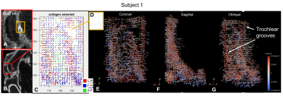 |
Magic Angle Directional Imaging (MADI) standard deviation maps correlate with knee pathology in a spontaneous injury model.
Karyn Elizabeth Chappell1, Djordje Brujic2, Mihailo Ristic2, Donald McRobbie3, Wladyslaw Gedroyc4, Catherine Van Der Straeten5, and Richard Meeson6
1Medicine, Orthopaedic Surgery, Stanford University, Redwood City, CA, United States, 2Mechanical Engineering, Imperial College London, London, United Kingdom, 3University of Adelaide, Adelaide, Australia, 4Imperial College London, London, United Kingdom, 5Imperial College London/Ghent University, Ghent, Belgium, 6Royal Veterinary College, London, United Kingdom
A proof of concept study in a spontaneous ligament rupture dog model assessed if the magic angle effect disappears in unhealthy tissue. Ten dogs were scanned using the MADI technique. Standard deviation (SD) maps of signal intensity were computed and measured in an interactive display. Three subjects showed abnormally high SD; two at the ACL origin, one in the trochlear articular cartilage. Fibre orientation maps showed either disorganised or highly organised collagen fibres in these subjects. On dissection two subjects had partially torn ACLs and the other a fibrocartilage lesion. There was excellent correlation between the MADI and dissection findings.
|
|
2804.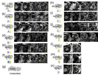 |
High-resolution 3T MRI of the dorsal and volar radioulnar ligament of the wrist
Saya Horiuchi1,2, Hon Yu1, Toshimi Tando1, Taiki Nozaki2, and Hiroshi Yoahioka1
1Department of radiological sciences, University of California, Irvine, Irvine, CA, United States, 2Radiology Department, St. Luke's International Hospital, Tokyo, Japan Poster Permission Withheld
It is technically challenging to diagnose injuries to the radioulnar ligaments (RUL) of the wrist on magnetic resonance imaging (MRI). We identified and classified the morphology of the dorsal and volar radioulnar ligaments (RUL) of the wrist into five types using 3T MRI. Two hundred and nine participants were retrospectively evaluated using 3T axial 2D and reformatted axial isotropic 3D images. Both dorsal and volar RULs attach to the radial to dorsal side of the ulnar styloid. The RUL was equally identified on high-resolution two-dimensional and on isotropic three-dimensional MRI.
|
|
2805.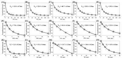 |
The Angular Dependence of 3D Ultrashort Echo Time Cones Adiabatic T1ρ (3D UTE Cones-AdiabT1ρ) Imaging of the Achilles Tendon
Mei Wu1,2, Mingxin Chen1, Yajun Ma1, Akhil Kasibhatla1, Lidi Wan1, Saeed Jerban1, Hyungseok Jang1, Eric Y Chang1,3, and Jiang Du1
1Department of Radiology, University of California, San Diego, San Diego, CA, United States, 2Department of Radiology, Guangzhou First People’s Hospital, School of Medicine, South China University of Technology, Guangzhou, China, 3Radiology Service, VA San Diego Healthcare System, San Diego, CA, United States
This study investigated the magic angle effect in three-dimensional ultrashort echo time Cones Adiabatic T1ρ (3D UTE Cones-AdiabT1ρ) imaging of the Achilles tendon on a clinical 3T scanner. The magic angle effect was investigated by repeated UTE Cones-AdiabT1ρ imaging of five human Achilles tendon samples at five angular orientations ranging from 0° to 90° relative to the B0 field. Conventional Cones continuous wave T1ρ (Cones-CW-T1ρ) and Cones-T2* sequences were also applied for comparison. Cones-AdiabT1ρ showed a much reduced magic angle effect as compared to regular Cones-CW-T1ρ and Cones-T2*, suggesting its potential use as a novel biomarker for musculoskeletal (MSK) imaging.
|
|
| 2806. | Quantitative ultrashort echo time (UTE)-Cones monitoring of tendon healing after arthroscopic rotator cuff repair (ARCR) surgery
xue yu xie1, puye wu2, and shuang chen3
1radiology, huashan hospital, shanghai, China, 2GE healthcare, beijing, China, 3huashan hospital, shanghai, China
A longitudinal monitoring of the supraspinatus tendon healing during the first year after arthroscopic rotator cuff repair (ARCR) surgery using quantitative ultrashort echo time (UTE)-Cones.
|
|
2807. |
The influence of the knee position on vTE-T2* values of patellar- and quadriceps tendon measured at 3T and 7T
Benedikt Hager1,2, Markus M. Schreiner3, Vladimir Mlynarik1, Vladimir Juras1, Xeni Deligianni4,5, Oliver Bieri4,5, Reinhard Windhager3, and Siegfried Trattnig1,2
1Department of Biomedical Imaging and Image-guided Therapy, High Field MR Centre, Medical University of Vienna, Vienna, Austria, 2CD Laboratory for Clinical Molecular MR Imaging, Vienna, Austria, 3Department of Orthopaedics and Trauma Surgery, Medical University of Vienna, Vienna, Austria, 4Department of Radiology, Division of Radiological Physics, University of Basel Hospital, Basel, Switzerland, 5Department of Biomedical Engineering, University of Basel, Allschwil, Switzerland
In our study in which we used a variable echo-time sequence for T2*-mapping of tendons in vivo at 3T and 7T, we showed that even small changes in the knee position in the knee coil can lead to significant changes in the T2*-values of the patellar tendon and quadriceps tendon. We have also shown that the T2*-values of the tendons correlate well with the angle of the bulk of the collagen fibers to the magnetic field. Based on these results, we recommend that for T2*-mapping it is important to examine the tendon in exactly the same position in the follow-up.
|
|
2808. |
Volumetric tendon segmentation using ultra-short echo-time (UTE) imaging and bivariate relaxation parameter-based histogram analysis
Martin Krämer1, Marta B Maggioni1, and Jürgen R Reichenbach1
1Medical Physics Group, Institute of Diagnostic and Interventional Radiology, Jena University Hospital - Friedrich Schiller University Jena, Jena, Germany
Segmentation of tendons based on MRI data is challenging because of their very short transverse relaxation times and typically curved structure as well as small diameter. In this work, we performed combined T2* and T1 mapping using multi-echo and variable flip angle ultra-short echo-time imaging. Based on the resulting relaxation parameter maps, bivariate histograms were calculated which showed distinct clusters that could be ascribed to various tissues. Ranges for both relaxation parameters were defined using these histograms and applied to volumetrically segment and subsequently visualize the patellar and quadriceps tendon.
|
|
2809. |
Ultrashort echo time adiabatic T1ρ (UTE-Ad-T1ρ) can predict the mechanical property differences in human anterior cruciate ligament (ACL)
Saeed Jerban1, Takehito Hananouchi2, Yajun Ma1, Erik Dorthe3, behnam namiranian1, Jonathan Wong4, Mei Wu1, Darryl D'lima3, Eric Y Chang4, and Jiang Du1
1Radiology, University of California, San Diego, San Diego, CA, United States, 2Department of Mechanical Engineering, Osaka Sangyo University, Daito, Osaka, Japan, 3Shiley Center for Orthopedic Research and Education at Scripps Clinic, La Jolla, CA, United States, 4Research Service, VA San Diego Healthcare System, San Diego, CA, United States
Clinical MRI techniques typically show the anterior cruciate ligament (ACL) as low in signal. Commonly used quantitative MRI techniques are sensitive to tissue orientation and magic angle effect. A recently developed ultrashort echo time (UTE)-based adiabatic T1ρ technique (UTE-Ad-T1ρ) can detect high signal in the ACL and may be less sensitive to magic angle effect. We have investigated for the first time the correlation between UTE-Ad-T1ρ and ACL mechanical properties. T1ρ showed significant strong correlation with average elastic modulus of ACL specimens. The UTE-Ad-T1ρ technique is highlighted as a potential tool to noninvasively assess the ACL mechanical properties.
|
|
2810. |
Comparison of Meniscal T2* Metrics in Elite Basketball Players and Swimmers
Erin C Argentieri1, James C Yoder1, Garry Gold2, Sharmila Majumdar3, Matthew F Koff1, and Hollis G Potter1
1Radiology and Imaging, Hospital for Special Surgery, New York, NY, United States, 2Stanford University, Stanford, CA, United States, 3University of California San Francisco, San Francisco, CA, United States
As basketball players represent a population with an inherently high risk of sustaining meniscal injuries, studying how basketball play can lead to changes in the meniscus is of clinical importance. To date, no studies have been performed to evaluate T2* values of the meniscus in high performance athletes. Therefore, the purpose of this study was to utilize ultra-short TE (UTE) MRI to compare meniscal T2* values between basketball players and swimmers. Significant differences of T2* values were found between the medial and lateral menisci. No significant difference of meniscal T2* values were found between basketball players and swimmers.
|
|
2811.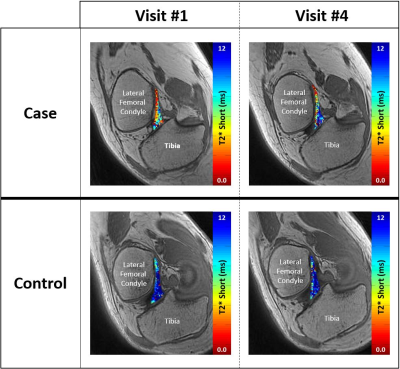 |
Pre- to post-ovulatory changes in ACL T2* metrics over the course of the female menstrual cycle: A new biomarker for ACL injury risk?
Erin C Argentieri1, Tatum W Braun1, Ryan E Breighner1, Bin Q Lin2, Ellen K Casey3, Shari T Jawetz1, Alissa J Burge1, Matthew F Koff1, and Hollis G Potter1
1Radiology and Imaging, Hospital for Special Surgery, New York, NY, United States, 2Biostatistics, Hospital for Special Surgery, New York, NY, United States, 3Physiatry, Hospital for Special Surgery, New York, NY, United States
This study evaluates changes in ACL T2* metrics over the course of the female menstrual cycle. In the pre-ovulatory phase, normally ovulating case subjects demonstrated significant shortening of T2*S and PS in comparison to visit #4 (post-ovulatory phase). Non-ovulatory control subjects displayed no significant changes over time. These findings suggest that shifts in collagen bound water occur within the ACL over the course of the menstrual cycle. Shifts in tissue water content have been associated with altered mechanical properties, and changes in ligament stiffness may alter proprioceptive sense, contribute to increases in laxity, and alter ACL-injury risk.
|
2812.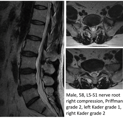 |
Compression of nerve root causes high degree of fat infiltration of lumbar multifidus muscle
Hui Hao1, Jia Yin Tong1, Xiao Cheng Wei2, and Jian Yang1
1Department of Radiology, the First Affiliated Hospital, Xi'an Jiaotong University, xi'an, China, 2MR Research China, GE Healthcare, Bei Jing, China
The increase of multifidus muscle fat infiltration will affect the postoperative rehabilitation of patients with lumbar spine surgery. In this study, reasons for fat infiltration on both sides of multifidus muscles were analyzed retrospectively. Through Priffman grading evaluating the intervertebral disc and Kader grading for fat infiltration, a retrospective analysis was conducted on 453 cases of intervertebral discs images. In our study, nerve root compression was found to be correlated with degree of multifidus muscle fat infiltration. This correlation support the hyposis that nerve root compression would result in an increase in the adiposity of multifarious muscles.
|
|
2813.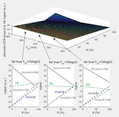 |
Stirring STIR contrast
Nick Zafeiropoulos1, Stephen Wastling1, Jasper Morrow2, Uros Klickovic1, Pietro Fratta1, Sachet Shah1, Enrico De Vita3, Mary M Reilly2, Tarek Yousry1, and John Thornton1
1UCL Queen Square Institute of Neurology, London, United Kingdom, 2MRC Centre for Neuromuscular Diseases, London, United Kingdom, 3King's College London, London, United Kingdom
In vivo lower-limb STIR images were compared with images simulated using a theoretical signal model with experimentally determined muscle-water T2 and fat fraction values. Nominally T2-weighted STIR contrast is seen to depend on changing tissue-water relative proton density as fat content increases in diseased muscle, in addition to expected T2 dependent changes. Imperfect inversion-recovery fat nulling may also cause unexpected hyper-intensity in regions of high fat content. These observations may have implications for the clinical interpretation of STIR signal intensity.
|
|
2814.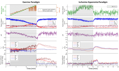 |
Dynamic multinuclear measurements in the calf with a dedicated RF coil while taking into account the nuclear Overhauser enhancement effect.
Alfredo Liubomir Lopez Kolkovsky1,2, Benjamin Marty1,2, Marc Lapert3, Eric Giacomini1, and Pierre G Carlier1,2
1NMR Laboratory, Institute of Myology, Paris, France, 2NMR Laboratory, CEA/DRF/IBFJ/MIRCen, Paris, France, 3Siemens Healthcare SAS, Saint-Denis, France
Interleaving multinuclear NMR measurements in the muscle during a transient state enriches the value of the exam by generating multiple data sets simultaneously. Here we show the results of our multinuclear interleaved approach using a 1H/31P dual-tune RF coil dedicated for calf experiments during an exercise and an ischemia-hyperemia paradigm. The nuclear Overhauser enhancement effect, which is dependent on the sequence and RF design, is characterized and its impact on rephosphorylation rate estimation and the [Pi]/[PCr] ratio is evaluated. The proposed methodology constitutes a powerful tool to evaluate mitochondrial function and metabolic efficiency in vivo.
|
|
2815.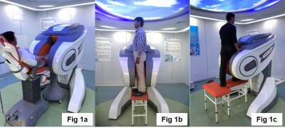 |
MRI of the knee during weight-bearing standing postures involving positions of joint extension and flexion
YueFen Zou1, Yu Zheng1, HongYuan Ding1, SiSeung Kim2, Shekhar Suresh Chandra3, Jurgen Fripp4, Jian Bao2, HuiYao Zhang2, Bing Keong Li2, and Craig Engstrom 5
1Medical Imaging, The First Affiliated Hospital of Nanjing Medical University, Nanjing, China, 2Jiangsu LiCi Medical Device Co. Ltd, LianYunGang, China, 3ITEE, The University of Queensland, Brisbane, Australia, 4The Australian e-Health Research Centre, CSIRO, Brisbane, Australia, 5School of Human Movement & Nutrition Science, The University of Queensland, Brisbane, Australia
Standing weight-bearing MR images of the knee joint in full extension and at various flexion angles (~15°, 30°) were acquired using 2D and 3D sequences on a newly developed open 0.25T extremity MRI system. 3D FLASH images were used to calculate measures of patella positioning used in previous clinical investigations as well as to produce MRI-based 3D bone models. The use of MRI and 3D modeling for topographical analyses of the bones and soft-tissues of the knee joint have the potential to provide more detailed assessment of pathoanatomical conditions involving structures such as the patella tendon and the menisci.
|
|
2816.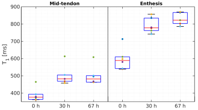 |
Immersion of ex vivo Achilles tendon in phosphate buffered saline influences their T1 and T2* relaxation times
Martin Krämer1, Matthias R Kollert2,3, Nicholas M Brisson2, Marta B Maggioni1, Georg N Duda2,3, and Jürgen R Reichenbach1
1Medical Physics Group, Institute of Diagnostic and Interventional Radiology, Jena University Hospital - Friedrich Schiller University Jena, Jena, Germany, 2Julius Wolff Institute and Center for Musculoskeletal Surgery, Charité – Universitätsmedizin Berlin, Berlin, Germany, 3Berlin-Brandenburg Center and School for Regenerative Therapies, Charité – Universitätsmedizin Berlin, Berlin, Germany
Controlling tissue integrity and hydration levels is a crucial step when preparing and measuring ex vivo samples of tendons. In this work, we immersed ex vivo Achilles tendons in phosphate buffered saline solution and measured T1 and T2* relaxation times at baseline, 30 h and 67 h after immersion using 3D ultra-short echo-time imaging with variable flip angles and echo-train shifted multi-echo acquisition, respectively. Results based on regions-of-interest in mid-tendon and enthesis areas showed a significant increase in both T1 and T2* after 30 h of immersion in the phosphate buffered saline solution.
|
|
2817.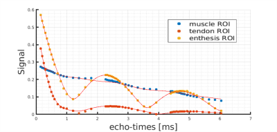 |
A three compartment complex signal model to describe the signal decay of ultrashort T2* tissues applied to ex vivo ovine Achilles tendons
Marta Brigid Maggioni1, Martin Krämer1, and Jürgen R. Reichenbach1,2,3,4
1Medical Physics Group, Institute of Diagnostic and Interventional Radiology, Jena University Hospital - Friedrich Schiller University, Jena, Germany, 2Michael Stifel Center for Data-driven and Simulation Science Jena, Friedrich Schiller University, Jena, Germany, 3Abbe School of Photonics, Friedrich Schiller University, Jena, Germany, 4Center of Medical Optics and Photonics, Friedrich Schiller University, Jena, Germany Poster Permission Withheld
Tendons are highly ordered tissues, mainly composed of collagen, and characterized by very short transverse T2* relaxation times. Even when using ultra-short echo-time imaging sequences, quantification of T2* is challenging as the origin and characteristics of the signal decay in tendons is still under debate. In this work, we finely sampled the decay of the transverse magnetization using an echo-train shifted multi-echo ultra-short echo-time sequence with 55 echoes and applied a complex tri-exponential model to extract T2* constants.
|
|
2818.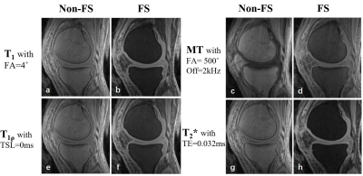 |
Fat suppression in 3D Quantitative Ultrashort Echo Time Cones (qUTE-Cones) imaging
Yanjun Chen1,2, Zhenyu Cai2, Liang Li2, Carolyn Xie2, Michael Carl3, Eric Y Chang4, Jiang Du2, and Yajun Ma2
1The Third Affiliated Hospital of Southern Medical University, Guangzhou, China, 2UC San Diego, San Diego, CA, United States, 3GE healthcare, San Diego, CA, United States, 4VA health system, San Diego, CA, United States
Strong fat signal contamination leads to significant errors in quantitative UTE (qUTE) imaging of musculoskeletal tissue. In this study, we used a fat suppression technique to investigate whether fat signals could be sufficiently suppressed in qUTE imaging and whether the fat saturation preparation would affect the resultant qUTE measures due to the induced water attenuation.
|
|
2819.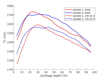 |
Effect of room temperature storage on relaxation properties of ex vivo articular cartilage
Olli Nykänen1, Nina Hänninen1,2, and Mikko J Nissi1,2
1Department of Applied Physics, University of Eastern Finland, Kuopio, Finland, 2Research Unit of Medical Imaging, Physics and Technology, University of Oulu, Oulu, Finland
Typical measurement protocols for quantitative MRI of ex vivo osteochondral samples may last hours when multiple parameters with high resolution are measured. However, knowledge about the changes in the relaxation properties of articular cartilage during long scans is inadequate. In this study, we measured T1, adiabatic T1rho and T2-relaxation times for two osteochondral samples directly after sample preparation and repeated the measurements after 15 hours of storage at room temperature. T1-relaxation time decreased noticeably at later timepoint while adiabatic T1rho and T2-relaxation times remained the same. Due to small sample size, statistical significance of the results could not be verified.
|
|
2820.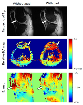 |
T1ρ imaging in 7T: evaluation of high dielectric pad for SAR reduction
Jeehun Kim1,2, Wanyong Shin2,3, Mei Li1,2, and Xiaojuan Li1,2,3
1Biomedical Engineering, Cleveland Clinic, Cleveland, OH, United States, 2Program of Advanced Musculoskeletal Imaging (PAMI), Cleveland Clinic, Cleveland, OH, United States, 3Department of Diagnostic Radiology, Cleveland Clinic, Cleveland, OH, United States
To fully utilize the superior signal-to-noise ratio of 7T MRI for cartilage T1ρ imaging, high specific-absorption ratio (SAR) needs to be accounted for. Previous researches showed the use of high dielectric pad could substantially decrease the SAR in 7T MRI. In this work, the effect of the use of high dielectric pad on T1ρ imaging in terms of SAR level and T1ρ relaxation time quantification.
|
|
2821.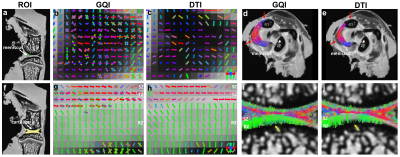 |
Resolving the Complex Fiber Structures in Knee Joint Using High Resolution Diffusion Imaging
Nian Wang1, Anthony J. Mirando2, Gary Cofer1, Yi Qi1, Matthew J. Hilton2, and G. Allan Johnson1
1Department of Radiology, Duke University, Durham, NC, United States, 2Department of Orthopaedic Surgery, Duke University, Durham, NC, United States
Using high-order diffusion tractography to map the complex collagen fibril structures in knee joint is still challenging, due to the limited spatial resolution, low angular resolution, and low signal-to-noise (SNR). The complex fiber distributions in different connective tissues in knee joint were investigated using high resolution diffusion imaging.
|
|
2822.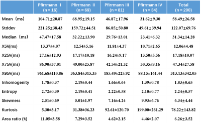 |
Histogram analysis of T2* value for lumbar intervertebral disc degeneration
Xiaoqing Liang1, Ruyi Xie1, Yitong Li1, Bowen Hou1, Weiyin Vivian Liu2, and Xiaoming Li1
1Tongji Hospital, Tongji Medical College, Huazhong University of Science and Technology, Wuhan, China, 2MR Research,GE Healthcare, Beijing, China
Low back pain (LBP) is a common spinal disease during the middle-to-old-aged population. Degeneration of lumbar intervertebral discs (IVDs) is recognized as the common and crucial cause of LBP. Histogram analysis is a recently popular method for assessing microstructural heterogeneity, however, its diagnostic performance in intervertebral disc degeneration hasn’t been proved. Histogram analysis on axial T2* mapping was applied to characterize degenerative degrees and heterogeneity of discs. All histogram parameters of T2* value were significantly related to Pfirrmann grading. Our study confirmed that the apparent “red zone” on T2* color maps was clearly related to degeneration.
|
|
2823.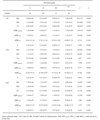 |
Diffusion kurtosis imaging and intravoxel incoherent motion in quantitative evalutation of lumbar intervertebral disc degeneration
Feifei Zeng1, Yunfei Zha2, and Weiyin Liu3
1Radiology, Renmin Hospital of Wuhan University, Wuhan, China, 2Renmin Hospital of Wuhan University, Wuhan, China, 3MR Research, GE Healthcare, Bejing, China
The purpose of this study is to compare the diagnostic value of diffusion kurtosis imaging (DKI) and intravoxel incoherent motion(IVIM) in assessing lumbar intervertebral disc degeneration. DKI parameters ( MK, MD, and FA), and IVIM parameters (ADCstandard, ADCslow, ADCfast, and f) of nucleus pulposus (NP), anterior annulus fibrosus (AAF) and posterior annulus fibrosus (PAF) were measured and correlated with Pfirrmann grades. It was found that DKI and IVIM parameters had significantly correlation with Pfirrmann grades. In addition, DKI was more sensitive than IVIM in quantitative detection of early lumbar intervertebral disc degeneration.
|
|
2824.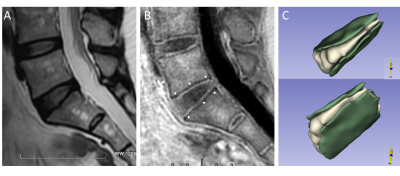 |
Imaging Cartilaginous Endplates Herniation in the Lumber Spine using ZTE-MRI
Yen-Chang Chen1, Chien-Yuan Lin1, Ching-Wei Gu2, Yu-Hsin Tang2, Wei-Min Hung2, Yi-Chih Hsu2, and Guo-Shu Huang2
1GE Healthcare, Taipei, Taiwan, 2Department of Radiology, Tri-Service General Hospital, National Defense Medical Center, Taipei, Taiwan
The cartilage endplate (CEP) plays an important role in the function and homeostasis of the intervertebral disc by serving as a mechanical stabilizer as well as a pathway for nutrient transport. There is a very little known about CEP due in part to the lack of suitable imaging technique to evaluate the CEP. The purposes of this study were to optimize the parameters of zero echo time (ZTE) MRI in enhancing CEP structure and then to evaluate the morphology of CEP in lumbar spine with herniation of disc using ZTE-MRI.
|
|
2825.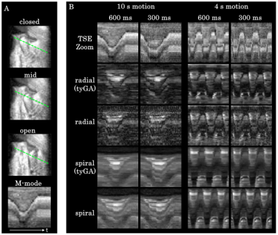 |
Real-time Imaging of the TMJ: Comparison of Methods
Kilian Stumpf1, Patrick Metze1, Tobias Speidel1, Thomas Hüfken1, and Volker Rasche1
1Department of Internal Medicine II, University Medical Center Ulm, Ulm, Germany
Disorders of the temporomandibular joint (TMJ), such as pain and blockage, usually arise while the joint is in motion. Capturing this dynamic process requires high temporal resolutions and good contrast between condyle and articular disk, which is possible with advanced sequences that often need sophisticated reconstruction algorithms. In this contribution, the widely available or easily implemented and reconstructed TSE-Zoom sequence was compared to advanced radial and spiral sequences employing uniform and tiny golden angle(tyGA) profile ordering. While tyGA sequences allow fluid rendering of the joint motion, TSE-Zoom can be considered a viable alternative with excellent contrast between condyle and disk.
|
|
2826. |
INCREASED PREVALENCE OF FEMOROACETABULAR IMPINGEMENT ANATOMY AMONG ATHLETES WHO TREAD WATER
Joanna Lind Langner1, Marianne Black1, James MacKay2, Kimberly Hall3, Marc Safran3, Feliks Kogan1, and Garry Gold1
1Radiology, Stanford University, Stanford, CA, United States, 2Cambridge University, Cambridge, United Kingdom, 3Stanford University, Stanford, CA, United States
Femoroacetabular impingement (FAI) is a disorder that causes hip pain and disability in young patients, particularly athletes. Treading water leads to increased stress on the hip, the effects of which are poorly understood. In this study, we used MRI to identify the prevalence of FAI anatomy in elite aquatic athletes that tread water. The prevalence of cam FAI was higher than that previously reported for any other sport. This study demonstrates that synchronized swimmers and water polo athletes are at increased risk of cam FAI, more than other sports.
|
2827.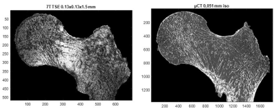 |
Trabecular Bone Microarchitecture: A Comparative Analysis between High Field, Ultra High Field MRI and X-ray micro CT in human anatomical samples
Enrico Soldati1, Martine Pithioux2, David Bendahan3, and Jerome Vicente1
1IUSTI, AixMarseille, Marseille, France, 2ISM, AixMarseille, Marseille, France, 3CRMBM, AixMarseille, Marseille, France
It has been previously suggested that trabecular bone could be assessed using ultra-high and high-field MRI. In the present study, human femurs head were scanned using with MRI at 3T and 7T MRI and the corresponding metrics were compared to those obtained using high resolution X-ray micro tomography.
|
|
2828.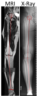 |
Feasibility of Rapid MRI Assessment of Leg Alignment
Feliks Kogan1, Scott Uhlrich2, Madeleine Berkson3, Akshay Chaudhari1, Marianne Black1, Valentina Mazzoli1, Lauren Watkins1, Garry E Gold1, and Brian Hargreaves1
1Department of Radiology, Stanford University, Stanford, CA, United States, 2Department of Mechanical Engineering, Stanford University, Stanford, CA, United States, 3Palo Alto Veterans Affairs Hospital, Palo Alto, CA, United States
Varus and valgus alignment are associated with the presence and progression of knee osteoarthritis. We assessed the feasibility of rapidly assessing leg alignment with a non-weight bearing MRI in under a minute. Good agreement was observed between alignment measurements from MRI and weight-bearing radiographs. This technique offers a sub-one-minute scan that can be incorporated into routine MRI knee scans to add important knee alignment information and potentially removes the need for additional scanning.
|
|
2829.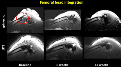 |
A rat model of periprosthetic joint infection (PJI) using 3D printed titium hip implants: Ultrashort echo time (UTE) MRI at 7T
Mariam Taha1,2, Greg O. Cron1,3,4, Gerd Melkus3,4, Peder E.Z. Larson5, Mazen Ibrahim2, Adam D.M. Paish6,7, Yusra Al Mosuli1,2, William Hadden2, David Holdsworth6,7, and Hesham Abdelbary2
1Ottawa Hospital Research Institute, Ottawa, ON, Canada, 2Orthopedic Surgery, The Ottawa Hospital, Ottawa, ON, Canada, 3Medical Imaging, The Ottawa Hospital, Ottawa, ON, Canada, 4Radiology, University of Ottawa, Ottawa, ON, Canada, 5Radiology, University of California at San Francisco, San Francisco, CA, United States, 6Western University, London, ON, Canada, 7Robarts Research Institute, London, ON, Canada Two to fourteen percent of primary total joint replacement cases develop periprosthetic joint infection (PJI), a devastating complication. PJI animal models for hip and knee are required to evaluate prevention and treatment methods. We developed a novel rat model for PJI using a 3D printed titanium hip implant. Implant stability in the rats was monitored with 7T MRI using Ultrashort Echo Time (UTE) imaging and classical spin echo imaging. UTE demonstrated superior image quality compared to spin echo imaging. This rat model will provide a unique opportunity to validate and study strategies focusing on the prevention and treatment of PJI. |
|
2830. |
Using Compressed Sense Technique for Lumbar Vertebra Imaging: Comparison with Conventional Parallel Imaging
Tianyang Gao1, Shinong Pan1, Zhao Lu1, Fengzhe Wang1, and Jiazheng Wang2
1Shengjing Hospital of China Medical University, Shenyang, China, 2Clinical Science, Philips Healthcare, Beijing, China
SENSE-TSE is widely used in routine joint scanning, which requires a considerable length of scan duration. Staying in the same position for a long time inevitably causes discomfort and unwanted movement of patients, resulting in image quality degradation. Therefore, shortening imaging time to reduce motion artifacts has become an important goal in the application of CS acceleration in clinical practice.This study aims to retrospectively compare the image quality and diagnosis accuracy of CS-TSE and SENSE-TSE in lumbar vertebra MRI.
|
|
2831.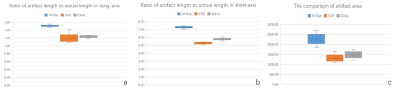 |
The evaluation of the metal artifact and SNR changes in multiple fat-suppression techniques:a phantom study
Xu Lulu1, Qi Liang1, Dou Weiqiang2, and Shen Yong3
1Radiology, The First Affiliated Hospital of Nanjing Medical University, Nanjing, China, 2MR Research China, GE Healthcare, Beijing, China, 3Enhanced MR application, GE Healthcare, Beijing, China
Finding the most suitable fat-suppression technique when acquiring the images with titanium alloy, the phantom was studied. We found the STIR was most potent when comparing the other methods. This result may provide the reference value when we offer the patients with metallic hardware after surgery with the proper fat-suppression technique.
|
|
2832. |
Study on the optimization of BOLD magnetic resonance ischemia-reactive hyperemia model of skeletal muscle of calf
Chunye Geng1 and Jianling Cui1
1The Third Hospital of Hebei Medical University, Shijiazhuang, China
In BOLD-MRI studies, cuff compression was often used to establish the ischemia-reactive hyperemia model of calf muscle.We found some BOLD signals did not decrease during ischemia, but increased or stay the same.This study was to find the reasons why BOLD signal showed multiple trends in ischemic period and to optimize the ischemia model. BOLD scan was performed on volunteers under two cuff pressures.BOLD signals of all muscles showed a downward trend in the P1 model of partly reduced blood supply. However, in P2 model of blood supply blocked completely, BOLD signals had mostly gone up during ischemia ,unexpectedly.
|
|
2833. |
Evaluation of Knee Osteoarthritis (OA) Using 3D Ultrashort-Echo-Time Cones Magnetization Transfer (3D UTE-Cones-MT) Imaging
Xiaodong Zhang1,2, Ya-jun Ma2, Aria Ashir2, Mei Wu2, Zhao Wei2, Eric Y Chang3, and Jiang Du2
1The third affilated hospital of Southern Medical Univerisity, Guangzhou, China, 2Department of Radiology, University of California, San Diego, CA, United States, 3Radiology Service, VA San Diego Healthcare System, San Diego, CA, United States
Magnetization transfer (MT) imaging has been used for indirect assessment of macromolecules in biological tissues. In this study, we explore the value of the 3D UTE-Cones-MT sequence for volumetric quantification of magnetization transfer ratio (MTR) and macromolecular proton fraction(f) in menisci of healthy volunteers and patients with different degrees of OA. The primary results demonstrate that this method can detect compositional changes in meniscus and can be used to differentiate healthy subjects from patients with mild or advanced OA.
|
|
2834.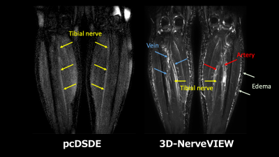 |
Phase-cycling diffusion-sensitized driven-equilibrium (pcDSDE) for MR neurography of the crus
HAJIME YOKOTA1, TAKAYUKI SAKAI2, MASAMI YONEYAMA3, AKIYO TAKADA4, and TAKASHI UNO1
1Diagnostic Radiology and Radiation Oncology, Graduate School of Medicine, Chiba University, Chiba, Japan, 2Radiology, Eastern Chiba Medical Center, Togane, Japan, 3Philips Japan, Tokyo, Japan, 4Radiology, Chiba University Hospital, Chiba, Japan
MR neurography for the crus is challenging because signals of the vessels and subcutaneous edema interrupt visualization of the nerve, and the water content of the nerve is small. Phase-cycling diffusion-sensitized driven-equilibrium (pcDSDE) can reduce signals of the neighboring structures including the vesssels, and visualize the nerves with diffusion contrast. pcDSDE was better than 3D-NerveVIEW for visualizing the tibial and sural nerves in the crus. The contrast ratio between the tibial nerve and neighboring muscle was significantly higher on pcDSDE than on 3D-NerveVIEW in both the proximal and distal crura.
|
|
2835.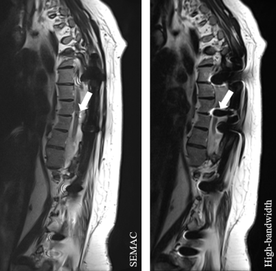 |
Can Accelerated SEMAC TSE Replace Conventional TSE in the Spine?
Ali Rashidi1, Shivani Ahlawat1, Rodrigo Luna1, and Jan Fritz1
1Russell H. Morgan Department of Radiology and Radiological Science, Johns Hopkins University School of Medicine, Baltimore, MD, United States
We investigated the exchangeability of accelerated SEMAC TSE and high-bandwidth TSE MRI in patients with spinal hardware. Two musculoskeletal radiologists found through quantitative and qualitative evaluations of 50 patients with cervical, thoracic, and lumbar spinal instrumentation that accelerated SEMAC resulted in fewer metal artifacts, better visibility of anatomical structures and abnormalities, only mildly increased blur and similar soft tissue and bone contrasts. In the spine, accelerated SEMAC TSE can be used as a replacement, rather than “add-on”, of high-bandwidth TSE.
|
|
2836.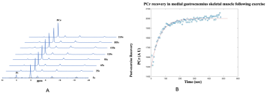 |
Investigating relationship between creatine kinase kinetics and total NAD levels in human skeletal muscle in vivo using 31P MRS at 7.0T
Deepa Thakuri1, Puneet Bagga1, Dushyant Kumar1, and Ravinder Reddy1
1Radiology, University of Pennsylvania, philadelphia, PA, United States
Nicotinamide adenine dinucleotide (NAD) plays an important role in cellular metabolism and it is important to study its correlation with other metabolites of the skeletal muscle. Phosphocreatine (PCr) present in abundant amount in the skeletal muscle facilitates the generation of ATP by conversion into creatine. 31P-MRS is a common technique used to study PCr recovery kinetics and its application has been shown in various diseases related to mitochondrial disorders. In this study, we aim to investigate the relationship between PCr recovery kinetics, total NAD concentration, and age.
|
|
2837.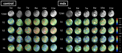 |
Assessment of skeletal muscle pathology in Dystrophin-deficient mice using structural and functional Diffusion MRI
Rina Ito1,2, Junichi Hata3,4, Mayu Iida1,2, Fumiko Seki2, Mitsuki Rikitake1,2, Yuji Komaki2, Chihoko Yamada2, Daisuke Nakashima4, Hirotaka James Okano3,4, and Takako Shirakawa1
1Department of Radiology, Faculty of Health Sciences, Tokyo Metropolitan University, Tokyo, Japan, 2Live Imaging Center, Central Institute for Experimental Animals, Kanagawa, Japan, 3Jikei University School of Medicine, Tokyo, Japan, 4Keio University School of Medicine, Tokyo, Japan
To analyze the disease of muscular dystrophy noninvasively, we assessed the degeneration of skeletal muscle and the distinction depending on types of muscle fibers, with changing parameters based on the sequence of diffusion weighted imaging. The mdx mice were used for longitudinal evaluation at the age from 5 to 10 weeks. We confirmed conventional diffusion tensor imaging could evaluate the pathological structure. Moreover, by applying the advanced imaging such as time-dependent diffusion MRI, we could demonstrate the functional characteristics of skeletal muscle cell and to detect micro-level condition of degeneration in mdx-mice.
|
|
 |
2838.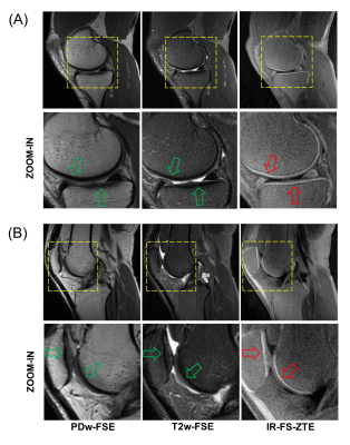 |
Osteochondral Junction Imaging Using Adiabatic Inversion Recovery Prepared Fat-Saturated Zero Echo Time MR Imaging: A Feasibility Study
Hyungseok Jang1, Yajun Ma1, Michael Carl2, Saeed Jerban1, Eric Chang1,3, and Jiang Du1
1University of California, San Diego, San Diego, CA, United States, 2GE Healthcare, La Jolla, CA, United States, 3VA San Diego Healthcare System, San Diego, CA, United States
Cartilage has a complex structure comprised of multiple zones with different MR signal properties. Among them, osteochondral junction (OCJ) cannot be directly imaged with conventional MR imaging techniques. In this study, we explore the feasibility of inversion recovery prepared fat-saturated zero echo time (IR-FS-ZTE) to image the human knee OCJ. To accentuate the signal from the OCJ region with short T2*, adiabatic inversion recovery along with fat-saturation preparation was applied, followed by continous, slient ZTE imaging. The feasibility and efficacy of IR-FS-ZTE were shown in ex vivo cadaveric human knee joints and in in vivo healthy volunteers.
|
2839.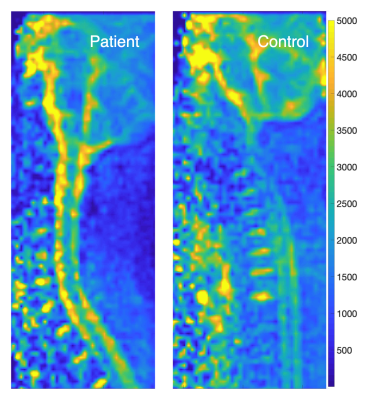 |
23Na MRI of human intervertebral cervical discs in Mucopolysaccharidosis type II
Lenka Minarikova1, Alena Svatkova2, Wolfgang Bogner1, Siegfried Trattnig1,3, Thomas Stulnig2, and Stephan Gruber1
1High Field MR Centre, Department of Biomedical Imaging and Image-guided Therapy, Medical University of Vienna, Vienna, Austria, 2Clinical Division for Endocrinology and Metabolism, Department of Medicine III, Medical University of Vienna, Vienna, Austria, 3CD Laboratory for Clinical Molecular MR Imaging, Vienna, Austria
Spinal skeletal disease is one of least therapy responding symptoms in MPS patients. Cervical spine of three MPS II patients and three matched controls was measured using 23Na MRI at 7T. There was a significant difference in intervertebral discs of MPS II patient compared to controls. 23Na MRI represents a potential quantitative method for assessment of cervical disc degeneration in MPS II patients and could serve as a potential marker used in monitoring of emerging therapies.
|
|
2840.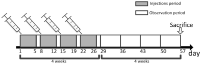 |
Gadolinium deposition in cortical Bone after repeated administration of Magnevist and Gadovist assessed by ultrashort echotime based T1 mapping
Kaixuan Zhao1, Shisi Li2, Keyan Yu2, Jian Wang2, Xiaodong Zhang2, Qinqin Yu2, Cuiling Zhu2, Yingjie Mei3,4, Pu Xu3, Peiwei Yi3, Jiang Du5, and Yanqiu Feng3
1Southern Medical University, Guang Zhou, China, 2Imaging department of Southern Medical University affiliated the third hospital, Guang Zhou, China, 3School of Biomedical Engineering, Guangdong Provincial Key Laborary of Medical Image Processing, Southern Medical University, Guang Zhou, China, 4Philips Healthcare, Guang Zhou, China, 5Department of Radiology, University of California San Diego, San Diego, CA, United States
In this preclinical study, we assessed feasibility of evaluating Gadolinium deposition in cortical bone by using recently developed actual flip angle variable repetition time 3D ultrashort echotime technique in rabbit model at 7T. Twenty times of administration of clinical equivalent dose of Magnevist and Gadovist that normalized according to body surface by U.S. FDA recommendation, and three times the dose of Gadovist (high-dose group) were investigated. Significant lower T1 values were observed in Magnevist administration group, Gadovist administration group and high-dose Gadovist administration group compared to control group, suggested T1 mapping might be a potential biomarker for evaluating Gadolinium deposition.
|
|
2841. |
Bilateral Sodium Magnetic Resonance Imaging of the Lower Extremity
Dimitri Alexander Kessler1, Mary A McLean2, Titus Lanz3, Frank Riemer4, Rolf F Schulte5, Andrew Grainger1, Fiona J Gilbert1, Martin J Graves1, and Joshua D Kaggie1
1Department of Radiology, University of Cambridge, Cambridge, United Kingdom, 2Cancer Research UK Cambridge Institute, University of Cambridge, Cambridge, United Kingdom, 3Rapid Biomedical GmbH, Rimpar, Germany, 4Mohn Medical Imaging and Visualization Centre, Department of Radiology, Haukeland University Hospital Helse Bergen, Bergen, Norway, 5General Electric Healthcare, Munich, Germany
We present a method for bilateral sodium magnetic resonance imaging (MRI) of the lower extremities. Sodium MRI can provide direct information on tissue biochemistry not available through standard proton MRI, and could therefore potentially assist in disease diagnosis. Our preliminary results demonstrate the application of large field-of-view sodium MRI to the musculoskeletal system for potential compositional assessment of multiple tissues in both legs including muscle, cartilage, synovium and arteries.
|
|
2842.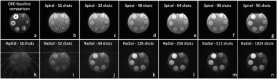 |
Initial Assessment of a novel hybrid ultrashort echo time (UTE) sequence
Lumeng Cui1, Emily J McWalter1,2, Gerald Moran3, and Niranjan Venugopal4
1Division of Biomedical Engineering, University of Saskatchewan, Saskatoon, SK, Canada, 2Department of Mechanical Engineering, University of Saskatchewan, Saskatoon, SK, Canada, 3Siemens Healthcare Limited, Oakville, ON, Canada, 4Department of Radiotherapy Physics, CancerCare Manitoba, Winnipeg, MB, Canada
Ultrashort echo time (UTE) pulse sequences have the unique ability to visualize short T2 tissues. While several UTE techniques have been implemented and demonstrated good clinical results, there currently does not exist a flexible and robust UTE pulse sequence which can easily manipulate multiple parameters “on-the-fly“ (i.e. selection of trajectories, excitation pulses, acquisition parameters, etc.), and obtain several complimentary scans in a single scan session. In this work we present a newly developed hybrid UTE pulse sequence that includes multiple excitation pulses, and varying trajectories, allowing for novel investigations in studying short T2 tissues.
|