
Digital Poster Session
Cancer: Diffusion, Perfusion, MRS & Machine Learning
Cancer
4764 -4778 Cancer Imaging: Diffusion, Perfusion, MRS & Machine Learning - Cancer Imaging: Diffusion
4779 -4789 Cancer Imaging: Diffusion, Perfusion, MRS & Machine Learning - Cancer Imaging: Clinical Diffusion
4790 -4804 Cancer Imaging: Diffusion, Perfusion, MRS & Machine Learning - Cancer Imaging: Perfusion & Permeability
4805 -4816 Cancer Imaging: Diffusion, Perfusion, MRS & Machine Learning - Cancer Imaging: MRS
4817 -4826 Cancer Imaging: Diffusion, Perfusion, MRS & Machine Learning - Cancer Imaging: Machine Learning & Radiomics
4764.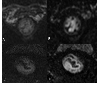 |
Rectal Carcinoma at 3T Assessed via iShim DWI: A Comparison of Image Quality with RESOLVE
Jinrong Qu1, Hongkai Zhang2, Xu Yan3, Shaoyu Wang4, and Thomas Benkert5
1Radiology, the Affiliated Cancer Hospital of Zhengzhou University & Henan Cancer Hospital, Zhengzhou, China, 2Radiology, the Affiliated Cancer Hospital of Zhengzhou University, Zhengzhou, China, 3MR Scientific Marketing, Siemens Healthcare, Shanghai, China, 4MR Scientific Marketing, Siemens Healthcare, Xi'an, China, 5Application Development, Siemens Healthcare GmbH, Erlangen, Germany
This study evaluated a new diffusion weighted imaging (DWI) technique for assessing rectal cancer. Based on a population of 12 patients diagnosed with rectal carcinoma, images were acquired using both Read-out Segmented Echo-Planar Imaging (RESOLVE) and integrated slice-specific shimming (iShim)-DWI sequences using the same field of view (FOV) at 3T in assessment of rectal carcinoma. The quality of the images was compared based on independent review by radiologists plus parameter data analysis. The results showed that the image quality and lesion conspicuity of the iShim-DWI sequence were significantly better than RESOLVE DWI sequence in assessment of rectal carcinoma at 3T.
|
|
4765.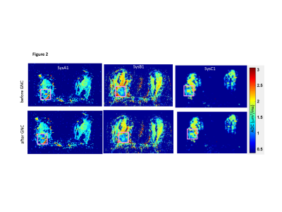 |
Retrospective ADC correction of gradient nonlinearity errors in multi-center breast DWI trials: ACRIN6698 multi-platform feasibility study
Dariya Malyarenko1, David C Newitt2, Lisa J Wilmes2, Ek Tsoon Tan3, Luca Marinelli4, Ajit Devaraj5, Johannes M Peeters6, Shivraman Giri7, Axel vom Endt8, Nola Hylton2, Savannah Partridge9, and Thomas L Chenevert1
1Radiology, University of Michigan, Ann Arbor, MI, United States, 2Radiology and Biomedical Imaging, University of California San Francisco, San Francisco, CA, United States, 3Hospital for Special Surgery, New York, NY, United States, 4GE Global Research, Niskayuna, NY, United States, 5Philips Research North America, Cambridge, MA, United States, 6Philips MR Clinical Science, Best, Netherlands, 7Siemens Medical Solutions, Boston, MA, United States, 8Siemens Healthcare GmbH, Erlangen, Germany, 9Radiology, University of Washington, Seattle, WA, United States
Multi-site, multi-platform clinical oncology trials seek to enhance quantitative utility of the apparent diffusion coefficient (ADC) metric by reducing technical cross-platform variability due to systematic gradient nonlinearity (GNL). Here we test feasibility of retrospective GNL correction implementation for a representative subset of subjects and systems from the ACRIN6698 breast cancer therapy response trial. GNL ADC correction based on previously developed formalism is demonstrated for trace-DWI DICOM using system-specific gradient-channel fields derived from vendor-provided spherical harmonic tables. Implemented correction substantially improves precision and removes ADC bias for DWI QC phantoms, and markedly changes ADC histogram percentiles for solid breast tumors.
|
|
4766.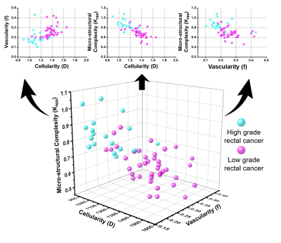 |
Combine the different DWI biological inspirations including cellularity, vascularity and structural complexity to grade rectal cancer
Zhijun Geng1, Yunfei Zhang2, Shaohan Yin1, Shanshan Lian1, Haoqiang He1, Hui Li1, Chuanmiao Xie1, and Yongming Dai2
1Sun Yat-sen University Cancer Center, Guangzhou, China, 2United Imaging Healthcare, Shanghai, China
Biological inspirations such as cellularity, vascularity and micro-structural complexity of diffusion weighted imaging (DWI) are of great significance during clinical practice. However, scarcely has the systematic integration of these biological inspirations been applied for clinical application. This research aims to systematically integrate cellularity, vascularity and micro-structural complexity derived from diffusion kurtosis imaging (DKI) and Intravoxel Incoherent Motions (IVIM) Imaging, explore their correlations and evaluate their diagnostic performance for grading rectal cancer. Results suggested that the integration of different DWI biological inspirations provided a more comprehensive characterization of tumor, holding great potential during clinical practice.
|
|
4767.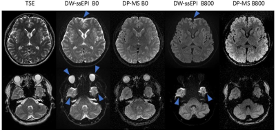 |
3D Isotropic Resolution Distortion-free Diffusion-Prepared Magnitude-Stabilized bSSFP Imaging at 1.5 Tesla
Yu Gao1, Ziwu Zhou1, Fei Han1, Xiaodong Zhong2, Yingli Yang1, and Peng Hu1
1Radiological Sciences, University of California, Los Angeles, Los Angeles, CA, United States, 2MR R&D Collaborations, Siemens Healthineers, Los Angeles, CA, United States
Strong spatial distortion of the DW-ssEPI sequence prevents its utilization in radiotherapy planning and treatment adaptation. In this work, a 3D diffusion-prepared magnitude-stabilized bSSFP sequence was developed and validated at 1.5T. A phase navigator was acquired during the catalyzation stage of the bSSFP readout to estimate the spatial variation of the signal phase, and a locally low-rank constrained reconstruction was developed to resolve the phase variation. The sequence was validated on a diffusion phantom and healthy volunteers. It provided submillimeter geometric fidelity and acceptable ADC accuracy, which makes it a promising candidate for treatment planning and adaptation of the brain.
|
|
4768. |
Measuring the repeatability of an automated Whole-Body Diffusion-Weighted MRI tumour segmentation approach in metastatic prostate cancer
Antonio Candito1, Nina Tunariu1, Richard Holbrey1, Sebastian Schäfer2, Matthew R Orton1, David Collins1, Fabio Zugni3, Martin O Leach1, Matthias Baumhauer2, Matthew D Blackledge1, and Dow-Mu Koh1
1The Institute of Cancer Research, London, United Kingdom, 2Mint Medical, Heidelberg, Germany, 3IEO, European Institute of Oncology IRCCS, Milan, Italy
There is currently no accepted biomarker for assessing response to treatment in patients with bone disease from advanced prostate cancer (APC). Whole-Body Diffusion-Weighted-MRI (WBDWI) is emerging as a bone response biomarker in APC. However, automatic segmentation of disease from WBDWI is needed to quantify changes in WBDWI-derived biomarkers (global tumour apparent diffusion coefficient (gADC) and total tumour volume, tDV) for more reliable response assessment. We investigate the repeatability of WBMRI parameters using automatic disease delineation in APC patients.
|
|
4769. |
A method for automatic segmentation of the spinal cord and surrounding cerebrospinal fluid using whole-body Diffusion-Weighted MRI
Antonio Candito1, Nina Tunariu1, Richard Holbrey1, Matthew R Orton1, David Collins1, Martin O Leach1, Matthew D Blackledge1, and Dow-Mu Koh1
1The Institute of Cancer Research, London, United Kingdom
Whole-body Diffusion-Weighted MRI (WBDWI) is an emerging tool for quantitative assessment of the response/progression of metastatic bone disease from advanced prostate cancer and myeloma. The tumour apparent diffusion coefficient (ADC) measured by WBDWI shows sensitivity for assessing treatment response, but requires accurate bone disease segmentation. Automatic delineation of the spinal cord and cerebrospinal fluid (CSF) on WBDWI enables clear separation of the spinal cord from disease within the adjacent vertebral column leading to accurate bone disease segmentation. We have developed a new edge-detection method for automatic delineation of the spinal cord and surrounding CSF from WBDWI.
|
|
4770. |
Controlling Apparent Diffusion and Kurtosis with Vesicle Size in Quantitative Diffusion Kurtosis Phantoms
Scott D. Swanson1, Dariya I. Malyarenko1, Alan M. McLean2, Mario L. Fabiilli1, and Thomas L. Chenevert1
1Department of Radiology, University of Michigan, Ann Arbor, MI, United States, 2Department of Chemistry, University of Michigan, Ann Arbor, MI, United States
Chemical composition of lamellar lipid vesicles determines particle diameter and hence the size of a restricted diffusion component. Phantoms made at 1% by weight solid using cetearyl alcohol and cetyl trimethyl ammonium bromide with varying molar ratios have different apparent diffusion constants and kurtosis values. Vesicle sizes are confirmed by dynamic light scattering and electron microscopy. These molecular vesicles provide a basis for a quantitative diffusion phantom and future MR studies of nanoscopic domains.
|
|
4771. |
Verifying the feasibility of automatic no-reference image quality metrics in evaluation the image quality of abdominal iSim at 3T
Fan WenWen1, Ou Yang Han1, Zhao XinMing1, Zhang HongMei1, Zhou Chun Wu1, Shi Qing Lei2, Feng Xiang2, Xu Xiao Juan1, Liu Kan1, Wang Xiao Ye3, Thomas Benkert Ph.D4, and Lu Tong Suo1
1National Cancer Center/Cancer Hospital, Chinese Academy of Medical Science and Peking Union Medical College, BeiJing, China, 2MR Scientfic marketing Siemens Healthcare, BeiJing, China, 3MR Application Siemens Healthcare, BeiJing, China, 4HC DI MR DL ONCO,Siemens Healthcare GmbH, BeiJing, China
This experiment proposed an automatic image quality evaluation method using no-reference image quality metrics of structural similarity index (SSIM), blind/referenceless image spatial quality evaluator (BRISQUE), perception based Image quality evaluator (PIQE) , SNR Wavelet and Contrast. The SNR Wavelet was calculated by the quotient of the image before wavelet filtering and the difference between the image before wavelet filtering and the image after wavelet filtering. The contrast was calculated using the average signal difference of three to five gray bars in the middle position. This study showed the automatic no-reference image quality metrics have potential in future in evaluation the image quality of abdominal DWI at 3T with a higher efficiency.
|
|
4772.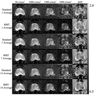 |
Reducing scan time of routine prostate diffusion-weighted imaging using random matrix theory reconstruction
Gregory Lemberskiy1,2, Yousef Mazaheri3, Herbert Alberto Vargas4, Ricardo Otazo3, Els Fieremans1, and Dmitry S Novikov1
1Radiology, New York University School of Medicine, New York, NY, United States, 2Microstructure Imaging INC, New York, NY, United States, 3Department of Medical Physics, Memorial Sloan Kettering Cancer Center, New York, NY, United States, 4Radiology, Memorial Sloan Kettering Cancer Center, New York, NY, United States We propose Random Matrix Theory (RMT) reconstruction to reduce the scan time of prostate diffusion (DWI) by using fewer averages and still maintain image quality. RMT leverages the joint redundancy across receiver coils, voxels, and measurements to identify and remove the universal noise-only Marchenko-Pastur distribution. We find that RMT can dramatically increase the SNR of the prostate protocol, where the coefficient of variation of the RMT reconstruction for 1 average is lower than the conventional reconstruction of 14 averages. Thereby, RMT allows to reduce scan time by over 5-fold with comparable image quality. |
|
4773.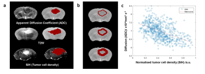 |
Novel glioblastoma infiltration biomarker validation by histopathologic assessment of the relation between ADC and tumor cellularity
Antoine Vallatos1, Haitham Al-Mubarak2, Joanna Birch3, Adam Waldman1, William Holmes2, Anthony Chalmers3, and Gerard Thompson1
1Centre for Clinical Brain Sciences, University of Edinburgh, Edinburgh, United Kingdom, 2Glasgow Experimental MRI Centre, Institute of Neuroscience and Psychology, University of Glasgow, Glasgow, United Kingdom, 3Wolfson Wohl Translational Cancer Research Center, Institute of Cancer Sciences, University of Glasgow, Glasgow, United Kingdom
This work quantitatively and qualitatively assesses the relation between tumor related cellularity variation and ADC values. We show that the ADC slope along linear profiles perpendicular to the boundary voxels of the ADC region of interest is a a biomarker of tumor infiltration beyond the contrast enhancing regions. We demonstrate the existence of a strong relation between ADC and tumor cellularity and show that a vectorial profile analysis of ADC maps could provide with a robust biomarker of glioblastoma cell infiltration.
|
|
4774.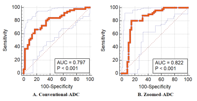 |
Improved Prostate Cancer Detection: Comparison of Zoomed Diffusion- Weighted Imaging (DWI) with Conventional DWI
Li ming Wei1, Shuhao Wang1, Caixia Fu2, Chunyu Jiang1, Ruiting Li1, Lei Hu1, Thomas Benkert3, and Jungong Zhao1
1Department of Diagnostic and Interventional Radiology, Shanghai Jiao Tong University Affiliated Sixt, shanghai, China, 2MR Application Development, Siemens Shenzhen magnetic Resonance Ltd, shenzhen, China, 3MR Application Predevelopment, Siemens Healthcare, Erlangen, Germany
This study compared zoomed diffusion- weighted imaging (DWI) and conventional DWI of prostate lesions with respect to image quality, lesion detection capability, and diagnostic accuracy. The results demonstrate that zoomed DWI can provide improved image quality, including reduced artifacts and higher contrast-to-noise ratio (CNR), and aid in improved prostate cancer detection.
|
|
4775.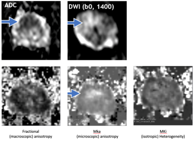 |
Preliminary feasibility study of MR Q-space trajectory imaging (QTI) for assessment of tissue microstructure in prostate cancer
Bjoern Jan Langbein1,2,3, Filip Szczepankiewicz1,2,4, Carl-Fredrik Westin 1,2, Adam Kibel2,5, Clare Tempany1,2, Martin Schostak3, Andriy Fedorov1,2, and Fiona Fennessy 1,2
1Brigham and Women’s Hospital, Department of Radiology, Boston, MA, United States, 2Harvard Medical School, Boston, MA, United States, 3Universitätsklink Magdeburg, Magdeburg, Germany, 4Clinical Sciences Lund, Lund University, Lund, Sweden, 5Brigham and Women’s Hospital, Division of Urology, Boston, MA, United States
Q-space trajectory imaging (QTI) has shown promise for characterisation of tissue microstructure. This study seeks to determine if QTI is feasible in assessment of prostate cancer (PCa) and to prefrom a preliminary evaluation of its role to help identify clinically significant prostate cancer (Gleason Grade group ≥2 lesions). QTI was performed in 40 men, 16 of which had biopsy-proven PCa and a PIRADS v.2.1 score ≥3. Preliminary data suggest that quantification of QTI values may indeed be helpful in men with clinically significant disease.
|
|
4776. |
Repeatability of ADC histogram metrics from the ACRIN6698 breast cancer therapy response trial
David C Newitt1, Dariya I Marlyarenko2, Nola M Hylton1, Brian D Ross2, Lisa Wilmes1, Savannah Partridge3, and Thomas L Chenevert2
1Radiology and Biomedical Imaging, University of California, San Francisco, CA, United States, 2Radiology, University of Michigan Medical School, Ann Arbor, MI, United States, 3Radiology, University of Washington, Seattle, WA, United States Repeatability of quantitative imaging metrics is important for establishing precision of diagnostic and prognostic measurements. In the multi-center ACRIN6698 trial, mean apparent diffusion coefficient (ADC) of breast tumors showed excellent repeatability but only moderate predictive power for breast cancer therapy response. Previous single-center studies have shown improved predictive performance using alternative ADC histogram metrics related to dense tumor volume. This study evaluates repeatability for a variety of alternative ADC histogram metrics to establish confidence intervals and inform predictive models for future breast cancer therapy response analysis. |
|
4777. |
Multi-site evaluation of MR breast phantom with internal MR-visible liquid crystal thermometer: initial temperature and DWI measurements
Lisa J Wilmes1, David C Newitt1, Wen Li1, Evelyn Proctor1, Natsuko Onishi1, Teffany Joy Bareng1, Tom L Chenevert 2, Dariya I Malyarenko2, Patrick J Bolan3, Todor Karaulanov4, Nola M Hylton1, and Kathryn E Keenan5
1University of California San Francisco, San Francisco, CA, United States, 2University of Michigan Medical School, Ann Arbor, MI, United States, 3University of Minnesota Medical School, Minneapolis, MN, United States, 4QalibreMD, Boulder, CO, United States, 5National Institute of Standards and Technology, Boulder, CO, United States Poster Permission Withheld
A multiparametric breast phantom with an integrated MRI-“visible” liquid crystal (LC) thermometer for real-time monitoring of phantom temperature was evaluated at three imaging sites, on MRI scanners from different vendors. Short-duration MRI acquisitions were optimized for visualization of LC elements and implemented at the sites for temperature measurements before and after DWI acquisitions. MRI-measured temperature differences between different imaging experiments were reflected in different water ADC values, calculated from DWI, that were consistent with the relationship between ADC and temperature described in the literature.
|
|
4778. |
Can fewer diffusion-weighting data points deliver similar diagnostic performance for breast IVIM-MRI?
Kurt Li1, Archana Machireddy2, Alina Tudorica2, Brendan Moloney2, Karen Oh2, Neda Jafarian2, Savannah Partridge3, Xin Li2, and Wei Huang2
1International School of Beaverton, Aloha, OR, United States, 2Oregon Health & Science University, Portland, OR, United States, 3University of Washington, Seattle, WA, United States
The goal of this study is to compare the diagnostic performance between intravoxel incoherent motion (IVIM) imaging parameters derived from standard biexponential IVIM model fitting and those from direct calculation using only 3 b-values. This was accomplished using Monte Carlo simulations and IVIM data acquisition and analysis from 27 patients with 28 suspicious breast lesions. Results show that at low signal-to-noise ratio, the 3 b-value approach improved parameter accuracy and provided comparable diagnostic performance in benign-malignant classification compared to the biexponential fitting approach.
|
 View the Poster
 Watch the Video
4779. |
Investigation of tumor grade and gadolinium enhancement by tensor-valued diffusion encoding and QTI analysis: an exploratory study of gliomas
Filip Szczepankiewicz1,2,3, Parikshit Juvekar2,4, Jan Brabec3, Pia C. Sundgren3, Markus Nilsson3, Alexandra Golby2,4, and Carl-Fredrik Westin1,2
1Radiology, Brigham and Women's Hospital, Boston, MA, United States, 2Harvard Medical School, Boston, MA, United States, 3Clinical Sciences Lund, Lund University, Lund, Sweden, 4Department of Neurosurgery, Brigham and Women's Hospital, Boston, MA, United States
Diffusion MRI is sensitive to the configuration of cell structures on the microscopic level, and may be a biomarker of tumor grade. High-grade gliomas are hypothesized to be more heterogeneous on a microscopic scale, when compared to low-grade; a feature that should be reflected in the diffusional kurtosis. To explore this premise, we use tensor-valued diffusion encoding to disentangle the isotropic and anisotropic kurtosis and relate them to tumor grade and tumor tissue that is enhancing vs. non-enhancing on post-gadolinium T1 maps.
|
|
4780.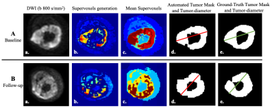 |
Automated RECIST Measurement and Therapeutic Response Evaluation in Osteosarcoma using Diffusion Weighted MRI
Amit Mehndiratta1, Esha Badiya Kayal1, Raju Sharma2, Sameer Bakhshi3, and Devasenathipathy Kandasamy2
1Centre for Biomedical Engineering, Indian Institute of Technology Delhi, New Delhi, India, 2Department of RadioDiagnosis, All India Institute of Medical Sciences, New Delhi, India, 3Dr. BRA Institute-Rotary Cancer Hospital, All India Institute of Medical Sciences, New Delhi, India
Accuracy and consistency in RECIST(Response evaluation criteria in solid tumors) measurements are crucial as it directly impacts patient treatment options. Manual RECIST measurement, requiring high expertise & attention, is time-expensive, prone-to-error, operator-subjective. we propose an automated tumor segmentation and RECIST score estimation method that uses MRI image slices as input, delineates the tumor in 3D, identifies the MRI slice with maximum-tumor-burden and then measures the tumor-diameter and RECIST1.1 score for treatment response assessment. Proposed method produced reliable and reproducible automated RECIST score measurements in current bone tumor dataset and might be useful as decision support tool saving manual-effort and reading-time.
|
|
4781. |
Comparison of ADC, IVIM and Kurtosis for differentiation of mesorectal lymph nodes in rectal cancer
Andrada Ianuș1,2, Inês Santiago3,4, Antonio Galzerano3, Paula Montesinos5, Nuno Loução5, Javier Sanchez-Gonzalez5, Daniel C. Alexander2, Celso Matos1,3, and Noam Shemesh1
1Champalimaud Research, Champalimaud Centre for the Unknown, Lisbon, Portugal, 2University College London, London, United Kingdom, 3Champalimaud Clinical Centre, Champalimaud Centre for the Unknown, Lisbon, Portugal, 4Nova Medical School, Lisbon, Portugal, 5Philips Healthcare Iberia, Madrid, Spain
In this study we employed a clinically feasible diffusion MRI acquisition and modelling approach at 1.5T to characterize perfusion (IVIM) and higher order diffusion (Kurtosis) properties of mesorectal lymph nodes in rectal cancer patients upon staging. The results showed that diffusivity estimated from the IVIM-Kurtosis model was the only parameter showing significant differences between benign and malignant lymph nodes. Moreover, ROC analysis evidenced improved differentiation when adding IVIM-Kurtosis to standard T2-weighted qualitative assessment by expert radiologists.
|
|
4782. |
Combined DWI and T2 mapping for the Differential Diagnosis of Malignant Liver Tumors
Jinrong Qu1, Hongkai Zhang2, Mingzhe Xu1, Xu Yan3, Shaoyu Wang4, and Fei Han5
1Radiology, the Affiliated Cancer Hospital of Zhengzhou University & Henan Cancer Hospital, Zhengzhou, China, 2Radiology, the Affiliated Cancer Hospital of Zhengzhou University, Zhengzhou, China, 3MR Scientific Marketing, Siemens Healthcare, Shanghai, China, 4MR Scientific Marketing, Siemens Healthcare, Xi'an, China, 5MR R&D Collaboration, Siemens Medical Solutions, Los Angeles, CA, United States
To improve the ability to differentially diagnose malignant liver tumors, quantitative apparent diffusion coefficients (ADCs) and T2 values were evaluated in 117 patients. We also included 59 patients with liver metastases (LMs) from different primary cancers. The results showed combining quantitative ADC and T2 values could make the differential diagnosis of various malignant liver tumors easier. However, this quantitative combination was not effective at improving the ability to diagnose patients with LMs.
|
|
4783.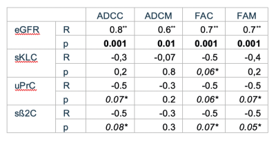 |
Evaluation of diffusion-weighted MR imaging for the characterization of myeloma-specific renal changes: initial results.
Alexandra Ljimani1, Lukas Prehm1, Julia Stabinska1, Eric Bechler1, Romans Zukovs2, Anja Müller-Lutz1, Miriam Frenken1, Elisabeth Appel1, and Hans-Jörg Wittsack1
1Department of Diagnostic and Interventional Radiology, University Duesseldorf, Duesseldorf, Germany, 2Department of Haematology, Oncology and Clinical Immunology, University Duesseldorf, Duesseldorf, Germany
Myeloma-specific renal changes are of particular interest for the prognosis and therapy monitoring of patients suffering from multiple myeloma. Here we present initial results for the evaluation of myeloma-specific renal changes by diffusion-weighted MR-imaging. Renal cortical and medullar ADC and FA values of myeloma kidney exhibit a good correlation with renal function. Cortical ADC and cortical and medullar FA values show trends to reflect myeloma-specific renal changes measured by proteinuria, serum kappa/lambda-ratio and ß2-microglobulin concentration. Diffusion-weighted MR-imaging could therefore be an important tool for monitoring myeloma-specific renal changes.
|
|
4784.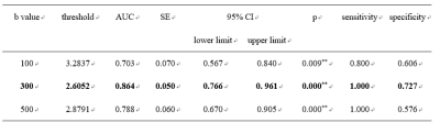 |
Diffusion-weighted imaging of thyroid nodules for the differentiation of benign from malignant lesions
Jie Liu1, Meng Dai1, and Fan Yang1
1Department of Radiology, Union Hospital, Tongji Medical College, Huazhong University of Science and Technology, Wuhan, China
To investigate the feasibility and effectiveness of diffusion-weighted imaging (DWI) in thyroid for differentiating benign and malignant thyroid nodules, determining the best b-value of DWI sequence for the diagnosis thyroid nodules.This study revealed that the ADC measurements in DWI is useful for quantitatively differentiating benign and malignant thyroid nodules, and should be potential for the assessment of thyroid nodules in addition to conventional MRI protocol.
|
|
| 4785. | Role of whole-body DWI in detection of tumour recurrence and metastasis by comparison with 18FDG PET-CT in patients with gastrointestinal cancer
Sikandar Mohd Shaikh1
1Yashoda Hospitals, Hyderabad, India
Twenty one patients with pathologically confirmed, newly diagnosed, untreated nodal metastases were included. DW images T2-weighted and T2-weighted SPAIR images were evaluated first, We used (18)F-FDG PET/CT as the standard of reference. True-positive, false-positive, and false-negative values were evaluated on a per-lesion basis. Tumor staging based on T2-weighted and T2-weighted SPAIR imaging without DWI and then with DWI was compared. True-positive lesions were increased from 89% to 97%, false-positive lesions were increased from 3% to 6%, and false-negative lesions were decreased from 11% to 3% by the addition of DWI.
|
|
4786. |
Five-level scale for post treatment whole body MRI-DWI interpretation in lymphoma (Minsk scale): comparison with PET/CT Deauville scale
Siarhei A Kharuzhyk1, Andrei V Dziuban 2, Edward A Zhavrid3, Elena V Sukolinskaja 3, and Olga A Kalenik 3
1Radiology, N.N. Alexandrov National Cancer Center, Lesnoy, Minsk District, Belarus, 2PET/CT, N.N. Alexandrov National Cancer Center, Lesnoy, Minsk District, Belarus, 3Chemotherapy, N.N. Alexandrov National Cancer Center, Lesnoy, Minsk District, Belarus
PET/CT is a standard imaging technique for tumor response assessment in lymphoma. Deauville scale is used to interpret post treatment PET/CT studies. We proposed 5-level scale for whole body MRI-DWI exams interpretation (Minsk scale) in patients with lymphoma after treatment. Five-level structure of Minsk scale makes it comparable to Deauville scale. Treatment response assessment using MRI-DWI and Minsk scale match PET/CT results in 93 of 105 (89%) patients. Survival analysis confirmed prognostic value of Minsk scale. MRI-DWI and Minsk scale could be recommended for tumor response evaluation in lymphoma as non-irradiative alternative to PET/CT.
|
|
4787.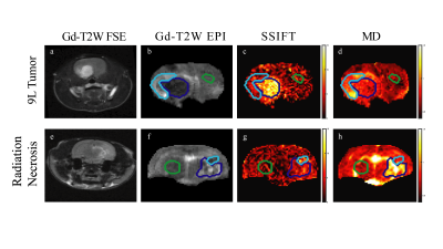 |
Validation of SSIFT MRI: Differentiation between brain tumor and radiation necrosis
Sean P Devan1,2, Xiaoyu Jiang1,3, Guozhen Luo4, Jing Cui1, Jingping Xie1, Zhongliang Zu1,3, Joel R Garbow5,6, James D Quirk5, John A Engelbach5, Austin N Kirschner4, John C Gore1,3, and Junzhong Xu1,3
1Institute of Imaging Science, Vanderbilt University Medical Center, Nashville, TN, United States, 2Chemical and Physical Biology Program, Vanderbilt University, Nashville, TN, United States, 3Department of Radiology and Radiological Sciences, Vanderbilt University Medical Center, Nashville, TN, United States, 4Department of Radiation Oncology, Vanderbilt University Medical Center, Nashville, TN, United States, 5Mallinckrodt Institute of Radiology, Washington University, St. Louis, MO, United States, 6Alvin J Siteman Cancer Center, Washington University, St. Louis, MO, United States
SSIFT is a recently developed MRI method that provides a specific means to detect brain tumors based on their differences in cell size. However, to date SSIFT has not been adequately validated. We therefore used computer simulations and studies of cultured cells in vitro and animal models in vivo to comprehensively validate SSIFT for brain cancer imaging. The results suggest SSIFT is highly specific for cell size and can differentiate brain tumors from other brain abnormalities such as peri-tumor edema and radiation necrosis, which cannot be reliably distinguished by other current MRI methods.
|
|
4788.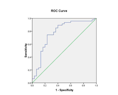 |
The value of ADC values and texture analysis in evaluating the differentiation degree of cervical squamous cell carcinoma
Mingxue Zheng1
1The First Affiliated Hospital of USTC, Hefei, China
Single ADC value can only distinguish low-differentiated CSCC from medium-well-differentiated squamous cell carcinoma, but it cannot distinguish medium-differentiated CSCC from highly-differentiated CSCC. ADC value combined with texture features can improve the efficacy of differentiation degree of CSCC before surgery.
|
|
4789.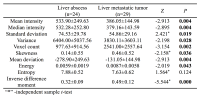 |
Differentiation of Liver Abscess and Liver Metastatic Tumor: Diffusion Weighted Images Texture Analysis
Nan Wang1, Ye Li1, Qingwei Song1, Xin Li2, Yan Guo3, Lizhi Xie3, Tingfan Wu2, and Ailian Liu1
1The First Affiliated Hospital of Dalian Medical University, Dalian, China, 2Translational Medicine Team, GE Healthcare, Shanghai, China, 3GE Healthcare, Beijing, China
Liver abscess is a serious abdominal infection that may be caused by bacteria, fungi, or parasites. The typical imaging findings of liver abscess on multiphase contrast-enhanced CT are well known, such as the “double target sign” in liver abscess, multilocular appearance, small bubbles or gas-liquid plane in the cavity. liver metastasis that develop central necrosis or cyst may mimic the appearance of liver abscess.
|
4790.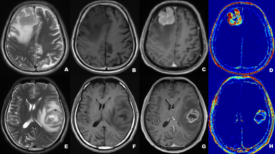 |
Differential diagnosis of central lymphoma and high-grade glioma: dynamic contrast enhanced histogram
Hanwen Zhang1, Guiwen Lv1, Wenjie He1, Yi Lei1, Fan Lin1, Mengzhu Wang2, Hong Zhang1, and Lihong Liang1
1Shenzhen Second People's Hospital, ShenZhen, China, 2MR Scientific Marketing, Siemens Healthineers, Guangzhou, China
Accurate identification between central nervous system lymphoma (CNSL) and high-grade glioma (HGG) has a great impact on the clinical treatment planning of the patients. This study uses the histogram analysis of dynamic contrast-enhanced (DCE) MRI to distinguish CNSL and HGG. Histogram features based on DCE pharmacokinetic parameters were used and achieved high diagnostic efficiency, which provides imaging features for guiding clinical treatment.
|
|
4791.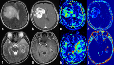 |
DSC and DCE histogram analyses of diffuse glioma biomarkers, including IDH, MGMT and TERT, on differentiation
Hanwen Zhang1, Guiwen Lv1, Wenjie He1, Yi Lei1, Fan Lin1, Mengzhu Wang2, Hong Zhang1, Lihong Liang1, and Siping Luo1
1Shenzhen Second People's Hospital, ShenZhen, China, 2Siemens Healthineers, Guangzhou, China
The molecular types of glioma including isocitrate dehydrogenase (IDH), O6-methylguanine-DNA methyltransferase (MGMT) and telomere reverse transcriptase (TERT), influence the therapeutic effect and prognosis of patients with diffuse gliomas. We combined DSC and histogram analysis of DCE methods in the diagnosis of diffuse glioma with different molecular types, in order to find biomarkers based on perfusion parameters to predict specific genotypes, and guide doctors to evaluate the prognosis of chemotherapy of patients.
|
|
4792.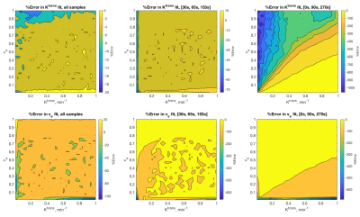 |
Estimation of Pharmacokinetic Parameters Using Three Carefully Selected DCE-MRI Timepoints for Breast Cancer Imaging
Julie C. DiCarlo1,2 and Thomas E. Yankeelov1,2,3,4
1Oden Institute for Computational Engineering and Sciences, The University of Texas at Austin, AUSTIN, TX, United States, 2Livestrong Cancer Institutes, The University of Texas at Austin, AUSTIN, TX, United States, 3Department of Biomedical Engineering, The University of Texas at Austin, AUSTIN, TX, United States, 4Department of Diagnostic Medicine, The University of Texas at Austin, AUSTIN, TX, United States
A new method based on analysis of simplicial complexes (ASC) is presented to select three time points at which to sample dynamic contrast-enhanced MRI uptake curves. The technique maps expected enhancement curve amplitudes to simplicial complex vertices and searches for the best-discriminating set of time samples. Simulation results indicate it should be possible to estimate Kety-Tofts kinetic parameters from images whose acquisition times are increased above 16 seconds per volume, in a 2-minute shorter imaging time than needed for signal enhancement ratio (SER) measurement.
|
|
4793.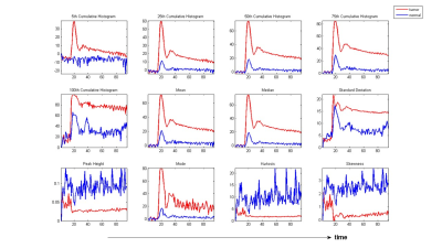 |
Radiogenomic Analysis using Dynamic Histogram Parameters of MR Perfusion-weighted Imaging in Glioblastoma
Kuan Chen1, Tzu-Wei Lee1, Chao-Wei Tso1, Larry Ying-Liang Lai2, Hui-Hsien Lin3, Fei-Ting Hsu4, and Hua-Shan Liu1
1Taipei Medical University, Taipei City, Taiwan, 2Taipei Medical University and National Health Research Institutes, Taipei City, Taiwan, 3CT/MR Division, Rotary Trading CO.,LTD, Taipei City, Taiwan, 4China Medical University, Taichung City, Taiwan Poster Permission Withheld
Imaging features contain information that reflects underlying pathophysiology but are distinct from that provided by assessments of tumor specimens, which can involve sampling errors because of selection bias of a localized sampling area. Most of the imaging assessment are based on the static and structural MRI and less has been done to perfusion-weighted imaging (PWI) which can reveal tumor vascularity. Time-dependent dynamic histogram parameters of PWI were derived and correlated with the survival rate and gene expression data. Our study demonstrated that the time-dependent information of dynamic histogram parameters can provide additional information regarding survival rate and GBM angiogenesis pathways.
|
|
4794.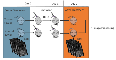 |
Multiparametric MRI to assess physiological changes induced by an anticancer therapy in neuroblastoma
Georgia Kanli1, Delphine Sauvage2, Elodie Viry2, Manon Bosseler 2, Bassam Janji2, and Olivier Keunen1
1Translational Radiomics Group, Quantitative Biology Unit, Luxembourg Institute of Health, Luxembourg, Luxembourg, 2Laboratory of Experimental Cancer Research, Department of Oncology, Luxembourg Institute of Health, Luxembourg, Luxembourg
Neuroblastoma (NB) is the most common extracranial solid tumor in childhood. High-risk patients have still a poor prognosis despite the therapeutic progress. The aim of this study is to assess by Magnetic Resonance Imaging (MRI) the physiological changes (perfusion and hypoxia) induced by an experimental anticancer therapy tested in a well-characterized preclinical transgenic neuroblastoma murine model. Our results show a decrease of the Ktrans parameter and relaxivity R2* value in treated mice compared to control, suggesting an impact of such therapy on vessels normalization.
|
|
4795.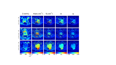 |
Multiparametric MRI as an early outcome predictor to chemotherapy and radiotherapy in cervical cancers
Jelena Mihailovic1,2, Aleksandar Tomasevic3, Fahmeed Hyder1,4, and Daniel Coman1
1(1) Magnetic Resonance Research Center (MRRC), Yale University, New Haven, CT, United States, 2(2) Department of Diagnostic Radiology, Yale University, New Haven, CT, United States, 3Institute for Oncology and Radiology Of Serbia, Belgrade, Yugoslavia, 4Center for Quantitative Neuroscience with Magnetic Resonance (QNMR), Yale University, New Haven, CT, United States
Pre/early intra-treatment prediction of patients with cervical cancer would enable treatment regimens to be changed at an early time point. We focused on diffusion-weighted imaging (DWI) and dynamic contrast-enhanced (DCE) MRI for quantifying of the tumor microenvironment in prediction of treatment response. Perfusion fraction multiplied by pseudo-diffusion coefficient, plasma flow, transfer constant between plasma and extracellular extravascular space were the parameters statistically significant associated with treatment outcome based on 95% CI in multivariate logistic regression model. Multi-parametric MRI techniques have the potential to assess tumor grade differentiation, and they showed additional value in detecting and therefore, predicting treatment response.
|
|
4796.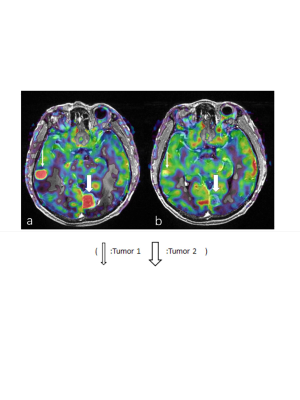 |
Assessment of the cerebral blood flow for brain metastasis after radiotherapy using 3D arterial spin labeling MRI
Chuanke Hou1, Guanzhong Gong1, Weiqiang Dou2, Weiyin Vivian Liu2, and Yong Yin1
1Department of Radiation Oncology, Shandong Cancer Hospital and Institute, Shandong First Medical University and Shandong Academy of Medical Sciences, Jinan 250117, China, Jinan, China, 2GE Healthcare, MR Research China, Beijing, China
In the study, the relationship between three brain regions (brain metastasis, normal brain region and perituberous edema region) and radiotherapy dose gradient has been explored. 3D arterial spin labeling perfusion imaging (3D-ASL) for quantitative cerebral blood flow measurement has been used in the clinical diagnosis, preoperative differentiation and grading of brain tumors. Therefore, individualized radiotherapy for BM patients could be optimized and improve the local control rate of BM.
|
|
4797.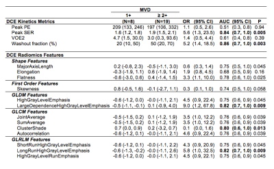 |
Breast MRI kinetic and radiomic features correlate with invasive breast cancer angiogenesis
Jennifer Xiao1, Michael Hirano1, Daniel Hippe1, Neal Shekar 1, Mara Rendi2, Kevin Cheung3, Habib Rahbar1, and Savannah Partridge 1
1Radiology, University of Washington, Seattle, WA, United States, 2Pathology, University of Washington, Seattle, WA, United States, 3Medicine, University of Washington, Seattle, WA, United States
Although kinetics are routinely incorporated into interpretation of contrast enhanced breast MRI, there is a paucity of literature supporting the association between the pathophysiologic basis of enhancement and its histopathologic correlate. Our study investigated DCE-MRI kinetic parameters and texture features analyzed with radiomics and their correlation with microvessel density as a surrogate for tumor angiogenesis. Twenty seven patients with invasive breast cancer were included in our study. Both peak SER and washout fraction and several texture features were significantly correlated with microvessel density (p<0.01), further supporting the biologic basis of malignant enhancement on breast MRI.
|
|
4798. |
Active Trans-Membrane Water Cycling Reflects Metabolic Activity in Breast Cancer MRI
Wei Huang1, Brendan Moloney1, Xin Li1, Manoj K Sammi1, Alina Tudorica1, Megan L Troxell1, Karen Y Oh1, Kathleen A Kemmer1, Arpana Naik1, Aneela Afzal1, and Charles S Springer, Jr.1
1Oregon Health & Science University, Portland, OR, United States
[Shutter-speed] SS-DCE-MRI allows access to the crucial on-going cell membrane sodium pump activity previously unreachable in vivo. Literature results indicate the latter is significantly increased early in the oncogenic transformation. This suggests an approach for early cancer detection with high-resolution imaging, a very important clinical goal. Here, we analyze SS-DCE-MRI and concomitant histopathological breast cancer data obtained during neo‑adjuvant chemotherapy, with this consideration in mind. The results support the contention, but indicate a non‑invasive, non-contrast DWI method under development is likely necessary to realize this clinical potential.
|
|
4799.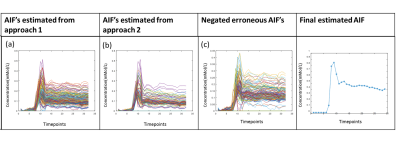 |
A Method to Improve Automated Subject Specific Arterial Input Function for DCE-MRI and to Evaluate its Effect on Glioma Grading at 3T
Dinil Sasi S1, Sameer Manickam2, Rakshit Dadarwal1, Rakesh K Gupta3, and Anup Singh4,5
1Center for Biomedical Engineering, Indian Institute of Technology Delhi, New Delhi, India, 2KTH Royal Institute of Technology, Stockholm, Sweden, 3Fortis Memorial Research Institute, Gurugram, India, 4Indian Institute of Technology Delhi, Hauz Khas, India, 5All India Institute of Medical Science, New Delhi, India
Arterial-input-function(AIF) or vascular-input-function is a prerequisite for quantitative analysis of dynamic-contrast-enhanced(DCE)-MRI data. For DCE-MRI data of human brain used in the current study, previously reported automatic AIF estimation approach resulted in large variations from theoretically expected shape. In this study, DCE-MRI data of 25 treatment-naïve glioma patients were included. Proposed optimization enabled the removal of wrongly selected voxels having distorted concentration curve and hence provided an improved AIF. A substantial change in the shape of AIF was observed on optimization. Corrected AIF also resulted in significant improvement in quantitative perfusion parameters and glioma gradin
|
|
4800.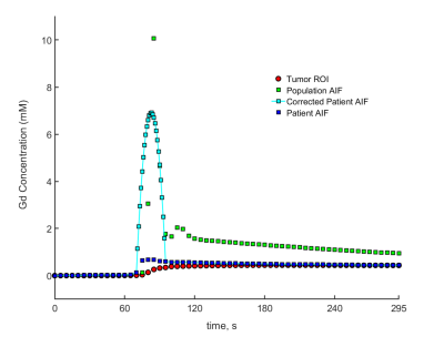 |
Personalized DCE-MRI parametric mapping of gynecological cancer using high spatiotemporal resolution GRASP
Nathanael Kim1, Yousef Mazaheri1, Yulia Lakhman2, Li Feng3, Ersin Bayram4, Alberto Vargas2, and Ricardo Otazo1,2
Video Permission Withheld
1Medical Physics, Memorial Sloan Kettering Cancer Center, New York, NY, United States, 2Radiology, Memorial Sloan Kettering Cancer Center, New York, NY, United States, 3Biomedical Engineering and Imaging Institute and Department of Radiology, Icahn School of Medicine at Mount Sinai, New York, NY, United States, 4GE Healthcare, Waukesha, WI, United States
Personalized estimation of the arterial input function (AIF) in DCE-MRI has been a relatively challenging task due to the slow imaging speed of conventional MRI. As a consequence, a population AIF is usually employed for parametric mapping, which represents a group effect rather than the long-desired personalized quantification. In this work, we use the GRASP method to perform DCE-MRI of gynecological tumors with high spatial and temporal resolution and to estimate the AIF directly from the data. The personalized AIF shows higher consistency with the tumor enhancement compared to the population AIF.
|
|
4801.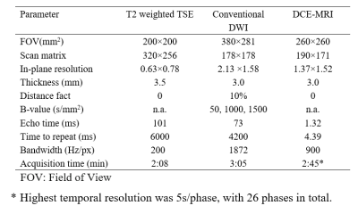 |
High-temporal-resolution dynamic contrast enhanced MRI helps the diagnosis of the clinically significant lesions of prostate
Lei Hu1, Li ming Wei1, Shuhao Wang1, Chunyu Jiang1, Caixia Fu2, and Jungong Zhao1
1Department of Diagnostic and Interventional Radiology, Shanghai Jiao Tong University Affiliated Sixt, shanghai, China, 2MR Application Development, Siemens Shenzhen magnetic Resonance Ltd, shenzhen, China
In clinical practice, definitions of the early enhancement are vague with different temporal resolution, and have caused some confusion. The aim of this study was to define the early enhancement in dynamic contrast enhanced (DCE)- MRI with high temporal resolution (5 sec), and to assess whether it can improve the detection of clinically significant lesions. The result indicated that the time to enhancement of clinically significant lesion was significantly different from those lesions with low grade or intermediate grade.
|
|
4802.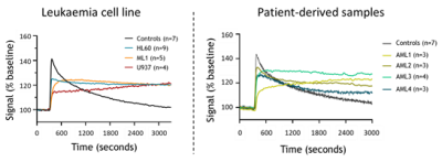 |
DCE-MRI for detection of leukaemia induced bone marrow vascular permeability
Ana Gomes1, Diana Passaro1, Dominique Bonnet1, and Bernard Siow1
1Francis Crick Institute, London, United Kingdom Poster Permission Withheld
Acute Myeloid Leukemia (AML) is the most common acute leukemia in adults. While the clinical presentation is quite uniform, it is a highly heterogeneous disease at the genetic level. Using intravital two-photon microscopy, we have previously showed that AML patient-derived samples belonging to different genetic subgroups induced a common pathologic bone marrow (BM) vascular phenotype. To understand the translational potential of our findings we have optimized DCE-MRI for the assessment of bone marrow vascular permeability upon leukaemia development.
|
|
4803.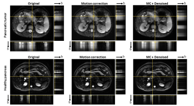 |
Reproducibility of intravoxel incoherent motion MRI of pancreas tumor and healthy pancreas
TING YIN1, Chao Ma2, Shiyue Chen2, Rolf Gruetter1, and Jianping Lu2
1CIBM-AIT, École polytechnique fédérale de Lausanne, Lausanne, Switzerland, 2Department of Radiology, Changhai Hospital of Shanghai, Shanghai, China
Intravoxel Incoherent Motion (IVIM) model has shown the potential of contrast free perfusion estimation in pancreatic diseases study, while IVIM quantification is challenging for conventional free-breathing DWI data due to the presence of high level noise. In this study, we demonstrated that motion correction and denoising can improve the sampling accuracy which allows for pixel-wise parametric maps with acceptable reproducibility.
|
|
4804. |
Evaluate the Feasibility of Delayed Contrast Extravasation MRI to Delineate Sub-volume Target in brain tumors Radiotherapy.
Yinxing Wang1, Guanzhong Gong1, Weiqiang Dou2, Weiyin Vivian Liu2, and Yong Yin1
1Department of Radiation Oncology, Shandong Cancer Hospital and Institute, Shandong First Medical University and Shandong Academy of Medical Sciences, Jinan, China, 2GE Healthcare, MR Research China, Beijing, China
Delayed contrast extravasation MRI (DCEM) could could differentiate regions of contrast agent clearance as an active tumor from regions of contrast agent accumulation as non-tumor tissues. By comparing the sub-volume of active tumor and non-tumor from DCEM with non-liquefaction necrosis and liquefaction from T2WI, our results showed that compared to liquefaction necrosis regions of the T2WI, the DCEM has advantages in distinguishing liquefaction area and could clearly differentiate sub-volume of active tumor from non-liquefaction. The application of DCEM was thus feasibly to guide the delineation of sub-volume target in brain tumor.
|
4805.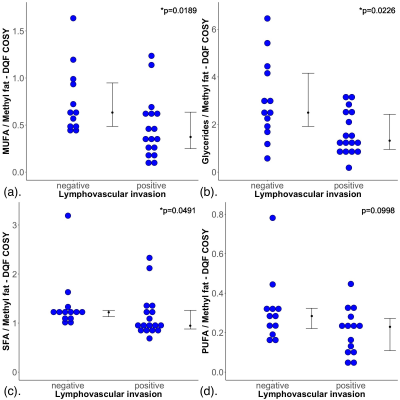 |
Lipid composition in breast tumours using double quantum filtered correlation spectroscopy is associated with lymphovascular invasion (LVI)
Sai Man Cheung1, Ehab Husain2, Yazan Masannat3, Vasiliki Mallikourti1, Steven Heys3, and Jiabao He1
1Aberdeen Biomedical Imaging Centre, University of Aberdeen, Aberdeen, United Kingdom, 2Pathology Department, Aberdeen Royal Infirmary, Aberdeen, United Kingdom, 3Breast Unit, Aberdeen Royal Infirmary, Aberdeen, United Kingdom
Lymphovascular invasion (LVI) is associated with increased recurrence and metastatic risk in breast cancer. LVI amplifies the impact of genetic mutations on lipid regulation, leading to the deregulation of lipid composition. Currently LVI is estimated using the biopsy sample, while definitive LVI can only be determined after surgery, precluding patients on neoadjuvant chemotherapy or hormonal treatment. Double quantum filtered correlation spectroscopy (DQF-COSY) is a method capable of accurate non-invasive lipid composition quantification. We therefore examined the role of lipid composition using DQF-COSY in differentiating the status of LVI.
|
|
4806.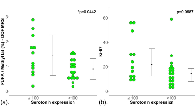 |
Polyunsaturated fatty acids (PUFA) depletion in high serotonin turnover breast tumours using advanced magnetic resonance spectroscopy (MRS)
Sai Man Cheung1, Ehab Husain2, Yazan Masannat3, Klaus Wahle4, Steven Heys3, and Jiabao He1
1Aberdeen Biomedical Imaging Centre, University of Aberdeen, Aberdeen, United Kingdom, 2Pathology Department, Aberdeen Royal Infirmary, Aberdeen, United Kingdom, 3Breast Unit, Aberdeen Royal Infirmary, Aberdeen, United Kingdom, 4Institute of Pharmacy and Biological Sciences, University of Strathclyde, Glasgow, United Kingdom
Locally advanced breast cancer is the most common cause of death in middle-aged women, with research focus shifted to preventative medicine. Polyunsaturated fatty acids (PUFA) is depleted in tumour initiation to sustain an inflammatory tumour microenvironment conducive to macrophage recruitment. Serotonin modulates macrophage activity and is a marker of poor 10-year survival in women with breast cancer. The relationship between serotonin and PUFA demands close examination for preventative treatment optimisation. We applied high sensitivity double quantum filtered MRS to accurately quantify PUFA between serotonin low and high breast tumours, and found PUFA depletion is associated with increased serotonin turnover.
|
|
4807. |
Deregulation of lipid metabolism in postmenopausal breast cancer patients using double quantum filtered correlation spectroscopy (DQF-COSY)
Sai Man Cheung1, Vasiliki Mallikourti1, Yazan Masannat2, Tanja Gagliardi3,4, Steven Heys2, and Jiabao He1
1Aberdeen Biomedical Imaging Centre, University of Aberdeen, Aberdeen, United Kingdom, 2Breast Unit, Aberdeen Royal Infirmary, Aberdeen, United Kingdom, 3Clinical Radiology, Aberdeen Royal Infirmary, Aberdeen, United Kingdom, 4Radiology, Royal Marsden Hospital, London, United Kingdom Deregulation of lipid metabolism has been shown in BRCA1/2 genetic mutation carriers. Mammary adipose tissues in postmenopausal women are the primary sites of oestrogen production linked to tumour initiation and progression. Therefore, lipid composition in postmenopausal breast plays a key role in breast cancer monitoring and subsequent development of prevention strategies. Previous studies focused on cell or animal models and invasive lipid extraction methods, while conventional MRS is inadequate in complete lipid composition measurement. We hypothesised that lipid composition in peri-tumoural breast adipose tissue is affected by the presence of tumour in postmenopausal women, using a non-invasive 2D MRS approach. |
|
4808.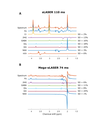 |
2-Hydroxyglutarate detection with semi-LASER and MEGA-sLASER at 7T
Zahra Shams1, Uzay Emir2, Wybe J.M. van der Kemp1, Dennis W.J. Klomp1, Jannie P. Wijnen1, and Evita Wiegers1
1Radiology, University Medical Center Utrecht, Utrecht, Netherlands, 2School of Health Sciences, College of Health and Human Sciences, Purdue University, West Lafayette, IN, United States
This study evaluates the performance of semi-LASER and MEGA-sLASER for 2-hydroxyglutarate (2-HG) detection at 7T. We compared a semi-LASER with TE of 110ms with editing of 2-HG at 4.02 ppm using MEGA-sLASER with TE of 74ms in phantoms with different concentrations of 2-HG. Both methods were able to detect 2-HG concentrations as low as 0.5mM. MEGA-sLASER provided a clean 2-HG signal. Contrary, the fact that the fitting accuracy for 2-HG in semi-LASER is similar to MEGA-sLASER promotes to choose for a semi-LASER implementation.
|
|
4809.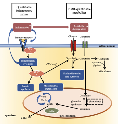 |
Association of tumor metabolites with inflammation in the glioma microenvironment studied with HRMAS NMR
Selin Ekici1 and Candace C. Fleischer1
1Department of Radiology and Imaging Sciences, Emory University School of Medicine, Atlanta, GA, United States
Evidence suggests that inflammation and metabolic dysregulation in the tumor microenvironment are related, yet the mechanistic relationship is not well characterized. Our goal was to utilize ex vivo 1H high resolution magic angle spinning (HRMAS) nuclear magnetic resonance (NMR) spectroscopy to study the relationship between metabolism and tumor inflammation in gliomas. We observed that multiple inflammatory markers were positively associated with glutamine, glutathione, and lactate. C-reactive protein was positively associated with myo-inositol and aspartate. These results support the hypothesis that inflammation may drive metabolic changes in gliomas and supports the use of HRMAS NMR in studies of the tumor microenvironment.
|
|
4810.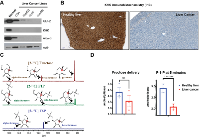 |
Quantification of Fructose Metabolism in Hepatocellular Carcinoma
Sui Seng Tee1, Roozbeh Eskandari2, Nathaniel Kim2, Arsen Mamakhanyan2, and Kayvan Keshari2
1Diagnostic Radiology and Nuclear Medicine, University of Maryland School of Medicine, Baltimore, MD, United States, 2Memorial Sloan Kettering Cancer Center, New York, NY, United States
Fructose metabolism utilizes a distinct set of transporters and enzymes and is limited to the liver, kidney and small intestines. Key to the ability to break down fructose is the enzyme ketohexokinase (KHK), that phosphorylates fructose to fructose-1-phosphate (F1P). In this study, we demonstrate that fructose metabolism is downregulated in a mouse model of hepatocellular carcinoma (HCC). Based on this novel observation, we use 13C nuclear magnetic resonance (NMR) to quantify altered hepatic fructose metabolism. We demonstrate significantly decreased F1P production in HCC, providing justification for developing non-invasive methods to detect fructose metabolism.
|
|
4811.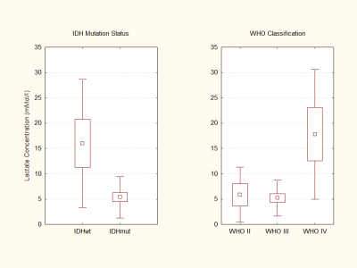 |
Elevation in lactate concentration and intracellular pH is less pronounced in IDH mutant compared to IDH wildtype glioma patients
Katharina Wenger1, Joachim P. Steinbach2, Oliver Bähr2, Ulrich Pilatus1, and Elke Hattingen1
1Neuroradiology, Goethe-University Frankfurt, Frankfurt, Germany, 2Neurooncology, Goethe-University Frankfurt, Frankfurt, Germany
Preclinical evidence points towards a metabolic reprogramming in isocitrate dehydrogenase mutated tumor cells with downregulation of the expression of genes that encode for glycolytic metabolism. We non-invasively investigated lactate concentrations, as well as intracellular pH using 1H and 31P MRS in a glioma patient cohort. At TE 97 ms, lactate peaks can be fitted with little impact of lipid/macromolecule contamination. We found a significant difference in lactate concentrations and intracellular pH comparing tumor voxels of IDH mutatet to IDH wildtype patients, with reduced lactate levels and near normal intracellular pH in IDHmut patients.
|
|
4812. |
Glioblastoma Characterization with Hyperpolarized [1-13C]Pyruvate MRSI, DWI and 18F-FDG PET
Geoffrey J. Topping1, Roland E. Kälin2, Linzhi Cai2, Rainer Glaß2, and Franz Schilling1
1Department of Nuclear Medicine, Klinikum rechts der Isar, Technical University of Munich, Munich, Germany, 2Neurosurgical Research, University Clinics Munich, Ludwig Maximilian University, Munich, Germany
Multimodal imaging has the potential for non-invasive assessment of imaging biomarkers that guide and monitor treatment of glioblastoma tumours. In this work, an imaging protocol for characterization of implanted patient-derived and murine glioblastoma tumours (GBM2, GBM14, and U87) was established using T2-weighted MRI, DWI, hyperpolarized 13C-pyruvate-lactate CSI, and 18F-FDG PET. Tumours were visible in T2-weighted MRI as high-signal regions with poorly defined borders. Compared with shams and non-tumour brain, all tumour lines had elevated ADC, 18F-FDG Ki, and lactate-to-pyruvate ratio. GBM2 had particularly high and variable lactate-to-pyruvate ratio, despite relatively low variability in ADC.
|
|
4813.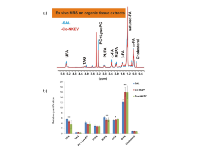 |
Antitumor effects of NK-derived nanovesicles in a B-cell lymphoma preclinical model by integrating in vivo MRI and ex vivo MRS
Rossella Canese1, Maria Elena Pisanu1, Mattea Chirico1, Daniele Macchia2, Massimo Spada2, Egidio Iorio1, Serena Cecchetti3, Cristina Federici4, and Luana Lugini4
1NMR and MRI unit, Core facilities, Istituto Superiore di Sanita', Rome, Italy, 2National Center for animal experimentation and welfare, Istituto Superiore di Sanita', Rome, Italy, 3Microscopy unit, Core facilities, Istituto Superiore di Sanita', Rome, Italy, 4Oncology and Molecular Medicine Department, Istituto Superiore di Sanita', Rome, Italy
NK cells (NK) are the first barrier of body defense from tumor cells. Individuals with low NK activity display an increased risk to develop cancer. Extracellular vesicles are secreted vesicles possessing immune regulatory properties. We demonstrated that exosomes produced by NK cells (isolated from blood of healthy donors) display a cytotoxic activity against tumors in vitro. In this work we monitor the effects of NKEVs treatment by in vivo MRI/MRS and ex vivo MRS metabolomics in a xenograft model of lymphoma. We found altered lipid and redox metabolism suggesting taurine and lipid signals as potential biomarkers of NKEVs response
|
|
4814.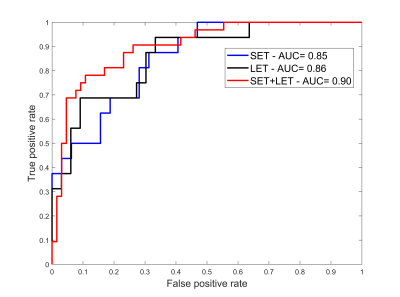 |
Augmented magnetic resonance spectra to differentiate metastasis from glioblastoma brain tumours
Nikolaos Dikaios1
1University of Surrey, Guildford, United Kingdom
Metabolic processes monitored by MRS precede micro-structural changes visualised by imaging. The high noise and the overlapping spectra of metabolites affect the accurate quantification of metabolite’s concentration. This work hypothesizes that each tissue has a unique metabolic fingerprint and a diagnostic model could be built based on tissue spectra. The MRS datasets however are usually small and acquired with different parameters. This work quantum-mechanically simulates spectra and uses the augmented spectra to train a model that can differentiate metastasis from glioblastoma brain cancer. The trained model was tested on acquired spectra from the INTERPRET single-voxel dataset and illustrated a ROC-AUC=0.90.
|
|
4815.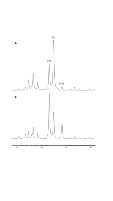 |
Effect of hypoxia and choline kinase inhibitor on choline metabolism of brain tumour cells using high resolution ¹H NMR
Claire Louise Kelly1, Sofya A Osharovich2, Marie Phelan3, Violaine See3, Edward J Delikatny2, and Harish Poptani1
1Center for Preclinical Imaging, University of Liverpool, Liverpool, United Kingdom, 2Department of Radiology, University of Pennsylvania, Philadelphia, PA, United States, 3Institute of Integrative Biology, University of Liverpool, Liverpool, United Kingdom
High resolution ¹H NMR of cell extracts was used to assess the effects of hypoxia on choline metabolism in four glioblastoma (GBM) cell lines (9L, F98, U87 and U251). In addition, the effects of hypoxia on the efficacy of JAS239, a choline kinase alpha (ChoKα) inhibitor, was also assessed in these cell lines. Hypoxic preconditioning resulted in a decrease in phosphocholine (PC) in 9L, F98 and U251 cells. JAS239 significantly decreased PC/glycerophosphocholine (GPC) ratio in the hypoxic 9L and U251 cells compared to when these cells were grown in normoxic conditions.
|
|
4816.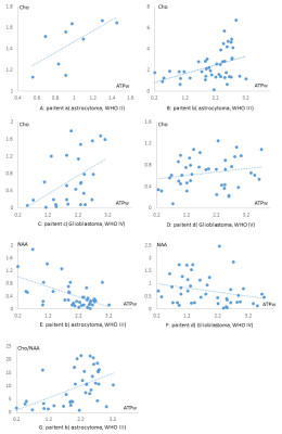 |
The correlation analysis of 3D APT with the parameters of 3D MR spectroscopy in glioma patients: an image-guided intraoperative pilot study
Xiaoluo Zhang1, Dongxiao Zhuang1, Jilei Zhang2, Jianqing Sun2, Weibo Chen2, and Jinsong Wu1
1Glioma Surgery Division, Neurologic Surgery Department, Huashan Hospital, Fudan University, Shanghai, China, 2Clinical Science, Philips Healthcare, Shanghai, China
Our study is the first research to investigate the relationship between 3D APT and 3D MRS on individual level. The APTw values were correlated with the metabolite concentrations or ratios of MRS, especially Cho concentration. We proposed that 3D APT can be potentially used in intra-operation system to guide surgical biopsy and tumor resection.
|
4817. |
Investigating the correlation and repeatability of radiomic features derived from Apparent Diffusion Coefficient maps of Soft-Tissue Sarcoma
Imogen Thrussell1, Jessica Winfield1,2, Matthew Orton1, Aisha Miah1,3, Shane Zaidi3, Amani Arthur1, Khin Thway3,4, Dirk Strauss5, David Collins1, Dow-Mu Koh1,2, Uwe Oelfke1, Paul Huang6, Christina Messiou1,2, and Matthew
Blackledge1
1Division of Radiotherapy and Imaging, The Institute of Cancer Research, London, United Kingdom, 2Department of Radiology, The Royal Marsden NHS Foundation Trust, London, United Kingdom, 3Sarcoma Unit, The Royal Marsden NHS Foundation Trust, London, United Kingdom, 4Department of Pathology, The Royal Marsden NHS Foundation Trust, London, United Kingdom, 5Department of Surgery, The Royal Marsden NHS Foundation Trust, London, United Kingdom, 6Division of Molecular Pathology, The Institute of Cancer Research, London, United Kingdom
Soft-tissue sarcomas (STS) are highly heterogeneous and, except for myxoid liposarcomas, changes in size following radiotherapy do not correlate with outcomes or histopathological response. Therefore, conventional size-based criteria fail to accurately assess response. In this study we explore the correlation and repeatability of radiomic features derived from Apparent Diffusion-Coefficient maps in a cohort of 27 patients with confirmed STS with a variety of histopathological subtypes. 19 features were identified that had good repeatability and were considered uncorrelated using hierarchical clustering. These features could be used in future studies to investigate heterogeneous response of STS to radiotherapy.
|
|
4818. |
Surveillance of the Morphologic and Metabolic Features of the Tumor Microenvironment During Progression of Glioblastoma
John J Walsh1, Maxime J Parent2, Lucas C Adam2, Muhammad H Khan1, Sandeep K Mishra2, Samuel K Maritim1, Daniel Coman2, and Fahmeed Hyder1,2
1Department of Biomedical Engineering, Yale University, New Haven, CT, United States, 2Department of Radiology and Biomedical Imaging, Yale University, New Haven, CT, United States
The morphologic and metabolic basis of the tumor microenvironment is multifaceted, and this complexity is magnified throughout tumor progression. Therefore longitudinal surveillance of this habitat during tumor growth will improve drug targeting. We used translational MRI methods in glioblastoma models to investigate time-dependent remodeling of the extracellular matrix and microvasculature in relation to interstitial acidification. With tumor progression, we detected extracellular volume reduction and vascular plasma flow increase, but extracellular pH and vascular permeability remained unchanged. These differential dynamic processes regulating morphologic and metabolic transformations of the tumor microenvironment suggest distinct time windows for mechanistic therapeutic targeting.
|
|
4819. |
A deep learning model to predict near pathological complete response for rectal cancer by the diffusion MRI data before chemoradiotherapy
Hai-Tao Zhu1, Xiao-Yan Zhang1, Yan-Jie Shi1, Xiao-Ting Li1, and Ying-Shi Sun1
1Peking University Cancer Hospital, BEIJING, China
A deep learning model is proposed to predict near pathological complete response by diffusion MRI data before chemoradiotherapy. 624 participants are included in this study with 424 for training and 200 for testing. The area under the curve of receiver operating characteristic is 0.800 (95%CI: 0.735-0.851). The sensitivity is 0.700 (95%CI: 0.568-0.812), the specificity is 0.871 (95%CI: 0.804-0.922). Compared with the strategy that uses both pre-NCRT and post-NCRT data, the method may predict the pathological results at an earlier time point before the initiation of NCRT, which enables a chance to modify the NCRT plan if needed.
|
|
4820. |
Prediction of therapeutic response of HCC to transcatheter arterial chemoembolization based on pretherapeutic dynamic enhanced MRI radiomics
Ying Zhao1, Ailian Liu1, Jingjun Wu1, Nan Wang1, Dahua Cui1, Tao Lin1, Qingwei Song1, Xin Li2, Tingfan Wu2, and Yan Guo3
1The First Affiliated Hospital of Dalian Medical University, Dalian, China, 2Translational Medicine Team, GE Healthcare, Shanghai, China, 3GE Healthcare, Beijing, China
In the current study, dynamic enhanced MRI radiomics was demonstrated to be capable to predict therapeutic response in hepatocellular carcinoma treated with transcatheter arterial chemoembolization, which will provide more prognostic information and facilitate clinical management.
|
|
4821. |
Differential diagnosis of hepatic benign and malignant tumors using texture features based on multiple high b-values DWI
Ying Zhao1, Jiazheng Wang2, Zhiwei Shen2, Zhongping Zhang2, Nan Wang1, Lihua Chen1, Dahua Cui1, Tao Lin1, Qingwei Song1, Renwang Pu1, Bingbing Gao1, and Ailian Liu1
1The First Affiliated Hospital of Dalian Medical University, Dalian, China, 2Philips Healthcare, Beijing, China
This work aimed for multiple high b-values DWI texture features based strategy to identify hepatic benign and malignant tumors, which may provide more abundant and comprehensive quantitative information and promote clinical decision-making. The results showed that texture features based on ultra-high b value (b = 3000 s/mm2) DWI images can achieve the best result (combined AUC: 0.960; sensitivity: 92.0%; specificity: 92.3%), forming a valuable strategy for clinical practice.
|
|
4822.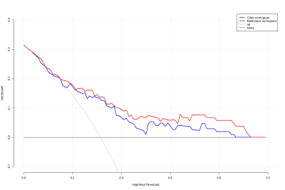 |
DWI-based Radiomics Nomogram Predicting Recurrence-Free Survival in Patients with Muscle-Invasive Bladder Cancer
Shan Zhang1, Guangyu Wu1, Guiqin Liu1, Yongming Dai2, and Yunfei Zhang2
1Renji Hospital Affiliated to Shanghai Jiaotong University School of Medicine, Shanghai, China, 2United Imaing Healthcare, Shanghai, China
We hypothesized that the radiomics features obtained from DWI holds great potential in improving the recurrence risk stratification of MIBC patients. Thus, we developed a radiomics nomogram and compared its performance with clinicopathological nomogram and radiomics signature in individual RFS prediction. Our results showed that the radiomics nomogram, which incorporates radiomics signatures, clinical characteristics and molecular characteristics, has greater potential to serve as a biomarker to estimate the RFS in MIBC patients. In conclusion, a radiomics nomogram may serve as a potential tool to facilitate individualized prediction of recurrence in patients with MIBC.
|
|
4823.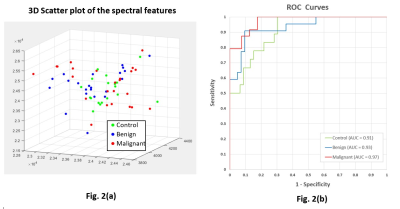 |
Neural-network discrimination of human plasma samples to detect pancreatic cancer
Meiyappan Solaiyappan1, Santosh K Bharti1, Mohamad Dbouk2, Paul T Winnard1, Michael Goggins2,3, and Zaver M. Bhujwalla1,3,4
1The Russell H. Morgan Dept of Radiology and Radiological Science, The Johns Hopkins University School of Medicine, Baltimore, MD, United States, 2Departments of Pathology and Medicine, The Sol Goldman Pancreatic Cancer Research Center, The Johns Hopkins University School of Medicine, Baltimore, MD, United States, 3Sidney Kimmel Comprehensive Cancer Center, The Johns Hopkins University School of Medicine, Baltimore, MD, United States, 43Radiation Oncology and Molecular Radiation Sciences, The Johns Hopkins University School of Medicine, Baltimore, MD, United States
The insidious growth of pancreatic cancer is a major factor contributing to its lethality. Only 10-15% of pancreatic cancers are resectable by the time they are detected. Early detection of pancreatic cancer through routine screening is clearly an unmet clinical need. Here we have applied neural network analysis to 1H magnetic resonance spectra of human plasma samples to differentiate between healthy subjects (control), subjects with benign lesions, and subjects with pancreatic ductal adenocarcinoma (PDAC). Our data support developing a neural-network approach to identify PDAC from 1H MRS of plasma samples.
|
|
4824.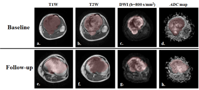 |
Texture Analysis in Chemotherapy Response Evaluation in Osteosarcoma: A Preliminary Study
Esha Baidya Kayal1, Devasenathipathy Kandasamy2, Raju Sharma2, Sameer Bakhshi3, and Amit Mehndiratta1,4
1Centre for Biomedical Engineering, Indian Institute of Technology, Delhi, New Delhi, India, 2Radio Diagnosis, All India Institute of Medical Sciences, New Delhi, New Delhi, India, 3Department of Medical Oncology, Dr. B.R. Ambedkar Institute-Rotary Cancer Hospital (IRCH), All India Institute of Medical Sciences, New Delhi, New Delhi, India, 4Department of Biomedical engineering, All India Institute of Medical Sciences, New Delhi, New Delhi, India Poster Permission Withheld
Recently texture analysis(TA) of MR images has also shown promising results in evaluating response to therapy in different types of cancer. TA uses mathematical approach to characterize the spatial distribution of signal intensity variations in an image and extracts quantitative features thus may infer clinically relevant information about tissue microstructure and its subtle changes during treatment. We examined efficacy of MRI based statistical TA methods in evaluating chemotherapy response in Osteosarcoma early in the course of treatment with correlation to histological response. Experimental results showed statistical TA methods may be effectively used in evaluating chemotherapy response in patient with Osteosarcoma.
|
|
4825.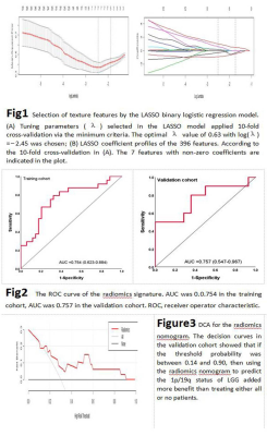 |
Application Value of Radiomics Approach in Predicting Chromosomal Arms 1p/19q in Low-Grade Gliomas
Wei Wei1, Fan Li Hua1, Nan Yu1, and Yong Yu1
1Affiliated Hospital of Shaanxi University of Chinese Medicine, XianYang, China
Genes play a crucial role in the development, progression and therapeutic outcome of tumors. Several studies have linked codeletion of chromosomal arms 1p/19q in LGG with positive response to chemotherapy and radiotherapy and longer progression-free survival, so 1p/19q status of LGG can impact optimal therapy options. Unfortunately, determining 1p/19q status requires surgical biopsy to identify chromosomal deletion. Radiomics, which extracts high-throughput features from medical images, showing great advantages in tumor phenotype classification, treatment options and prognosis analysis.
|
|
 |
4826. |
Deep Learning to Discriminate Nasopharyngeal Carcinoma and Benign Hyperplasia on MRI
Lun M. Wong1, Ann D. King1, and Qiyong Ai1
1Department of Imaging and Interventional Radiology, The Chinese University of Hong Kong, Shatin, Hong Kong
Benign hyperplasia is a common finding in the adenoid and walls of the nasopharynx and may hamper the detection of early-stage nasopharyngeal carcinoma (NPC) on MRI. In this study we aim to utilize deep learning to discriminate early-stage NPC from benign hyperplasia using T2-weighted-fat-suppressed MR images. We tested our method on a dataset of 413 cases, comprising 203 with early-stage NPC confined to the nasopharynx and 210 with benign hyperplasia. After training with validation (n=350 and n=13 respectively) followed by testing (n=50), the network achieved a promising result with a sensitivity of 100% and specificity of 83% for NPC detection.
|