
Digital Poster Session
Cancer: Therapy Planning, Response & Breast
Cancer
4827 -4840 Cancer Imaging: Therapy Planning, Response & Breast - Cancer Imaging: Therapy Planning
4841 -4854 Cancer Imaging: Therapy Planning, Response & Breast - Cancer Imaging: Therapy Response
4855 -4870 Cancer Imaging: Therapy Planning, Response & Breast - Cancer Imaging: Miscellaneous 2
4871 -4886 Cancer Imaging: Therapy Planning, Response & Breast - Cancer Imaging: Breast
4827.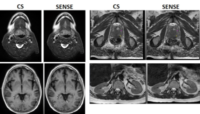 |
Combined SENSE and Compressed Sensing to Enhance Radiation Therapy MR Simulation
Kristen Zakian1, Neelam Tyagi1, Victoria Yu1, Alex Dresner2, Paul Romesser3, and Ricardo Otazo1
1Medical Physics, Memorial Sloan Kettering Cancer Center, New York, NY, United States, 2Philips Healthcare, Best, Netherlands, 3Radiation Oncology, Memorial Sloan Kettering Cancer Center, New York, NY, United States
We evaluated a combination of SENSE acceleration and compressed sensing in the MR simulation workflow to reduce scan time and improve image quality and anatomical coverage. Imaging series were performed with SENSE-only and with a combined SENSE+compressed sensing (CS) software package. CS resulted in an average reduction of 28% in single-series scan time with negligible changes in image quality and the ability to contour structures for RT treatment planning. In addition, CS permitted pulse sequence modifications which reduced respiratory artifacts, improved visibility of the prostatic urethra, and eliminated the need for a Foley catheter in prostate cancer patients.
|
|
4828.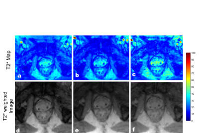 |
Accelerated T2* Mapping in prostate cancer on an MR-Linac
Wajiha Bano1, Mohammad Golbabaee2, and Andreas Wetscherek1
1Joint Department of Physics, The Institute of Cancer Research and The Royal Marsden NHS Foundation Trust, London, United Kingdom, 2Computer Science Department, University of Bath, Bath, United Kingdom
T2* mapping can be used to characterize tumour hypoxia, which is associated with therapy resistance. We show the feasibility of fast T2* mapping in prostate cancer patients on a 1.5T MR-Linac. The undersampled data was reconstructed by combining parallel imaging and a sparsifying transform with an iterative model-based reconstruction. The method was tested on a multi-compartment phantom and two prostate cancer patients with retrospective undersampling. With the proposed method T2* maps can be acquired in under five minutes. We demonstrated the feasibility of daily quantitative MRI over the course of radiotherapy on an MR-Linac, to characterize treatment response.
|
|
4829.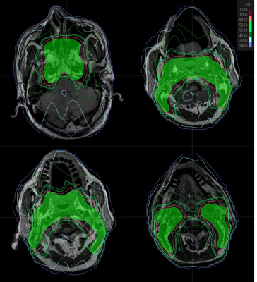 |
Study on Intensity Modulated Radiotherapy Plan Design of Nasopharyngeal Carcinoma based on MR Images
Xinsen Yao1, Guanzhong Gong1, Jianxin Ren1, and Yong Yin1
1Shandong Cancer Hospital and Institute, Jinan, China
To study the feasibility of intensity modulated radiotherapy (IMRT) plan which designed for nasopharyngeal carcinoma (NPC) based on MRI-only comparing the dosimetric difference of different CT value assignment methods on dose calculation. Pseudo CT were generated by different assignment methods based on simulation CT. The areas with a difference of more than 1% were mainly distributed in the air cavity, bone periphery and the skin. The assignment method of each tissue and organs were set to Populate CT value compared with other methods has the least influence on the dose calculation of NPC-IMRT plan, which could meet the clinical requirements.
|
|
4830. |
Planning Reconstruction of Interstitial High Dose Rate Brachytherapy Needles based on MRI Data
Victoria Newton1, William McGuire2, Rachel Wills1, Gerry Lowe1, and N. Jane Taylor2
1Radiotherapy Physics, Mount Vernon Hospital, Northwood, United Kingdom, 2Paul Strickland Scanner Centre, Mount Vernon Hospital, Northwood, United Kingdom
Interstitial brachytherapy needles in High Dose Rate radiotherapy patients can be visualised clearly and accurately within clinical tolerance values using just a proton-density StarVIBE sequence instead of registered CT and MR images. This could lead to a reduced radiation dose MR-only workflow without compromising the quality of treatment and a reduction of errors in image registration.
|
|
4831.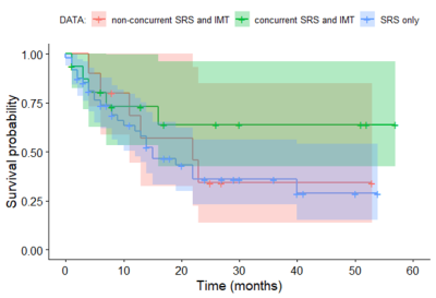 |
Does the timing of immunotherapy with stereotactic radiosurgery affect the outcome of patients with brain metastases?
Endre Grøvik1, Line Brennhaug Nilsen1, Ingrid Digernes1, Cathrine Saxhaug1, Anna Latysheva1, Oliver Geier1, Birger Breivik2, Dag Ottar Sætre3, Kari Dolven Jacobsen1, Åslaug Helland1, and Kyrre Eeg Emblem1
1Oslo University Hospital, Oslo, Norway, 2Hospital of Southern Norway, Kristiansand, Norway, 3Østfold Hospital Trust, Kalnes, Norway
The introduction of immunotherapy (IMTx) has led to a paradigm shift in the treatment of patients with metastatic cancer. Particular interest has been directed towards combining IMTx with stereotactic radiosurgery (SRS) to improve treatment response. Lately, studies have indicated that the timing of IMTx with SRS may be crucial for the patient outcome, suggesting that there is a window of opportunity to induce an optimal synergy between irradiation and immune agents. To this end, this work investigates whether the timing of IMTx with SRS affects the outcome of patients with brain metastases.
|
|
4832.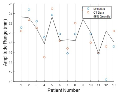 |
COMPARISON OF 4D-CT AND 4D- MRI FOR LUNG TUMOUR MOTION MEASUREMENT: A PHANTOM STUDY.
Terry Edward Perkins1, Jonathan Goodwin2, and Peter Greer3
1Sydney university, Rankin Park, Australia, 2Radiation Oncology, Calvary Mater Newcastle, Newcastle, Australia, 3Radiation Oncology, Calvary Mater Hopsital Newcastle, Newcastle, Australia
Using a golden angle radial sampling technique (Siemens WIP#1104), the motion range detection capabilities of 4D-MRI were compared to 4D-CT simulating lung tumour motion using a MODUS QA 4D MRI phantom. Thirteen patient respiratory patterns were used to simulate realistic breathing motion. Repeated measures ANOVA (F=0.052, p=0.948), a post-hoc paired t-test (all p>0.677) and the RMSE (RMSE12 = 1.9 mm) were used to evaluate differences between the detected motion ranges. Results showed no statistically significant difference between the motion range detection capabilities of 4D-CT and 4D-MRI in the phantom. This observation provides evidence for further evaluation in a patient population.
|
|
4833.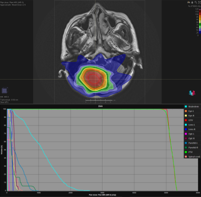 |
Magnetic resonance imaging in the evaluation of dose distribution of a radiotherapy plan for brain metastases
Jianxin Ren1, Guanzhong Gong1, Xinsen Yao1, Weiqiang Dou2, Weiyin Vivian Liu2, and Yong Yin1
1Department of Radiation Oncology, Shandong Cancer Hospital and Institute, Shandong First Medical University and Shandong Academy of Medical Sciences, Jinan, China, 2GE Healthcare, MR Research China, Beijing, China
In this study, a protocol of pseudo CT values obtained on MRI for treatment planning was proposed. The tissue or organ-specific CT values in representation of the electron density acquired from 35 patients with brain metastases were assigned to MRI. The dose distribution was recalculated, and the dosimetric differences between a new plan and an original plan were compared. The results showed that the dose distribution errors were basically controlled under 2.0% with pseudo CT values to bones, cavities and soft tissues, and lowered than 1.5% with pseudo CT values to different tissues and organs.
|
|
4834. |
MR-guided-radiotherapy gating using 3D positional probability volume derived from time-resolved volumetric MRI: A conceptual study
Jing Yuan1, Oi Lei Wong1, Yihang Zhou1, Yick Wing Ho1, Kin Yin Cheung1, and Siu Ki Yu1
1Medical Physics and Research Department, Hong Kong Sanatorium & Hospital, Happy Valley, Hong Kong
In this study, we proposed a conceptual MRgRT gating strategy using the organ’s motion positional probability volume (PPV) derived from the time-resolved volumetric MRI, aiming to improve treatment accuracy and efficiency. We demonstrated it for the scenario of kidney MRgRT using prospectively acquired CAIPIRINHA-accelerated VIBE MRI data at 1.5T from 7 healthy volunteers. Gating efficiencies of the proposed method were compared to those using the conventional 40%-60% respiratory phase gating. The results showed that the proposed method achieved significantly higher gating efficiency with similar target positional accuracy, indicating its potential value in the future individualized adaptive MRgRT.
|
|
4835.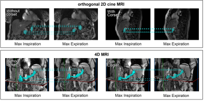 |
MRI based measurement of pancreas motion reduction with an MR- and particle therapy compatible abdominal corset
Sergej Schneider1,2,3, Sarah Stefanowicz1,3,4, Christina Jentsch1,4,5, Fabian Lohaus4, Chiara Valentini4, Esther G. C. Troost1,3,4,5,6, and Aswin L. Hoffmann1,3,4
1Oncoray – National Center for Radiation Research in Oncology, Faculty of Medicine and University Hospital Carl Gustav Carus, Technische Universität Dresden, Helmholtz-Zentrum Dresden-Rossendorf, Dresden, Germany, 2Technische Universität Dresden, Carl Gustav Carus Faculty of Medicine, Dresden, Germany, 3Institute of Radiooncology-OncoRay, Helmholtz-Zentrum Dresden-Rossendorf, Dresden, Germany, 4Department of Radiotherapy and Radiation Oncology, Faculty of Medicine and University Hospital Carl Gustav Carus, Technische Universität Dresden, Dresden, Germany, 5National Center for Tumor Diseases (NCT), Partner Site Dresden, Germany, German Cancer Research Center (DKFZ), Heidelberg, Germany, Faculty of Medicine and University Hospital Carl Gustav Carus, Technische Universität Dresden, and Helmholtz Association / Helmholtz-Zentrum Dresden-Rossendorf (HZDR), Dresden, Germany, 6German Cancer Consortium (DKTK), partner site Dresden, and German Cancer Research Center (DKFZ), Heidelberg, Germany
Particle beam therapy (PBT) is extremely sensitive to patient setup uncertainties and anatomical variations prior to and during dose delivery. Organ motion mitigation strategies are hence required to deliver the dose with high precision and accuracy. In this study, an MR- and PBT-compatible patient-individualized abdominal corset was developed and tested in 9 patients with abdominal cancer for its efficacy to reduce respiratory-induced motion. Pancreas motion was analyzed with and without corset by means of orthogonal 2D-cine and 4D-MRI.
|
|
4836.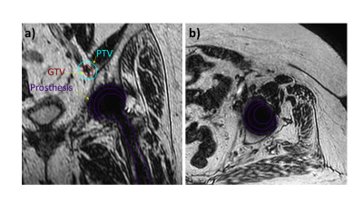 |
MRI guided radiation therapy near metal implants in a 1.5T MR-Linac system
Fahed Alsanea1, Teodor Stanescu2, Yao Ding1, Sastry Vedam1, Jinzhong Yang1, Seungtaek Choi1, Anuja Jhingran1, and Jihong Wang1
1UT MD Anderson Cancer Center, Houston, TX, United States, 2Princess Margaret Cancer Centre, Toronto, ON, Canada
MR-Linac systems are useful to visualize small soft-tissue targets for radiotherapy (RT) treatments. Geometric distortion caused by metal implants can interfere with the accuracy of the RT plan design and delivery. In this study, we simulated the spatial distortions caused by a hip prosthesis and determined its impact on surrounding tissues.
|
|
4837.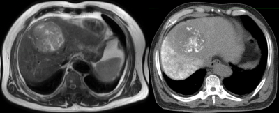 |
The Investigation of Internal Target Volume(ITV)Definition For Radiation Therapy of Hepatocellular Carcinoma Guided Using 4D-MR-T2 Images
Yinxing Wang1, Jianxin Ren1, Guanzhong Gong1, Weibo Chen2, Marijn Kruiskamp3, and Yong Yin1
1Department of Radiation Oncology, Shandong Cancer Hospital and Institute, Shandong First Medical University and Shandong Academy of Medical Sciences, Jinan, China, 2Philips Healthcare, Shanghai, China, 3Philips Healthcare, BIU ADI MR Therapy Clinical Science, Amsterdam, Netherlands
In this study, we did the research of the feasibility and advantage in defining the ITV for RT of HCC guided by 4D-MR-T2 images compared to 4D-CT images. It had been found that gross tumor target volume and internal tumor target volume boundaries could be displayed clearly on 4D-MR-T2 images. The position and deformation status of liver and tumors could be monitored easily on 4D-MR-T2 images. 4D-MR-T2 Images should be the first choice for simulation of HCC to define the ITV, and establish adaptive RT approach for HCC.
|
|
4838.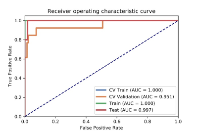 |
Evaluating the pairwise AdaBoost model in predicting the efficacy of chemo-radiotherapy for advanced rectal cancer under small sample size
Qinglei Shi1, Xiaoming Xi*2, Yilong Yin3, Jie Kuang4, Gaofeng Shi4, Xu Yan5, Yi Qu6, and Dongsheng Zhou7
1School of Software, Shan Dong University, Jinan, China, 2School of Computer Science and Technology, Shandong Jianzhu University, Jinan, China, 3School of Software, Software Park Campus, Shandong University, Jinan, China, 4CT Room, Radiology Department, Hebei Medical University Affilited 4th Hospital, Shi Jiazhuang, China, 5MR Scientific Marketing, Siemens Healthcare, Shanghai, China, Shanghai, China, 6Department of Geriatrics, Qilu Hospital of Shandong University, Jinan, China, 7Department of Breast and Thyroid Surgery, Shandong Provinvial Qianfoshan Hospital, The First Hospital Affiliated with Shandong First Medical University, Jinan, China
This paper proposed a pairwise AdaBoost model in predicting the therapeutic effect of non-metastatic LARC treated with neoadjuvant chemotherapy-radiation therapy based on radiomics signatures coming from ADC maps. Compared with traditional models, the pairwise AdaBoost model has ability to enlarge the number of training samples, which is useful to improve the generalization ability of the model. The experimental results demonstrated that the pairwise AdaBoost model seems can improve the accuracy and robustness of the model in predicting the treatment effect for locally LARC treated with neoadjuvant chemotherapy-radiation therapy.
|
|
4839.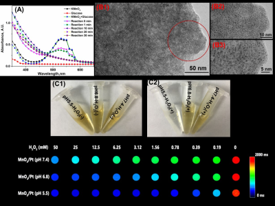 |
MRI Guided and Enhanced Radiosensitization by MnO2/Pt Nanosheets with Tumor Microenvironment-Triggered in Cancer Radiotherapy
Lijuan Chen1, Mingbo Liu1, Wei wei1, Yan Bai1, Haiyan Gao1, and Meiyun Wang1
1Henan Provincial People’s Hospital, Zhengzhou, China
We design the MnO2/Pt system to enhance the radiosensitization. 1-2 nm of platinum are uniformly dispersed on the as-prepared MnO2 nanosheet. The as-prepared MnO2/Pt are degraded by H2O2 and GSH into magnetic resonance functional imaging materials Mn2+. Moreover, MnO2/Pt can degrade GSH to GSSG, which is obstructed DNA repair of cancer cells after radiotherapy. From the results of colony formation, MnO2/Pt can improve the radiotherapy effect on U87 cells with the treatments of X-ray (8 Gy). MnO2/Pt are expected to achieve integrated magnetic resonance guided sensitization radiotherapy in vivo.
|
|
4840. |
Intrinsically Registered Fat Saturated and Unsaturated T2W Images Using a Keyhole Method
Steven Jackson1,2, Hanna Hanson2, David Cobben2, Kathryn Banfill2, Ahmed Salem2, Lisa McDaid1, Marcel van Herk2, and Benjamin Rowland2
1The Christie NHS Foundation Trust, Manchester, United Kingdom, 2Division of Cancer Sciences, The University of Manchester, Manchester, United Kingdom
Fat saturated and unsaturated T2W images of the same anatomy are required for MRI lung radiotherapy treatment planning. A prospective 'keyhole' pulse sequence that produces both image contrasts at full resolution via acquisition of a subset of FS k-space substituted into the non FS k-space was simulated. Simulated FS images with 50% of FS k-space substituted in a cross hair pattern were found to be only slightly inferior to acquired FS images by four clinicians based on scoring for delineation suitability. The prospective pulse sequence would save MR protocol time and improve the patient experience.
|
 View the Poster
 Watch the Video
4841.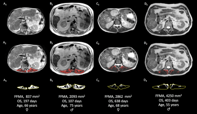 |
Yttrium-90 Radioembolization for Hepatocellular Carcinoma: Outcome Prediction with MRI Derived Fat-Free Muscle Area
Anton Faron1, Alois M. Sprinkart1, Claus C. Pieper1, Rolf Fimmers2, Wolfgang Block1, Carsten Meyer1, Daniel Thomas1, Ulrike Attenberger1, and Julian A. Luetkens1
1Radiology, University Hospital Bonn, Bonn, Germany, 2Department of Medical Biometry, Informatics, and Epidemiology, University Hospital Bonn, Bonn, Germany
Magnetic resonance imaging (MRI) allows for body composition assessment, including determination of fat-free muscle area (FFMA). This study aimed to evaluate the prognostic value of FFMA, opportunistically measured from pre-interventional liver MRI, to predict outcome in patients receiving radioembolization (RE) for treatment of hepatocellular carcinoma (HCC). Patients with high FFMA showed significantly increased overall survival following treatment. On multivariate analysis, FFMA was an independent predictor of survival and thereby may reveal additional prognostic information in these patients. FFMA may become a promising new biomarker, since it is easy to assess from routine clinical imaging and represents a potential therapeutic target.
|
|
4842. |
A descriptive study to evaluate radiotherapy-related response for myxoid liposarcomas using functional MRI
Evanthia Kousi1, Christina Messiou1, Aisha Miah2, Matthew Orton1, Rick Haas3,4, Khin Thway2, Georgina Hopkinson1, Shane Zaidi2, Myles Smith2, Elizabeth Barquin2, Eleanor Moskovic2, Nikolaos Fotiadis2, Dirk Strauss2, Andrew
Hayes1, and Maria A Schmidt1
1The Royal Marsden NHS Foundation Trust and Institute of Cancer Research, London, United Kingdom, 2The Royal Marsden NHS Foundation Trust, London, United Kingdom, 3The Netherlands Cancer Institute, Amsterdam, Netherlands, 4The Leiden University Medical Center, Leiden, Netherlands Summary: Myxoid liposarcomas (MLS) show enhanced radiotherapy response owing to their distinctive vasculature. We explore the role of functional MRI in identifying MLS response to radiotherapy. Methods: Ten patients with histologically-proven MLS received radiotherapy. Diffusion, T2* and vascularity estimates were assessed at pre-, during and post-radiotherapy. Results: Baseline values and post-radiotherapy changes of [Gd], IAUGC60 and Ktrans were statistically different between responders and non-responders but not ADC. Responders demonstrated statistically significant early tumour volume and post-radiotherapy T2* reductions. Conclusion: Baseline vascularity estimates and their post-radiotherapy changes could predict MLS response. Early volume changes precede changes in MLS functionality. |
|
4843.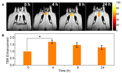 |
Magnetic Resonance Image guided radiation therapy with nanoparticles improves radiation response of Oral Tumor Xenograft
Gayatri Sharma1, Abdul K Abdul K. Parchur2, Jaidip M. Jagtap2, brian M. Fish2, Bergom Carmen 3, Michael Flister4, Meetha M. Medhora5, and Amit Joshi1,6
1Biomedical Engineering, Medical College of Wisconsin, MILWAUKEE, WI, United States, 2Medical College of Wisconsin, MILWAUKEE, WI, United States, 3Radiation Oncology, Medical College of Wisconsin, MILWAUKEE, WI, United States, 4Physiology, Medical College of Wisconsin, MILWAUKEE, WI, United States, 5Medical College of Wisconsin, Medical College of Wisconsin, MILWAUKEE, WI, United States, 6Biophysics, Medical College of Wisconsin, MILWAUKEE, WI, United States
X-ray CT or MR Image-guided radiation therapy (IGRT) is routinely employed for oral cancer treatment. Enhancing tumor dose while limiting collateral damage to salivary glands is of critical importance for oral cancer patients. Here, we demonstrate the efficacy of multifunctional Theranostic nanoparticles (TNPs) for MR image guided radiation therapy in a rat xenograft model of oral cancer. These TNPs composed of Gold (Au) core and Gd(III) shell with combined X-ray and MR contrast can enable pre-procedure radiotherapy planning via T1-MR imaging, as well as enhance radiation treatment efficacy by increasing the tumor deposited radiation dose.
|
|
4844.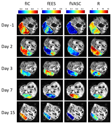 |
VERDICT and kurtosis modelling of diffusion MRI for early assessment of radiotherapy response in a model of human neuroendocrine tumour
Lukas Carl Lundholm1, Mikael Montelius1, Oscar Jalnefjord1, Emman Shubbar1, Eva Forssell-Aronsson1, and Maria Ljungberg1
1Department of Radiation Physics, Institute of Clinical Sciences, Göteborg, Sweden
Diffusion MRI methods to characterize tumour tissue could facilitate early assessment of radiotherapy response. In this study mouse models of human small intestine neuroendocrine tumours were irradiated externally, and post-treatment effects were studied over two weeks using VERDICT and kurtosis analysis of diffusion MRI measurements. Results showed multiple correlations between tumour treatment response and parameters estimated from the models, suggesting that a decrease of cell density and microstructural barriers early after treatment indicates a more substantial response. Both VERDICT and kurtosis analysis shows great potential for early assessment of treatment response to external radiation therapy in the studied tumour model.
|
|
4845.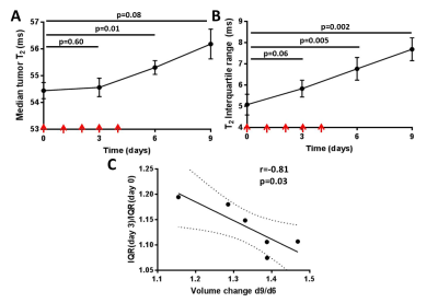 |
Heterogeneity analysis in T2 mapping enables highly sensitive radiotherapy response measurement and prediction in pancreatic allografts.
Michal Tomaszewski1, William Dominguez Viqueira1, Heiko Enderling2, and Robert J Gillies1
1Cancer Physiology, H. Lee Moffitt Cancer Center, Tampa, FL, United States, 2Institute of Mathematical Oncology, H. Lee Moffitt Cancer Center, Tampa, FL, United States
Tumor response to radiotherapy (XRT) varies significantly between patients, calling for improved response quantification methods. Tumor response to fractionated XRT was quantified in a mouse model of pancreatic cancer with multiple MR sequences. Texture feature analysis of quantitative T2 maps revealed that histogram width information (Interquartile range metric) provided a more sensitive measure of XRT response than first order metrics, reflecting the structural heterogeneity increase due to necrosis, which presaged tumor volume changes. Thus, radiomic analyses of qT2 maps show significant promise for early response prediction, which will be crucial for future development of dynamic adaptive radiotherapy.
|
|
4846.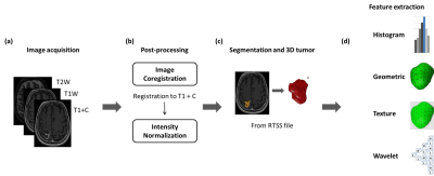 |
Prediction of Treatment Outcome after Radiosurgery in Brain Metastases from Lung Cancer Using Preradiosurgical MR Radiomics
Chien-Yi Liao1, Cheng-Chia Lee2,3,4, Huai-Che Yang2,3, Wen-Yuh Chung2,3, Hsiu-Mei Wu3,5, Wan-Yuo Guo3,5, Ren-Shyan Liu1,6, and Chia-Feng Lu1,7
1Department of Biomedical Imaging and Radiological Sciences, National Yang-Ming University, Taipei, Taiwan, 2Department of Neurosurgery, Neurological Institute, Taipei Veteran General Hospital, Taipei, Taiwan, 3School of Medicine, National Yang-Ming University, Taipei, Taiwan, 4Brain Research Center, National Yang-Ming University, Taipei, Taiwan, 5Department of Radiology, Taipei Veteran General Hospital, Taipei, Taiwan, 6Department of Nuclear Medicine and National PET/Cyclotron Center, Taipei Veterans General Hospital, Taipei, Taiwan, 7Institute of Biophotonics, National Yang-Ming University, Taipei, Taiwan
The development of brain metastases is a devastating consequence of disease progression of advanced non-small cell lung cancer patients. This study proposed an approach to predict the treatment outcome after gamma knife radiosurgery based on preradiosurgical MR radiomics. We suggested that imaging characteristics extracted from preradiosurgical MRIs can be potential image biomarkers for the outcome prediction.
|
|
4847.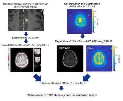 |
Evolution of the tissue sodium concentration in brain metastases undergoing stereotactical radiosurgery: a feasilbility study
Anne Adlung1, Michaela A. U. Hoesl1, Sherif Mohamed2, Arne M. Ruder3, Frank A. Giordano3, Eva Neumaier Probst2, and Lothar R. Schad1
1Computer Assisted Clinical Medicine, Medical Faculty Mannheim, Heidelberg University, Mannheim, Germany, 2Department of Neuroradiology, Medical Faculty Mannheim, Heidelberg University, Mannheim, Germany, 3Department of Radiation Oncology, Medical Faculty Mannheim, Heidelberg University, Mannheim, Germany
Brain metastases are the most common malignancy in the brain and can be caused by different primary tumors. The Sodium-SRS study is a prospective feasibility study to observe TSC changes in brain metastases around ablative stereotactical radiosurgery (SRS) treatment with 22 Gy single dose using GammaKnife. TSC was measured with 23Na MRI in two patients at three different time points. The prelimenary results showed feasibility to detect TSC dynamics in brain metastases treated with SRS, which warrants further evaluation.
|
|
4848.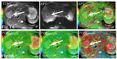 |
The application of APT and IVIM in predicting the recurrence of early stage hepatocellular carcinoma treated with TACE: A pilot study
Jia fei1, Han dongming1, and Wang Kaiyu2
1The First Affiliated Hospital of Xinxiang Medical University, Xin xiang, China, 2GE Healthcare ,MR Research China, Beijing, China
This work assessed the feasibility of quantitative parameters derived from APT and IVIM in the predicting the recurrence of early stage Hepatocellular carcinoma (HCC) treated with transcatheter arterial chemoembolization (TACE). We find that combined quantitative parameters of APT and IVIM can be used to predict the recurrence. Therefore, APT and IVIM can be used as imaging biomarkers to predict the recurrence of early stage of HCC after TACE treatment.
|
|
4849.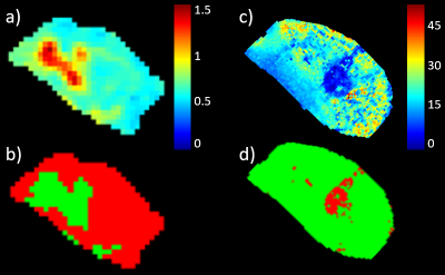 |
Multiparametric MRI with cluster analysis for early assessment of radiotherapy response in a human small-intestine neuroendocrine tumor model
Mikael Montelius1, Lukas Lundholm1, Oscar Jalnefjord1, Emman Shubbar1, Eva Forssell-Aronsson1, and Maria Ljungberg1
1Radiation Physics, Clinical Sciences, Gothenburg, Sweden
The diffusion coefficient (D) and T2* reflect clinically significant tumor characteristics, including cellularity and hypoxia. Methods to non-invasively assess D and T2* at diagnosis and early follow up are desired. The aim of this work was to evaluate the sensitivity of D and T2* to radiotherapy effects in a neuroendocrine tumor model, and to evaluate clustering as an objective method to facilitate D and T2* analysis. 20 mice were examined using MRI before and repeatedly for 2 weeks after tumor irradiation (8Gy) . We show that D and T2* are potential response biomarkers, and that clustering improves response prediction for T2*.
|
|
4850.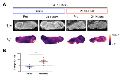 |
Intrinsic susceptibility MRI can inform on tumour vascular decompression in response to the stromal targeted therapy PEGPH20
Emma L. Reeves1, Jin Li1, Jessica K. R. Boult1, Barbara Blouw2, David Kang2, Jeffrey C. Bamber1, Yann Jamin1, and Simon P. Robinson1
1Radiotherapy and Imaging, Institute of Cancer Research, London, United Kingdom, 2Halozyme Therapeutics, San Diego, CA, United States
PEGPH20 has been shown to decompress blood vessels and improve tumour drug delivery. Given the sensitivity of R2* to paramagnetic deoxyhaemoglobin and reflection of RBC content in tumour vasculature, we hypothesised that IS-MRI may provide a useful biomarker of PEGPH20 response. We performed IS-MRI before and after PEGPH20 treatment in 4T1 HAS3 and MDA-MB-231 LM2-4 orthotopic breast tumours. R2* significantly increased following PEGPH20 treatment in 4T1 HAS3 tumours, however, R2* was unable to detect a similar response to PEGPH20 in the relatively hypovascular MDA-MB-231 LM2-4 tumours. Another vascular biomarker may provide greater sensitivity and detect PEGPH20 response in hypovascular tumours.
|
|
4851.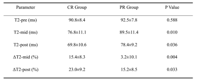 |
Quantitative T2 Mapping in Advanced Cervical Cancer: A Pilot Study of Patients’ Concurrent Chemoradiotherapy Response
Shujian Li1, Jie Liu1, Jinxia Zhu2, Han Fei3, and Jingliang Cheng1
1Department of Magnetic Resonance, the First Affiliated Hospital of Zhengzhou University, Zhengzhou, China, 2MR Collaboration, Siemens Healthcare Ltd, Beijing, China, 3MR R&D Collaboration, Siemens Healthineers, Los Angeles, CA, United States
This study determined the value of quantitative transverse relaxation time (T2) mapping as an alternative method to conventional magnetic resonance imaging (MRI) for evaluating the response to concurrent chemoradiotherapy (CCRT) in 27 patients with advanced cervical cancer (CC). Response values were calculated at three time points (before CCRT, 4 weeks after CCRT, and at the end of CCRT) and then compared. The results showed that quantitative T2 mapping can be effectively used for monitoring treatment response during and after treatment, but not for pre-treatment monitoring.
|
|
4852.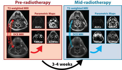 |
Dynamic Contrast Enhanced MRI Parametric Maps as Indicators of Lymph Node Radiotherapy Response in Head and Neck Cancer
Kareem Wahid1, Abdallah Mohammed1, Lisanne Van Dijk1, Sara Ahmed1, Renjie He1, Baher Elgohari1, Yao Ding2, Jihong Wang2, Stephen Lai3, and Clifton Fuller1
1Radiation Oncology, MD Anderson Cancer Center, Houston, TX, United States, 2Radiation Physics, MD Anderson Cancer Center, Houston, TX, United States, 3Head and Neck Surgery, MD Anderson Cancer Center, Houston, TX, United States
Current methodology in analyzing pathological lymph node radiotherapy response in head and neck cancer relies on qualitative visual inspection of geometrical features. A quantitative approach that takes into account dynamic changes in underlying lymph node pathology could be an important supplemental clinical tool in radiotherapy treatment planning. Herein, we investigate dynamic contrast enhanced MRI quantitative parametric maps as possible biomarker candidates for lymph node radiotherapy response by analyzing multi-timepoint images of head and neck cancer patients.
|
|
4853.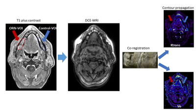 |
Prospective assessment of DCE-MRI parameters associated with advanced mandibular osteoradionecrosis after IMRT of head and neck cancer
Abdallah Sherif Radwan Mohamed1, Renjie He1, Yao Ding1, Jihong Wang1, Joly Fahim1, Baher Elgohari1, Hesham Elhalawani1, Jason Johnson2, Jason Stafford3, Jim Bankson3, Mark Chambers4, Vlad Sandulache5, Clifton Fuller1, and
Stephen Lai4
1Radiation Oncology, MD Anderson Cancer Center, Houston, TX, United States, 2Diagnostic imaging, MD Anderson Cancer Center, Houston, TX, United States, 3Imaging physics, MD Anderson Cancer Center, Houston, TX, United States, 4Head and Neck Surgery, MD Anderson Cancer Center, Houston, TX, United States, 5Baylor College of Medicine, Houston, TX, United States
Our aim was to characterize the quantitative DCE-MRI parameters associated with advanced mandibular osteoradionecrosis (ORN) in comparison with normal mandible. Thirty patients with advanced ORN after radiation of head and neck cancer were prospectively enrolled. Our results confirm there is a quantitatively significant higher degree of leakiness in the mandibular vasculature as measured using DCE-MRI parameters of areas affected with advanced grade of ORN versus healthy mandible. We were able to measure significant increases in quantitative parameters (3.3 fold Ktrans, 3 fold ve) compared to non-ORN mandibular bone.
|
|
4854.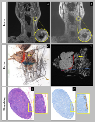 |
Validation of USPIO-MRI for detection of lymph node metastases in head and neck cancer: feasibility and workflow.
Daphne A.J.J. Driessen1, Tim Dijkema1, Sjoert A.H. Pegge2, Patrik Zámecnik2, Adriana C.H. van Engen-van Grunsven3, Willem L.J. Weijs4, Robert P. Takes5, Tom W.J. Scheenen2, and Johannes H.A.M. Kaanders1
1Radiation Oncology, Radboud university medical centre, Nijmegen, Netherlands, 2Radiology and Nuclear Medicine, Radboud university medical centre, Nijmegen, Netherlands, 3Pathology, Radboud university medical centre, Nijmegen, Netherlands, 4Oral- and Maxillofacial Surgery and Head and Neck Surgery, Radboud university medical centre, Nijmegen, Netherlands, 5Otorhinolaryngology and Head and Neck Surgery, Radboud university medical centre, Nijmegen, Netherlands
Head and neck cancers have a high propensity to metastasize to the lymph nodes of the neck. Accurate knowledge of this regional nodal status is of great importance for therapy selection and prognosis. Ultrasmall superparamagnetic iron oxide (USPIO) particles (Ferumoxtran-10 / Ferrotran®) are a promising contrast agent that can be used to detect nodal metastases with MRI. This study aims to validate USPIO-MRI for the detection of nodal metastases in head and neck cancer patients with histopathology as the reference standard. The workflow and preliminary results in 5 patients are shown in this abstract.
|
4855.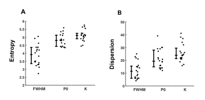 |
Heterogeneity of cellularity in breast cancer from diffusion q-space MRI
Nicholas Senn1, Ehab Husain2, Yazan Masannat3, Sai Man Cheung1, Bernard Siow4, Steven D Heys5, and Jiabao He1
1Aberdeen Biomedical Imaging Centre, University of Aberdeen, Aberdeen, United Kingdom, 2Pathology department, Aberdeen Royal Infirmary, Aberdeen, United Kingdom, 3Breast Unit, Aberdeen Royal Infirmary, Aberdeen, United Kingdom, 4Francis Crick Institute, London, United Kingdom, 5School of Medicine, University of Aberdeen, Aberdeen, United Kingdom
Breast cancers often present a spatially heterogenous response to treatment manifesting as diverse variations in the underlying distribution of cellularity. We hypothesised that kurtosis from diffusion q-space imaging provides a higher effect gradient to assess the amount of entropy and dispersion in breast cancer cellularity than measures of diffusion displacement. We investigated whole breast tumours excised from surgery, with imaging performed same day overnight on a clinical 3T MRI system. The entropy and dispersion obtained from kurtosis were significantly higher than that obtained from displacement measurements, yielding a higher effect gradient, and significantly correlated with the histologic cellularity.
|
|
4856.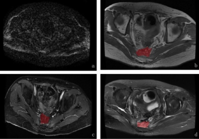 |
MRI texture analysis based on 3D tumor measurement in differential diagnosis of benign and malignant presacral lesion
jian jiang1 and ailian liu1
1The first affiliated hospital of Dalian medical university, Dalian, China
My study is to evaluate the diagnostic value of 3D MRI texture analysis based on T1WI, T2WI, DKI and contrasted-enhanced T1WI MRI sequences
in differential diagnosis of benign and malignant presacral lesion after resection of rectal carcinoma
|
|
4857.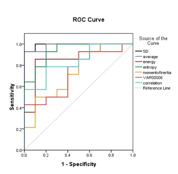 |
The value of whole tumor volume measurement texture analysis based on ADC images in differentiating DCIS from DCISM: Apreliminary study.
Ye Ju1, Shifeng Tian1, Ailian Liu1, Lina Zhang1, Lizhi Xie2, Yan Guo2, and Xin Li2
1First affiliated hospital of dalian medical university, Dalian, China, 2GE Healthcare, Beijing, China
In this study, we used the whole tumor volume measurement of texture analysis based on ADC images to identify breast ductal carcinoma in situ (DCIS) and DCIS with microinvasion (DCISM), which revealed to be feasible approach for differentiating between DCIS and DCISM.
|
|
4858.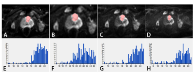 |
Texture analyses based on multiple high b-value diffusion-weighted imaging (DWI) for evaluation of tumor heterogeneity in prostate cancer
Jingjun Wu1, Ailian Liu1, Jiazheng Wang2, Liangjie Lin2, Lihua Chen1, Qingwei Song1, Renwang Pu1, and Bingbing Gao1
1Department of Radiology, the First Affiliated Hospital of Dalian Medical University, Dalian, China, 2Philips Healthcare, Beijing, China
In the current study, multiple high b-value DWI-based texture analysis was applied to evaluate tumor heterogeneity in prostate cancer patients. Parameters, including MinIntensity, MedianIntensity, MeanValue, and Skewnesswere were significantly different among DWI images with different b values. Therefore, texture analysis based on DWI maps with different b values may serve as a preoperative and non-invasive method for evaluating heterogeneity of prostate cancer.
|
|
4859.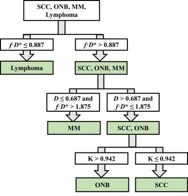 |
Diffusion Kurtosis Imaging and Intravoxel Incoherent Motion MR Imaging in the Differentiation of Sinonasal Malignant Tumors
Zuohua Tang1, Zebin Xiao1, Peng Wang1, and Zhongshuai Zhang2
1Radiology, Fudan University Eye & ENT Hospital, Shanghai, China, 2MR Scientific Marketing, Siemens Healthcare, Shanghai, China
A reliable differentiation of different histological types of sinonasal malignancies is usually difficult by using conventional CT and MR imaging but plays an important role in determining the treatment strategies for the patients. This is the first study which combined DKI and IVIM to distinguish among four histological types of sinonasal malignant tumors, including squamous cell carcinomas (SCCs), olfactory neuroblastomas (ONBs), malignant melanomas (MMs), and lymphomas. Our study showed that these sinonasal malignant tumors had distinct profiles in DKI and IVIM parameters, determined by different proportions of tissue complexity, capillary perfusion, or water diffusion components within tumor.
|
|
4860. |
Quantitative Classification of Clinically Significant Prostate Cancer Using a Biexponential Model of Extended b-value Diffusion-Weighted MRI
Christine H Feng1, Christopher C Conlin2, Ana E Rodríguez-Soto2, Roshan Karunamuni1, Aaron B Simon1, Rebecca Rakow-Penner2, Michael E Hahn2, Anders M Dale2,3, and Tyler Seibert1,4
1Radiation Medicine & Applied Sciences, UC San Diego, La Jolla, CA, United States, 2Radiology, UC San Diego, La Jolla, CA, United States, 3Neurosciences, UC San Diego, La Jolla, CA, United States, 4Bioengineering, UC San Diego, La Jolla, CA, United States
Prostate cancer is the second most frequent malignancy in men worldwide—novel strategies are needed to better identify patients with clinically significant disease. We developed a biexponential linear regression model using data from 36 men who underwent prostate MRI with 5 distinct b-values. We evaluated quantitative detection of clinically significant prostate cancer. The biexponential model and derivative lookup table outperformed simple ADC and kurtosis in quantitative classification of benign tissue and cancer (AUC 0.958 and 0.976 vs 0.632 and 0.409, respectively).
|
|
4861. |
Parameter Induced Variations in DCE-MRI Analysis: Monitoring Treatment Response for Head and Neck Tumours
Madeline Carr1,2,3, Michael Jameson1,2,3,4, Christopher Rumley2,4,5, Gary Liney2,3,4, Mark Lee3,4, Phillip Chlap2,4, Peter Metcalfe1,2, and Lois Holloway1,2,3,4,6
1Centre for Medical and Radiation Physics, University of Wollongong, Wollongong, Australia, 2Medical Physics, Ingham Institute of Applied Medical Research, Liverpool, Australia, 3Liverpool and Macarthur Cancer Therapy Centres, Liverpool, Australia, 4South Western Sydney Clinical School, University of New South Wales, Liverpool, Australia, 5Townsville Hospital and Health Service, Townsville, Australia, 6Institute of Medical Physics, University of Sydney, Camperdown, Australia
The effect of altering the input parameters into a Pharmacokinetic (PK) model on output parameter values generated was investigated. This included determining the variations induced by using individualized versus population based Arterial Input Function and haematocrit values in the model. This was completed for multiple DCE-MRI scans acquired along the course of radiotherapy treatment for 5 head and neck cancer patients. Qualitatively, most patients had similar trendlines when comparing between each combination of parameter inputs. However quantitatively, the %difference between baseline and subsequent weeks highlighted significant impacts caused by input parameter selection; having implications for applications in treatment response monitoring.
|
|
| 4862. | Preoperative Prediction of Axillary Lymph Node Metastasis in Breast Carcinoma Using Radiomics Features Based on the Fat-suppressed T2 Sequence
Hongna Tan1, Fuwen Gan2, Yaping Wu3, Yusong Lin4, and Meiyun Wang3
1Radiology, Department of Radiology, Henan Provincial People’s Hospital & Imaging Diagnosis of Neurological Diseases and Research Laboratory of Henan Province & People's Hospital of Zhengzhou University, Henan, China, 450003, Zhengzhou, China, 2Collaborative Innovation Center for Internet Healthcare & School of Software and Applied Technology, Zhengzhou University, Zhengzhou, Henan,China, 450052, Zhengzhou, China, 3Department of Radiology, Henan Provincial People’s Hospital & Imaging Diagnosis of Neurological Diseases and Research Laboratory of Henan Province & People's Hospital of Zhengzhou University, Henan, China, 450003, Zhengzhou, China, 4Collaborative Innovation Center for Internet Healthcare & School of Software, Zhengzhou University, Zhengzhou, Henan,China, 450052, Zhengzhou, China
Currently, a noninvasive and high diagnostic sensitivity model for preoperative predicting the status of ALN is need. Our aim is to investigate the value of radiomics method based on the fat-suppressed T2 sequence for preoperative predicting axillary lymph node (ALN) metastasis in breast carcinoma.
|
|
4863. |
Optimisation of whole-body diffusion weighted MRI for imaging patients with metastatic melanoma treated with immunotherapy
Annemarie Knill1, Matthew Blackledge1, Jessica Winfield1,2, Andra Curcean2, James Larkin3, Samra Turajlic3, Dow-Mu Koh1,2, and Christina Messiou1,2
1Division of Radiotherapy and Imaging, The Institute of Cancer Research, London, United Kingdom, 2Department of Radiology, The Royal Marsden NHS Foundation Trust, London, United Kingdom, 3Renal and Melanoma Unit, The Royal Marsden NHS Foundation Trust, London, United Kingdom
Whole body MRI, including diffusion weighted imaging, has potential to unravel complex response patterns in patients with metastatic melanoma receiving immunotherapy. To minimise the effect of noise on resultant apparent diffusion coefficient (ADC) maps, images should be acquired using optimal b-values. These values have been calculated from MRI scans of 11 patients, with 99 metastases. The optimal pair of b-values is found to be 50 and 1250 s/mm2. A significant difference is reported between the distributions of the ADC values before and after treatment with immunotherapy. This provides preliminary evidence that ADC could provide a biomarker of response to immunotherapy.
|
|
4864. |
The value of multiparametric MRI-based radiomics in diagnosing of benign and malignant prostatic diseases
Junjia Luo1, Shuangfeng Dai2, Zebin Luo3, Wubiao Chen3, Xiaodong Chen3, and Wenxuan Luo3
1Guangdong Medical University, Zhanjiang, China, 2Huiying Medical Technology Co., Ltd., Beijing, China, 3Affiliated Hospital of Guangdong Medical University, Zhanjiang, China
Radiomics, as it refers to the extraction and analysis of large number of advanced quantitative radiological features from medical images using high throughput methods, it provides a new evaluation method for detection of the suspicious lesion of cancer. The present study explores the value of radiomics based on the mulitparameter magnetic resonance imaging (mpMRI) for diagnosis of the benign and malignant prostatic diseases.
|
|
| 4865. | Histogram Analysis Parameters ADC for Distinguishing Orbital Tumors of Lymphoma,Pleomorphic Adenoma and Adenoid Cystic Carcinoma
Chen Chen1 and Cuiping Ren1
1the First Affiliated Hospital of Zhengzhou University, ZhengZhou, China
To determine if histograms of ADC can be used to differentiate orbital lymphoma,pleomorphic adenoma(PA) and adenoid cystic carcinoma(ACC)
|
|
4866. |
T2-mapping of MR imaging for discriminating mucinous adenocarcinoma from nomucinous adenocarcinoma in rectal cancer and compared with diffusion weighted image
yuxi ge1 and weiqiang dou2
1Wuxi Fourth People's Hospital, wuxi, China, 2GE Healthcare, MR Research, Beijing, China
T2-mapping, as a quantitative and accurate measurement that can be used as a noninvasive biological marker to predict the pathological classification of rectal cancer, may be beneficial to making an individualized treatment strategy before operation of rectal cancer.
|
|
4867. |
Radiotherapy for prostate cancer: Effects of fiducial gold marker on diffusion-weighted magnetic resonance imaging
Osamu Tanaka1, Takuya Taniguchi1, Kousei Ono1, and Masayuki Matsuo2
1Radiation Oncology, Asahi University Hospital, Gifu, Japan, 2Radiation Oncology, Gifu University Hospital, Gifu, Japan
Gold markers showed little effect on the quality of DWI. Therefore, despite using iron-containing markers and the size of marker < 0.5 mm being available, MRI, particularly DWI, may be used during follow-up imaging.
|
|
4868. |
Quantify breast stiffness in breast carcinoma patients using Magnetic Resonance Elastography at 3T and its comparison with clinical grading
Prateek Kalra1, Ashonti Harper1, Jeffrey Hawley 1, Brian Raterman1, and Arunark Kolipaka1
1Radiology, Ohio State University Wexner Medical Center, Columbus, OH, United States
Dynamic contrast-enhanced MRI kinetic parameters have been investigated to assess the response to neoadjuvant chemotherapy (NAC). However, those studies have predictive value only after one or two cycles of NAC treatment and involves injection of contrast agent. Moreover, breast tumor sample is graded clinically using biopsy and is invasive. Aim of this study is to evaluate any correlation between MRE stiffness and clinical grading in breast carcinoma and if MRE can predict the response to NAC. Preliminary results show moderate correlation between MRE-derived stiffness and clinical grading in breast carcinoma patients.
|
|
4869.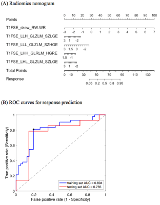 |
Prediction of response to neoadjuvant chemotherapy for nasopharyngeal carcinoma using pretreatment MRI: a validated radiomics nomogram
Junqi Sun1, Shenglan Chen2, Jie Ding2,3, Feifei Shan1, Yi Kang1, Xiaoting Tang1, and Chuan Huang2,4,5
1Yuebei People’s Hospital, Shaoguan, China, 2Biomedical Engineering, Stony Brook University, Stony Brook, NY, United States, 3Diagnostic Radiology, Li Ka Shing Faculty of Medicine, The University of Hong Kong, Hong Kong, Hong Kong, 4Radiology, Stony Brook Medicine, Stony Brook, NY, United States, 5Psychiatry, Stony Brook Medicine, Stony Brook, NY, United States
Nomograms are widely adopted as prognostic tools for clinical practice. In this study, we developed and temporally validated a radiomics nomogram to predict early response to neoadjuvant chemotherapy among patients with nasopharyngeal carcinoma using pretreatment MRI.
|
|
4870. |
IVIM & Diffusion Kurtosis MR Imaging on Interim Response Assessment of Hodgkin Lymphoma
Archana Vadiraj Malagi1, Devasenathipathy Kandasamy2, Kedar Khare3, Deepam Pushpam4, Rakesh Kumar5, Sameer Bakhshi4, and Amit Mehndiratta1,6
1Centre for Biomedical Engineering, Indian Institute of Technology Delhi, New Delhi, India, 2Department of Radiodiagnosis, All India Institute of Medical Sciences Delhi, New Delhi, India, 3Indian Institute of Technology Delhi, New Delhi, India, 4Department of Medical Oncology, Dr. B.R. Ambedkar Institute-Rotary Cancer Hospital (IRCH), All India Institute of Medical Sciences Delhi, New Delhi, India, 5Department of Nuclear Medicine, All India Institute of Medical Sciences Delhi, New Delhi, India, 6Department of Biomedical Engineering, All India Institute of Medical Sciences Delhi, New Delhi, India
PET/CT plays an important role in assessment of treatment response in Hodgkin lymphoma (HL). This study goal was to evaluate the role of IVIM and DKI parameters role in interim assessment of HL. ADC, D(BE+TV), Dk and f showed reduction in interim which was in accordance with changes of SUVmax and SUVmean whereas D* and k showed increase against treatment response. In future, IVIM and DKI can have the potential in accurate assessment of interim monitoring in HL.
|
4871.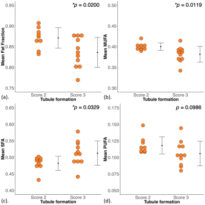 |
Lipid composition mapping in whole breast tumour using chemical shift encoded imaging
Sai Man Cheung1, Kwok Shing Chan2, Nicholas Senn1, Yazan Masannat3, Ehab Husain4, Steven Heys3, and Jiabao He1
1Aberdeen Biomedical Imaging Centre, University of Aberdeen, Aberdeen, United Kingdom, 2Donders Institute for Brain, Cognition and Behaviour, Radboud University, Nijmegen, Netherlands, 3Breast Unit, Aberdeen Royal Infirmary, Aberdeen, United Kingdom, 4Pathology Department, Aberdeen Royal Infirmary, Aberdeen, United Kingdom Lipid composition in breast has a major role in breast cancer prevention, with deregulation of lipid metabolism identified in BRCA1/2 genetic mutation carriers. Neoplastic tubule formation can infiltrate adipose tissue in peri-tumoural region, with low tubular differentiation indicating a poorer prognosis. Lipid composition measurement through biochemical extraction is invasive, while conventional chemical shift imaging demands an intolerably long acquisition time. Recent development in gradient-echo (GRE) based imaging allows lipid composition mapping of the whole breast in a clinically acceptable timeframe. We set out to examine the relationship between peri-tumoural lipid composition and tubule formation using GRE-based imaging in breast tumours. |
|
4872. |
A Pilot Evaluation of Intravoxel Incoherent Motion Imaging Features of Stage T1 HER2-positive breast carcinomas
Nan Zhang1, Qingwei Song1, Lina Zhang1, Ailian Liu1, Yu Song1, and Lizhi Xie2
1The First Affiliated Hospital of Dalian Medical University, Dalian, China, 2GE Healthcare, MR Research China, Beijing, China Poster Permission Withheld
Intravoxel incoherent motion (IVIM) imaging provides quantitative measurement of ADCslow for cellularity and ADCfast and ffast for vascularity. The parameters derived from IVIM can distinguish lesions of different aggressive pathological subtypes of Stage T1 breast carcinomas[1]. This study concerned clinical features, perfusion as well as diffusion parameters using IVIM imaging and then compared these parameters from conventional MR images on the classification of Stage T1 HER2-positive and HER2-negative breast carcinoma lesions .
|
|
4873.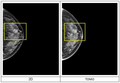 |
Diagnostic efficiency of DBT compared to conventional MRI and FFDM in diagnosing breast cancer
Fan WenWen1, Fan WenWen1, Ou Yang Han2, Zhao XinMing2, Zhang HongMei2, and Xie LiZhi3
1Department of Imaging Diagnosis, National Cancer Center/Cancer Hospital, Chinese Academy of Medical Science and Peking Union Medical College, BeiJing, China, 2National Cancer Center/Cancer Hospital, Chinese Academy of Medical Science and Peking Union Medical College, BeiJing, China, 3MR Research, GE Healthcare, BeiJing, China
The aim of this study was to evaluate the technical characteristics and diagnostic efficiency of Digital Breast Tomosynthesis (DBT) compared to conventional MRI approach and full-field digital mammography (FFDM) in screening breast cancer (BC) patients. Briefly, 253 female patients who underwent Digital Mammary Gland 3D Tomosynthesis, MR scan and FFDM were enrolled in this study. Briefly, the DBT, FFDM and MRI showed a sensitivity of 90.9%, 85.6% and 94.1%; specificity of 89.7%, 80.7% and 83.3%; and coincidence rate of 89.3%, 83.4% and 89.7%, respectively, and the difference was statistically significant (p<0.001).
|
|
4874.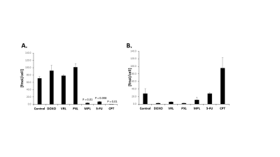 |
Molecular Effects of Chemotherapeutic Agents on Choline Phospholipid Metabolism of Triple Negative Breast Cancer Cells
Caitlin M Tressler1, Kanchan Sonkar1, Vinay Ayyappan1, Menglin Cheng1, and Kristine Glunde1
1Johns Hopkins University, Baltimore, MD, United States
Magnetic resonance spectroscopy (MRS) of breast cancer patients has shown a significant decrease in total choline (tCho) signal in patients responding to chemotherapy.1-4 Despite the clinical development of tCho as an imaging biomarker for response to chemotherapy, little is known about the molecular mechanism driving this observed change. We examined six chemotherapy agents in two triple negative breast cancer (TNBC) cell lines by high resolution (HR) 1H MRS to determine the impact of chemotherapy treatment on choline metabolites. Furthermore, we have begun to probe the impact of these chemotherapy agents on important genes in choline metabolism using quantitative real-time PCR.
|
|
4875.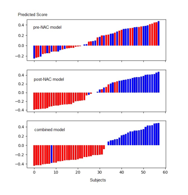 |
Prediction of pathological complete response to neoadjuvant chemotherapy in breast cancer using deep learning method
Yuhong Qu1, Haitao Zhu1, Kun Cao1, Xiaoting Li1, and Ying-shi Sun1
1Beijing cancer hospital, Beijing, China
This study established a deep learning model to predict PCR status after neoadjuvant therapy by combining pre-NAC and post-NAC MRI data.The area under the receiver operating characteristic (ROC) curve (AUC) of the models are 0.968 for post-NAC and 0.970 for the combined data.
|
|
4876.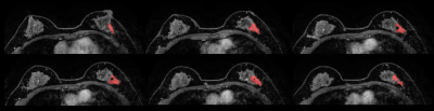 |
Can radiomics and machine learning capture the unique differences between invasive lobular and invasive ductal carcinoma of the breast?
Carolina Rossi Saccarelli1,2, Peter Gibbs3, Almir G V Bitencourt1, Isaac Daimiel1, Roberto Lo Gullo1, Sunitha B Thakur3, Elizabeth A Morris1, and Katja Pinker 1
1breast radiology, MSKCC, New York, NY, United States, 2breast radiology, Hospital Sirio-Libanes, Sao Paulo, Brazil, 3MSKCC, New York, NY, United States In this study, we hypothesized that the specific genomic profiles of invasive lobular carcinoma (ILC) can be captured with radiomics analysis and machine learning (ML) from standardized dynamic contrast-enhanced breast MRI. Three-dimensional tumor segmentation of the first post-contrast T1-weighted sequence was conducted and included the entire mass and non-mass enhancement lesions, unifocal and multifocal/multicentric lesions. This supervised ML model produced an accuracy of 76.6%, sensitivity of 72.7%, specificity of 80.6%, PPV of 79.1% and NPV of 74.5%. Our preliminary results indicate that radiomics analysis coupled with supervised ML allows a non-invasive differentiation between ILC and invasive ductal carcinoma. |
|
4877. |
Characterization of Sub-centimeter Enhancing Breast Masses on MRI with Radiomics and Machine Learning in BRCA Mutation Carriers
Roberto Lo Gullo1, Isaac Daimiel 1, Carolina Saccarelli1, Almir Bitencourt1, Peter Gibbs2, michael James Fox2, Sunitha B. Thakur2,3, Danny F. Martinez1, Elisabeth A. Mossir1, and Katja Pinker1
1Radiology, Breast imaging, Memorial Sloan Kettering Cancer Center, New York, NY, United States, 2Radiology, Memorial Sloan Kettering Cancer Center, New York, NY, United States, 3medical physics, Memorial Sloan Kettering Cancer Center, New York, NY, United States
The purpose of our study was to investigate whether radiomics features extracted from MRI of BRCA-positive patients with breast masses smaller than 1 cm coupled with machine learning can differentiate benign from malignant lesions using model-free parameter maps. We included 96 patients with 116 lesions assessed by two readers according to the BI-RADS lexicon. Radiomics features were calculated and included in a machine learning model, along with clinical factors, to discriminate between malignant and benign lesions. The machine learning model, combining two clinical and three radiomics features, achieved higher diagnostic accuracy (81.5%) compared to morphological assessment alone (53.4%)
|
|
| 4878. | MR-based machine learning radiomics can predict tumor heterogeneity and pathologic response after neoadjuvant therapy in HER2 breast cancer
Almir Bitencourt1,2, Peter Gibbs3, Carolina Rossi Saccarelli3, Isaac Daimiel3, Roberto Lo Gullo3, Michael Fox3, Sunitha Thakur3, Katja Pinker3, Elizabeth A Morris3, Monica Morrow4, and Maxime Jochelson3
1Breast Radiology, MSKCC, New York, NY, United States, 2Department of Imaging, A.C. Camargo Cancer Center, Sao Paulo, Brazil, 3MSKCC, New York, NY, United States, 4Breast Surgery, MSKCC, New York, NY, United States
In this study, we used magnetic resonance (MR)-based clinical and radiomic features to assess tumor heterogeneity in 311 HER2 overexpressing breast cancer patients receiving neoadjuvant chemotherapy (NAC), and correlated these findings with tumor heterogeneity and pathologic response. Tumor heterogeneity was evaluated based on the HER2 expression (IHC vs. FISH) . Pathologic complete response (pCR) was defined as no residual invasive carcinoma in the breast or axillary lymph nodes (ypT0/isN0). Radiomics analysis and machine learning with MRI were able to assess tumor heterogeneity and predict pCR after neoadjuvant chemotherapy in these patients, with a diagnostic accuracy of 97.4% and 85.2%, respectively.
|
|
4879.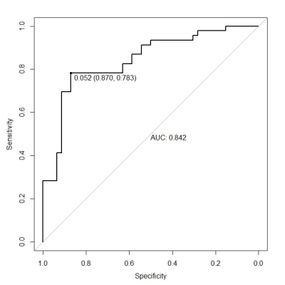 |
Radiomics analysis on magnetic resonance diffusion weighted imagefor prediction of Ki-67 expression in breast cancer
Qinglin Wang 1, Ning Mao1, Haizhu Xie1, Fengjie Liu1, and Jingjing Cui2
1Yantai Yuhuangding Hospital, Yantai, China, 2Huiying Medical Technology Co., Beijing, China
The expression level of Ki-67 has an important reference value for the diagnosis, treatment and prognosis evaluation of breast cancer. To explore the feasibility of diffusion-weighted imaging for prediction of Ki-67 expression. In this study, the expression of Ki-67 in breast cancer was differentiated by semi-automatic extraction of the image parameters of diffusion-weighted(DWI) before treatment. The results of this study show that Ki-67-negative and Ki-67-positive breast cancer have different imaging characterization values in DWI images. The imaging of DWI is feasible in identifying the two, which is helpful to predict the expression level of Ki-67 in breast cancer before operation.
|
|
4880.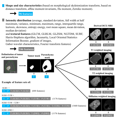 |
Prediction of pathologic complete response of neoadjuvant chemotherapy for early triple negative breast cancer using pre-therapeutic MRI
Angeline Nemeth1, Pierre Chaudet2, Benjamin Leporq1, Pierre-Etienne Heudel3, Olivier Tredan3, Isabelle Treilleux4, Frank Pilleul2, Agnès Coulon2, and Olivier Beuf1
1Univ Lyon, INSA‐Lyon, Université Claude Bernard Lyon 1, UJM-Saint Etienne, CNRS, Inserm, CREATIS UMR 5220, U1206, F69621, Lyon, France, 2Department of Radiology, Centre Léon Bérard, Lyon, France, 3Department of Medical Oncology, Centre Léon Bérard, Lyon, France, 4Department of Pathology, Centre Léon Bérard, Lyon, France
A total of 76 patients were enrolled in this retrospective monocentric study. All patients had a non-metastatic triple negative breast cancer and underwent a pre-therapeutic MRI protocol (T1-weighted, T2-weigthed, diffusion-weighted and dynamic-contrast-enhanced imaging) before a neoadjuvant chemotherapy. Results of radiomic analyses based on multiple contrast images and using 3 classifiers (support vector machine, Random forest and multilayer perceptron) were leading to a relative interest of using a combination of features extracted from DCE-MRI, T1W and T2W in the aim to predict responders and non-responders.
|
|
4881.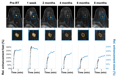 |
Breast tumor response on dynamic contrast-enhanced MRI after single dose neoadjuvant partial breast irradiation
Maureen Groot Koerkamp1, Jeanine Vasmel1, Stefano Mandija1,2, Marielle Philippens1, Ramona Charaghvandi3, Maaike Moman4, Wouter Veldhuis4, Celien Vreuls5, Paul van Diest5, Arjen Witkamp6, Ronald Koelemij7, Annemiek Doeksen7, Thijs van Dalen8,
Elsken van der Wall9, Helena Verkooijen10, Jan Lagendijk1, Bram van Asselen1, Desiree van den Bongard1, and Anette Houweling1
1Radiotherapy, UMC Utrecht, Utrecht, Netherlands, 2Computational Imaging Group for MR diagnostic & therapy, UMC Utrecht, Utrecht, Netherlands, 3Radiotherapy, Radboudumc, Nijmegen, Netherlands, 4Radiology, UMC Utrecht, Utrecht, Netherlands, 5Pathology, UMC Utrecht, Utrecht, Netherlands, 6Surgical Oncology, UMC Utrecht, Utrecht, Netherlands, 7Surgery, St. Antonius, Utrecht, Netherlands, 8Surgery, Diakonessenhuis, Utrecht, Netherlands, 9Medical Oncology, UMC Utrecht, Utrecht, Netherlands, 10Epidemiology, UMC Utrecht, Utrecht, Netherlands
Radiologist assessment and quantitative dynamic contrast-enhanced (DCE) MRI analysis were used to determine if changes in these parameters could predict pathologic complete response to single ablative dose neoadjuvant partial breast irradiation. 3.0T DCE MRI scans in prone position were obtained pre-radiotherapy (RT), and 1 week, 2, 4, 6, and, if applicable, 8 months post-RT. Quantitative analysis showed an increase of relative enhancement 1 week after RT followed by a decrease at later time points which indicates acute tissue response and tumor regression, respectively. Both radiologist assessment (n=36) and quantitative analysis (n=27) could not predict pathologic response.
|
|
4882.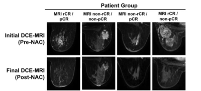 |
Tumor Characteristics of Discordant MRI Imaging and Pathology Following Neoadjuvant Chemotherapy in Non-metastatic Breast Cancer
Alexandra Besser1, Ana Rodriguez-Soto1, Helen Park2, Andrew Park2, Grace Ahn Sora2, Haydee Ojeda-Fournier1, and Rebecca Rakow-Penner2
1Radiology, University of California, San Diego, La Jolla, CA, United States, 2University of California, San Diego, La Jolla, CA, United States
For patients with non-metastatic or locally advanced breast cancer, MRI imaging is used to follow response to neoadjuvant chemotherapy (NAC) and determine treatment plan. However, MRI radiological complete response is not always concordant with pathological complete response. In a cohort of patients imaged at our institution with non-metastatic/locally advanced breast cancer, we report that 24% of post-NAC final MRI imaging is discordant with the final pathology report. We also show that many of these MRI false negatives demonstrate residual intermediate to high grade DCIS and contain microcalcifications.
|
|
4883. |
Diffusion fractional anisotropy in peritumoral edema of triple negative breast cancer
Benjamin Charles Musall1, Abeer H Abdelhafez2, Hagar S Mahmoud2, Ken-Pin Hwang1, Jong Bum Son1, Mark D Pagel3, Lumarie Santiago4, Gary J Whitman4, Huong Le-Petross4, Tanya W Moseley4, Rosalind P Candelaria4, Thorunn Helgason5, Elizabeth E Ravenberg6,
Jennifer K Litton6, Stacy L Moulder6, Wei T Yang4, Jingfei Ma1, Gaiane M Rauch2, and Beatriz E Adrada4
1Imaging Physics, MD Anderson Cancer Center, Houston, TX, United States, 2Abdominal Imaging, MD Anderson Cancer Center, Houston, TX, United States, 3Cancer Systems Imaging, MD Anderson Cancer Center, Houston, TX, United States, 4Breast Imaging, MD Anderson Cancer Center, Houston, TX, United States, 5Investigational Cancer Therapeutics, MD Anderson Cancer Center, Houston, TX, United States, 6Breast Medical Oncology, MD Anderson Cancer Center, Houston, TX, United States
Diffusion fractional anisotropy and ADC were compared between tumors, peritumoral edema, and fibroglandular tissue in triple negative breast cancer. Fractional anisotropy in the peritumoral edema of triple negative breast cancer did not correlate strongly with ADC and may offer additional characterization of peritumoral edema.
|
|
4884. |
Diffusion tensor distribution imaging of breast tumors: Initial findings
Isaac Damiel1, Daniel Topgaard2,3, Karin Bryske2, Sunitha Thakur1, and Katja Pinker-Domenig1
1Memorial Sloan Kettering Cancer Center, New York, NY, United States, 2Random Walk Imaging, Lund, Sweden, 3Lund University, Lund, Sweden Diffusion tensor distribution (DTD) imaging was applied in a pilot study to investigate the potential for breast tumor grading in a clinical setting. The method relies on advanced gradient waveforms to encode the signal with information about cell densities, shapes, and orientations, and quantify tissue composition as a probability distribution in a space with dimensions analogous to the cellular ones. Five patients with ductal carcinoma underwent a <4 min DTD protocol giving voxel-resolved distributions and parameter maps with microstructural information not accessible with conventional methods, thereby encouraging future studies with larger patient groups and comparison with current gold standards. |
|
4885.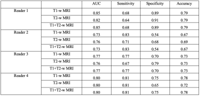 |
MRI and PET for staging of axillary lymph nodes in breast cancer at diagnosis
Pauline Huang1, Renee Cattell1, Meghan Moriarty1, Kwan Chen1, Lev Bangiyev1, Cliff Bernstein1, Jules Cohen1, Lea Baer1, Dinko Franceschi1, Haifang Li1, and Tim Duong1
1Radiology, Renaissance School of Medicine, Stony Brook, NY, United States
In breast cancer, lymph node status is highly related to prognosis. We evaluated the use of MRI of axillary lymph nodes (aLNs) to detect metastasis in pre-chemo breast cancer with PET as reference standard. For 4 readers scoring on T1-weighted DCE and T2-weighted MRI, the AUC, sensitivity, specificity and accuracy ranged from 0.73-0.85, 64%-83%, 54-91%, and 67-79%, respectively. Readers showed significant agreement (Spearman’s Rho 0.35-0.65). Though improvement is needed, this approach presents a less invasive method of assessing all aLNs and avoiding their unnecessary removal.
|
|
4886.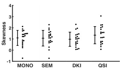 |
q-Space imaging is more sensitive to breast tumour cellularity than conventional diffusion-weighted imaging methods at 3T
Nicholas Senn1, Yazan Masannat2, Ehab Husain3, Bernard Siow4, Steven D Heys5, and Jiabao He1
1Aberdeen Biomedical Imaging Centre, University of Aberdeen, Aberdeen, United Kingdom, 2Breast Unit, Aberdeen Royal infirmary, Aberdeen, United Kingdom, 3Pathology department, Aberdeen Royal Infirmary, Aberdeen, United Kingdom, 4Francis Crick Institute, London, United Kingdom, 5School of Medicine, University of Aberdeen, Aberdeen, United Kingdom
q-Space imaging (QSI) was compared against conventional DWI and non-Gaussian diffusion models of diffusion kurtosis imaging (DKI) and stretched-exponential model (SEM) to evaluate the skewness in histogram distribution of diffusion displacement and diffusivity for profiling breast tumour cellularity. We investigated whole breast tumours excised from surgery, with imaging performed same day overnight on a clinical 3T MRI system. We found QSI to yield a higher effect gradient to assess cellularity in breast cancer compared with conventional diffusion-weighted imaging methods. The skewness obtained from QSI further showed fidelity with the skewness of cellularity obtained from histology.
|