
Digital Poster Session
Spectroscopy:
Spectroscopy
1785 -2933 Proton MRS and Other Nuclei MR - 1H-MRS Non-Cancer Applications 1
2934 -2947 Proton MRS and Other Nuclei MR - 1H-MRS Non-Cancer Applications 2
2948 -2960 Proton MRS and Other Nuclei MR - MRS Cancer Applications
2961 -2974 Proton MRS and Other Nuclei MR - 31P MRS & MRI
2975 -2987 Proton MRS and Other Nuclei MR - X-Nuclei MRI & MRS, 23Na, 39K & 2H
2988 -2999 Proton MRS and Other Nuclei MR - X-Nuclei MRI & MRS & 31P, 13C, 19F, 17O & 2H
2921.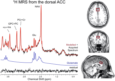 |
Excitability of the Glutamatergic Neurotransmission in the Anterior Cingulate During Appraisal of Emotional Stimuli
Paul R Burghardt1, Dalal Khatib1, Andrew Neff1, Katherine Nowak1, Katlin Chappelle1, and Jeffrey Stanley1
1Wayne State University, Detroit, MI, United States
Personality and fitness impact emotional regulation, however the neurobiological mechanisms underlying these relationships are poorly understood. dACC glutamate modulation was quantified during appraisal of emotional images using ¹H fMRS. Additionally, aerobic fitness and trait personality were assessed in participants. The dACC glutamate was lower during positive images compared to neutral images, which was associated with trait openness. Glutamate levels during appraisal of negative emotional images did not differ from neutral images, but was negatively associated with aerobic fitness. These results shed light on the mechanisms that modulate the impact of trait personality and fitness on ACC response during emotional regulation.
|
|
 |
2922.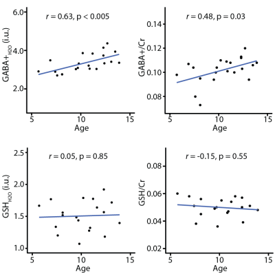 |
Effect of age on in vivo GABA and glutathione levels in a pediatric sample
Muhammad Gulamabbas Saleh1, Afroditi Papantoni2, Georg Oeltzschner1, Mark Mikkelsen1, Nicolaas A Puts1, Richard A Edden1, and Susan Carnell2
Video Permission Withheld
1Russell H. Morgan Department of Radiology and Radiological Science, The Johns Hopkins University School of Medicine, Baltimore, MD, United States, 2Department of Psychiatry and Behavioral Sciences, The Johns Hopkins University School of Medicine, Baltimore, MD, United States
GABA is the primary inhibitory neurotransmitter in the human brain and is implicated in several neuropathologies. Glutathione is a major antioxidant in the brain and is considered a marker of oxidative stress. Several studies have reported age-related declines in GABA levels in adulthood, but the aging dynamics of both GABA and glutathione have not been well explored in childhood. We demonstrate increases in GABA and no differences in glutathione with age in a healthy pediatric sample (5.7-13.9 years). This study provides insight into neuronal maturation in children and may facilitate better understanding of the pathophysiology of developmental disorders.
|
|
2923. |
Detection of acetyl-carnitine in the human heart in vivo using long echo-time 1H-MRS at 3T
Joe Pollacco1,2, William Clarke3, Aaron Hess1, Dragana Savic1,4, Christopher T Rodgers1,5, Damian Tyler1,4, Jack JJ Miller1,2,4, and Ladislav Valkovic1,6
1Oxford Centre for Clinical Magnetic Resonance Research, RDM Cardiovascular Medicine, University of Oxford, Oxford, United Kingdom, 2Department of Physics, University of Oxford, Oxford, United Kingdom, 3Wellcome Centre for Integrative Neuroimaging, University of Oxford, Oxford, United Kingdom, 4Department of Physiology, Anatomy and Genetics, University of Oxford, Oxford, United Kingdom, 5Wolfson Brain Imaging Centre, Department of Clinical Neurosciences, University of Cambridge, Cambridge, United Kingdom, 6Department of Imaging Methods, Slovak Academy of Sciences, Bratislava, Slovakia
Cardiac acetyl-carnitine plays an important role in fat metabolism. Decreased carnitine concentration has been reported in heart failure, but it has previously been difficult to measure acetyl-carnitine in-vivo. We show for the first time that it is possible to detect a clear acetyl-carnitine resonance at δ=2.1ppm without lipid contamination within the heart using a long echo time semi-LASER sequence at 3T. This validates the finding in skeletal muscle that the T2 of acetyl-carnitine is much longer than its surrounding lipid signals. Further work is underway to investigate variability of the acetyl-carnitine signal postprandially.
|
|
2924.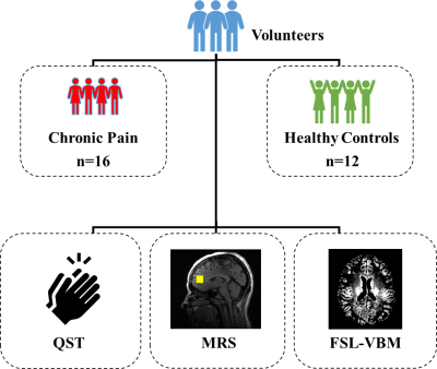 |
Association of brain metabolite concentrations and pain perception in a cohort of patients with chronic pain
Maame Owusu-Ansah1, Candace C. Fleischer1, and Daniel E. Harper2
1Department of Radiology and Imaging Sciences, Emory University School of Medicine, Atlanta, GA, United States, 2Department of Anesthesiology, Emory University School of Medicine, Atlanta, GA, United States
A proof-of-concept generalized linear model to characterize the relationships between brain metabolite concentrations and pain perception is presented, explicitly accounting for differences in regional grey matter (GM) density. Brain metabolite concentrations measured with magnetic resonance spectroscopy (MRS), and pain perception characterized using quantitative sensory testing (QST), were significantly associated in a cohort of patients with chronic pain. These results contribute to the expanding literature that supports the utility of neuroimaging to understand the underlying mechanisms of centralized chronic pain.
|
2925. |
Ultrashort-TE Liver 1H MR Spectroscopy in Alcohol Use Disorder at 3 T
Martin Gajdosik1, Jonathan Wai2,3, Karl Landheer1, Diana Martinez2,4, and Christoph Juchem1,5
1Department of Biomedical Engineering, Columbia University, New York City, NY, United States, 2Department of Psychiatry, Columbia University Irving Medical Center, New York City, NY, United States, 3New York State Psychiatric Institute, New York City, NY, United States, 4Department of Psychiatry, New York State Psychiatric Institute, New York City, NY, United States, 5Department of Radiology, Columbia University College of Physicians and Surgeons, New York City, NY, United States Poster Permission Withheld
Accurate detection of hepatocellular lipid content (HLC) in individuals with alcohol use disorder (AUD) is needed for early diagnosis of hepatic steatosis, a harbinger of serious liver diseases such as cirrhosis. Using a standard clinical 3T system and ultrashort-TE liver MRS, the proposed sequence employed asymmetric RF pulses with spoiler gradient cycling to achieve minimal TE of 5ms and minimal T2 errors for reliable signal quantification. No difference was found in HCL and choline levels between controls and AUD subjects. After a 3-week abstinence period, changes in hepatic fat and choline levels were observed in subjects with AUD.
|
|
2926.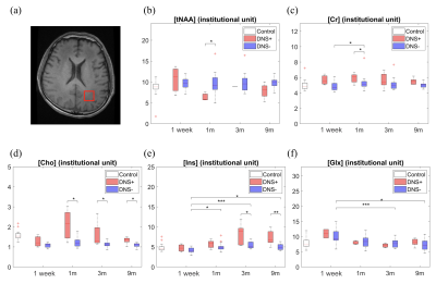 |
A 9-month follow-up study in patients with delayed neurologic sequelae after CO intoxication using absolute quantification of 1H MRS and DKI
Yi-Chen Tsai1, Hui-Ru Tsai1, Ming-Chung Chou2, Ping-Hong Lai3,4, Jie-Yuan Li5,6, and Cheng-Wen Ko1
1Dept. of Computer Science and Engineering, National Sun Yat-sen University, Kaohsiung, Taiwan, 2Dept. of Medical Imaging and Radiological Sciences, Kaohsiung Medical University, Kaohsiung, Taiwan, 3Dept. of Radiology, Veterans General Hospital-Kaohsiung, Kaohsiung, Taiwan, 4School of Medicine, National Yang-Ming University, Taipei, Taiwan, 5Dept. of Neurology, E-Da Hospital, Kaohsiung, Kaohsiung, Taiwan, 6School of Medicine, I-Shou University, Kaohsiung, Kaohsiung, Taiwan
In this study, we explored the metabolic changes in WM of patients with and without DNS using 1H MRS and DKI longitudinally at 1 week and 1, 3, and 9 months after CO intoxication. Increased Cr and Cho were observed at early stage in WM of patients with DNS. Decreased tNAA showed higher correlation with the presence of WM lesion than of DNS. The chronic change of Ins and Glx in patients with DNS implies themselves as potential indices to provide valuable information for monitoring DNS development.
|
|
2927.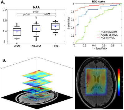 |
Neurometabolic Mapping of RRMS patients using multi-slice Spiral-MRSI and support vector machine techniques
Oun Al-iedani1,2, Karen Ribbons2, Jeannette Lechner-Scott2,3,4, Neda Gholizadeh1, Scott Quadrelli5, Rodney Lea2, Ovidiu Andronesi6, and Saadallah Ramadan1,2
1School of Health Sciences, University of Newcastle, Newcastle, Australia, 2Hunter Medical Research Institute (HMRI), Newcastle, Australia, 3School of Medicine and Public Health, University of Newcastle, Newcastle, Australia, 4Department of Neurology, John Hunter Hospital, Newcastle, Australia, 5Faculty of Medicine, University of Queensland, Herston, Australia, 6Harvard Medical School, Massachusetts General Hospital, Boston, MA, United States
The study designed a novel neurometabolic mapping using multi-slice Spiral-MRSI with multi-voxel segmentation and support vector machine(SVM) techniques to demonstrate the true nature of NAWM and WML of RRMS patients, compared to HCs. 3D-Spiral-MRSI covering 75% of the brain on 16 RRMS and 9 HCs were used. Multi-slice-MRSI was processed using novel pipeline with 3-model SVM classifications. Neurometabolic mapping revealed that (NAA/tCr) in WM-lesions was significantly lower than NAWM-MS and HCs with HCs vs WML model achieving highest sensitivity and specificity. Multi-slice-spiral-MRSI may enhance diagnosis and clinical monitoring of RRMS patients, and is sensitive in diagnosing RRMS even in NAWM.
|
|
2928. |
Respiratory-triggered quantitative MR spectroscopy of the human spinal cord at 7 T
Tangi Roussel1,2, Yann Le Fur1,2, Jean-Philippe Ranjeva1,2, and Virginie Callot1,2
1Aix-Marseille Univ, CNRS, CRMBM, Marseille, France, 2APHM, Hôpital Universitaire Timone, CEMEREM, Marseille, France
1H MR spectroscopy (MRS) is of great interest to help characterizing human spinal cord pathologies. However, few studies have been reported so far because of challenging experimental difficulties caused by the small size of the structure, static and radiofrequency field heterogeneities, as well as physiological motion and especially breathing. In this work, we demonstrate the necessity of respiratory-gated acquisition to collect robust 1H MRS data from the cervical spinal cord at 7T. We also present dedicated post-processing and quantitative approaches for SC metabolic assessment.
|
|
2929. |
1H MRS at 3T reveals reduced GABA and glutamate levels in the visual cortex induced by pharmacologically increased dopamine
Ralf Mekle1, Jochen B Fiebach1, and Heiner Stuke2
1Center for Stroke Research Berlin, Charité – Universitätsmedizin Berlin, Berlin, Germany, 2Department of Psychiatry and Psychotherapy, Charité – Universitätsmedizin Berlin, Berlin, Germany
Schizophrenia frequently manifests psychotic symptoms, such as delusions and hallucinations. However, its neurochemical mechanisms are not well deciphered, though dysfunctional dopaminergic neurotransmission is suggested. In this study, single volume 1H MRS using MEGA-PRESS at 3 T was applied to investigate possible neurochemical changes in the visual cortex induced by pharmacologically increased dopamine levels in healthy volunteers that also performed a visual detection task. Increased dopamine yielded decreased GABA and reduced glutamate quantities. Furthermore, an inverse correlation of glutamate with false perceptions in the detection task supports the theory that glutamate hypofunction might contribute to the formation of hallucinations in schizophrenia.
|
|
2930.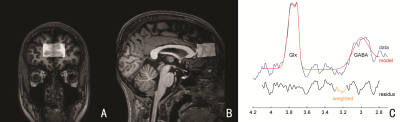 |
Effect of DNA methylation levels of dopamine D4 receptor gene on GABA concentrations in the mPFC in children with primary nocturnal enuresis
Zijun Li1, Xiaoyang Li1, and Bing Yu1
1Shengjing Hospital of China Medical University, Shenyang, China In the current study we investigated the effect of DRD4 DNA methylation levels on the GABA concentrations in mPFC in PNE children. Our results suggested the DNA methylation level of DRD4 is associated with increased GABA concentrations in the mPFC in PNE children. The effects of epigenetic variation of the DRD4 DNA on GABA concentrations in mPFC may help us understand the epigenetic susceptibility of the DRD4 DNA methylation to PNE. |
|
2931.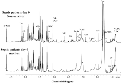 |
Metabolic Profile Differences in Serum of Sepsis Patients may Identify Survivors and Non-Survivors
Virendra Kumar1, Naveen Kumar MS2, sujeet kumar mewar1, Pradeep Kumar1, Naveet Wig2, Prayas Sethi2, and Sanjeev Sinha2
1Department of NMR, All India Institute of Medical Sciences, New Delhi, India, 2Department of Medicine, All India Institute of Medical Sciences, New Delhi, India
The present study reported the differences in metabolic profile of serum samples from patient with sepsis to identify survivor and non-survivor (day 0) using NMR metabolomics. A significantly higher concentration of tyrosine, histidine, methionine, betaine, creatine, phosphocreatine and choline in the serum of sepsis patients who did not survive septic shock compared to survivors. These findings suggest that metabolic alterations at day 0 may predict the survival of the septic patients.
|
|
2932. |
Proton magnetic resonance spectroscopy (1H-MRS) in degenerative cervical spinal cord compression.
Petr Bednarik1, Tomas Horak2, Magda Horakova3, Alena Svatkova4, Zdenek Kadanka3, Zdenek Kadanka jr3, Petr Kudlicka5, Jan Valosek6, Dinesh Deelchand7, Pierre-Gilles Henry7, and Josef Bednarik3
1Departement of Medical Imaging and Image-guided Therapy, High Field MR Center, Vienna, Austria, 2Masaryk University, Brno, Czech Republic, 3Department of Neurology, University Hospital Brno, Brno, Czech Republic, 4Department of Internal Medicine, Medical University of Vienna, Vienna, Austria, 5Multimodal and Functional Imaging Laboratory, Central European Institute of Technology, Brno, Czech Republic, 6Departments of Neurology and Biomedical Engineering, University Hospital Olomouc, Olomouc, Czech Republic, 7Center for Magnetic Resonance Research, University of Minnesota, Minneapolis, MN, United States
While cervical spinal cord (CSC) compression occurs almost ubiquitously with aging, early metabolic changes in CSC that might develop into irreversible clinical myelopathy symptoms have not been described yet. Thus, we utilized fine-tuned semi-LASER 3T MRS protocol that addresses limitations of standard MRS methods to unravel metabolic damage in the cranial parts of the CSC in 50 patients with non-myelopathic degenerative cervical cord compression and 10 with clinically symptomatic degenerative cervical myelopathy in comparison to 35 healthy controls. Higher tNAA/tCr and myo-Ins/tNAA levels suggest axonal loss above the level of stenosis, indicating that CSC impairment exceeds the level of compression.
|
|
2933. |
Detection of metabolic abnormalities in different genotypes of Shank3 deficiency in mice using MRS at 11.7T
Ying Liu1,2, Lei Wei1,2, and Xiaoyong Zhang1,2
1Institute of Science and Technology for Brain-Inspired Intelligence, Fudan University, Shanghai, China, 2Key Laboratory of Computational Neuroscience and Brain-Inspired Intelligence (Fudan University), Ministry of Education, Shanghai, China
Shank3 plays an important role in the functioning of glutamatergic synapses. In this work, we investigated whether Shank3 deficiency could induce neuroimaging changes in a transgenic autism mouse model using structural MRI and MRS at 11.7T. Our data showed that the neurochemical metabolites in medial prefrontal cortex (mPFC) are significantly changed in Shank3 deficiency models of heterozygous and homozygous genotypes, relative to wildtype controls. However, the volume of mPFC does not show significant alteration among three groups. We concluded that metabolic abnormalities are closely related to different phenotypes of Shank3 deficiency in mice.
|
|
1785.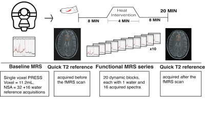 |
Metabolite activity in the anterior cingulate cortex during a painful stimulus using functional MRS
Jessica Archibald1,2, Erin L MacMillan3,4,5, Carina Graf2,6, Piotr Kozlowski2,7,8, Cornelia Laule2,9,10, and John LK Kramer1,2,11
1Experimental Medicine, University of British Columbia, Vancouver, BC, Canada, 2International Collaboration on Repair Discoveries (ICORD), Vancouver, BC, Canada, 3Radiology, University of British Columbia, Vancouver, BC, Canada, 4Image Tech Lab, Simon Fraser University, Vancouver, BC, Canada, 5Philips Healthcare Canada, Vancouver, BC, Canada, 6Physics and Astronomy, University of British Columbia, Vancouver, BC, Canada, 7Radiology, University of British Columbia, vancouver, BC, Canada, 8UBC MRI Research Centre, Vancouver, BC, Canada, 9Physics and Astronomy, University of British Columbia, vancouver, BC, Canada, 10Pathology and Laboratory Medicine, University of British Columbia, Vancouver, BC, Canada, 11School of Kinesiology, University of British Columbia, Vancouver, BC, Canada
Current treatment evaluation procedures of pain conditions are dependent on self-reported measures. The objective of this study was to determine changes in excitatory neurotransmitters (i.e., glutamate and glutamate+glutamine) in the anterior cingulate cortex as an objective measure of pain during a painful stimulus using single voxel functional magnetic resonance spectroscopy (fMRS). Glutamate concentration changes during the painful stimulus suggest a role for glutamate in detecting pain which was not related to self-reported pain ratings. An exploratory analysis on sex revealed an 8.63% (p=0.08) increase in glutamate at pain onset in female participants compared with a 7.45% (p=0.31) increase in males.
|
 View the Poster
 Watch the Video
2934. |
Metabolomic profiling of wildtype and transgenic Giardia lamblia strains by 1H Magic Angle Spinning NMR spectroscopy.
Martina Vermathen1, Norbert Mueller2, David Leitsch3, Damian Hertig4, Peter Vermathen4, and Joachim Mueller2
1Department of Chemistry and Biochemistry, University Bern, Bern, Switzerland, 2Institute of Parasitology, Vetsuisse Faculty, University Bern, Bern, Switzerland, 3Institute of Specific Prophylaxis and Tropical Medicine, Medical University of Vienna, Vienna, Austria, 4DBMR & DIPR, University Bern, Bern, Switzerland
The intestinal protozoan parasite Giardia lamblia is a major cause of persistent diarrhea in humans and animals worldwide. Treatment of giardiasis with nitro-prodrugs relies on intrinsic enzymes in G.lamblia that are involved in activating or deactivating the prodrugs. To elucidate more about the physiologic function of these pathways and their potential roles in drug resistance, G.lamblia wildtype and transgenic strains were metabolomically characterized based on 1H HR-MAS NMR data and specific metabolites that may be related to nitroreductase activity were identified and discussed.
|
|
2935. |
Diurnal fluctuation of glutathione measured with MR spectroscopy
Shin-ichi Urayama1, Yujiro Yoshihara2, Masaki Fukunaga3, and Toshiya Murai2
1Human Brain Research Center, Kyoto University, Kyoto, Japan, 2Department of Psychiatry, Kyoto University, Kyoto, Japan, 3National Institute for Physiological Sciences, Okazaki, Japan
Glutathione (GSH) is a powerful and general antioxidant found in cell of various animals. Since its concentration in central nervous system has been thought to related to psychiatric disease, several studies has carried out but the results are still controversial. Here, we investigated GSH diurnal fluctuation of healthy subjects and compared with the results of the reproducibility study. Our results shows GSH concentration in brain fluctuates by 10 to 20% during the daytime. This finding indicates that conditions of MRS examination must be regulated strictly and the fluctuation should be took into account at interpretation of GSH studies.
|
|
2936.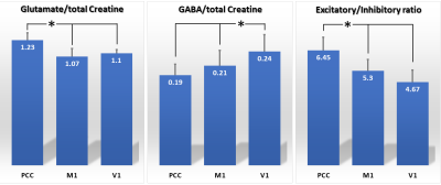 |
Glutamate, GABA and Excitatory/Inhibitory Ratios observed by Short-TE STEAM proton MRS measurements of young healthy subjects at 7T.
Tomohisa Okada1, Koji Fujimoto1, Dinh Ha Duy Thuy1, Hideto Kuribayashi2, Yuta Urushibata2, Ravi Teja Seethamraju3, Sinyeob Ahn4, and Tadashi Isa1
1Kyoto University, Kyoto, Japan, 2Siemens Healthcare K.K., Tokyo, Japan, 3Siemens Healthineers USA, Burlington, MA, United States, 4Siemens Healthineers USA, Berkeley, CA, United States
Relationship between glutamate and GABA as well as their regional differences have not been much investigated. Focused on the posterior cingulate cortex (PCC), the primary motor area (M1) and the primary visual area (V1), this study found significantly higher glutamate concentration and E/I ratios at PCC than others, whereas GABA concentration was significantly higher at V1 than others. Region-wise correlation analysis between glutamate and GABA found statistically significant correlation only at M1 (r = 0.69, p = 0.0005). These differences may reflect difference in functional status at rest, but further investigation is required to clarify the reasons.
|
|
2937.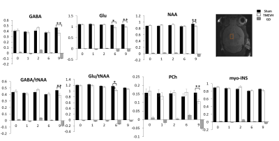 |
Detection of metabolic alterations in the thalamus of the TMEV mouse model of multiple sclerosis at 9.4 Tesla
Poonam Choudhary1,2, Suyog Pol2, Marilena Preda2,3, Robert Zivadinov2,3, and Ferdinand Schweser2,3
1Department of Medical Physics, Jacobs School of Medicine and Biomedical Sciences, University at Buffalo, The State University of New York, Buffalo, NY, United States, 2Buffalo Neuroimaging Analysis Center, Department of Neurology, Jacobs School of Medicine and Biomedical Sciences, University at Buffalo, The State University of New York, Buffalo, NY, United States, 3Center for Biomedical Imaging at the Clinical and Translational Science Institute, University at Buffalo, The State University of New York, Buffalo, NY, United States
This study aimed to characterize the metabolic alterations in the thalamus throughout the disease course of Theiler's murine encephalomyelitis virus (TMEV) in mice using 9.4T MRS, and relate the findings to the known effects of microglia activation on neurotransmitter homeostasis. Our study confirmed that TMEV models metabolite alterations in Multiple Sclerosis. Dynamics of neurotransmitter imbalance suggest that early thalamic dyshomeostatis is driven by neuronal loss in basal ganglia input.
|
|
2938. |
Effects of lisinopril on arterial stiffness, hippocampal blood flow, N-acetyl aspartate and cortical thickness in hypertensive Dahl-s rats
Samuel Ajamu1, Rachel Fenner1, Nikkita Khattar2, Yulia Grigorova1, Edward Lakatta1, Ondrej Juhasz1, Peter Rapp3, Mustapha Bouhrara2, Richard Spencer2, Olga Fedorova1, and Kenneth Fishbein2
1Laboratory of Cardiovascular Science, National Institute on Aging, Baltimore, MD, United States, 2Laboratory of Clinical Investigation, National Institute on Aging, Baltimore, MD, United States, 3National Institute on Aging, Baltimore, MD, United States
Central arterial stiffness (CAS), associated with hypertension, is likely associated with attendant cerebral hypoperfusion, neuronal density loss and cognitive decline, and stiffening of cerebral arterial wall. We previously found associations between pulse wave velocity (PWV), a marker of CAS, and hippocampal cerebral blood flow (CBF) and neuronal density in 6 months old hypertensive Dahl salt-sensitive (Dahl-S) rats, which exhibit age-associated memory loss. The present study showed the ACE inhibitor, lisinopril, resulted in stabilized hippocampal blood flow and NAA concentration compared to nontreated age-matched animals. We also observed significant changes in cortical thickness for treated animals compared to nontreated control.
|
|
2939.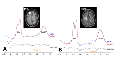 |
Association of multiple sclerosis central fatigue with inhibitory and excitatory neurotransmitters
Jameen ARM1,2, Georg Oeltzschner3,4, Oun Al-Iedani1,2, Rodney Lea5,6, Jeannette Lechner-Scott5,7,8, and Sadallah Ramadan1,5
1School of Health Sciences, University of Newcastle, Newcastle, Australia, 2Imaging centre, Hunter Medical Research Institute, Newcastle, Australia, 3The Russell H. Morgan Department of Radiology and Radiological Science, Johns Hopkins University School of Medicine, Baltimore, MD, United States, 4F. M. Kirby Research Center for Functional Brain Imaging, Kennedy Krieger Institute, Baltimore, MD, United States, 5HMRI Imaging centre, Hunter Medical Research Institute, Newcastle, Australia, 6School of Biomedical Sciences, Queensland University of Technology, Brisbane, Australia, 7Faculty of Health and Medicine, University of Newcastle, Newcastle, Australia, 8Department of Neurology, John Hunter Hospital, Newcastle, Australia
Despite neuro metabolic and morphological alterations linked to central fatigue in multiple sclerosis (MS), the pathophysiology of this symptom is not fully understood. Dysfunction of the GABAergic/Glutamatergic pathways involving inhibitory and excitatory neurotransmitters such as γ-aminobutyric acid (GABA) and glutamine+glutamate pools (Glx) have been implicated in several neurological disorders, including MS. In this study, we evaluated if GABA and Glx levels are associated with central fatigue in MS. Our results showed significant correlations of GABA and Glx levels with fatigue scores which suggest dysregulation of GABAergic/glutamatergic neurotransmission is possibly implicated in the mechanisms of mediating central fatigue in MS
|
|
2940.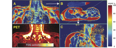 |
In-vivo 1H-MRS Shows Differences in Polyunsaturated Fatty Acid Content between White and Brown Adipose Tissue in Humans.
Ronald Ouwerkerk1, Jatin Raj Matta1, Ahmed Hamimi1, Aaron M Cypess2, Kong Y Chen3, and Ahmed Medhat Gharib1
1Biomedical and Metabolic Imaging Branch, NIDDK/NIH, Bethesda, MD, United States, 2Translational Physiology Section, Diabetes, Endocrinology, and Obesity Branch, NIDDK/NIH, Bethesda, MD, United States, 3Energy Metabolism Section, Diabetes, Endocrinology, and Obesity Branch, NIDDK/NIH, Bethedsa, MD, United States
Localized 1H-MRS was used to compare the fatty acid composition and MR relaxation in PET proven metabolically active brown adipose tissue (BAT) in humans with white adipose tissue (WAT). Paired comparison of BAT and WAT results from the same individuals the showed differences in MR relaxation of fatty acid resonances and unsaturated fatty acid content between metabolically activatable brown fat and white adipose tissue
|
|
2941.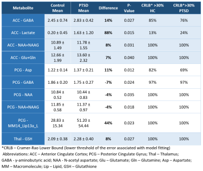 |
Neurochemical Deregulation in Post-traumatic Stress Disorder
Nathan Tosh1,2, Scott Quadrelli2, Chris Foster2, Graham Galloway2, David Crompton2,3, and Carolyn Mountford2
1School of Clinical Sciences, Queensland University of Technology, Brisbane, Australia, 2Translational Research Institute, Woolloongabba, Australia, 3Griffith University, Brisbane, Australia
2D L-COSY has previously been used to demonstrate neurochemical differences between PTSD and healthy cohorts but requires a 19 minute acquisition. Single Voxel Spectroscopy (SVS), acquired in 3 minutes, was used to collect data from 3 brain regions in healthy controls and patients with Post-traumatic Stress Disorder. Neurochemical differences were seen in all the ACC, Thalamus and PCG. Data, analysed using LC Model, showed elevated levels of NAA, Lactate, GABA and Glutathione as well as glutamatergic dysfunction. Thus, SVS can be used to identify PTSD in a shorter time frame than 2D L-COSY.
|
|
2942. |
Lactate change during visual stimuli detected by MEGA-PRESS at 3T
Ya-Tien Liu1, Dian-Han Yang1, Cheng-Wen Ko1, Tzu-Chao Chuang2, and Shang-Yueh Tsai3,4
1Department of Computer Science and Engineering, National Sun Yat-sen University, Kaohsiung, Taiwan, 2Department of Electrical Engineering, National Sun Yat-sen University, Kaohsiung, Taiwan, 3Graduate Institute of Applied Physics, National Chengchi University, Taipei, Taiwan, 4Research Center of Mind, National Chengchi University, Taipei, Taiwan
In this study, we investigate the feasibility of using MEGA-PRESS to detect Lac response to the visual stimuli in visual cortex in fMRS experiment at 3T. The significant raise in Lac level was found either by averaging the whole fMRS scan or by averaging over the stimuli blocks. Our results show consistency with the published results. More sophisticated approach to analyze fMRS data may be necessary to facilitate reliable metabolic quantification.
|
|
2943.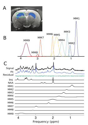 |
Simultaneous characterization of individual macromolecule resonances and metabolites in the Fischer 344 rat during healthy aging
Caitlin Fowler1, Dan Madularu2, Masoumeh Dehghani2, Gabriel Devenyi2, and Jamie Near3
1Biological and Biomedical Engineering, McGill University - Douglas Hospital Research Centre, Verdun, QC, Canada, 2Douglas Hospital Research Centre, Verdun, QC, Canada, 3Psychiatry, McGill University - Douglas Hospital Research Centre, Montréal, QC, Canada
To better understand pathological aging, we must first have a thorough understanding of changes that occur in the brain during healthy aging. This project employs Magnetic Resonance Spectroscopy to characterize longitudinal changes in the neurochemical profile of healthy Fischer 344 rats. In addition to the commonly reported metabolites, nine macromolecular resonances were included in the basis set and quantified, based on parameterization of a group-averaged metabolite-nulled spectrum. For the first time, longitudinal age-related changes in metabolites and individual macromolecules were simultaneously characterized in the Fischer rat. Sex-specific differences were also identified in metabolites and macromolecules.
|
|
2944. |
Effects of High Fat Diet on the adipose tissues and brain metabolism of a transgenic mouse which over-expresses human hydrolase hMTH1
Rossella Canese1, Gabriele De Luca2, Ambra Dell'Orso3, Egidio Iorio1, Mattea Chirico1, Maria Elena Pisanu1, Paola Fortini3, and Valeria Simonelli3
1NMR and MRI unit, Core facilities, Istituto Superiore di Sanita', Rome, Italy, 2Oncology and Molecular Medicine Department, Istituto Superiore di Sanita', Rome, Italy, 3Environmental and Health department, Istituto Superiore di Sanita', Rome, Italy
Oxidative stress is implicated in the pathogenesis of cancer, neurodegeneration and aging. hMTH1 is a hydrolase able to protect cells by oxidative damage. Overexpression of hMTH1 in transgenic mice (hMTH1‐Tg) confers significant protection against neurodegeneration and motor impairment. In this study, we use the hMTH1‐Tg mouse model and we found that oxidative damage is able to affect brain metabolism and adipose organ composition and extension. Moreover, we investigated the protective role of hMTH1-tg against an environmental oxidative stimulus like the assumption of a diet at high content of fat.
|
|
2945.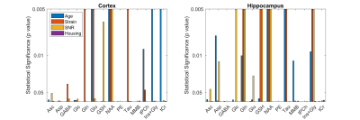 |
Effects of Housing, Age, Strain, and Signal to Noise Ratio on Magnetic Resonance Spectroscopy in the Rat Brain
Wendy Oakden1 and Greg J Stanisz1,2,3
1Physical Sciences, Sunnybrook Research Institute, Toronto, ON, Canada, 2Medical Biophysics, University of Toronto, Toronto, ON, Canada, 3Neurosurgery and Paediatric Neurosurgery, Medical University of Lublin, Lublin, Poland
Changes in metabolite levels as a result of treatment can be quite small. This study investigated the variability of MRS detectable metabolites so that the effects relating to the treatment would not be confounded by other experimental factors. A four-way ANOVA was used to separate the effects of differences in housing conditions (single housing, pairs, or enriched environment) as well as age (5-15 weeks), strain (Long Evans vs Sprague Dawley), and SNR. Significant changes in MRS were observed due to age, strain, and SNR, but any changes due to housing conditions were small in comparison.
|
|
2946. |
Evaluating Apparent T2* values of Metabolites in Healthy Adults and Youths using TIDEL-COSY
Zohaib Iqbal1, Rajakumar Nagarajan2, Manoj Sarma3, Andres Saucedo3, and Michael Albert Thomas3
1Radiation Oncology, UT Southwestern Medical Center, Dallas, TX, United States, 2Human Magnetic Resonance Center, UMass, Amherst, MA, United States, 3UCLA, Los Angeles, CA, United States
For decades, one-dimensional (1D) spectroscopy has aided in the diagnosis of several pathologies. However, 1D approaches suffer from severe spectral overlap, possibly limiting their application. Two-dimensional (2D) spectroscopy allows for greater spectral separation, and many studies have successfully quantified metabolites with the localized correlated spectroscopy (L-COSY). Here, we combine the L-COSY technique with a T2* weighted deconvolution (TIDE) approach and develop the TIDEL-COSY method. We show the capability of the novel TIDEL-COSY method and compare the apparent T2* values between healthy youths and adults for a variety of metabolites.
|
|
2947. |
Detection of beta-hydroxybutyrate in human brain after oral administration of a ketone ester
Jan Willem van der Veen1, Corinde Wiers2, and Jun Shen3
1Magnetic Resonance Spectroscopy Core, NIMH, NIH, Bethesda, MD, United States, 2NIH, NIAAA, Bethesda, MD, United States, 3Magnetic Resonance Spectroscopy Core, NIH, NIMH, Bethesda, MD, United States
Beta-Hydroxybutyrate (BHB) is a marker of metabolic ketosis in brain. To study ketogenic treatment spectral editing was developed to invert the signals in the 3.2-4.6 ppm spectral region for detecting BHB and co-edited signals. Using this spectral editing technique we found markedly elevated BHB signal in the human brain shortly after oral administration of 1,3-butanediol monoester of beta-hydroxybutyrate as compared to the control scan without consumption of the ketone ester.
|
2948. |
Downfield Magnetic Resonance Spectroscopy in a mouse model of Brain Glioma
Sónia Isabel Gonçalves1, Rui V. Simões1, and Noam Shemesh1
1Champalimaud Research, Champalimaud Centre for the Unknown, Lisbon, Portugal
MRS allows non-invasive in-vivo exploration of tissue metabolism. However, about half of the proton spectrum (downfield of water) has been nearly ignored over the decades of MRS application due to water suppression. We show that ISIS-based Relaxation Enhanced MRS (iRE-MRS) which uses frequency selective excitation and ISIS localization offers short echo times and enhances exchange-broadened resonances. For the first time, we measure in-vivo downfield spectra in mouse glioma tumors (and controls) and show remarkable spectral signatures for the tumor downfield.
|
|
2949.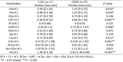 |
MR Spectroscopic Differences of Low and High Grade TERTp-only Gliomas
Banu Sacli-Bilmez1, Ayhan Gursan2, Ayca Ersen Danyeli3, Cengiz Yakicier4, M.Necmettin Pamir5,6, Koray Ozduman5,6, Alp Dincer5,7, and Esin Ozturk-Isik1
1Institute of Biomedical Engineering, Bogazici University, Istanbul, Turkey, 2Department of Radiology, University Medical Center Utrecht, Utrecht, Netherlands, 3Department of Medical Pathology, Acibadem Mehmet Ali Aydinlar University, Istanbul, Turkey, 4Department of Molecular Biology and Genetics, Acibadem Mehmet Ali Aydinlar University, Istanbul, Turkey, 5Center for Neuroradiological Applications and Reseach, Acibadem Mehmet Ali Aydinlar University, Istanbul, Turkey, 6Department of Neurosurgery, Acibadem Mehmet Ali Aydinlar University, Istanbul, Turkey, 7Department of Radiology, Acıbadem Mehmet Ali Aydinlar University, Istanbul, Turkey
Gliomas that only have the telomerase reverse transcriptase promoter mutation (TERTp-only) are known to have the worst overall survival. The aim of this study was to analyze magnetic resonance spectroscopic (MRS) differences of different grades of TERTp-only gliomas, and classify these groups using machine learning. The results indicated that the ratios of glycerophosphocholine (GPC), glutathione (GSH), total choline (tCho), and glutamine and glutamate complex (Glx) to total creatine increased along with the tumor grade, and machine learning models classified low- and high-grade TERTp-only gliomas with a high accuracy.
|
|
|
2950. |
GSH and GABA decreases in IDH1 mutated low-grade gliomas detected by HERMES spectral editing at 3 T in vivo
Tao Gong1, Richard Edden2,3, and Guangbin Wang1
1Shandong Medical Imaging Research Institute, Jinan, China, 2Russell H. Morgan Department of Radiology and Radiological Science, Johns Hopkins University School of Medicine, Baltimore, MD, United States, 3F. M. Kirby Research Center for Functional Brain Imaging, Kennedy Krieger Institute, Baltimore, MD, United States
IDH1 mutation could result in better prognosis compared with wild-type, but the mechanisms remain largely unknown. GABA and GSH are low-concentration metabolites, could not be detected using conventional MRS, but playing an important role in energy production of glioma. HERMES can be thought of as two MEGA-PRESS experiments with the benefit of saving half the acquisition time to simultaneously detect GABA and GSH. The results demonstrated that HERMES is a reliable tool for the simultaneous detection of GABA and GSH signals, and both of them decreased significantly in IDH1 mutated low-grade gliomas.
|
2951. |
High-grade gliomas: Metabolic and structural assessment using high resolution 3D-MRSI at 7T
Gilbert Hangel1,2, Cornelius Cadrien1,2, Philipp Lazen1, Alexandra Lipka1, Philipp Moser1, Eva Hečková1, Lukas Hingerl1, Stanislav Motyka1, Stephan Gruber1, Bernhard Strasser3, Georg Widhalm2, Barbara Kiesel2, Mario Mischkulnig2,
Julia Furtner4, Thomas Rötzer5, Karl Rössler2, Siegfried Trattnig1,6, and Wolfgang Bogner1
1High Field MR Centre, Department of Biomedical Imaging and Image-guided Therapy, Medical University of Vienna, Vienna, Austria, 2Department of Neurosurgery, Medical University of Vienna, Vienna, Austria, 3Athinoula A. Martinos Center for Biomedical Imaging, Department of Radiology, Massachusetts General Hospital, Harvard Medical School, Boston, MA, United States, 4Division of Neuroradiology and Musculoskeletal Radiology, Department of Biomedical Imaging and Image-guided Therapy, Medical University of Vienna, Vienna, Austria, 5Clinical Institute of Neurology, Medical University of Vienna, Vienna, Austria, 6Christian Doppler Laboratory for Clinical Molecular MR Imaging, Vienna, Austria
We applied high resolution 3D-MRSI covering the whole brain at 7T to 16 high-grade glioma measurements and evaluated our findings in regards of quantifiable metabolites and their structure in the glioma compared to histology and clinical imaging. Our findings include 14 apparently quantifiable metabolites that resolve glioma structure. Especially the pattern of Glycine to myo-Inositol could be indicative of glioma type and proliferation beyond morphological visibility, making 3D-MRSI an interesting lead for preoperative biomarkers with spatial resolution. Other less-researched metabolites such as Serine and Cysteine could lead to new investigations of glioma metabolism.
|
|
2952. |
Identification of Potential Biomarkers for Human Esophageal Cancer: NMR-based metabolomics of paired tissue-serum samples
Yan Lin1, Wei Ye1, Jiayun Zhao1, Ouyang Ting1, and Wan Tang1
1Radiology Department, Second Affiliated Hospital of Shantou University Medical College, Shantou City, China
This was a parallel investigation of esophageal tumor tissues and adjacent normal mucosal tissues alongside patient-matched serum samples by nuclear magnetic resonance (NMR)-based metabolomics, to investigate how serum metabolic phenotypes were linked to the changes in the biochemical landscape of esophageal tumors. These associations provide evidence of distinct metabolic signatures and pathway disturbances between tumor tissues and serum of esophageal cancer patients, and changes in serum metabolic signature could reflect reprogramming of the aforementioned metabolic pathways in EC tissues.
|
|
2953.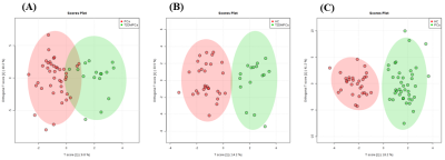 |
Metabolic Differences in Blood Plasma of Type 2 Diabetic and Non-diabetic Prostate Cancer Patients using NMR Spectroscopy
Virendra Kumar1, Pradeep Kumar1, Rajeev Kumar2, Sanjay Sharma3, Sanjay Thulkar4, Siddhartha Dattagupta5, S Senthil Kumaran1, Rama Jayasundar1, and N R Jagannathan6
1Department of NMR, All India Institute of Medical Sciences, New Delhi, India, 2Department of Urology, All India Institute of Medical Sciences, New Delhi, India, 3Department of Radio-diagnosis, RPC, All India Institute of Medical Sciences, New Delhi, India, 4Department of Radio-diagnosis, IRCH, All India Institute of Medical Sciences, New Delhi, India, 5Department of Pathology, All India Institute of Medical Sciences, New Delhi, India, 6Present address: Department of Radiology, Chettinad Academy of Research and Education, Kelambakkam, Tamil Nadu, India
The present study evaluates the metabolic profile of blood plasma for distinguishing prostate cancer (PCa) patients with type 2 diabetes mellitus (T2DM), PCa and healthy controls using 1H-NMR spectroscopy. Our data showed significant lower concentration of glycine, glycerophosphocholine, choline and higher concentration of glucose in PCa patients with T2DM as compared to PCa patients and healthy control. Results provided an insight into the alterations in the metabolic pathways in T2DM-PCa and indicated that NMR spectroscopy may help in determining metabolic changes associated with PCa patients with T2DM and may help in diagnosis.
|
|
2954.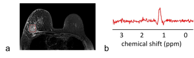 |
Lactate detection in breast cancer using optimised multiple quantum coherence MR spectroscopy and effective phased-array signal combination
Vasiliki Mallikourti1, Sai Man Cheung1, Yazan Masannat2, Tanja Gagliardi 3,4, Steven Heys1,2, and Jiabao He1
1Institute of Medical Sciences, School of Medicine, University of Aberdeen, Aberdeen, United Kingdom, 2Breast Unit, Aberdeen Royal Infirmary, Aberdeen, United Kingdom, 3Department of Clinical Radiology, Aberdeen Royal Infirmary, Aberdeen, United Kingdom, 4Department of Radiology, Royal Marsden Hospital, London, United Kingdom
Lactate, as a central metabolite in aerobic glycolysis, is associated with cancer aggressiveness and disease progression. The non-invasive detection of lactate in breast cancer requires the minimal contamination signal from high amplitude lipid signal and maximal amplitude lactate signal, demanding the optimisation of coherent pathway encoding gradients and sequence timing in multiple quantum coherence (MQC) MRS. Furthermore, phased-array coils allows additional signal gain through appropriate combination algorithms. Adaptively optimised combination (AOC) coupled with the optimal MQC gradient at 50 ms mT m-1 provided more than 40% improvement of lactate SNR with complete lipid suppression.
|
|
2955.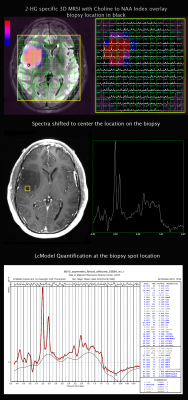 |
Metabolic Characterization of Pre-Surgical Lower Grade Glioma Using Spectroscopic Imaging Optimized for 2-HG Detection
Marisa Lafontaine1, Adam Autry1, Llewellyn Jalbert1, Elizabeth Phillips1, Susan Chang2, and Yan Li1
1Radiology and Biomedical Imaging, UCSF, San Francisco, CA, United States, 2Neurological Surgery, UCSF, San Francisco, CA, United States
2-HG optimized MR spectroscopic imaging showed 2HG was detected in 37 out of 40 IDH1+ patients. Although it was not significantly different between IDH+ and IDH- gliomas, significant correlation was found with progression free survival in newly-diagnosed grade II and grade III gliomas.
|
|
2956.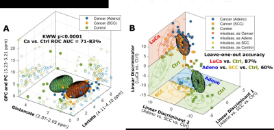 |
Investigation of lung cancer early detection with serum MRS biomarkers
Leo Ling Cheng1, Tjada A. Schult1,2, Mara J. Lauer1,3, Lindsey A. Vandergrift1, Mari A. Mino-Kenudson4, and David C. Christiani5,6
1Pathology, Massachusetts General Hospital, Charlestown, MA, United States, 2Charite Medical University, Berlin, Germany, 3Julius-Maximilians University, Wuerzburg, Germany, 4Pathology, Massachusetts General Hospital, Boston, MA, United States, 5Environmental Health, Harvard T.H. Chan School of Public Health, Boston, MA, United States, 6Department of Medicine, Massachusetts General Hospital, Boston, MA, United States
To improve the survival rate of non-small cell lung cancer (LuCa) patients, an economically efficient screening method without radiation hazard is preferred. Based on our previously reported potential serum biomarkers for predicting survival and differentiating LuCa stages and types, we investigated the potential for these biomarkers to be used for early detection screening using LuCa serum samples from patients prior to their diagnoses and compared with those measured at the time of diagnosis. If developed, our findings could be used as a minimally-invasive early diagnosis scheme.
|
|
2957. |
A metabolomics approach for phenotyping IDH mutant gliomas using 1 and 2D NMR spectroscopy
Thomas Leather Leather1, Marie Phalen2, Khaja Sayed3, Nitika Rathi3, Michael Jenkinson4, Kumar Das4, and Harish Leather Poptani1
1University of Liverpool, Liverpool, United Kingdom, 2NMR Centre, University of Liverpool, Liverpool, United Kingdom, 3Neuropathology, Walton Centre NHS Foundation Trust, Liverpool, United Kingdom, 4Walton Centre NHS Foundation Trust, Liverpool, United Kingdom
The world health organisation has recognised mutations in the IDH1/2 gene as integral in diagnosis and prognostication of gliomas. The mutation facilitates production of the oncometabolite 2-hydroxygluarate (2-HG). The direct mechanisms behind the production and accumulation of 2-HG are well known, however we are yet to understand its effect on prognosis. Although the focus of in vivo studies has been on detection of the 2-HG peak, it is often impeded by spectral overcrowding. Here we used an NMR metabolomics approach to assess metabolome-wide alterations in the presence of the mutation to seek additionally detectable changes in metabolites in these tumours.
|
|
2958. |
Detection of 2-Hydroxyglutarate using JPRESS Magnetic Resonance Spectroscopy
Yan Zhang1 and Jun Shen1
1National Institute of Mental Health, Bethesda, MD, United States
A J-resolved PRESS (JPRESS) technique is proposed for detecting 2-hydroxyglutarate (2HG) in IDH mutant glioma patients at 3T. Spin density simulations and phantom experiments show that the spectral overlap between 2HG and glutamate at ~2.3 ppm is substantially reduced using the one-dimensional cross-sections at J = 0 and J = 7.5 Hz. These two cross-sections can be combined to quantify the 2HG concentration.
|
|
2959. |
Metabolic Biomarkers of TERT expression in glioblastoma
Vinay Ayyappan1, Georgios Batsios2, Nick Stevers3, Abigail R Molloy2, Aliya A Lakhani2, Joseph F Costello3, Pavithra Viswanath2, and Sabrina M Ronen2
1John Hopkins University, Baltimore, MD, United States, 2Radiology and Biomedical Imaging, University of California San Francisco, San Francisco, CA, United States, 3Neurological Surgery, University of California San Francisco, San Francisco, CA, United States Poster Permission Withheld
Expression of telomerase reverse transcriptase (TERT) is essential for tumor proliferation, including in primary glioblastomas. Inhibiting TERT expression is also a therapeutic strategy for glioblastomas. The goal of this study was to identify non-invasive magnetic resonance spectroscopy (MRS)-detectable metabolic biomarkers of TERT expression in glioblastoma cells. Our studies indicate that TERT expression is linked to redox, as indicated by higher 1H MRS-detectable reduced glutathione, and higher 13C MRS-detectable flux of glucose via the pentose phosphate pathway in TERT-expressing GBM cells. Hyperpolarized [U-13C, U-2H]glucose can monitor TERT expression in GBM cells, and may serve to noninvasively probe TERT expression in glioblastomas.
|
|
2960. |
Metabolic profiles of blood plasma in prostate cancer patients having different dietary patterns
Pradeep Kumar1, Rajeev Kumar 2, Sanjay Sharma3, Sanjay Thulkar4, S. Datta Gupta5, S. senthil Kumaran1, Rama Jayasundar1, N. R. Jagannathan6, and Virendra Kumar1
1Department of NMR, All India Institute of Medical Sciences, New Delhi, India, Ansari nagar, India, 2Department of Urology, All India Institute of Medical Sciences, New Delhi, India, Ansari nagar, India, 3Department of Radio-diagnosis, RPC, All India Institute of Medical Sciences, New Delhi, India, Ansari nagar, India, 4Department of Radio-diagnosis, IRCH, All India Institute of Medical Sciences, New Delhi, India, Ansari nagar, India, 5Department of Pathology, All India Institute of Medical Sciences, New Delhi, India, Ansari nagar, India, 6Present address: Department of Radiology, Chettinad Academy of Research and Education, Kelambakkam, Tamil Nadu, India
In the present study we reported the effect of dietary lifestyle on metabolic profile of blood plasma in prostate cancer (PCa) patients using NMR spectroscopy the long term effects of dietary patterns our metabolic profiles and PCa have been reported in literatures. A significantly higher concentration of choline, glutamate, creatine, succinate, 1-methylhistidine and methionine were observed in non-vegetarians patients compared to vegetarians. Our results suggested that the metabolic pathway alterations seen in amino acids, phospholipids and energy may be related to changes due diet to and PCa progression.
|
2961.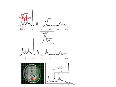 |
31P MRSI of the human brain: do we need 7T to detect two Pi pools, UDPG signals and determine the NAD+/NADH ratio?
Arend Heerschap1, Tom H Peeters2, Andor Veltien2, and Tom W.J, Scheenen2
1Radiology, Radboud University Nijmegen Medical centre, Nijmegen, Netherlands, 2Radboud University Nijmegen Medical centre, Nijmegen, Netherlands All resonances in human brain 31P MR spectra detectable at 7T and higher can also be detected at 3T provided that 1H decoupling, 1H-31P NOE and a 31P receive-array are used. This includes a alkaline peak for extracellular Pi and peaks assigned to UDPG. A NAD+/NADH redox state similar to that reported at 7T was observed. A comparison of measured T1 values with those reported at different field strengths and NOEs measured at 3 and 7T indicates that CSA is not dominant in 31P relaxation and hence there is no sensitivity benefit from shorter T1s at higher fields. |
|
2962.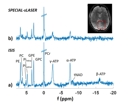 |
Quantification of 31P Human Brain spectra at 9.4T using ISIS and SPECIAL-semiLASER
Johanna Dorst1, Loreen Ruhm1, Nikolai Avdievich1, Wolfgang Bogner2, and Anke Henning1,3
1High-Field MR Center, Max Planck Institute for Biological Cybernetics, Tübingen, Germany, 2High-Field MR Center, Department of Biomedical Imaging and Image-guided Therapy, Medical University of Vienna, Vienna, Austria, 3Advanced Imaging Research Center, UT Southwestern Medical Center, Dallas, TX, United States
For phosphorus single voxel MR spectroscopy, ISIS is often the method of choice, but it requires an 8 steps encoding scheme and post-acquisition signal combination which makes it prone to motion. Therefore, a SPECIAL-semiLASER sequence for 31P MRS was implemented in this study, which only needs two encoding steps for 3D localization. Spectra from the human brain acquired at 9.4T with both sequences are compared in terms of absolute SNR and SNR efficiency. Absolute SNR as well as SNR efficiency is lower for SPECIAL-semiLASER, but calculated concentrations are comparable.
|
|
2963.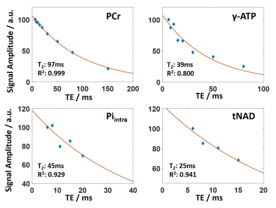 |
31P Transversal Relaxation Times in the Human Brain at 9.4T
Johanna Dorst1, Tamas Borbath1, Loreen Ruhm1, Nikolai Avdievich1, and Anke Henning1,2
1High-Field MR Center, Max Planck Institute for Biological Cybernetics, Tübingen, Germany, 2Advanced Imaging Research Center, UT Southwestern Medical Center, Dallas, TX, United States
31P transversal relaxation times in the human brain at 9.4T are reported. These values are useful to optimize measurement protocols, and to perform absolute quantification. Measurements were performed using a STEAM sequence. To account for J-evolution of homonuclear spin-spin coupled metabolites, basis sets were modeled in VeSPA and spectra were fitted in LCModel. The measured T2 relaxation times are between 93ms and 116ms for phosphomonoesters and –diesters and PCr, and between 25ms and 45ms for Piintra, ATP and tNAD.
|
|
2964.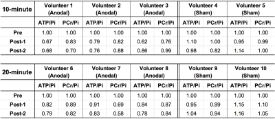 |
Differential modulation of the cerebral energy consumption by two different tDCS settings as measured by 31P MR spectroscopy
Chang-Hoon Choi1, Elene Iordanishvili2, Lea-Sophie Stollberg2, Harshal Patel2, N. Jon Shah1,3,4,5, and Ferdinand Binkofski1,2,4
1INM-4, Forschungszentrum Juelich, Juelich, Germany, 2Department of Neurology, RWTH Aachen University Hospital, Aachen, Germany, 3INM-11, Forschungszentrum Juelich, Juelich, Germany, 4JARA-BRAIN-Translational Medicine, Aachen, Germany, 5Department of Neurology, RWTH Aachen University, Aachen, Germany
Abnormalities in energy regulation are thought to be linked to neurodegenerative and neuropsychiatric disorders. Here, we modulated the brain activity in M1 using sham/anodal tDCS with two different settings, and examined the effect on high-energy metabolites using in vivo 31P-MRS. PCr/Pi and ATP/Pi values showed a decreasing trend following the stimulation compared to the sham measurements and a statistically significant difference was shown between Pre-stimulation and Post-stimulations. However, differences in both PCr/Pi and ATP/Pi were not statistically significant when either a 10- or 20-minute stimulation protocol was applied, but longer stimulation showed a prolonged decrease in both PCr/Pi and ATP/Pi.
|
|
2965.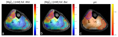 |
Volumetric mapping of the intracellular magnesium ion concentration in human calf muscles using 31P MRSI at 7 Tesla
Vanessa L. Franke1, Mark E. Ladd1, Peter Bachert1, and Andreas Korzowski1
1Division of Medical Physics in Radiology, German Cancer Research Center (DKFZ), Heidelberg, Germany
Intracellular free magnesium ion concentration [Mg2+free] is an important parameter in the regulation of the energy metabolism and can be assessed non-invasively by the use of 31P MRSI. The purpose of this study is to show that volumetric mapping of [Mg2+free] in the human calf muscle is possible at B0=7T with a voxel size of 1 ml. The [Mg2+free] maps of healthy volunteers showed local differences, presumably resulting from different muscle fiber compositions. Two different approaches employed for the calculation of [Mg2+free] yielded comparable maps but
different absolute values, indicating the need for further investigation.
|
|
2966. |
Accelerating volumetric 31P MRSI of the human calf muscle at 7 Tesla: can low-rank denoising filters replace the need for signal averaging?
Andreas Korzowski1, Johannes Breitling1, Vanessa L. Franke1, Mark E. Ladd1, and Peter Bachert1
1Medical Physics in Radiology, German Cancer Research Center (DKFZ), Heidelberg, Germany
Volumetric 31P MRSI in the human calf muscle at 7T in principle enables acquisitions with high spatial resolution, but may require signal averaging to improve signal-to-noise ratios of low-concentrated metabolites. To minimize the acquisition duration of volumetric 31P MRSI, in this study we demonstrate that the acquisition of signal averages can be reliably substituted by application of low-rank denoising filters without the introduction of a quantification bias. This allows acquisition of 31P MRSI data with 1 ml voxels in measurement durations of only 14 minutes that have the same quality as if acquired in 56 minutes with signal averaging.
|
|
2967. |
Under-sampled spiral 31P-MRSI for dynamic exercise applications at 3T
Jabrane Karkouri1,2,3, Magalie Viallon1, Arthur Coste1, Josef Pfeuffer4, Thomas Troalen2, Sylvain Grange1, Remy Prost1, Fabien Millioz1, Pierre Croisille1, and Helene Ratiney1
1Université de Lyon, INSA-Lyon, Université Claude Bernard Lyon 1, UJM-Saint Etienne, CNRS, Inserm, CREATIS UMR 5220, U1206, Lyon, France, Lyon, France, 2SIemens Healthcare SAS, Saint-Denis, France, 3Wolfson Brain Imaging Center, University of Cambridge, Cambridge, United Kingdom, 4SIemens Healthcare SAS, Erlangen, Germany
In this abstract, a novel approach based on spiral MRSI and under-sampling of the temporal dimension is applied in the case of dynamic phosphorus at 3T. The goal was to assess mitochondrial capacity of different individual muscles within the calf at once.
|
|
2968. |
Feasibility of 31P MR Imaging at 7T
Tijl van der Velden1, Mark Gosselink1, Giel Mens1, Hans Hoogduin1, Jeanine Prompers1, Dennis Klomp1, and Martijn Froeling1
1Center of Image Sciences, UMC Utrecht, Utrecht, Netherlands
Typical 31P CSI acquisitions have long scan times with a course resolution. Recent technical developments have incorporated a transmit body coil and receiver array for 31P in 7T systems. This opens up the possibility to acquire 31P data similar to conventional 1H imaging. In this study, we investigated the feasibility of 31P gradient echo MRI to measure the 31P distribution in human muscle.
|
|
2969.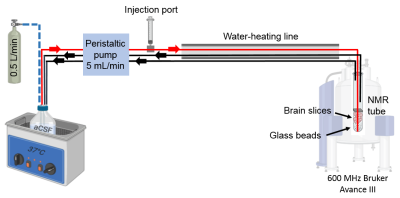 |
Perfusion system for studying dynamic metabolomics in neonatal rat brain slices exposed to oxygen deprivation using 1H and 31P NMR
Alicja Molska1, Deborah Katherine Hill1, Trygve Andreassen1, and Marius Widerøe1
1Department of Circulation and Medical Imaging, Norwegian University of Science and Technology (NTNU), Trondheim, Norway
We designed an NMR-compatible bioreactor that allows real-time metabolic measurements of viable rat brain slices under normal conditions and under conditions mimicking hypoxic-ischemic (HI) brain injury. Special emphasis was put on providing physiological temperature in the system, a significant factor in the HI model. 1H NMR spectra were acquired to assess changes in brain metabolites. Peaks of lactic acid, NAA, glutamate, aspartate, and creatine were detected. By acquiring a time series of proton spectra, we observed significantly increased lactate levels over time when switching from normoxia to hypoxia while other metabolites remained stable over the whole experiment.
|
|
2970.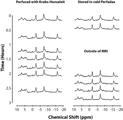 |
A Perfusate Comparison for Ex-Vivo Lung Preservation: Using 31P MRS to Assess Cold Perfadex and Modified Krebs–Henseleit Buffer
Jonathan Snow1, Mehrdad Pourfathi1, Sarmad Siddiqui1, Ian Duncan1, Harrilla Profka1, Federico Sertic1,2, Stephen J Kadlecek1, Gabriel Unger1, Hooman Hamedani1,3, Yi Xin1,3, Luis Loza1, Faraz Amzajerdian 1,3, Tahmina Achekzai1,
Kai Ruppert1, Ryan Baron1, Yiwen Qian1,3, Michael Rosalino1, Maurizio Cereda1,4, Shampa Chatterjee5, and Rahim R. Rizi1
1Radiology, University of Pennsylvania, Philadelphia, PA, United States, 2Surgery, University of Pennsylvania, Philadelphia, PA, United States, 3Bioengineering, University of Pennsylvania, Philadelphia, PA, United States, 4Anesthesiology and Critical Care, University of Pennsylvania, Philadelphia, PA, United States, 5Physiology, University of Pennsylvania, Philadelphia, PA, United States
Ex-vivo lung perfusion (EVLP) is a procedure used in clinical care to support lung viability during transplantation. Here, we used magnetic resonance spectroscopy (MRS) to monitor and compare the energy status of ex-vivo rat lungs flushed and stored in cold Perfadex or perfused with a modified warm Krebs-Henseleit buffer. The results demonstrate decreasing energy status over the course of the 3hr experiment under both conditions, with a slightly lower rate of decline in lung viability using cold Perfadex.
|
|
2971.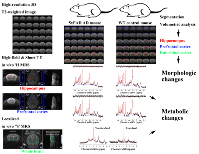 |
Neurodegenerative changes in the brain of Alzheimer’s mice investigated by high-field 1H-31P MRS and segmentation on high-resolution MRI
Chi-Hyeon Yoo1,2, Hyeon-Man Baek2, and Bo-Young Choe1
1The Catholic University of Korea, Seoul, Republic of Korea, 2Gacheon University, Incheon, Republic of Korea
The aim of this study was to investigate morphological and metabolic changes in the brain of model mice with AD by using combined 1H-31P MRS and 3D T2 MRI in an integrated way. To investigate the AD-related metabolic changes, mean concentrations and /PCr relative ratios were compared between the groups for all major metabolites. In addition, a volumetric analysis was conducted on the volume of hippocampus, entorhinal cortex, and PFC between the groups. The AD-related changes in brain can be investigated by high-field and short-TE 1H MRS and high-resolution T2-weighted MRI in conjugate with the altas-based automatic brain segmentation.
|
|
2972.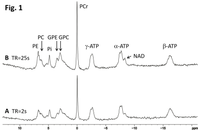 |
Probing Creatine Phosphorylation in Human Brain by Combining Phosphorus and Proton MRS
Shizhe Li1, Jan Willem van der Veen1, JoEllyn Stolinski1, Christopher Johnson1, Maria Ferraris-Araneta1, Milalynn Victorino1, and Jun Shen1
1National Institutes of Health, Bethesda, MD, United States
Cerebral creatine is affected by many CNS disorders. Combining phosphorus and proton MRS to evaluate both phosphocreatine and creatine may provide important insight into brain energetics associated with abnormal creatine levels as the creatine kinase reaction is strongly coupled to energy metabolism in the CNS. Here we show that the PCr to total phosphate ratio is highly immune to T1 saturation effect. By combining phosphorus and proton MRS of healthy subjects we demonstrate that it is feasible to characterize creatine phosphorylation with high immunity to T1 saturation.
|
|
2973. |
Bloch-Siegert flip angle calibration for phosphorus at the human brain at 9.4 T using ISIS localization
Loreen Ruhm1, Johanna Dorst1, Nikolai Avdievich1, and Anke Henning1,2
1Max Planck Institute for Biological Cybernetics, Tuebingen, Germany, 2Advanced Imaging Research Center, UT Southwestern Medical Center, Dallas, TX, United States
Correct calibration of the transmit field B1+ is crucial to achieve optimal SNR. However, fast and robust B1+ calibration is difficult for X-nuclei due to the low signal sensitivity. In this work, we proposed a fast B1+ calibration method based on the Bloch-Siegert shift and single voxel ISIS localization. With the proposed sequence, the B1+ calibration can be done in less than 5 min for the human brain at B0 = 9.4 T.
|
|
2974.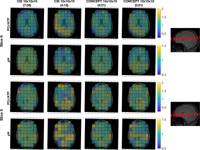 |
Fast 31P-MRSI of the brain at 3T using concentric ring (CONCEPT) MRSI
William T Clarke1, Lukas Hingerl2, Wolfgang Bogner2, Christopher T Rodgers3,4, and Ladislav Valkovic4,5
1Wellcome Centre for Integrative Neuroimaging, NDCN, University of Oxford, Oxford, United Kingdom, 2High-field MR Centre, Department of Biomedical Imaging and Image-guided Therapy, Medical University of Vienna, Vienna, Austria, 3Wolfson Brain Imaging Centre, Department of Clinical Neurosciences, University of Cambridge, Cambridge, United Kingdom, 4Oxford Centre for Clinical Magnetic Resonance Research, Radcliffe Department of Medicine, University of Oxford, Oxford, United Kingdom, 5Department of Imaging Methods, Institute of Measurement Science, Slovak Academy of Sciences, Bratislava, Slovakia
A density-weighted concentric ring trajectory MRSI sequence is created for 31P-MRSI of the human brain at 3T. The sequence is assessed in three subjects at five different acquisition times (7:28 – 1:07 minutes) and compared against resolution and time-matched 3D cartesian CSI sequences. The proposed CONCEPT MRSI sequences robustly measure 3D localised PCr/ATP ratios of the human brain 1.5 times faster than the minimum possible with CSI at a matched repetition time. This opens the possibility of fast dynamic measurements of PCr/ATP in whole human cerebrum at 3T.
|
2975.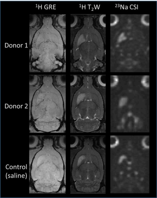 |
Therapeutic Efficacy of Stem Cell Donors for Stroke as Determined by 23Na MRI and 1H MRS at 21.1 T
Shannon Helsper1,2, Xuegang Yuan1,2, F. Andrew Bagdasarian1,2, and Samuel Colles Grant1,2
1National High Magnetic Field Laboratory, Florida State University, Tallahassee, FL, United States, 2Chemical & Biomedical Engineering, FAMU-FSU College of Engineering, Tallahassee, FL, United States
This study evaluates the therapeutic efficacy of human mesenchymal stem cells (hMSC) from different donors applied to an ischemic rat model. Biochemical markers of tissue recovery were measured longitudinally over 21 d using sodium chemical shift imaging, relaxation enhanced MR spectroscopy, and T2-weighted proton imaging at 21.1 T. Ultra-high field provided increased sensitivity, enabling insight into ionic and metabolic regulation while demonstrating differential recovery based on donor characteristics evident only after extended culture. With trophic and immunomodulation effects, hMSC hold promise for cell-based stroke therapy, but donor variance must be evaluated for translation. MR metrics classify donor quality and effectiveness.
|
|
2976. |
Comparison Between Single Channel Dual Loop 23Na Coil and 6-Channel Flexible Wraparound 23Na Coil for Renal Sodium Imaging
Benjamin L Prestwich1, Matthew Clemence2, and Susan T Francis1
1University of Nottingham, Nottingham, United Kingdom, 2Philips Healthcare, Guildford, United Kingdom
Sodium (23Na) MRI has the potential to provide insight to many diseases however 23Na MRI requires nonstandard hardware. This work compares two different designs of RF coil; a single channel pair of transmit-receive (Tx/Rx) loops and a 1Tx-6Rx flexible wraparound coil. Firstly, a 3D gradient-recalled-echo (GRE) sequence was optimised for each coil, then the signal-to-noise ratio (SNR) and field of view (FOV) was compared in both a phantom and in-vivo in a healthy control. SNR was 7±3 and 12±4 for the loops coil and the 6-channel coil respectively.
|
|
2977.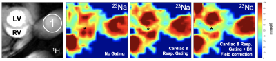 |
Corrections of cardiac 23Na-MRI measurements in mice with an in-house build surface coil at 7T
Martin Christa1, Ibrahim A. Elabyad2, Wolfgang Rudolf Bauer1, and Maxim Terekhov2
1Comprehensive Heart Failure Center (CHFC), Department of Internal Medicine I, University Hospital Wuerzburg, Wuerzburg, Germany, 2Comprehensive Heart Failure Center (CHFC), Chair of Cellular and Molecular Imaging, University Hospital Wuerzburg, Wuerzburg, Germany
With higher field strength and, thus, better SNR available, 23Na-MRI is becoming increasingly popular in (pre-) clinical studies. Improving accuracy and repeatability will help to translate 23Na-MRI into routine. Therefore, we investigated the effect of respiratory and cardiac gating, as well as the influence of B1-field correction on cardiac total sodium quantification with a 2D radial UTE sequence in mice using a surface coil at 7T. We found that ungated and uncorrected measurements overestimate cardiac sodium concentration. Combined respiratory and cardiac gating and B1-field correction significantly decreased the measured cardiac tissue sodium concentration (-40.6%).
|
|
2978.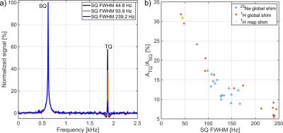 |
Sodium TQ signal cancelation due to B0 inhomogeneities despite the use of a refocusing pulse
Dennis Kleimaier1 and Lothar R. Schad1
1Computer Assisted Clinical Medicine, Heidelberg University, Mannheim, Germany
The sodium TQ signal is sensitive to B0 inhomogeneities leading to TQ signal cancelation. In this study, we evaluated three different shim routines to assess the robustness to B0 inhomogeneities and the reproducibility of the sodium TQ signal. Using an optimized shim routine, the TQ signal dependence on potassium concentration was investigated. Severe B0 inhomogeneities caused a reduction in the TQ signal, despite using a refocusing pulse, without affecting the transversal relaxation times. To achieve reproducible TQ measurements, the FWHM of the sodium SQ signal must be specified. Addition of different amounts KCl caused a reduction in the TQ signal.
|
|
2979.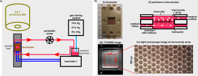 |
Intracellular sodium changes in cancer cells using a microcavity array-based bioreactor and sodium triple-quantum signal
Dennis Kleimaier1, Victor Schepkin2, Cordula Nies3, Eric Gottwald3, and Lothar R. Schad1
1Computer Assisted Clinical Medicine, Heidelberg University, Mannheim, Germany, 2National High Magnetic Field Laboratory, Florida State University, Tallahassee, FL, United States, 3Institute of Functional Interfaces, Karlsruhe Institut of Technology, Karlsruhe, Germany
This study demonstrates the possibility of a microcavity array-based bioreactor to investigate intracellular sodium changes using sodium TQ signal. The response of HepG2 cells during 60min Na+/K+-ATPase inhibition by 1mM ouabain or K+-free medium was monitored. An improved TQTPPI sequence with almost four times TQ SNR increase allowed achieving cell sensitivity of 14*106. During cell experiments, the TQ signal increased to 128.9±3.9% and 165.0±7.4% for 1mM ouabain and K+-free medium, respectively. After reperfusion, the TQ signal recovered to 129.5±4.5%. This bioreactor design with improved TQ signal detection provides the capability to investigate a variety of cells using sodium TQ signal.
|
|
|
2980.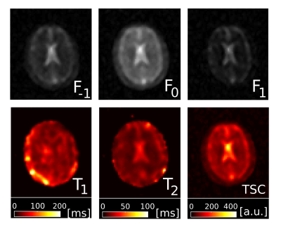 |
Configuration Space Imaging of 23Na at 3T
Jessica Schäper1,2, Claudia Weidensteiner1,2, Philipp Madörin2, and Oliver Bieri1,2
1Department of Biomedical Engineering, University of Basel, Basel, Switzerland, 2Department of Radiology, University Hospital Basel, Basel, Switzerland Poster Permission Withheld
Configuration space is explored for sodium MRI with bSSFP in phantoms and for the brain of a healthy volunteer. To this end, a set of eight phase-cycled bSSFP scans are recorded and from the lowest order configuration modes, quantitative T1, T2 and sodium concentration maps are heuristically derived using the framework valid for spin-1/2 particles.
|
2981.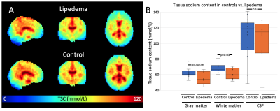 |
Reduced brain sodium and elevated cerebral blood flow in women with lipedema
Kalen J. Petersen1, Maria Garza1, Paula Donahue2, Kevin Harkins3, Manus Donahue1,4,5, and Rachelle Crescenzi1
1Radiology, Vanderbilt University Medical Center, Nashville, TN, United States, 2Physical Medicine and Rehabilitation, Vanderbilt University Medical Center, Nashville, TN, United States, 3Biomedical Engineering, Vanderbilt University, Nashville, TN, United States, 4Neurology, Vanderbilt University Medical Center, Nashville, TN, United States, 5Psychiatry, Vanderbilt University Medical Cener, Nashville, TN, United States
Patients with the adipose disorder lipedema exhibit elevated sodium in the extremities, potentially due to blood or lymphatic vasculopathy. Despite evidence of psychological symptoms, these parameters have not been examined in the brain. We utilized multi-nuclear imaging (23Na-MRI) and arterial spin labeling to test whether dysregulation of brain sodium and perfusion is present in women with (n=15) versus without (n=18) lipedema. We observed lower brain sodium (61.3±6.9 vs. 67.9±5.8mmol/L; p=0.03) and higher cerebral blood flow (43.3±7.0 vs. 37.9±6.3ml blood/100mg tissue/min; p=0.03) in lipedema compared to control participants. Results suggest that brain sodium and hemodynamic dysregulation may exist in lipedema patients.
|
|
2982.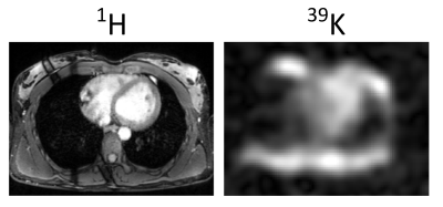 |
Quantitative Human Cardiac 39K MRI at 7T: What is feasible?
Johanna Lott1,2, Tanja Platt1, Sebastian C Niesporek1, Daniel Wenz3, Peter Bachert1,2, Mark E Ladd1,2,4, Thoralf Niendorf5,6, and Armin M Nagel1,7,8
1Medical Physics in Radiology, German Cancer Research Center (DKFZ), Heidelberg, Germany, 2Faculty of Physics and Astronomy, University of Heidelberg, Heidelberg, Germany, 3Center for Biomedical Imaging - Animal Imaging and Technology (CIBM-AIT), École Polytechnique Fédérale de Lausanne (EPFL), Lausanne, Switzerland, 4Faculty of Medicine, University of Heidelberg, Heidelberg, Germany, 5Berlin Ultrahigh Field Facility (B.U.F.F.), Max Delbrueck Center for Molecular Medicine in the Helmholtz Association, Berlin, Germany, 6MRI. TOOLS GmbH, Berlin, Germany, 7Institute of Radiology, University Hospital Erlangen, Erlangen, Germany, 8Institute of Medical Physics, Friedrich-Alexander-Universität Erlangen-Nürnberg (FAU), Erlangen, Germany
Monitoring the viability of myocardial tissue is of prognostic value after an ischemic event1-3. Ultrahigh fields (B0≥7T) facilitates cardiac 39K MRI. However, short relaxation times, low signal-to-noise ratio, and low spatial resolution render the quantitative determination of the myocardial tissue potassium concentration (mTPC) challenging. We analyze the feasibility of quantitative cardiac 39K MRI at 7T with simulations. For realistic noise conditions the mTPC can be determined with a deviation of 14% from the ground truth by applying a partial volume correction. Quantitative cardiac 39K MRI would further benefit e.g. from higher magnetic field strengths (B0>7T) or iterative reconstruction techniques.
|
|
2983.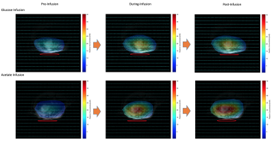 |
Non-Invasive Dynamic Assessment of Heart Energy Metabolisms using in vivo Deuterium MRS Imaging and Close Chest Model
Tao Wang1,2, Xiao-Hong Zhu1, Byeong-Yeul Lee1, Wei Zhu1,2, Hannes M Wiesner1, Yi Zhang1, and Wei Chen1
1Center for Magnetic Resonance Research, Radiology, University of Minnesota, Minneapolis, MN, United States, 2Medical Physics, Radiation Oncology, University of Minnesota, Minneapolis, MN, United States
Continuation from our previous open-chest rat heart project to exploit the in-vivo deuterium (2H) MRS (DMRS) or imaging (DMRSI) methods for dynamic measurement of the myocardial energy metabolism in rat heart at 16.4 T, we further tested its sensitivity and feasibility in response to cardiac glucose metabolism as compared with acetate using a non-invasive method on close-chest rats by using a 1H/2H dual-frequency surface coil. This work demonstrates that both fatty acids and glucose are the main sources of fuel for cardiac activities with a preference for fatty acids.
|
|
2984.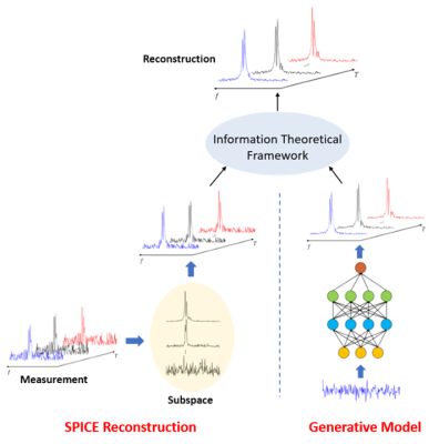 |
Rapid Dynamic Deuterium MR Spectroscopic Imaging Using Deep-SPICE
Yudu Li1,2, Yibo Zhao1,2, Rong Guo1,2, Fanyang Yu2,3, Xiao-Hong Zhu4, Wei Chen4, and Zhi-Pei Liang1,2
1Department of Electrical and Computer Engineering, University of Illinois at Urbana-Champaign, Urbana, IL, United States, 2Beckman Institute for Advanced Science and Technology, University of Illinois at Urbana-Champaign, Urbana, IL, United States, 3Department of Bioengineering, University of Illinois at Urbana-Champaign, Urbana, IL, United States, 4Center for Magnetic Resonance Research, Department of Radiology, University of Minnesota, Minneapolis, MN, United States
Dynamic deuterium MR spectroscopic imaging (2H-MRSI) is emerging as a powerful tool for measurement of metabolic changes using deuterated substrates. In this work, we propose a novel method to reconstruct the often extremely noisy dynamic 2H-MRSI data, incorporating both physics-based subspace spectral model and deep learning-based data priors via an information-theoretical framework. The proposed method has been validated using both simulated and experimental data, showing a significant improvement over the conventional reconstruction and processing method.
|
|
2985.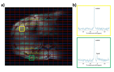 |
Development of a 4-channel body coil for Deuterium Metabolic Imaging of the liver at 7T
Ayhan Gursan1, Arjan D. Hendriks1, Dimitri Welting1, Pim A. de Jong1, Dennis W. Klomp1, and Jeanine J. Prompers1
1Department of Radiology, University Medical Center Utrecht, Utrecht, Netherlands
Deuterium Metabolic Imaging (DMI) is a new technique, which could potentially be used to study liver metabolism in vivo. The aim of this study was to develop a 4-channel 2H transmit/receive array for DMI of the liver at 7T, combined with 4 1H dipole elements for conventional anatomical MRI. Natural abundance DMI water and lipid maps measured on a phantom showed good correspondence with 1H Dixon water and lipid images. The multi-channel setup allowed us to pick up the natural abundance 2H water signal throughout the whole liver in a 3D DMI acquisition with a nominal resolution of 20x20x20 mm3.
|
|
2986.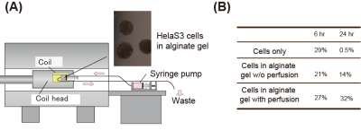 |
NMR and MRS studies of proteins delivered into cultured cells, towards MR analyses of proteins under physiological conditions
Airi Higashi1, Mitsuhiro Takeda1, Sosuke Yoshinaga1, and Hiroaki Terasawa1
1Faculty of Life Sciences, Kumamoto University, Kumamoto, Japan Poster Permission Withheld
We seek to study proteins under physiological conditions, by delivering 13C-labeled proteins into mouse brains and observing them by 13C MRS. We previously observed 13C-labeled a-Synuclein (α-Syn) embedded in an agarose gel by 13C MRS. To examine the feasibility of MRS detection under more in vivo-like conditions, we delivered α-Syn into cultured mammalian cells and observed the cells by NMR, revealing that the delivered α-Syn underwent intracellular interactions and N-terminal acetylation. For MRS experiments with the cells, we constructed a system that perfuses the cells, to prevent cell death over a longer time period.
|
|
2987.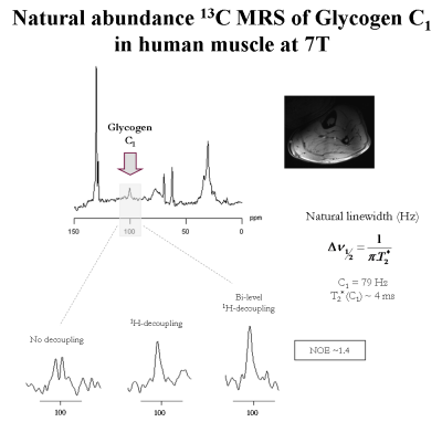 |
Towards structure and metabolism of glycogen C1-C6 by 13C MRS at 7T using broadband 1H decoupling and low-power NOE by means of bi-level WALTZ cycles
Eulalia Serés Roig1 and Rolf Gruetter1,2,3
1Laboratory of Functional and Metabolic Imaging (LIFMET) - Ecole Polytechnique Fédérale de Lausanne (EPFL), Lausanne, Switzerland, 2Centre d'Imagerie BioMédicale - Animal and Imaging Technology (CIBM-AIT) - Ecole Polytechnique de Lausanne (EPFL), Lausanne, Switzerland, 3Department of Radiology, Universities of Lausanne (UNIL) and Geneva (UNIGE), Lausanne, Switzerland Poster Permission Withheld
Glycogen metabolism is essential for glucose homeostasis in humans and may be disrupted in a variety of diseases. Largely neglected due to its low concentration, yet brain glycogen plays an active role in brain energy metabolism, such as during hypo-glycemia. Carbon-13 Magnetic Resonance Spectroscopy (13C MRS) allows the non-invasive detection of glycogen in vivo, while is typically done via the C1 carbon signal. In this study, we explore the potential of 13C MRS at 7T using uniform 13C excitation and bi-level broadband 1H-decoupling towards simultaneous detection of glycogen C1-C6 in
vivo in human muscle, as a step towards brain studies.
|
2988. |
Comparison of low-rank denoising methods for accelerating the acquisition of 31P-MRSI
William T Clarke1 and Mark Chiew1
1Wellcome Centre for Integrative Neuroimaging, NDCN, University of Oxford, Oxford, United Kingdom
Two new low-rank denoising methods are compared to an existing low-rank denoising method in 31P-MRSI data of human skeletal muscle and brain. All three methods increase the SNR of the noisy data above that of high SNR data acquired in 4-times the duration. Denoising algorithm parameters are examined using a grid search. Optimal algorithm choice is dataset dependent and incorrect selection of parameters can bias spectra.
|
|
2989. |
Development of a 2H birdcage volume coil for homogeneous deuterium metabolic imaging of the human brain at 7T
Ayhan Gursan1, Arjan D. Hendriks1, Stefan R. van Genderen1, Dimitri Welting1, Dennis W. Klomp1, and Jeanine J. Prompers1
1Department of Radiology, University Medical Center Utrecht, Utrecht, Netherlands
Deuterium metabolic imaging (DMI) is a new emerging technique, which can measure the metabolism in the brain non-invasively. In this work, a shielded 2H birdcage volume coil for the head was developed and compared with a 2H surface coil setup. In contrast to the surface coil, the 2H birdcage coil enabled high SNR DMI measurements over the entire brain. The homogeneous B1+ field of the birdcage coil allowed for T1 mapping of deuterated water in the brain. Overall, the developed 2H birdcage coil shows high potential for non-invasive metabolic imaging of the brain.
|
|
2990. |
Application of a BIlinear Rotation Decoupling (BIRD) filter for indirect 13C measurements using J-difference editing in the liver
Pandichelvam Veeraiah1, Kim Brouwers1, Joachim E Wildberger1, Vera B Schrauwen-Hinderling1,2, and Lucas Lindeboom1,2
1Departments of Radiology and Nuclear Medicine, Maastricht University Medical Center, Maastricht, Netherlands, 2Nutrition and Movement Sciences, Maastricht University Medical Center, Maastricht, Netherlands
Indirect 13C measurements provide a large signal gain when compared to direct 13C-MRS. The regular J-difference editing (JDE) techniques are quite challenging to apply in the liver, as respiratory motion will result in imperfect subtraction of the huge overlapping 1H-[12C] signal, which hampers accurate quantification of the 1H-[13C] signals. Here, we demonstrated that the application of a BIlinear Rotation Decoupling filter prior to a regular JDE (STEAM-ACED) to pre-suppress the hindering 1H-[12C] signal, leads to a robust detection of the 1H-[13C] signal irrespective of respiratory motion. We showed an application of
measuring 1H-[13C] lipid signals from the liver in vivo.
|
|
2991.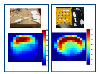 |
A Large FOV 3.0T 16Ch Highly Flexible Array for Body 13C Metabolic Imaging Studies.
Dashen Chu1, Bahareh Behzadnezhad2,3, Scott Lindsay1, Victor Taracila2, Galen Reed4, Albert Chen2, Robert Stormont1, and Fraser Robb2
1GE Healthcare, Inc, Waukesha, WI, United States, 2GE Healthcare, Inc, Aurora, OH, United States, 3University of Wisconsin (Madison), Madison, WI, United States, 4GE Healthcare, Inc, Dallas, TX, United States
Over the last decade, there has been a move toward ultra-flexible conformal coil technology. Similar to flexible H1 coils, there is an interest in developing multi-nuclear coil arrays that are more conformal to human anatomy with highly decoupled elements. In this work, we demonstrate a highly flexible 13C coil array with highly decoupled elements, showing significant improvements in SNR
|
|
2992.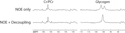 |
Detection of Human Muscle Glycogen by Natural Abundance Carbon-13 MRS at 7 T with Short Duration Proton Decoupling
Li An1, Shizhe Li1, Kalpana Manthiram2, Jyoti Singh Tomar1, Hirotsugu Oda2, Carlos R Ferreira2, and Jun Shen1
1National Institute of Mental Health, National Institutes of Health, Bethesda, MD, United States, 2National Human Genome Research Institute, National Institutes of Health, Bethesda, MD, United States
In this work, a continuous wave (CW) proton decoupling with a short duration of 15 ms was used to detect natural abundance 13C glycogen C1 signals in the human calf at 7 T. This short duration CW proton decoupling dramatically reduced RF power deposition, which allowed the TR to be shortened to 310 ms and additional NOE pulses to be used to ensure optimal SNR performance. The reconstructed glycogen spectra and the peak area ratio between decoupled and non-decoupled glycogen C1 resonances demonstrated that adequate proton decoupling was achieved.
|
|
2993.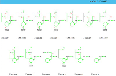 |
tcaCALC: MATLAB Software for Quantitative Analysis of 13C Metabolic Isotopic Tracer Experiments
Jeffry R. Alger1,2,3, Gaurav Sharma1, A. Dean Sherry1,4, and Craig R. Malloy1,5
1Advanced Imaging Research Center, University of Texas, Southwestern Medical Center, Dallas, TX, United States, 2Neurology, University of California, Los Angeles, Los Angeles, CA, United States, 3NeuroSpectroScopics LLC, Sherman Oaks, CA, United States, 4Department of Chemistry, Universiy of Texas at Dallas, Richardson, TX, United States, 5Department of Internal Medicine, University of Texas-Southwestern Medical Center, Dallas, TX, United States
tcaCALC has been developed as a MATLAB tool that facilitates the quantitative analysis of metabolic tracing experiments that use 13C enriched tracers. The most recent version of tcaCALC facilitates analysis by incorporating spectral fitting of J-coupled 13C NMR multiplets and modeling complex metabolic situations in which specific metabolic fluxes may be active or inactive. The MATLAB source code and a compiled version are available for sharing to interested research teams.
|
|
2994.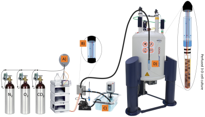 |
Life monitoring of cellular metabolism and mitochondrial respiration in 3D cell culture system using NMR Spectroscopy
Damian Hertig1,2,3, Sally Maddah1,2, Roman Memedovski1,2, Andrea Debora Felser2, Aitor Moreno4, Matteo Pennestri4, Jean-Marc Nuoffer2,5, and Peter Vermathen1
1Departments of Clinical Research and Radiology, University of Bern, Bern, Switzerland, 2Institute of Clinical Chemistry, University Hospital Bern, Bern, Switzerland, 3Graduate School for Cellular and Biomedical Sciences, University of Bern, Bern, Switzerland, 4Bruker Biospin AG, Fällanden, Switzerland, 5Department of Pediatric Endocrinology, University Hospital Bern, Bern, Switzerland
In this study we show the feasibility of simultaneous measure of oxygen consumption and metabolic data in living 3D cell culture in standard 5mm NMR tube. Using high cell density of 20 million fibroblasts we demonstrate high viability and reproducibility of the measurements over 12 hours. We describe the effect of the flow rate on the metabolic activity. Importantly, we show the required sensitivity to detect substrate degradation rates of major mitochondrial fuel pathways and ability to measure rapid O2 and lactate changes as surrogate marker of oxidative phosphorylation and anaerobic glycolysis.
|
|
2995.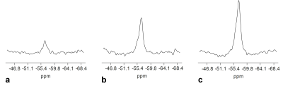 |
In vivo assessment of fluoxetine human brain concentrations using fluorine MR Spectroscopy at 3T
Chu-Yu Lee1, Phil Lee1,2, Jean C. Dinh3, Andrew T Fox1, Richard M. Dubinsky4, William M. Brooks1,4, J. Steven Leeder3,5, and In-Young Choi1,4,6
1Hoglund Brain Imaging Center, University of Kansas Medical Center, Kansas City, KS, United States, 2Department of Radiology, University of Kansas Medical Center, Kansas City, KS, United States, 3Division of Clinical Pharmacology, Toxicology & Therapeutic Innovation, Department of Pediatrics, Children’s Mercy, Kansas City, MO, United States, 4Department of Neurology, University of Kansas Medical Center, Kansas City, KS, United States, 5School of Medicine, University of Missouri-Kansas City, Kansas City, MO, United States, 6Department of Molecular & Integrative Physiology, University of Kansas Medical Center, Kansas City, KS, United States
Fluoxetine is one of the most commonly used medications to treat mental disorders, however, 30-50% of patients fail to respond to the treatment. Thus, direct noninvasive measurements of Fluoxetine concentration in the brain may give insights into individual variations in their clinical responses. This study aimed to quantify fluoxetine concentrations in the human brain using 19F MRS at 3T. 19F MRS measurements were performed on 20 subjects with stable doses of fluoxetine over a month. The fluoxetine concentration was 30±28.2 μM and significantly correlated with their daily doses; r=0.78, p<0.0001, which promises the utility of 19F MRS for personalized medicine.
|
|
2996.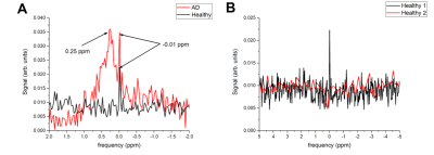 |
19F MRS Detection of Tau Aggregates Using Lansoprazole in an Ex Vivo Rat Model of Alzheimer’s Disease
Sarah K Yeo1, Yurii Shepelytskyi2, Vira Grynko2, Francis T Hane3,4, Tao Li3, and Mitchell S Albert3,4,5
1Biology, Lakehead University, Thunder Bay, ON, Canada, 2Chemistry and Materials Science, Lakehead University, Thunder Bay, ON, Canada, 3Thunder Bay Regional Health Research Institute, Thunder Bay, ON, Canada, 4Chemistry, Lakehead University, Thunder Bay, ON, Canada, 5Northern Ontario School of Medicine, Thunder Bay, ON, Canada
Neurofibrillary tangles (NFTs) composed of hyperphosphorylated tau protein are pathological characteristics of Alzheimer’s Disease (AD). Thus, the use of biosensors that bind to NFTs are beneficial for in vivo detection of tauopathy. This study evaluates the use of Lansoprazole (LSZ) as an indicator of NFTs in AD brains. 19F MRS of AD and control brains with LSZ was acquired using a 3.0T clinical MRI scanner. We demonstrate that the 19F signal from LSZ interacts with NFTs in a rat model of AD. This shows potential in using LSZ to distinguish between AD and healthy brains with a clinical 3.0T scanner.
|
|
2997.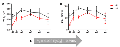 |
Determination of the oxygen partial pressure in hind limb ischemia by 19F MRI
Tuba Gueden-Silber1, Nicolas Stumpe1, Rebekka Schnekkmann2, Maria Grandoch2, and Ulrich Floegel1
1Molecular Cardiology, University Clinic Duesseldorf, Düsseldorf, Germany, 2Pharmacology and Clinical Pharmacology, University Clinic Duesseldorf, Düsseldorf, Germany
Tissue hypoxia induces a variety of detrimental processes. Therefore, the determination of oxygen partial pressure can be helpful to assess the current oxygen supply of an organ. 19F MRI showed to be useful for the quantification of tissue pO2 by exploiting the paramagnetic effect of O2 on the longitudinal relaxation rate R1 of perfluorocarbon nanoemulsions. Thus, allowing the calculation of pO2 through experimental R1 data. In the present study, we applied this approach for monitoring the gradual recovery of tissue oxygenation in a murine model of hind limb ischemia. We observed tissue oxygenation recovery within 2 weeks post-occlusion.
|
|
2998.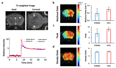 |
Dynamic Oxygen-17 MRI with Adaptive Reconstruction using Golden-Means-Based 3D Radial Sampling
Yuning Gu1, Huiyun Gao2, Kihwan Kim1, Ciro Ramos-Estebanez3, Yunmei Wang2, and Xin Yu1,4,5
1Department of Biomedical Engineering, Case Western Reserve University, Cleveland, OH, United States, 2Case Cardiovascular Research Institute, Case Western Reserve University, Cleveland, OH, United States, 3Department of Neurology, Case Western Reserve University, Cleveland, OH, United States, 4Department of Radiology, Case Western Reserve University, Cleveland, OH, United States, 5Department of Physiology and Biophysics, Case Western Reserve University, Cleveland, OH, United States
A golden-means-based 3D radial sampling and k-space weighted image reconstruction method was developed for dynamic tracking of intravenously injected 17O-water in mouse brain. The method enabled image reconstruction with adaptive temporal resolution of 3 to 15 s to capture the regional differences in 17O-water uptake and washout kinetics in post-stroke mice, with an isotropic voxel size of 1.77 µL on a 9.4T scanner.
|
|
2999.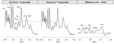 |
A new approach involving combination of 1H MRS and [2,2,2′-2H3]acetate administration to assess cerebral neurotransmission in vivo
Puneet Bagga1, Laurie J Rich1, Neil E Wilson1, Mark Elliott1, Mitch D Schnall1, John A Detre2, Mohammad Haris3,4, and Ravinder Reddy1
1Department of Radiology, University of Pennsylvania, Philadelphia, PA, United States, 2Department of Neurology, University of Pennsylvania, Philadelphia, PA, United States, 3Sidra Medicine, Doha, Qatar, 4LARC, Qatar University, Doha, Qatar
1H MR spectroscopy is currently the only technique that allows the non-invasive detection and quantification of a wide range of neurochemicals. In this study, we performed 1H MRS in conjunction with the administration of [2,2,2′-2H3]acetate to measure turnover kinetics of glutamate, glutamine and GABA in rat brain. As 2H is invisible on 1H MRS, the turnover of metabolites will lead to a corresponding drop in their 1H MR signal visualized by subtraction of the post-[2,2,2′-2H3]acetate administration from the Pre-administration 1H MR spectra. The fractional enrichment data can be fitted to evaluate the rates of
cerebral glutamatergic and GABAergic neurotransmitter cycling.
|